Search This Blog

Monday, March 9, 2015

Tissue engineering


From Wikipedia, the free encyclopedia


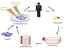
Principle of tissue engineering

Tissue engineering is the use of a combination of cells, engineering and materials methods, and suitable biochemical and physicochemical factors to improve or replace biological functions. While it was once categorized as a sub-field of biomaterials, having grown in scope and importance it can be considered as a field in its own right.

While most definitions of tissue engineering cover a broad range of applications, in practice the term is closely associated with applications that repair or replace portions of or whole tissues (i.e., bone, cartilage, blood vessels, bladder, skin, muscle etc.). Often, the tissues involved require certain mechanical and structural properties for proper functioning. The term has also been applied to efforts to perform specific biochemical functions using cells within an artificially-created support system (e.g. an artificial pancreas, or a bio artificial liver). The term regenerative medicine is often used synonymously with tissue engineering, although those involved in regenerative medicine place more emphasis on the use of stem cells or progenitor cells to produce tissues.

Overview


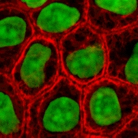
Micro-mass cultures of C3H-10T1/2 cells at varied oxygen tensions stained with Alcian blue

A commonly applied definition of tissue engineering, as stated by Langer[1] and Vacanti,[2] is "an interdisciplinary field that applies the principles of engineering and life sciences toward the development of biological substitutes that restore, maintain, or improve tissue function or a whole organ".[3] Tissue engineering has also been defined as "understanding the principles of tissue growth, and applying this to produce functional replacement tissue for clinical use."[4] A further description goes on to say that an "underlying supposition of tissue engineering is that the employment of natural biology of the system will allow for greater success in developing therapeutic strategies aimed at the replacement, repair, maintenance, and/or enhancement of tissue function."[4]

Powerful developments in the multidisciplinary field of tissue engineering have yielded a novel set of tissue replacement parts and implementation strategies. Scientific advances in biomaterials, stem cells, growth and differentiation factors, and biomimetic environments have created unique opportunities to fabricate tissues in the laboratory from combinations of engineered extracellular matrices ("scaffolds"), cells, and biologically active molecules. Among the major challenges now facing tissue engineering is the need for more complex functionality, as well as both functional and biomechanical stability in laboratory-grown tissues destined for transplantation. The continued success of tissue engineering, and the eventual development of true human replacement parts, will grow from the convergence of engineering and basic research advances in tissue, matrix, growth factor, stem cell, and developmental biology, as well as materials science and bio informatics.

In 2003, the NSF published a report entitled "The Emergence of Tissue Engineering as a Research Field", which gives a thorough description of the history of this field.[5]

Examples

Cells as building blocks


Stained cells in culture

Tissue engineering utilizes living cells as engineering materials. Examples include using living fibroblasts in skin replacement or repair, cartilage repaired with living chondrocytes, or other types of cells used in other ways.

Cells became available as engineering materials when scientists at Geron Corp. discovered how to extend telomeres in 1998, producing immortalized cell lines.[citation needed] Before this, laboratory cultures of healthy, noncancerous mammalian cells would only divide a fixed number of times, up to the Hayflick limit.

Extraction

From fluid tissues such as blood, cells are extracted by bulk methods, usually centrifugation or apheresis. From solid tissues, extraction is more difficult. Usually the tissue is minced, and then digested with the enzymes trypsin or collagenase to remove the extracellular matrix (ECM) that holds the cells. After that, the cells are free floating, and extracted using centrifugation or apheresis.
Digestion with trypsin is very dependent on temperature. Higher temperatures digest the matrix faster, but create more damage. Collagenase is less temperature dependent, and damages fewer cells, but takes longer and is a more expensive reagent.

Types of cells


Mouse embryonic stem cells

Cells are often categorized by their source:
  • Autologous cells are obtained from the same individual to which they will be reimplanted. Autologous cells have the fewest problems with rejection and pathogen transmission, however in some cases might not be available. For example in genetic disease suitable autologous cells are not available. Also very ill or elderly persons, as well as patients suffering from severe burns, may not have sufficient quantities of autologous cells to establish useful cell lines. Moreover since this category of cells needs to be harvested from the patient, there are also some concerns related to the necessity of performing such surgical operations that might lead to donor site infection or chronic pain. Autologous cells also must be cultured from samples before they can be used: this takes time, so autologous solutions may not be very quick. Recently there has been a trend towards the use of mesenchymal stem cells from bone marrow and fat. These cells can differentiate into a variety of tissue types, including bone, cartilage, fat, and nerve. A large number of cells can be easily and quickly isolated from fat, thus opening the potential for large numbers of cells to be quickly and easily obtained.
  • Allogeneic cells come from the body of a donor of the same species. While there are some ethical constraints to the use of human cells for in vitro studies, the employment of dermal fibroblasts from human foreskin has been demonstrated to be immunologically safe and thus a viable choice for tissue engineering of skin.
  • Xenogenic cells are these isolated from individuals of another species. In particular animal cells have been used quite extensively in experiments aimed at the construction of cardiovascular implants.
  • Syngenic or isogenic cells are isolated from genetically identical organisms, such as twins, clones, or highly inbred research animal models.
  • Primary cells are from an organism.
  • Secondary cells are from a cell bank.
  • Stem cells are undifferentiated cells with the ability to divide in culture and give rise to different forms of specialized cells. According to their source stem cells are divided into "adult" and "embryonic" stem cells, the first class being multipotent and the latter mostly pluripotent; some cells are totipotent, in the earliest stages of the embryo. While there is still a large ethical debate related with the use of embryonic stem cells, it is thought that another alternative source - induced stem cells may be useful for the repair of diseased or damaged tissues, or may be used to grow new organs.

Scaffolds

Cells are often implanted or 'seeded' into an artificial structure capable of supporting three-dimensional tissue formation. These structures, typically called scaffolds, are often critical, both ex vivo as well as in vivo, to recapitulating the in vivo milieu and allowing cells to influence their own microenvironments. Scaffolds usually serve at least one of the following purposes:
  • Allow cell attachment and migration
  • Deliver and retain cells and biochemical factors
  • Enable diffusion of vital cell nutrients and expressed products
  • Exert certain mechanical and biological influences to modify the behaviour of the cell phase
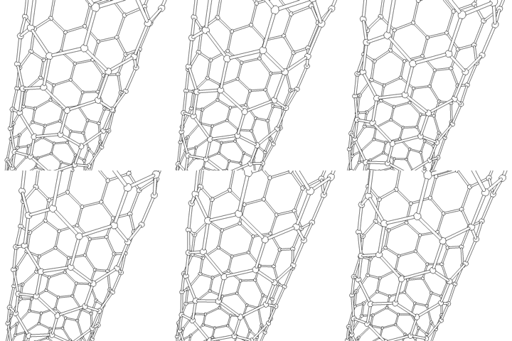
This animation of a rotating Carbon nanotube shows its 3D structure. Carbon nanotubes are among the numerous candidates for tissue engineering scaffolds since they are biocompatible, resistant to biodegradation and can be functionalized with biomolecules. However, the possibility of toxicity with non-biodegradable nano-materials is not fully understood.[16]

To achieve the goal of tissue reconstruction, scaffolds must meet some specific requirements. A high porosity and an adequate pore size are necessary to facilitate cell seeding and diffusion throughout the whole structure of both cells and nutrients. Biodegradability is often an essential factor since scaffolds should preferably be absorbed by the surrounding tissues without the necessity of a surgical removal. The rate at which degradation occurs has to coincide as much as possible with the rate of tissue formation: this means that while cells are fabricating their own natural matrix structure around themselves, the scaffold is able to provide structural integrity within the body and eventually it will break down leaving the neotissue, newly formed tissue which will take over the mechanical load. Injectability is also important for clinical uses. Recent research on organ printing is showing how crucial a good control of the 3D environment is to ensure reproducibility of experiments and offer better results.

Materials

Many different materials (natural and synthetic, biodegradable and permanent) have been investigated. Most of these materials have been known in the medical field before the advent of tissue engineering as a research topic, being already employed as bioresorbable sutures. Examples of these materials are collagen and some polyesters.

New biomaterials have been engineered to have ideal properties and functional customization: injectability, synthetic manufacture, biocompatibility, non-immunogenicity, transparency, nano-scale fibers, low concentration, resorption rates, etc. PuraMatrix, originating from the MIT labs of Zhang, Rich, Grodzinsky and Langer is one of these new biomimetic scaffold families which has now been commercialized and is impacting clinical tissue engineering.

A commonly used synthetic material is PLA - polylactic acid. This is a polyester which degrades within the human body to form lactic acid, a naturally occurring chemical which is easily removed from the body. Similar materials are polyglycolic acid (PGA) and polycaprolactone (PCL): their degradation mechanism is similar to that of PLA, but they exhibit respectively a faster and a slower rate of degradation compared to PLA.

Scaffolds may also be constructed from natural materials: in particular different derivatives of the extracellular matrix have been studied to evaluate their ability to support cell growth. Proteic materials, such as collagen or fibrin, and polysaccharidic materials, like chitosan[17] or glycosaminoglycans (GAGs), have all proved suitable in terms of cell compatibility, but some issues with potential immunogenicity still remains. Among GAGs hyaluronic acid, possibly in combination with cross linking agents (e.g. glutaraldehyde, water soluble carbodiimide, etc...), is one of the possible choices as scaffold material. Functionalized groups of scaffolds may be useful in the delivery of small molecules (drugs) to specific tissues. Another form of scaffold under investigation is decellularised tissue extracts whereby the remaining cellular remnants/extracellular matrices act as the scaffold.

A 2009 study by Ratmir et al. aimed to improve in vivo-like conditions for 3D tissue via "stacking and de-stacking layers of paper impregnated with suspensions of cells in extracellular matrix hydrogel, making it possible to control oxygen and nutrient gradients in 3D, and to analyze molecular and genetic responses".[18] It is possible to manipulate gradients of soluble molecules, and to characterize cells in these complex gradients more effectively than conventional 3D cultures based on hydrogels, cell spheroids, or 3D perfusion reactors.[19] Different thicknesses of paper and types of medium can support a variety of experimental environments. Upon deconstruction, these sheets can be useful in cell-based high-throughput screening and drug discovery.[19]

Synthesis


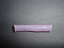
Tissue engineered vascular graft

Tissue engineered heart valve

A number of different methods have been described in literature for preparing porous structures to be employed as tissue engineering scaffolds. Each of these techniques presents its own advantages, but none are free of drawbacks.
Nanofiber Self-Assembly
Molecular self-assembly is one of the few methods for creating biomaterials with properties similar in scale and chemistry to that of the natural in vivo extracellular matrix (ECM), a crucial step toward tissue engineering of complex tissues.[20] Moreover, these hydrogel scaffolds have shown superiority in in vivo toxicology and biocompatibility compared to traditional macroscaffolds and animal-derived materials.
Textile technologies
These techniques include all the approaches that have been successfully employed for the preparation of non-woven meshes of different polymers. In particular, non-woven polyglycolide structures have been tested for tissue engineering applications: such fibrous structures have been found useful to grow different types of cells. The principal drawbacks are related to the difficulties in obtaining high porosity and regular pore size.
Solvent Casting & Particulate Leaching (SCPL)
This approach allows for the preparation of structures with regular porosity, but with limited thickness. First, the polymer is dissolved into a suitable organic solvent (e.g. polylactic acid could be dissolved into dichloromethane), then the solution is cast into a mold filled with porogen particles. Such porogen can be an inorganic salt like sodium chloride, crystals of saccharose, gelatin spheres or paraffin spheres. The size of the porogen particles will affect the size of the scaffold pores, while the polymer to porogen ratio is directly correlated to the amount of porosity of the final structure. After the polymer solution has been cast the solvent is allowed to fully evaporate, then the composite structure in the mold is immersed in a bath of a liquid suitable for dissolving the porogen: water in the case of sodium chloride, saccharose and gelatin or an aliphatic solvent like hexane for use with paraffin. Once the porogen has been fully dissolved, a porous structure is obtained. Other than the small thickness range that can be obtained, another drawback of SCPL lies in its use of organic solvents which must be fully removed to avoid any possible damage to the cells seeded on the scaffold.
Gas Foaming
To overcome the need to use organic solvents and solid porogens, a technique using gas as a porogen has been developed. First, disc-shaped structures made of the desired polymer are prepared by means of compression molding using a heated mold. The discs are then placed in a chamber where they are exposed to high pressure CO2 for several days. The pressure inside the chamber is gradually restored to atmospheric levels. During this procedure the pores are formed by the carbon dioxide molecules that abandon the polymer, resulting in a sponge-like structure. The main problems resulting from such a technique are caused by the excessive heat used during compression molding (which prohibits the incorporation of any temperature labile material into the polymer matrix) and by the fact that the pores do not form an interconnected structure.
Emulsification/Freeze-drying
This technique does not require the use of a solid porogen like SCPL. First, a synthetic polymer is dissolved into a suitable solvent (e.g. polylactic acid in dichloromethane) then water is added to the polymeric solution and the two liquids are mixed in order to obtain an emulsion. Before the two phases can separate, the emulsion is cast into a mold and quickly frozen by means of immersion into liquid nitrogen. The frozen emulsion is subsequently freeze-dried to remove the dispersed water and the solvent, thus leaving a solidified, porous polymeric structure. While emulsification and freeze-drying allow for a faster preparation when compared to SCPL (since it does not require a time consuming leaching step), it still requires the use of solvents. Moreover, pore size is relatively small and porosity is often irregular. Freeze-drying by itself is also a commonly employed technique for the fabrication of scaffolds. In particular, it is used to prepare collagen sponges: collagen is dissolved into acidic solutions of acetic acid or hydrochloric acid that are cast into a mold, frozen with liquid nitrogen and then lyophilized.
Thermally Induced Phase Separation (TIPS)
Similar to the previous technique, this phase separation procedure requires the use of a solvent with a low melting point that is easy to sublime. For example dioxane could be used to dissolve polylactic acid, then phase separation is induced through the addition of a small quantity of water: a polymer-rich and a polymer-poor phase are formed. Following cooling below the solvent melting point and some days of vacuum-drying to sublime the solvent, a porous scaffold is obtained. Liquid-liquid phase separation presents the same drawbacks of emulsification/freeze-drying.
Electrospinning
A highly versatile technique that can be used to produce continuous fibers from submicrometer to nanometer diameters. In a typical electrospinning set-up, a solution is fed through a spinneret and a high voltage is applied to the tip. The buildup of electrostatic repulsion within the charged solution, causes it to eject a thin fibrous stream. A mounted collector plate or rod with an opposite or grounded charge draws in the continuous fibers, which arrive to form a highly porous network. The primary advantages of this technique are its simplicity and ease of variation. At a laboratory level, a typical electrospinning set-up only requires a high voltage power supply (up to 30 kV), a syringe, a flat tip needle and a conducting collector. For these reasons, electrospinning has become a common method of scaffold manufacture in many labs. By modifying variables such as the distance to collector, magnitude of applied voltage, or solution flow rate—researchers can dramatically change the overall scaffold architecture.
CAD/CAM Technologies
Because most of the above techniques are limited when it comes to the control of porosity and pore size, computer assisted design and manufacturing techniques have been introduced to tissue engineering. First, a three-dimensional structure is designed using CAD software. The porosity can be tailored using algorithms within the software.[21] The scaffold is then realized by using ink-jet printing of polymer powders or through Fused Deposition Modeling of a polymer melt.[22]
A 2011 study by El-Ayoubi et al. investigated "3D-plotting technique to produce (biocompatible and biodegradable) poly-L-Lactide macroporous scaffolds with two different pore sizes" via solid free-form fabrication (SSF) with computer-aided-design (CAD), to explore therapeutic articular cartilage replacement as an "alternative to conventional tissue repair".[23] The study found the smaller the pore size paired with mechanical stress in a bioreactor (to induce in vivo-like conditions), the higher the cell viability in potential therapeutic functionality via decreasing recovery time and increasing transplant effectiveness.[23]
Laser-assisted BioPrinting (LaBP)
In a 2012 study,[24] Koch et al. focused on whether Laser-assisted BioPrinting (LaBP) can be used to build multicellular 3D patterns in natural matrix, and whether the generated constructs are functioning and forming tissue. LaBP arranges small volumes of living cell suspensions in set high-resolution patterns.[24] The investigation was successful, the researchers foresee that "generated tissue constructs might be used for in vivo testing by implanting them into animal models" (14). As of this study, only human skin tissue has been synthesized, though researchers project that "by integrating further cell types (e.g. melanocytes, Schwann cells, hair follicle cells) into the printed cell construct, the behavior of these cells in a 3D in vitro microenvironment similar to their natural one can be analyzed", useful for drug discovery and toxicology studies.[24]

Assembly methods

One of the continuing, persistent problems with tissue engineering is mass transport limitations. Engineered tissues generally lack an initial blood supply, thus making it difficult for any implanted cells to obtain sufficient oxygen and nutrients to survive, and/or function properly.

Self-assembly

Self-assembly may play an important role here, both from the perspective of encapsulating cells and proteins, as well as creating scaffolds on the right physical scale for engineered tissue constructs and cellular ingrowth. The micromasonry is a prime technology to get cells grown in a lab to assemble into three-dimensional shapes. To break down tissue into single-cell building blocks, researchers have to dissolve the extracellular mortar that normally binds them together. But once that glue is removed, it's quite difficult to get cells to reassemble into the complex structures that make up our natural tissues. While cells aren't easily stackable, building blocks are. So the micromasonry starts with the encapsulation of living cells in polymer cubes. From there, the blocks self-assemble in any shape using templates.

Liquid-based template assembly

The air-liquid surface established by Faraday waves is explored as a template to assemble biological entities for bottom-up tissue engineering. This liquid-based template can be dynamically reconfigured in a few seconds, and the assembly on the template can be achieved in a scalable and parallel manner. Assembly of microscale hydrogels, cells, neuron-seeded micro-carrier beads, cell spheroids into various symmetrical and periodic structures was demonstrated with good cell viability. Formation of 3D neural network was achieved after 14-day tissue culture.[25]

Additive manufacturing

It might be possible to print organs, or possibly entire organisms using additive manufacturing techniques. A recent innovative method of construction uses an ink-jet mechanism to print precise layers of cells in a matrix of thermoreversible gel. Endothelial cells, the cells that line blood vessels, have been printed in a set of stacked rings. When incubated, these fused into a tube.[22][26]

The field of three-dimensional and highly accurate models of biological systems is pioneered by multiple projects and technologies including a rapid method for creating tissues and even whole organs involves a 3D printer that can print the scaffolding and cells layer by layer into a working tissue sample or organ. The device is presented in a TED talk by Dr. Anthony Atala, M.D. the Director of the Wake Forest Institute for Regenerative Medicine, and the W.H. Boyce Professor and Chair of the Department of Urology at Wake Forest University, in which a full bladder is printed on stage during the seminar and then presented to the crowd.[27][28] This specific technology has been used to print a bladder for a young man that would have otherwise died without the availability of a transplant, and is anticipated to be able to print kidneys and perhaps livers in the future for transplantation and theoretically for toxicology and other biological studies as well.

Scaffolding

In 2013, using a 3-d scaffolding of Matrigel in various configurations, substantial pancreatic organoids was produced in vitro. Clusters of small numbers of cells proliferated into 40,000 cells within one week. The clusters transform into cells that make either digestive enzymes or hormones like insulin, self-organizing into branched pancreatic organoids that resemble the pancreas.[29]
The cells are sensitive to the environment, such as gel stiffness and contact with other cells.
Individual cells do not thrive; a minimum of four proximate cells was required for subsequent organoid development. Modifications to the medium composition produced either hollow spheres mainly composed of pancreatic progenitors, or complex organoids that spontaneously undergo pancreatic morphogenesis and differentiation. Maintenance and expansion of pancreatic progenitors require active Notch and FGF signaling, recapitulating in vivo niche signaling interactions.[30]

The organoids were seen as potentially offering mini-organs for drug testing and for spare insulin-producing cells.[29]

Tissue culture

In many cases, creation of functional tissues and biological structures in vitro requires extensive culturing to promote survival, growth and inducement of functionality. In general, the basic requirements of cells must be maintained in culture, which include oxygen, pH, humidity, temperature, nutrients and osmotic pressure maintenance.

Tissue engineered cultures also present additional problems in maintaining culture conditions. In standard cell culture, diffusion is often the sole means of nutrient and metabolite transport. However, as a culture becomes larger and more complex, such as the case with engineered organs and whole tissues, other mechanisms must be employed to maintain the culture, such as the creation of capillary networks within the tissue.

Bioreactor for cultivation of vascular grafts

Another issue with tissue culture is introducing the proper factors or stimuli required to induce functionality. In many cases, simple maintenance culture is not sufficient. Growth factors, hormones, specific metabolites or nutrients, chemical and physical stimuli are sometimes required. For example, certain cells respond to changes in oxygen tension as part of their normal development, such as chondrocytes, which must adapt to low oxygen conditions or hypoxia during skeletal development. Others, such as endothelial cells, respond to shear stress from fluid flow, which is encountered in blood vessels. Mechanical stimuli, such as pressure pulses seem to be beneficial to all kind of cardiovascular tissue such as heart valves, blood vessels or pericardium.

Bioreactors

A bioreactor in tissue engineering, as opposed to industrial bioreactors, is a device that attempts to simulate a physiological environment in order to promote cell or tissue growth in vivo. A physiological environment can consist of many different parameters such as temperature and oxygen or carbon dioxide concentration, but can extend to all kinds of biological, chemical or mechanical stimuli. Therefore, there are systems that may include the application of forces or stresses to the tissue or even of electrical current in two- or three-dimensional setups.
In academic and industry research facilities, it is typical for bioreactors to be developed to replicate the specific physiological environment of the tissue being grown (e.g., flex and fluid shearing for heart tissue growth).[31] Several general-use and application-specific bioreactors are also commercially available, and may provide static chemical stimulation or combination of chemical and mechanical stimulation.

The Bioreactors used for 3D cell cultures are small plastic cylindrical chambers with regulated internal humidity and moisture specifically engineered for the purpose of growing cells in three dimensions.[32] The bioreactor uses bioactive synthetic materials such as polyethylene terephthalate membranes to surround the spheroid cells in an environment that maintains high levels of nutrients.[19][33] They are easy to open and close, so that cell spheroids can be removed for testing, yet the chamber is able to maintain 100% humidity throughout.[34] This humidity is important to achieve maximum cell growth and function. The bioreactor chamber is part of a larger device that rotates to ensure equal cell growth in each direction across three dimensions.[34] MC2 Biotek has developed a bioreactor known as ProtoTissue[32] that uses gas exchange to maintain high oxygen levels within the cell chamber; improving upon previous bioreactors, because the higher oxygen levels help the cell grow and undergo normal cell respiration.[35]
Long fiber generation

In 2013, a group from the University of Tokyo developed cell laden fibers up to a meter in length and on the order of 100 µm in size.[36] These fibers were created using a microfluidic device that forms a double coaxial laminar flow. Each 'layer' of the microfluidic device (cells seeded in ECM, a hydrogel sheath, and finally a calcium chloride solution). The seeded cells culture within the hydrogel sheath for several days, and then the sheath is removed with viable cell fibers. Various cell types were inserted into the ECM core, including myocytes, endothelial cells, nerve cell fibers, and epithelial cell fibers. This group then showed that these fibers can be woven together to fabricate tissues or organs in a mechanism similar to textile weaving. Fibrous morphologies are advantageous in that they provide an alternative to traditional scaffold design, and many organs (such as muscle) are composed of fibrous cells.

Bioartificial organs

An artificial organ is a man-made device that is implanted or integrated into a human to replace a natural organ, for the purpose of restoring a specific function or a group of related functions so the patient may return to a normal life as soon as possible. The replaced function doesn't necessarily have to be related to life support, but often is. The ultimate goal of tissue engineering as a discipline is to allow both 'off the shelf' bioartificial organs and regeneration of injured tissue in the body. In order to successfully create bioartificial organs from a patients stem cells, researchers continue to make improvements in the generation of complex tissues by tissue engineering. For example, much research is aimed at understanding nanoscale cues present in a cell’s microenvironment.[20]

Artificial uterus


From Wikipedia, the free encyclopedia

An artificial uterus (or womb) is a theoretical device that would allow for extracorporeal pregnancy or extrauterine fetal incubation (EUFI)[1] by growing an embryo or fetus outside of the body of a female organism that would normally internally carry the embryo or fetus to term.
An artificial uterus, as a replacement organ, would have many applications. It could be used to assist male or female couples in the development of a fetus.[1] This can potentially be performed as a switch from a natural uterus to an artificial uterus, thereby moving the threshold of fetal viability to a much earlier stage of pregnancy.[1] In this sense, it can be regarded as a neonatal incubator with very extended functions. Also, it can potentially be used for initiation of fetal development.[1] Furthermore, it could avail for performing, for example, fetal surgery procedures at an early stage instead of having to postpone them until term of pregnancy.[1]

Components

An artificial uterus would have to provide nutrients and oxygen to nurture a fetus, as well as dispose of waste material. The scope of an artificial uterus (or "artificial uterus system" to emphasize a broader scope) may also include the interface serving the function otherwise provided by the placenta, an amniotic tank functioning as the amniotic sac, as well as an umbilical cord.

Nutrition, oxygen supply and waste disposal

A human may still supply nutrients and dispose of waste products if the artificial uterus is connected to her.[1] Also, it may provide immune protection against diseases by passing of IgG antibodies to the embryo or fetus.[1]

Artificial supply and disposal have the potential advantage of allowing the fetus to develop in an environment that is not influenced by the presence of disease, environmental pollutants, alcohol, or drugs which a human may have in the circulatory system.[1] Also, there is no risk of an immune reaction towards the embryo or fetus that could otherwise arise from insufficient gestational immune tolerance.[1] Following are aspects of individual functions of an artificial supplier and disposer:
  • Waste disposal may be performed through dialysis.[1]
  • For oxygenation of the embryo or fetus, and removal of carbon dioxide, extracorporeal membrane oxygenation (ECMO) is a functioning technique, having successfully kept goat fetuses alive for up to 237 hours in amniotic tanks.[2] ECMO is currently a technique used in selected neonatal intensive care units to treat term infants with selected medical problems that result in the infant's inability to survive through gas exchange using the lungs.[3] However, the cerebral vasculature and germinal matrix are poorly developed in fetuses, and subsequently, there is an unacceptably high risk for intraventricular hemorrhage (IVH) if administering ECMO at a gestational age less than 32 weeks.[4] Liquid ventilation has been suggested as an alternative method of oxygenation, or at least providing an intermediate stage between the womb and breathing in open air.[1]
  • For artificial nutrition, current techniques are problematic.[1] Total parenteral nutrition, as studied on infants with severe short bowel syndrome, has a 5 year survival of approximately 20%.[1][5]
  • Issues related to hormonal stability also remain to be addressed.[1]
Theoretically, animal suppliers and disposers may be used, but when involving an animal's uterus the technique may rather be in the scope of interspecific pregnancy.

Uterine wall

Naturally, the myometrium of the uterine wall functions to expel the fetus at the end of a pregnancy, and the endometrium plays a role in forming the placenta.

An artificial uterus may include components of equivalent function. Also, methods have been considered to connect an artificial placenta and other "inner" components directly to an external circulation.[1]

Interface (artificial placenta)

An interface between the supplier and the embryo or fetus may be entirely artificial, e.g. by using one or more semipermeable membranes such as is used in extracorporeal membrane oxygenation (ECMO).[2]

There is also potential to grow a placenta using human endometrial cells. In 2002, it was announced that tissue samples from cultured endometrial cells removed from a human donor had successfully grown.[6][7] The tissue sample was then engineered to form the shape of a natural uterus, and human embryos were then implanted into the tissue. The embryos correctly implanted into the artificial uterus' lining and started to grow. However, the experiments were halted after six days to stay within the permitted legal limits of in vitro fertilisation (IVF) legislation in the United States.[1]

Also, a human placenta may theoretically be transplanted to inside an artificial uterus, but the passage of nutrients across this artificial uterus remains an unsolved issue.[1]

Amniotic tank (artificial amniotic sac)

The main function of an amniotic tank would be to fill the function of the amniotic sac in physically protecting the embryo or fetus, optimally allowing it to move freely. It should also be able to maintain an optimal temperature. Lactated Ringer's solution can be used as a substitute for amniotic fluid.[2]

Umbilical cord

Theoretically, in case of premature removal of the fetus from the natural uterus, the natural umbilical cord could be used, kept open either by medical inhibition of physiological occlusion, by anti-coagulation as well as by stenting or creating a bypass for sustaining blood flow between the mother and fetus.[1]

Bioethical considerations

The development of artificial uteri and ectogenesis raises a number of bioethical and legal considerations.

The artificial uterus has important implications for reproductive rights and the abortion debate. Artificial uteri may expand the range of fetal viability, raising questions about the role that fetal viability plays within abortion law. Within severance theory, for example, abortion rights only include the right to remove the fetus and do not extend to terminate the life of the fetus. If a fetus is recognized as a person, and transferring the fetus from a woman's womb to an artificial uterus is possible, the choice to terminate a pregnancy may not necessitate the termination of the fetus.[8][9]

There are also concerns that children who develop in an artificial uterus may lack "some essential bond with their mothers that other children have."[10]

In the 1970 book The Dialectic of Sex, feminist Shulamith Firestone wrote that differences in biological reproductive roles are a source of gender inequality. Firestone singled out pregnancy and childbirth, making the argument that an artificial womb would free "women from the tyranny of their reproductive biology."[11][12] Equally, this would free the "male sex," from the tyranny of the female reproductive biology.

In fiction

The use of the artificial uterus has played a significant role in science fiction:
  • The first work of science fiction to propose the idea of ectogenesis was the science fiction essay Daedalus or Science and the Future by Geneticist J.B.S Haldane.[citation needed]
  • The most famous depiction was by Aldous Huxley in his 1932 novel, Brave New World. In Huxley's dystopian future, children are grown in artificial wombs before being decanted into the world. It described a centrifugal pump that kept a liquid of hog's stomach extract and fetal foal’s liver moving over the placenta and driving it through a synthetic lung and waste product filter. This approach is not applicable to clinical medicine because a placenta attached to a uterus is not detachable as a viable organ.[13]
  • Philip K. Dick discusses synthetic wombs in his novel The Divine Invasion.
  • In Frank Herbert's Dune, axlotl tanks are semi-artificial uteri, women turned into biological factories used to create ghola clones and later the spice melange.
  • In Frank Herbert's 1966 novel Destination:Void plants are bio-engineered to serve as wombs.
  • Though a documentary based on fact, the final scene of the 1974 film Birds Do It, Bees Do It showed a speculative future where human fetuses are grown in artificial wombs, apparently for the vast majority of human reproduction.
  • In Marge Piercy's 1976 science fiction novel, Woman on the Edge of Time, babies are grown in artificial uteri.
  • In Star Wars: Episode II on the planet Kamino a vast complex makes hundreds of thousands of human clones. It has revolving hubs of laboratory flasks (artificial uteri) containing developing embryos in nutrient solution. They will serve as soldiers for the Republic and to aid the Jedi, who would otherwise be largely outnumbered against the separatist droid armies.
  • The 1982 movie Tomorrow's Child[1] plotline is about the first baby born from an artificial uterus.
  • In the short-lived 1990s science fiction television series Space: Above and Beyond, the InVitros are a genetically engineered race of people gestated in large laboratory flasks that serve as artificial uteri.
  • The 1999 movie The Matrix also features the artificial gestation of humans.
  • In Pokémon: The First Movie, Mewtwo was created through artificially bringing a mew fetus to birth. Through a system of unexplained machines.
  • The artificial uterus has made an appearance in the Gundam series: in Gundam Wing, one of the main characters Quatre Raberba Winner has 29 sisters that were born from artificial uterus; in Gundam SEED, Kira Yamato is designated the Ultimate Coordinator because he was grown from an artificial uterus.
  • In the NOW Comics The Terminator comic book series in the 1980s, John Connor's resistance forces utilize artificial uteri to continue human reproduction so that the women in their fighting force do not need to be immobilized by pregnancy.
  • In David Weber's Honorverse series, fetuses are routinely "tubed" in artificial uterus. Some characters, such as Allison Harrington, refrain from using this option because of moral scruples.
  • In Lois McMaster Bujold's Vorkosigan Saga, artificial uteri, called uterine replicators are widely used, and body births are considerably out of favor on most technologically advanced worlds, to the extent that Miles Vorkosigan disgusts some Cetagandan women by mentioning that his cousin Ivan was born from his mother's body. Miles was himself gestated in a uterine replicator. Ethan of Athos features an all-male world in which men use artificial uteri to reproduce. Children are grown in and birthed from uterine replicators.
  • In The Island, cloned humans are grown to adults in artificial uteri to harvest organs.
  • In Neon Genesis Evangelion, Ayanami Rei is cloned from Ikari Yui and grown through some method of artificial reproduction. Much of the pseudo-technology in this series is a product of utilizing artificial propagation.
  • In the anime Ergo Proxy, artificial wombs are featured, which are the origin of all inhabitants of the dome city Romdo.
  • In the BattleTech Universe, almost every warrior of each of the Clan factions is born in an artificial uteri. In development they undergo a process that ensures their complete genetic health. They call themselves Truebirths, and feel they are superior to all who were born naturally, whom they call Freebirths.
  • In Kyle XY Kyle and Jessi are grown in an artificial womb created by Adam Baylin - Kyle's Genetic Donator.
  • In the video game Hitman franchise, Dr.Ort-Meyer's clones are made out of artificial wombs.
  • In the 1995 movie Species, Sil, an alien-human hybrid, is grown in an artificial womb.
  • In the 2009 movie Splice, Dren, an animal-human hydrid, is also grown in an artificial womb.
  • In the 2009 movie Avatar, The Avatars are grown in artificial amnio tanks during the travel time to Pandora.
  • In 2011 Book Wombs, Unoccupied space craft equipped with artificial wombs are dispatched to find and inhabit new planets.
  • In The Naked Sun, a 1957 novel by Isaac Asimov, chapter 11, we find that every fetus on planet Solaria is grown in such devices starting from one month after conception.
  • In Babylon 5, Lyta Alexander's memories reveal that the Vorlons have been using artificial wombs to grow foetuses of several species, including Humans, Drazi and Centauri, as a way of introducing the gene for telepathy into those species.
  • Damian Wayne, Bruce Wayne & Talia al Ghul's son is a product of this and genetic manipulation. Talia hid Damian's existence from his father, until she found it reasonable for her own means. He is currently acting as his father's sidekick Robin and in an alternate future shown to take up his father's title.
  • In the British comic 2000AD, the character Rogue Trooper was genetically engineered to survive on the incredibly polluted planet Nu Earth, and was grown in an incubation vat.

In mythology

In the Mahabharata, Queen Gandhari of Gandhara (existing on the border between Afghanistan and Pakistan), the wife of King Dhrita-rashtra was unable to deliver a baby though several months elapsed. Meanwhile her brother-in-law's wife Kunthi delivered 5 babies who were to become Pandavas afterwards. Then Gandhari got jealous of Kunthi and hysterically hit against her own belly resulting in a miscarriage. The embryo got disintegrated into 100 pieces. They were then placed in "jars of ghee" (clarified butter) by Saint Veda Vyasa, her father-in-law and grown to term as 100 Kaurava princes.

One step closer to artificial photosynthesis and 'solar fuels'

Original link:  http://phys.org/news/2015-03-closer-artificial-photosynthesis-solar-fuels.html

One step closer to artificial photosynthesis and 'solar fuels'
Credit: Lance Hayashida/Caltech Marcomm
Caltech scientists, inspired by a chemical process found in leaves, have developed an electrically conductive film that could help pave the way for devices capable of harnessing sunlight to split water into hydrogen fuel.

When applied to semiconducting materials such as silicon, the film prevents rust buildup and facilitates an important chemical process in the solar-driven production of fuels such as methane or hydrogen.

"We have developed a new type of protective coating that enables a key process in the solar-driven production of fuels to be performed with record efficiency, stability, and effectiveness, and in a system that is intrinsically safe and does not produce explosive mixtures of hydrogen and oxygen," says Nate Lewis, the George L. Argyros Professor and professor of chemistry at Caltech and a coauthor of a new study, published the week of March 9 in the online issue of the Proceedings of the National Academy of Sciences (PNAS), that describes the film.

The development could help lead to safe, efficient artificial photosynthetic systems—also called solar-fuel generators or "artificial leaves"—that replicate the natural process of photosynthesis that plants use to convert sunlight, water, and carbon dioxide into oxygen and fuel in the form of carbohydrates, or sugars.

The artificial leaf that Lewis' team is developing in part at Caltech's Joint Center for Artificial Photosynthesis (JCAP) consists of three main components: two electrodes—a photoanode and a —and a membrane. The photoanode uses sunlight to oxidize water molecules to generate oxygen gas, protons, and electrons, while the photocathode recombines the protons and electrons to form hydrogen gas. The membrane, which is typically made of plastic, keeps the two gases separate in order to eliminate any possibility of an explosion, and lets the gas be collected under pressure to safely push it into a pipeline.
One step closer to artificial photosynthesis and 'solar fuels' 
George L. Argyros Professor and Professor of Chemistry Nate Lewis and postdoc Ke Sun, who together have helped develop a protective film that is rust-resistant, highly transparent, and highly catalytic. This new thin-film could help pave the …more
Scientists have tried building the electrodes out of common semiconductors such as silicon or gallium arsenide—which absorb light and are also used in solar panels—but a major problem is that these materials develop an oxide layer (that is, rust) when exposed to water.

Lewis and other scientists have experimented with creating protective coatings for the electrodes, but all previous attempts have failed for various reasons. "You want the coating to be many things: chemically compatible with the semiconductor it's trying to protect, impermeable to water, electrically conductive, highly transparent to incoming light, and highly catalytic for the reaction to make oxygen and fuels," says Lewis, who is also JCAP's scientific director. "Creating a protective layer that displayed any one of these attributes would be a significant leap forward, but what we've now discovered is a material that can do all of these things at once."

The team has shown that its nickel oxide film is compatible with many different kinds of semiconductor materials, including silicon, indium phosphide, and cadmium telluride. When applied to photoanodes, the nickel oxide film far exceeded the performance of other similar films—including one that Lewis's group created just last year. That film was more complicated—it consisted of two layers versus one and used as its main ingredient titanium dioxide (TiO2, also known as titania), a naturally occurring compound that is also used to make sunscreens, toothpastes, and white paint.

"After watching the photoanodes run at record performance without any noticeable degradation for 24 hours, and then 100 hours, and then 500 hours, I knew we had done what scientists had failed to do before," says Ke Sun, a postdoc in Lewis's lab and the first author of the new study.

Lewis's team developed a technique for creating the nickel oxide film that involves smashing atoms of argon into a pellet of nickel atoms at high speeds, in an oxygen-rich environment. "The nickel fragments that sputter off of the pellet react with the oxygen atoms to produce an oxidized form of nickel that gets deposited onto the semiconductor," Lewis says.
One step closer to artificial photosynthesis and 'solar fuels' 
Ke Sun, a Caltech postdoc in the lab of George L. Argyros Professor and Professor of Chemistry Nate Lewis, peers into a sample of a new, protective film that he has helped develop to aid in the process of harnessing sunlight to generate fuels. Credit: Lance Hayashida/Caltech Marcomm
Crucially, the team's nickel works well in conjunction with the membrane that separates the photoanode from the photocathode and staggers the production of hydrogen and oxygen gases.

"Without a membrane, the photoanode and photocathode are close enough to each other to conduct electricity, and if you also have bubbles of highly reactive hydrogen and oxygen gases being produced in the same place at the same time, that is a recipe for disaster," Lewis says. "With our film, you can build a safe device that will not explode, and that lasts and is efficient, all at once."

Lewis cautions that scientists are still a long way off from developing a commercial product that can convert sunlight into fuel. Other components of the system, such as the photocathode, will also need to be perfected.

"Our team is also working on a photocathode," Lewis says. "What we have to do is combine both of these elements together and show that the entire system works. That will not be easy, but we now have one of the missing key pieces that has eluded the field for the past half-century."


More information: Stable solar-driven oxidation of water by semiconducting photoanodes protected by transparent catalytic nickel oxide films, PNAS, www.pnas.org/cgi/doi/10.1073/pnas.1423034112

Bionics


From Wikipedia, the free encyclopedia

Robot behaviour (bottom) modeled after that of a cockroach (top) and a gecko (middle).

Bionics (also known as bionical creativity engineering) is the application of biological methods and systems found in nature to the study and design of engineering systems and modern technology.[citation needed]

The word bionic was coined by Jack E. Steele in 1958, possibly originating from the technical term bion (pronounced BEE-on; from Ancient Greek: βίος), meaning 'unit of life' and the suffix -ic, meaning 'like' or 'in the manner of', hence 'like life'. Some dictionaries, however, explain the word as being formed as a portmanteau from biology and electronics. It was popularized by the 1970s television series The Six Million Dollar Man and The Bionic Woman, which were based upon the novel Cyborg by Martin Caidin, which was influenced by Steele's work, and feature humans given superhuman powers by electromechanical implants

The transfer of technology between lifeforms and manufactures is, according to proponents of bionic technology, desirable because evolutionary pressure typically forces living organisms, including fauna and flora, to become highly optimized and efficient. A classical example is the development of dirt- and water-repellent paint (coating) from the observation that the surface of the lotus flower plant is practically unsticky for anything (the lotus effect).[citation needed].

Ekso Bionics is currently developing and manufacturing intelligently powered exoskeleton bionic devices that can be strapped on as wearable robots to enhance the strength, mobility, and endurance of soldiers and paraplegics.

The term "biomimetic" is preferred when reference is made to chemical reactions.[citation needed] In that domain, biomimetic chemistry refers to reactions that, in nature, involve biological macromolecules (for example, enzymes or nucleic acids) whose chemistry can be replicated using much smaller molecules in vitro.

Examples of bionics in engineering include the hulls of boats imitating the thick skin of dolphins; sonar, radar, and medical ultrasound imaging imitating the echolocation of bats.

In the field of computer science, the study of bionics has produced artificial neurons, artificial neural networks,[1] and swarm intelligence. Evolutionary computation was also motivated by bionics ideas but it took the idea further by simulating evolution in silico and producing well-optimized solutions that had never appeared in nature.

It is estimated by Julian Vincent, professor of biomimetics at the University of Bath's department of mechanical engineering Biomimetics group, that "at present there is only a 12% overlap between biology and technology in terms of the mechanisms used".[2]

History

The name biomimetics was coined by Otto Schmitt in the 1950s. The term bionics was coined by Jack E. Steele in 1958 while working at the Aeronautics Division House at Wright-Patterson Air Force Base in Dayton, Ohio. However, terms like biomimicry or biomimetics are more preferred in the technology world in efforts to avoid confusion between the medical term bionics. Coincidentally, Martin Caidin used the word for his 1972 novel Cyborg, which inspired the series The Six Million Dollar Man. Caidin was a long-time aviation industry writer before turning to fiction full-time.

Methods


Velcro was inspired by the tiny hooks found on the surface of burs.

Often, the study of bionics emphasizes implementing a function found in nature rather than just imitating biological structures. For example, in computer science, cybernetics tries to model the feedback and control mechanisms that are inherent in intelligent behavior, while artificial intelligence tries to model the intelligent function regardless of the particular way it can be achieved.

The conscious copying of examples and mechanisms from natural organisms and ecologies is a form of applied case-based reasoning, treating nature itself as a database of solutions that already work. Proponents argue that the selective pressure placed on all natural life forms minimizes and removes failures.

Although almost all engineering could be said to be a form of biomimicry, the modern origins of this field are usually attributed to Buckminster Fuller and its later codification as a house or field of study to Janine Benyus.

Roughly, we can distinguish three biological levels in the fauna or flora, after which technology can be modeled:

Examples

  • Velcro is the most famous example of biomimetics. In 1948, the Swiss engineer George de Mestral was cleaning his dog of burrs picked up on a walk when he realized how the hooks of the burrs clung to the fur.
  • The horn-shaped, saw-tooth design for lumberjack blades used at the turn of the 19th century to cut down trees when it was still done by hand was modeled after observations of a wood-burrowing beetle. It revolutionized the industry because the blades worked so much faster at felling trees.
  • Cat's eye reflectors were invented by Percy Shaw in 1935 after studying the mechanism of cat eyes. He had found that cats had a system of reflecting cells, known as tapetum lucidum, which was capable of reflecting the tiniest bit of light.
  • Leonardo da Vinci's flying machines and ships are early examples of drawing from nature in engineering.
  • Resilin is a replacement for rubber that has been created by studying the material also found in arthropods.
  • Julian Vincent drew from the study of pinecones when he developed in 2004 "smart" clothing that adapts to changing temperatures. "I wanted a nonliving system which would respond to changes in moisture by changing shape", he said. "There are several such systems in plants, but most are very small — the pinecone is the largest and therefore the easiest to work on". Pinecones respond to higher humidity by opening their scales (to disperse their seeds). The "smart" fabric does the same thing, opening up when the wearer is warm and sweating, and shutting tight when cold.
  • "Morphing aircraft wings" that change shape according to the speed and duration of flight were designed in 2004 by biomimetic scientists from Penn State University. The morphing wings were inspired by different bird species that have differently shaped wings according to the speed at which they fly. In order to change the shape and underlying structure of the aircraft wings, the researchers needed to make the overlying skin also be able to change, which their design does by covering the wings with fish-inspired scales that could slide over each other. In some respects this is a refinement of the swing-wing design.

Lotus leaf surface, rendered: microscopic view
  • Some paints and roof tiles have been engineered to be self-cleaning by copying the mechanism from the Nelumbo lotus.[3]
  • Cholesteric liquid crystals (CLCs) are the thin-film material often used to fabricate fish tank thermometers or mood rings, that change color with temperature changes. They change color because their molecules are arranged in a helical or chiral arrangement and with temperature the pitch of that helical structure changes, reflecting different wavelengths of light. Chiral Photonics, Inc. has abstracted the self-assembled structure of the organic CLCs to produce analogous optical devices using tiny lengths of inorganic, twisted glass fiber.
  • The wing structure of the blue morpho butterfly was studied and the way it reflects light was mimicked to create an RFID tag that can be read through water and on metal.[4]
  • The wing structure of butterflies has also inspired the creation of new nanosensors to detect explosives.[5]
  • Technoecosystems or 'EcoCyborg' systems involve the coupling of natural ecological processes to technological ones which mimic ecological functions. This results in the creation of a self-regulating hybrid system.[6] Research into this field was initiated by Howard T. Odum,[7] who perceived the structure and emergy dynamics of ecosystems as being analogous to energy flow between components of an electrical circuit.
  • Medical adhesives involving glue and tiny nano-hairs are being developed based on the physical structures found in the feet of geckos.
  • Computer viruses also show troubling similarities with biological viruses in their way to curb program-oriented information towards self-reproduction and dissemination.
  • The cooling system of the Eastgate Centre building, in Harare was modeled after a termite mound to achieve very efficient passive cooling.
  • Adhesive which allows mussels to stick to rocks, piers and boat hulls inspired bioadhesive gel for blood vessels.[8]
  • Through the field of bionics, new aircraft designs with far greater agility and other advantages may be created. This has been described by Geoff Spedding and Anders Hedenström in an article in Journal of Experimental Biology. Similar statements were also made by John Videler and Eize Stamhuis in their book Avian Flight [9] and in the article they present in Science about LEVs.[10] John Videler and Eize Stamhuis have since worked out real-life improvements to airplane wings, using bionics research. This research in bionics may also be used to create more efficient helicopters or miniature UAVs. This latter was stated by Bret Tobalske in an article in Science about Hummingbirds.[11] Bret Tobalske has thus now started work on creating these miniature UAVs which may be used for espionage. UC Berkeley as well as ESA have finally also been working in a similar direction and created the Robofly [12] (a miniature UAV) and the Entomopter (a UAV which can walk, crawl and fly).[13]

Specific uses of the term

In medicine

Bionics is a term which refers to the flow of concepts from biology to engineering and vice versa. Hence, there are two slightly different points of view regarding the meaning of the word.

In medicine, bionics means the replacement or enhancement of organs or other body parts by mechanical versions. Bionic implants differ from mere prostheses by mimicking the original function very closely, or even surpassing it.

Bionics' German equivalent, Bionik, always adheres to the broader meaning, in that it tries to develop engineering solutions from biological models. This approach is motivated by the fact that biological solutions will usually be optimized by evolutionary forces.

While the technologies that make bionic implants possible are still in a very early stage, a few bionic items already exist, the best known being the cochlear implant, a device for deaf people. By 2004 fully functional artificial hearts were developed. Significant further progress is expected to take place with the advent of nanotechnologies. A well-known example of a proposed nanodevice is a respirocyte, an artificial red cell, designed (though not built yet) by Robert Freitas.

Kwabena Boahen from Ghana was a professor in the Department of Bioengineering at the University of Pennsylvania. During his eight years at Penn, he developed a silicon retina that was able to process images in the same manner as a living retina. He confirmed the results by comparing the electrical signals from his silicon retina to the electrical signals produced by a salamander eye while the two retinas were looking at the same image.

In 2007 the Scottish company Touch Bionics launched the first commercially available bionic hand, named "i-Limb Hand". According to the firm, by May 2010 it has been fitted to more than 1,200 patients worldwide.[14]

The Nichi-In group is working on bimomimicking scaffolds in tissue engineering, stem cells and regenerative medicine have given a detailed classification on biomimetics in medicine.[15]

Politics

A political form of biomimicry is bioregional democracy, wherein political borders conform to natural ecoregions rather than human cultures or the outcomes of prior conflicts.

Critics of these approaches often argue that ecological selection itself is a poor model of minimizing manufacturing complexity or conflict, and that the free market relies on conscious cooperation, agreement, and standards as much as on efficiency – more analogous to sexual selection. Charles Darwin himself contended that both were balanced in natural selection – although his contemporaries often avoided frank talk about sex, or any suggestion that free market success was based on persuasion, not value.

Advocates, especially in the anti-globalization movement, argue that the mating-like processes of standardization, financing and marketing, are already examples of runaway evolution – rendering a system that appeals to the consumer but which is inefficient at use of energy and raw materials. Biomimicry, they argue, is an effective strategy to restore basic efficiency.

Biomimicry is also the second principle of Natural Capitalism.

Other uses

Business biomimetics is the latest development in the application of biomimetics. Specifically it applies principles and practice from biological systems to business strategy, process, organisation design and strategic thinking. It has been successfully used by a range of industries in FMCG, defence, central government, packaging and business services. Based on the work by Phil Richardson at the University of Bath[16] the approach was launched at the House of Lords in May 2009.

In a more specific meaning, it is a creativity technique that tries to use biological prototypes to get ideas for engineering solutions. This approach is motivated by the fact that biological organisms and their organs have been well optimized by evolution. In chemistry, a biomimetic synthesis is a chemical synthesis inspired by biochemical processes.

Another, more recent meaning of the term bionics refers to merging organism and machine. This approach results in a hybrid system combining biological and engineering parts, which can also be referred as a cybernetic organism (cyborg). Practical realization of this was demonstrated in Kevin Warwick's implant experiments bringing about ultrasound input via his own nervous system.

Satan

From Wikipedia, the free encyclopedia https://en.wikipedia.org/wiki/Satan Illustration of the Devil on fo...