Search This Blog

Thursday, November 7, 2024

Mind-wandering

From Wikipedia, the free encyclopedia

Mind-wandering is loosely defined as thoughts that are not produced from the current task. Mind-wandering consists of thoughts that are task-unrelated and stimulus-independent. This can be in the form of three different subtypes: positive constructive daydreaming, guilty fear of failure, and poor attentional control.

In general, a folk explanation of mind-wandering could be described as the experience of thoughts not remaining on a single topic for a long period of time, particularly when people are engaged in an attention-demanding task.

One context in which mind-wandering often occurs is driving. This is because driving under optimal conditions becomes an almost automatic activity that can require minimal use of the task positive network, the brain network that is active when one is engaged in an attention-demanding activity. In situations where vigilance is low, people do not remember what happened in the surrounding environment because they are preoccupied with their thoughts. This is known as the decoupling hypothesis.

Studies using event-related potentials (ERPs) have quantified the extent that mind-wandering reduces the cortical processing of the external environment. When thoughts are unrelated to the task at hand, the brain processes both task-relevant and unrelated sensory information in a less detailed manner.

Mind-wandering appears to be a stable trait of people and a transient state. Studies have linked performance problems in the laboratory and in daily life. Mind-wandering has been associated with possible car accidents. Mind-wandering is also intimately linked to states of affect. Studies indicate that task-unrelated thoughts are common in people with low or depressed mood. Mind-wandering also occurs when a person is intoxicated via the consumption of alcohol.

Studies have demonstrated a prospective bias to spontaneous thought because individuals tend to engage in more future than past related thoughts during mind-wandering. The default mode network is thought to be involved in mind-wandering and internally directed thought, although recent work has challenged this assumption.

History

The history of mind-wandering research dates back to 18th century England. British philosophers struggled to determine whether mind-wandering occurred in the mind or if an outside source caused it. In 1921, Varendonck published The Psychology of Day-Dreams, in which he traced his "'trains of thoughts' to identify their origins, most often irrelevant external influences".

Wallas (1926) considered mind-wandering as an important aspect of his second stage of creative thought – incubation. It was not until the 1960s that the first documented studies were conducted on mind-wandering. John Antrobus and Jerome L. Singer developed a questionnaire and discussed the experience of mind-wandering.

This questionnaire, known as the Imaginal Processes Inventory (IPI), provides a trait measure of mind-wandering and it assesses the experience on three dimensions: how vivid the person's thoughts are, how many of those thoughts are guilt- or fear-based, and how deep into the thought a person goes. As technology continues to develop, psychologists are starting to use functional magnetic resonance imaging to observe mind-wandering in the brain and reduce psychologists' reliance on verbal reports.

Research methods

Jonathan Smallwood and colleagues popularized the study of mind-wandering using thought sampling and questionnaires. Mind-wandering is studied using experience sampling either online or retrospectively. One common paradigm within which to study mind-wandering is the SART (sustained attention to response task).

In a SART task there are two categories of words. One of the categories are the target words. In each block of the task a word appears for about 300 ms, there will be a pause and then another word. When a target word appears the participant hits a designated key. About 60% of the time after a target word a thought probe will appear to gauge whether thoughts were on task. If participants were not engaged in the task they were experiencing task-unrelated thoughts (TUTs), signifying mind-wandering.

Another task to judge TUTs is the experience sampling method (ESM). Participants carry around a personal digital assistant (PDA) that signals several times a day. At the signal a questionnaire is provided. The questionnaire questions vary but can include: (a) whether or not their minds had wandered at the time of the (b) what state of control they had over their thoughts and (c) about the content of their thoughts.

Questions about context are also asked to measure the level of attention necessary for the task. One process used was to give participants something to focus on and then at different times ask them what they were thinking about. Those who were not thinking about what was given to them were considered "wandering". Another process was to have participants keep a diary of their mind-wandering. Participants are asked to write a brief description of their mind-wandering and the time in which it happened. These methodologies are improvements on past methods that were inconclusive.

Neuroscience

Mind-wandering is important in understanding how the brain produces what William James called the train of thought and the stream of consciousness. This aspect of mind-wandering research is focused on understanding how the brain generates the spontaneous and relatively unconstrained thoughts that are experienced when the mind wanders.

One candidate neural mechanism for generating this aspect of experience is a network of regions in the medial frontal and medial parietal cortex known as the default network. This network of regions is highly active even when participants are resting with their eyes closed suggesting a role in generating spontaneous internal thoughts. One relatively controversial result is that periods of mind-wandering are associated with increased activation in both the default and executive system a result that implies that mind-wandering may often be goal oriented.

It is commonly assumed that the default mode network is known to be involved during mind-wandering. The default mode network is active when a person is not focused on the outside world and the brain is at wakeful rest because experiences such as mind-wandering and daydreaming are common in this state.

It is also active when the individual is thinking about others, thinking about themselves, remembering the past, and planning for the future. However, recent studies show that signals in the default mode network provide information regarding patterns of detailed experience in active tasks states. This data suggests that the relationship between the default mode network and mind-wandering remains a matter of conjecture.

In addition to neural models, computational models of consciousness based on Bernard Baars' Global Workspace theory suggest that mind-wandering, or "spontaneous thought" may involve competition between internally and externally generated activities attempting to gain access to a limited capacity central network.

Individual differences

There are individual differences in some aspects of mind-wandering between older and younger adults. Although older adults reported less mind-wandering, these older participants showed the same amount of mind-wandering as younger adults. There were also differences in how participants responded to an error.

After an error, older adults took longer to return focus back to the task when compared with younger adults. It is possible that older adults reflect more about an error due to conscientiousness. Research has shown that older adults tend to be more conscientious than young adults. Personality can also affect mind-wandering.

People that are more conscientious are less prone to mind-wandering. Being more conscientious allows people to stay focused on the task better which causes fewer instances of mind-wandering. Differences in mind-wandering between young and older adults may be limited because of this personality difference.

Mental disorders such as ADHD (attention deficit hyperactivity disorder) are linked to mind-wandering. Seli et al. (2015) found that spontaneous mind-wandering, the uncontrolled or unwarranted shifting of attention, is a characteristic of those who have ADHD. However, they note that deliberate mind-wandering, or the purposeful shifting of one's attention to different stimuli, is not a consistent characteristic of having ADHD.

Franklin et al. (2016) arrived at similar conclusions; they had college students take multiple psychological evaluations that gauge ADHD symptom strength. Then, they had the students read a portion of a general science textbook. At various times and at random intervals throughout their reading, participants were prompted to answer a question that asked if their attention was either on task, slightly on task, slightly off task, or off task prior to the interruption.

In addition, they were asked if they were aware, unaware, or neither aware nor unaware of their thoughts as they read. Lastly, they were tasked to press the space bar if they ever caught themselves mind-wandering. For a week after these assessments, the students answered follow-up questions that also gauged mind-wandering and awareness.

This study's results revealed that students with higher ADHD symptomology showed less task-oriented control than those with lower ADHD symptomology. Additionally, those with lower ADHD symptomology were more likely to engage in useful or deliberate mind-wandering and were more aware of their inattention. One of the strengths of this study is that it was performed in both lab and daily-life situations, giving it broad application.

Mind-wandering in and of itself is not necessarily indicative of attention deficiencies. Studies show that humans typically spend 25-50% of their time thinking about thoughts irrelevant to their current situations.

In many disorders it is the regulation of the overall amount of mind-wandering that is disturbed, leading to increased distractibility when performing tasks. Additionally, the contents of mind-wandering is changed; thoughts can be more negative and past-oriented, particularly unstable or self-centered.

Working memory

Recent research has studied the relationship between mind-wandering and working memory capacity. Working memory capacity represents personal skill to have a good command of individual's mind. This relationship requires more research to understand how they influence one another. It is possible that mind-wandering causes lower performance on working memory capacity tasks or that lower working memory capacity causes more instances of mind-wandering.

Only the second of these has actually been proven. Reports of task-unrelated thoughts are less frequent when performing tasks that do not demand continuous use of working memory than tasks which do. Moreover, individual difference studies demonstrate that when tasks are non-demanding, high levels of working memory capacity are associated with more frequent reports of task-unrelated thinking especially when it is focused on the future. By contrast, when performing tasks that demand continuous attention, high levels of working memory capacity are associated with fewer reports of task-unrelated thoughts.

Together these data are consistent with the claim that working memory capacity helps sustain a train of thought whether it is generated in response to a perceptual event or is self-generated by the individual. Therefore, under certain circumstances, the experience of mind-wandering is supported by working memory resources. Working memory capacity variation in individuals has been proven to be a good predictor of the natural tendency for mind-wandering to occur during cognitively demanding tasks and various activities in daily life.

Mind-wandering sometimes occurs as a result of saccades, which are the movements of one's eyes to different visual stimuli. In an antisaccade task, for example, subjects with higher working memory capacity scores resisted looking at the flashing visual cue better than participants with lower working memory capacity. Higher working memory capacity is associated with fewer saccades toward environmental cues.

Mind-wandering has been shown to be related to goal orientation; people with higher working memory capacity keep their goals more accessible than those who have lower working memory capacity, thus allowing these goals to better guide their behavior and keep them on task.

Another study compared differences in speed of processing information between people of different ages. The task they used was a go/no go task where participants responded if a white arrow moved in a specific direction but did not respond if the arrow moved in the other direction or was a different color. In this task, children and young adults showed similar speed of processing but older adults were significantly slower.

Speed of processing information affects how much information can be processed in working memory.  People with faster speed of processing can encode information into memory better than people that have slower speed of processing. This can lead to memory of more items because more things can be encoded.

Retention

Mind-wandering affects retention where working memory capacity is directly related to reading comprehension levels. Participants with lower working memory capacity perform worse on comprehension-based tests.

When investigating how mind-wandering affects retention of information, experiments are conducted where participants are asked a variety of questions about factual information, or deducible information while reading a detective novel. Participants are also asked about the state of their mind before the questions are asked.

Throughout the reading itself, the author provides important cues to identify the villain, known as inference critical episodes (ICEs). The questions are asked randomly and before critical episodes are reached. It was found that episodes of mind-wandering, especially early on in the text led to decreased identification of the villain and worse results on both factual and deducible questions.

Therefore, when mind-wandering occurs during reading, the text is not processed well enough to remember key information about the story. Furthermore, both the timing and the frequency of mind-wandering helps determine how much information is retained from the narrative.

Reading comprehension

Reading comprehension must also be investigated in terms of text difficulty. To assess this, researchers provide an easy and hard version of a reading task. During this task, participants are interrupted and asked whether their thoughts at the time of interruption had been related or unrelated to the task. What is found is that mind-wandering has a negative effect on text comprehension in more difficult readings.

This supports the executive-resource hypothesis which describes that both task related and task-unrelated thoughts (TUT) compete for executive function resources. Therefore, when the primary task is difficult, little resources are available for mind-wandering, whereas when the task is simple, the possibility for mind-wandering is abundant because it takes little executive control to focus on simple tasks.

However, mind-wandering tends to occur more frequently in harder readings as opposed to easier readings. Therefore, it is possible that similar to retention, mind-wandering increases when readers have difficulty constructing a model of the story.

Happiness

As part of his doctoral research at Harvard University, Matthew Killingsworth used an iPhone app that captured a user's feelings in real time. The tool alerts the user at random times and asks: "How are you feeling right now?" and "What are you doing right now?" Killingsworth and Gilbert's analysis suggested that mind-wandering was much more typical in daily activities than in laboratory settings.

They also describe that people were less happy when their minds were wandering than when they were otherwise occupied. This effect was somewhat counteracted by people's tendency to mind-wander to happy topics, but unhappy mind-wandering was more likely to be rated as more unpleasant than other activities.

The authors note that unhappy moods can also cause mind-wandering, but the time-lags between mind-wandering and mood suggests that mind-wandering itself can also lead to negative moods. Furthermore, research suggests that regardless of working memory capacity, subjects participating in mind-wandering experiments report more mind-wandering when bored, stressed, or unhappy.

Executive functions

Executive functions (EFs) are cognitive processes that make a person pay attention or concentrate on a task. Three executive functions that relate to memory are inhibiting, updating and shifting. Inhibiting controls a person's attention and thoughts when distractions are abundant. Updating reviews old information and replaces it with new information in the working memory. Shifting controls the ability to go between multiple tasks. All three EFs have a relationship to mind-wandering.

Executive functions have roles in attention problems, attention control, thought control, and working memory capacity. Attention problems relate to behavioral problems such as inattention, impulsivity and hyperactivity. These behaviors make staying on task difficult leading to more mind-wandering. Higher inhibiting and updating abilities correlates to lower levels of attention problems in adolescence.

The inhibiting executive function controls attention and thought. The failure of cognitive inhibition is a direct cause of mind-wandering. Mind-wandering is also connected to working memory capacity (WMC). People with higher WMC mind-wander less on high concentration tasks no matter their boredom levels. People with low WMC are better at staying on task for low concentration tasks, but once the task increases in difficulty they had a hard time keeping their thoughts focused on task.

Updating takes place in the working memory, therefore those with low WMC have a lower updating executive function ability. That means a low performing updating executive function can be an indicator of high mind-wandering. Working memory relies on executive functions, with mind-wandering as an indicator of their failure. Task-unrelated thoughts (TUTs) are empirical behavioral manifestations of mind-wandering in a person. The longer a task is performed the more TUTs reported. Mind-wandering is an indication of an executive control failure that is characterized by TUTs.

Metacognition serves to correct the wandering mind, suppressing spontaneous thoughts and bringing attention back to more "worthwhile" tasks.

Fidgeting

Paul Seli and colleagues have shown that spontaneous mind-wandering is associated with increased fidgeting; by contrast, interest, attention and visual engagement lead to Non-Instrumental Movement Inhibition. One possible application for this phenomenon is that detection of non-instrumental movements may be an indicator of attention or boredom in computer aided learning.

Traditionally teachers and students have viewed fidgeting as a sign of diminished attention, which is summarized by the statement, “Concentration of consciousness, and concentration of movements, diffusion of ideas and diffusion of movements go together.” However, James Farley and colleagues have proposed that fidgeting is not only an indicator of spontaneous mind-wandering, but is also a subconscious attempt to increase arousal in order to improve attention and thus reduce mind-wandering.

Default mode network

From Wikipedia, the free encyclopedia
https://en.wikipedia.org/wiki/Default_mode_network

Default mode network
fMRI scan showing regions of the default mode network; the dorsal medial prefrontal cortex, the posterior cingulate cortex, the precuneus and the angular gyrus
Default mode network connectivity. This image shows main regions of the default mode network (yellow) and connectivity between the regions color-coded by structural traversing direction (xyz → rgb).

In neuroscience, the default mode network (DMN), also known as the default network, default state network, or anatomically the medial frontoparietal network (M-FPN), is a large-scale brain network primarily composed of the dorsal medial prefrontal cortex, posterior cingulate cortex, precuneus and angular gyrus. It is best known for being active when a person is not focused on the outside world and the brain is at wakeful rest, such as during daydreaming and mind-wandering. It can also be active during detailed thoughts related to external task performance. Other times that the DMN is active include when the individual is thinking about others, thinking about themselves, remembering the past, and planning for the future. The DMN creates a coherent "internal narrative" control to the construction of a sense of self.

The DMN was originally noticed to be deactivated in certain goal-oriented tasks and was sometimes referred to as the task-negative network, in contrast with the task-positive network. This nomenclature is now widely considered misleading, because the network can be active in internal goal-oriented and conceptual cognitive tasks. The DMN has been shown to be negatively correlated with other networks in the brain such as attention networks.

Evidence has pointed to disruptions in the DMN of people with Alzheimer's disease and autism spectrum disorder. Psilocybin produces the largest changes in areas of the DMN associated with neuropsychiatric disorders.

History

Hans Berger, the inventor of the electroencephalogram, was the first to propose the idea that the brain is constantly busy. In a series of papers published in 1929, he showed that the electrical oscillations detected by his device do not cease even when the subject is at rest. However, his ideas were not taken seriously, and a general perception formed among neurologists that only when a focused activity is performed does the brain (or a part of the brain) become active.

But in the 1950s, Louis Sokoloff and his colleagues noticed that metabolism in the brain stayed the same when a person went from a resting state to performing effortful math problems, suggesting active metabolism in the brain must also be happening during rest. In the 1970s, David H. Ingvar and colleagues observed blood flow in the front part of the brain became the highest when a person is at rest. Around the same time, intrinsic oscillatory behavior in vertebrate neurons was observed in cerebellar Purkinje cells, inferior olivary nucleus and thalamus.

In the 1990s, with the advent of positron emission tomography (PET) scans, researchers began to notice that when a person is involved in perception, language, and attention tasks, the same brain areas become less active compared to passive rest, and labeled these areas as becoming "deactivated".

In 1995, Bharat Biswal, a graduate student at the Medical College of Wisconsin in Milwaukee, discovered that the human sensorimotor system displayed "resting-state connectivity," exhibiting synchronicity in functional magnetic resonance imaging (fMRI) scans while not engaged in any task.

Later, experiments by neurologist Marcus E. Raichle's lab at Washington University School of Medicine and other groups showed that the brain's energy consumption is increased by less than 5% of its baseline energy consumption while performing a focused mental task. These experiments showed that the brain is constantly active with a high level of activity even when the person is not engaged in focused mental work. Research thereafter focused on finding the regions responsible for this constant background activity level.

Raichle coined the term "default mode" in 2001 to describe resting state brain function; the concept rapidly became a central theme in neuroscience. Around this time the idea was developed that this network of brain areas is involved in internally directed thoughts and is suspended during specific goal-directed behaviors. In 2003, Greicius and colleagues examined resting state fMRI scans and looked at how correlated different sections in the brain are to each other. Their correlation maps highlighted the same areas already identified by the other researchers. This was important because it demonstrated a convergence of methods all leading to the same areas being involved in the DMN. Since then other networks have been identified, such as visual, auditory, and attention networks. Some of them are often anti-correlated with the default mode network.

Until the mid-2000s, researchers labeled the default mode network as the "task-negative network" because it was deactivated when participants had to perform external goal-directed tasks. DMN was thought to only be active during passive rest and inactive during tasks. However, more recent studies have demonstrated the DMN to be active in certain internal goal-directed tasks such as social working memory and autobiographical tasks.

Around 2007, the number of papers referencing the default mode network skyrocketed. In all years prior to 2007, there were 12 papers published that referenced "default mode network" or "default network" in the title; however, between 2007 and 2014 the number increased to 1,384 papers. One reason for the increase in papers was the robust effect of finding the DMN with resting-state scans and independent component analysis (ICA). Another reason was that the DMN could be measured with short and effortless resting-state scans, meaning they could be performed on any population including young children, clinical populations, and nonhuman primates. A third reason was that the role of the DMN had been expanded to more than just a passive brain network.

Anatomy

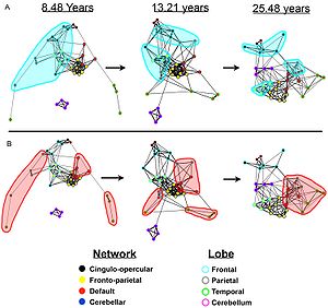
Graphs of the dynamic development of correlations between brain networks. (A) In children the regions are largely local and are organized by their physical location; the frontal regions are highlighted in light blue. (B) In adults the networks become highly correlated despite their physical distance; the default network is highlighted in light red. This result is now believed to have been confounded by artifactual processes attributable to the tendency of younger subjects to move more during image acquisition, which preferentially inflates estimates of connectivity between physically proximal regions (Power 2012, Satterthwaite 2012).

The default mode network is an interconnected and anatomically defined set of brain regions. The network can be separated into hubs and subsections:

Functional hubs: Information regarding the self

  • Posterior cingulate cortex (PCC) & precuneus: Combines bottom-up (not controlled) attention with information from memory and perception. The ventral (lower) part of PCC activates in all tasks which involve the DMN including those related to the self, related to others, remembering the past, thinking about the future, and processing concepts plus spatial navigation. The dorsal (upper) part of PCC involves involuntary awareness and arousal. The precuneus is involved in visual, sensorimotor, and attentional information.
  • Medial prefrontal cortex (mPFC): Decisions about self-processing such as personal information, autobiographical memories, future goals and events, and decision making regarding those personally very close such as family. The ventral (lower) part is involved in positive emotional information and internally valued reward.
  • Angular gyrus: Connects perception, attention, spatial cognition, and action and helps with parts of recall of episodic memories.

Dorsal medial subsystem: Thinking about others

Medial temporal subsystem: Autobiographical memory and future simulations

The default mode network is most commonly defined with resting state data by putting a seed in the posterior cingulate cortex and examining which other brain areas most correlate with this area. The DMN can also be defined by the areas deactivated during external directed tasks compared to rest. Independent component analysis (ICA) robustly finds the DMN for individuals and across groups, and has become the standard tool for mapping the default network.

It has been shown that the default mode network exhibits the highest overlap in its structural and functional connectivity, which suggests that the structural architecture of the brain may be built in such a way that this particular network is activated by default. Recent evidence from a population brain-imaging study of 10,000 UK Biobank participants further suggests that each DMN node can be decomposed into subregions with complementary structural and functional properties. It has been a widespread practice in DMN research to treat its constituent nodes to be functionally homogeneous, but the distinction between subnodes within each major DMN node has mostly been neglected. However, the close proximity of subnodes that propagate hippocampal space-time outputs and subnodes that describe the global network architecture may enable default functions, such as autobiographical recall or internally-orientated thinking.

In the infant's brain, there is limited evidence of the default network, but default network connectivity is more consistent in children aged 9–12 years, suggesting that the default network undergoes developmental change.

Functional connectivity analysis in monkeys shows a similar network of regions to the default mode network seen in humans. The PCC is also a key hub in monkeys; however, the mPFC is smaller and less well connected to other brain regions, largely because human's mPFC is much larger and well developed.

Diffusion MRI imaging shows white matter tracts connecting different areas of the DMN together. The structural connections found from diffusion MRI imaging and the functional correlations from resting state fMRI show the highest level of overlap and agreement within the DMN areas. This provides evidence that neurons in the DMN regions are linked to each other through large tracts of axons and this causes activity in these areas to be correlated with one another. From the point of view of effective connectivity, many studies have attempted to shed some light using dynamic causal modeling, with inconsistent results. However, directionality from the medial prefrontal cortex towards the posterior cingulate gyrus seems confirmed in multiple studies, and the inconsistent results appear to be related to small sample size analysis.

Function

The default mode network is thought to be involved in several different functions:

It is potentially the neurological basis for the self:

  • Autobiographical information: Memories of collection of events and facts about one's self
  • Self-reference: Referring to traits and descriptions of one's self
  • Emotion of one's self: Reflecting about one's own emotional state

Thinking about others:

  • Theory of mind: Thinking about the thoughts of others and what they might or might not know
  • Emotions of others: Understanding the emotions of other people and empathizing with their feelings
  • Moral reasoning: Determining a just and an unjust result of an action
  • Social evaluations: Good-bad attitude judgements about social concepts
  • Social categories: Reflecting on important social characteristics and status of a group
  • Social isolation: A perceived lack of social interaction

Remembering the past and thinking about the future:

  • Remembering the past: Recalling events that happened in the past
  • Imagining the future: Envisioning events that might happen in the future
  • Episodic memory: Detailed memory related to specific events in time
  • Story comprehension: Understanding and remembering a narrative
  • Replay: Consolidating recently acquired memory traces

The default mode network is active during passive rest and mind-wandering which usually involves thinking about others, thinking about one's self, remembering the past, and envisioning the future rather than the task being performed. Recent work, however, has challenged a specific mapping between the default mode network and mind-wandering, given that the system is important in maintaining detailed representations of task information during working memory encoding. Electrocorticography studies (which involve placing electrodes on the surface of a subject's cerebral cortex) have shown the default mode network becomes activated within a fraction of a second after participants finish a task. Additionally, during attention demanding tasks, sufficient deactivation of the default mode network at the time of memory encoding has been shown to result in more successful long-term memory consolidation.

Studies have shown that when people watch a movie, listen to a story, or read a story, their DMNs are highly correlated with each other. DMNs are not correlated if the stories are scrambled or are in a language the person does not understand, suggesting that the network is highly involved in the comprehension and the subsequent memory formation of that story. The DMN is shown to even be correlated if the same story is presented to different people in different languages, further suggesting the DMN is truly involved in the comprehension aspect of the story and not the auditory or language aspect.

The default mode network is deactivated during some external goal-oriented tasks such as visual attention or cognitive working memory tasks. However, with internal goal-oriented tasks, such as social working memory or autobiographical tasks, the DMN is positively activated with the task and correlates with other networks such as the network involved in executive function. Regions of the DMN are also activated during cognitively demanding tasks that require higher-order conceptual representations. The DMN shows higher activation when behavioral responses are stable, and this activation is independent of self-reported mind wandering. Meditation, which involves focusing the mind on breathing and relaxation, is associated with reduced activity of the DMN.

Tsoukalas (2017) links theory of mind to immobilization, and suggests that the default network is activated by the immobilization inherent in the testing procedure (the patient is strapped supine on a stretcher and inserted by a narrow tunnel into a massive metallic structure). This procedure creates a sense of entrapment and, not surprisingly, the most commonly reported side-effect is claustrophobia.

Gabrielle et al. (2019) suggests that the DMN is related to the perception of beauty, in which the network becomes activated in a generalized way to aesthetically moving domains such as artworks, landscapes, and architecture. This would explain a deep inner feeling of pleasure related to aesthetics, interconnected with the sense of personal identity, due to the network functions related to the self.

Clinical significance

The default mode network has been hypothesized to be relevant to disorders including Alzheimer's disease, autism, schizophrenia, major depressive disorder (MDD), chronic pain, post-traumatic stress disorder (PTSD) and others. In particular, the DMN has also been reported to show overlapping yet distinct neural activity patterns across different mental health conditions, such as when directly comparing attention deficit hyperactivity disorder (ADHD) and autism.

People with Alzheimer's disease show a reduction in glucose (energy use) within the areas of the default mode network. These reductions start off as slight decreases in patients with mild symptoms and continue to large reductions in those with severe symptoms. Surprisingly, disruptions in the DMN begin even before individuals show signs of Alzheimer's disease. Plots of the peptide amyloid-beta, which is thought to cause Alzheimer's disease, show the buildup of the peptide is within the DMN. This prompted Randy Buckner and colleagues to propose the high metabolic rate from continuous activation of DMN causes more amyloid-beta peptide to accumulate in these DMN areas. These amyloid-beta peptides disrupt the DMN and because the DMN is heavily involved in memory formation and retrieval, this disruption leads to the symptoms of Alzheimer's disease.

DMN is thought to be disrupted in individuals with autism spectrum disorder. These individuals are impaired in social interaction and communication which are tasks central to this network. Studies have shown worse connections between areas of the DMN in individuals with autism, especially between the mPFC (involved in thinking about the self and others) and the PCC (the central core of the DMN). The more severe the autism, the less connected these areas are to each other. It is not clear if this is a cause or a result of autism, or if a third factor is causing both (confounding).

Although it is not clear whether the DMN connectivity is increased or decreased in psychotic bipolar disorder and schizophrenia, several genes correlated with altered DMN connectivity are also risk genes for mood and psychosis disorders.

Rumination, one of the main symptoms of major depressive disorder, is associated with increased DMN connectivity and dominance over other networks during rest. Such DMN hyperconnectivity has been observed in first-episode depression and chronic pain. Altered DMN connectivity may change the way a person perceives events and their social and moral reasoning, thus increasing their susceptibility to depressive symptoms.

Lower connectivity between brain regions was found across the default network in people who have experienced long-term trauma, such as childhood abuse or neglect, and is associated with dysfunctional attachment patterns. Among people experiencing PTSD, lower activation was found in the posterior cingulate gyrus compared to controls, and severe PTSD was characterized by lower connectivity within the DMN.

Adults and children with ADHD show reduced anticorrelation between the DMN and other brain networks. The cause may be a lag in brain maturation. More generally, competing activation between the DMN and other networks during memory encoding may result in poor long-term memory consolidation, which is a symptom of not only ADHD but also depression, anxiety, autism, and schizophrenia.

Modulation

The default mode network (DMN) may be modulated by the following interventions and processes:

  • Acupuncture – Deactivation of the limbic brain areas and the DMN. It has been suggested that this is due to the pain response.
  • Antidepressants – Abnormalities in DMN connectivity are reduced following treatment with antidepressant medications in PTSD.
  • Attention Training Technique - Research shows that even a single session of Attention Training Technique changes functional connectivity of the DMN.
  • Deep brain stimulation – Alterations in brain activity with deep brain stimulation may be used to balance resting state networks.
  • Meditation – Structural changes in areas of the DMN such as the temporoparietal junction, posterior cingulate cortex, and precuneus have been found in meditation practitioners. There is reduced activation and reduced functional connectivity of the DMN in long-term practitioners. Various forms of nondirective meditation, including Transcendental Meditation and Acem Meditation, have been found to activate the DMN.
  • Physical Activity and Exercise – Physical Activity, and more likely Aerobic Training, may alter the DMN. In addition, sports experts are showing networks differences, notably of the DMN.
  • Psychedelic drugs – Reduced blood flow to the PCC and mPFC was observed under the administration of psilocybin. These two areas are considered to be the main nodes of the DMN. One study on the effects of LSD demonstrated that the drug desynchronizes brain activity within the DMN; the activity of the brain regions that constitute the DMN becomes less correlated.
  • Psychotherapy – In PTSD, the abnormalities in the default mode network normalize in individuals who respond to psychotherapy interventions.
  • Sleep deprivation – Functional connectivity between nodes of the DMN in their resting-state is usually strong, but sleep deprivation results in a decrease in connectivity within the DMN. Recent studies suggest a decrease in connectivity between the DMN and the task-positive network as a result of sleep loss.
  • Sleeping and resting wakefulness
    • Onset of sleep – Increase in connectivity between the DMN and the task-positive network.
    • REM sleep – Possible increase in connectivity between nodes of the DMN.
    • Resting wakefulness – Functional connectivity between nodes of the DMN is strong.
    • Stage N2 of NREM sleep – Decrease in connectivity between the posterior cingulate cortex and medial prefrontal cortex.
    • Stage N3 of NREM sleep – Further decrease in connectivity between the PCC and MPFC.

Criticism

Some have argued the brain areas in the default mode network only show up together because of the vascular coupling of large arteries and veins in the brain near these areas, not because these areas are actually functionally connected to each other. Support for this argument comes from studies that show changing in breathing alters oxygen levels in the blood which in turn affects DMN the most. These studies however do not explain why the DMN can also be identified using PET scans by measuring glucose metabolism which is independent of vascular coupling and in electrocorticography studies measuring electrical activity on the surface of the brain, and in MEG by measuring magnetic fields associated with electrophysiological brain activity that bypasses the hemodynamic response.

The idea of a "default network" is not universally accepted. In 2007 the concept of the default mode was criticized as not being useful for understanding brain function, on the grounds that a simpler hypothesis is that a resting brain actually does more processing than a brain doing certain "demanding" tasks, and that there is no special significance to the intrinsic activity of the resting brain.

Nomenclature

The default mode network has also been called the language network, semantic system, or limbic network. Even though the dichotomy is misleading, the term task-negative network is still sometimes used to contrast it against other more externally-oriented brain networks.

In 2019, Uddin et al. proposed that medial frontoparietal network (M-FPN) be used as a standard anatomical name for this network.

Temporoparietal junction

From Wikipedia, the free encyclopedia
 
Temporoparietal junction
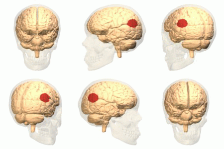
Side view of the human brain. TPJ is indicated by red circle.
 
Side view of the human brain. TPJ is indicated by red circle.

The temporoparietal junction (TPJ) is an area of the brain where the temporal and parietal lobes meet, at the posterior end of the lateral sulcus (Sylvian fissure). The TPJ incorporates information from the thalamus and the limbic system as well as from the visual, auditory, and somatosensory systems. The TPJ also integrates information from both the external environment as well as from within the body. The TPJ is responsible for collecting all of this information and then processing it.

This area is also known to play a crucial role in self–other distinctions processes and theory of mind (ToM). Furthermore, damage to the TPJ has been implicated in having adverse effects on an individual's ability to make moral decisions and has been known to produce out-of-body experiences (OBEs). Electromagnetic stimulation of the TPJ can also cause these effects. Apart from these diverse roles that the TPJ plays, it is also known for its involvement in a variety of widespread disorders including anxiety disorders, amnesia, Alzheimer's disease, autism spectrum disorder, and schizophrenia.

Anatomy and function

Animation. Both left and right temporoparietal junctions are shown in red.

The brain contains four main lobes: temporal lobe, parietal lobe, frontal lobe, and the occipital lobe. The temporoparietal junction lies in the region between the temporal and parietal lobes, near the lateral sulcus (Sylvian fissure). Specifically, it is composed of the inferior parietal lobule and the caudal parts of the superior temporal sulcus. There are two halves to the temporoparietal junction, with each component in their respective hemispheres of the brain. Each half of the TPJ pertains to various aspects of cognitive function. Often, however, the separate halves of the TPJ will work in coordination. The TPJ is mainly involved in information processing and perception.

Right temporoparietal junction

The right temporoparietal junction (rTPJ) is involved in the processing of information in terms of the ability of an individual to orient attention to new stimuli. Evidence from neuroimaging studies as well as lesion studies revealed that the rTPJ plays a pivotal role in analyzing signals from self-produced actions as well as with signals from the external environment. For example, an individual with lesions in their rTPJ would more than likely exhibit a sense of hemi-neglect, wherein they would no longer be able to pay attention to anything they observe on the left. So, if someone were to have a lesion in their rTPJ, then over time the awareness of the left limbs may fade without treatment. Visual signals provide the sensory information necessary for the brain to process spatial recognition of the world. When vision is limited, knowledge of existence begins to fade away since as far as the brain is concerned the object does not exist. Furthermore, the rTPJ plays a role in the way individuals observe and process information, thus impacting social interaction. Empathy and sympathy require an individual to simultaneously distinguish between different possible perspectives on the same situation. Imaging studies show that this ability depends upon the coordinated interaction of the rTPJ to identify and process the social cues presented to it. This rapid process allows for an individual to quickly react to situations.

Left temporoparietal junction

The left temporoparietal junction (lTPJ) contains both Wernicke's area and the angular gyrus, both prominent anatomical structures of the brain that are involved in language cognition, processing, and comprehension of both written and spoken language. Steven Pinker discusses this brain region, theorising that it underlies an amodal 'language of thought' or Mentalese. The lTPJ, in this account, takes in observations from external environments, such as conversations, makes connections in the brain regarding memories or incidents and then converts those thoughts and connections to written and spoken language. Pinker's full account of this is explained in The Language Instinct: How the Mind Creates Language. The lTPJ also plays an important role in reasoning of other's beliefs, intentions, and desires. Activation of the lTPJ was observed in patients processing mental states such as beliefs when an fMRI was used on patients as they were asked to make inferences regarding the mental states of others such as lying. This study was further supplemented by a study which identified that lesions to the left TPJ can impair cognitive processes specifically involved in the inference of someone else's belief, intention, or desire. Individuals with lesions in the lTPJ were no longer able to correctly identify when someone was lying or insinuating a false sense of belief or desire. The lTPJ is also involved in the processing of associating and remembering the names of individuals and objects.

Disorders

The dopaminergic-serotonergic system mediates our ability to distinguish and understand others’ beliefs as well as predict their behavior in light of that understanding. In certain disorders involving the dopaminergic-serotonergic system, this mentalizing process is disrupted and part or all of the process is impaired; this includes amnesia, Alzheimer's disease, and schizophrenia.

Amnesia

Amnesia is a deficit in memory caused by brain damage, disease, or physiological trauma. Amnesia is best understood via Henry Molaison, or patient H.M., who suffered from severe epilepsy and eventually had a temporal lobectomy. After surgery, his epilepsy improved but then he had anterograde amnesia, wherein long-term memory formation is inhibited. Short-term memory remained normal except that he could never remember anything that had happened after his surgery for very long. Based on general known roles of the TPJ, it is known that the TPJ is involved in the memory processing system of the body. Studies have also revealed that certain types of epileptic amnesia could be attributed to TPJ. fMRI studies indicated that there was lower activation of the rTPJ in patients with epileptic amnesia. Furthermore, it was noticed the autobiographical memories were affected in these patients. As such, the rTPJ along with the right cerebellum were identified as core components of autobiographical memory.

In terms of treatment, most forms of amnesia fix themselves without actually undergoing treatment. However, options such as cognitive therapy or occupational therapy have proved to help. Therapy will focus on various methods to improve a patient's memory and with repetition over time, a patient's memory as a whole will improve and eventually become close to normal.

Alzheimer's disease

Alzheimer's disease is the most common form of dementia and is also the sixth leading cause of death in the United States. This disease has no known cure and is a disease that worsens as it progresses and eventually leads to death. Reduced metabolism in the TPJ, along with the superior frontal sulcus, correlates with Alzheimer's patients’ inability to perceive themselves as others do (with a third-person point of view); the discrepancy between a patient's understanding of their own cognitive impairment and the actual extent of their cognitive impairment increases as metabolism in the TPJ decreases. Additionally, the TPJ contains the praxicon, a dictionary of representations of different human actions, which is necessary to distinguishing between actions of the self and other people. Because dementia (including Alzheimer's) patients with anosognosia are unable to distinguish between the normal actions of other people and their own diminished abilities, it is expected that damage to the TPJ is arresting this cognitive function.

Autism spectrum disorder

There may be a connection between the temporoparietal junction and how individuals with autism spectrum disorder's recognition of socially awkward situations may differ from neurotypicals’. Research reported in 2015 from an experiment in which participants, high-functioning adults with autism spectrum disorder (ASD) and neurotypical (NT) controls, were asked to watch socially awkward situations (a complete episode of the sitcom The Office) under an fMRI, which measured their brain activity. Several brain regions implicated in social perceptual and cognitive processes were of interest: "the dorsal, middle and ventral parts of medial prefrontal cortex (DMPFC, MMPFC and VMPFC), right and left temporo-parietal junctions (RTPJ and LTPJ), right superior temporal sulcus (RSTS) and temporal pole, and posterior medial cortices [posterior cingulate, precuneus (PC)]." In general, participants’ activity in several of those brain regions tracked the episode's socially awkward moments to similar extents—the results were evidence of a lack of group difference except in one region: their activity near the RTPJ, spanning into the posterior end of the RSTS, showed notable quantitative differences between the ASD and NT groups (with ASD group showing lower activity).

Research reported in 2016 on ASC-related structural or physiological differences found using neuroimaging noted that results are often inconsistent across the literature, which could be caused by a variety of variance sources. (Re-)analysis using a technique they developed to reduce one common external source of variance showed group differences in TPJ. However, although statistically significant, results did not display the discriminative power sufficient to classify diagnostic groups, instead yielding accuracy results close to random. They concluded that ASD is a highly heterogeneous syndrome/diagnostic category whose differences from NT controls are difficult to characterize globally using neuroimaging.

Schizophrenia

The decreased ability for schizophrenia patients to function in social situations has been related to a deficit within the theory of mind process. There have been relatively few studies that have examined the role of theory of mind in schizophrenia patients; the findings of these studies as they relate to the activation of the TPJ are varied. Some studies have found decreased activation of the TPJ in schizophrenia patients who were asked to make inferences about other peoples' social intentions based on cartoons; other studies, however, performed similar assessments of schizophrenia patients and found that the TPJ actually became hyperactive, compared to control individuals without schizophrenia, in the TPJ. This indicates that there is abnormal activation of the TPJ in these patients while performing tasks that involving understanding social intention of others, but the directionality of this abnormal activity is not clear, or possibly not universal throughout schizophrenia patients. It was found that the changes in activation in the TPJ were lateralized; they found that there was reduced activity in only the right TPJ and proposed that based on previous research about the different roles of the right and left TPJ the findings indicated that there was a more general deficit in the overall mentalizing process for these patients, but their ability to understand other individuals' basic social intentions through observing interaction is not impaired.

A study found that there was a connection between the auditory hallucinations in schizophrenia and the TPJ; the TPJ has been determined as a critical node in the auditory-verbal hallucination system. This study found that there was a significant decrease in the connectivity between the left TPJ and the right hemispheric homotope of the Broca's area, which is related to the production of language that is also characteristic of AVH events. This aspect of impairment seen in schizophrenia patients may also be related to the involvement of the TPJ with producing out of body experiences.

Anxiety disorders

A recent study showed reduced activity in the TPJ of adolescents compared to adults during an extinction task, suggesting a role for the TPJ in anxiety disorders.

Future of possible treatments

Vasopressin is a neuropeptide that is involved in regulating social behaviors, including social memory and recognition. One study examined the connection between vasopressin and cortical areas that are involved in processing social interactions including the TPJ. This study looked specifically at the brain regions that were active in men who were given vasopressin and tested based on familiarity related tasks. They found that the introduction of vasopressin caused a localized specific change in social recognition-related activity in the left TPJ/Brodmann area 39; the presence of vasopressin diminishes the heightened activity in the left TPJ that is present upon exposure to an unfamiliar social stimulus indicating that the presence of vasopressin leads individuals to associate an unfamiliar face with a familiar category more readily. While recognizing that this is the first study that has looked into this connection, the authors propose that it has potential to lead into further research about regulating the TPJ with vasopressin or a similar compound, which could allow pharmacologists to target this area of the brain and help with certain disorders including autism, social anxiety disorder. Perhaps such an approach could also be used to treat certain symptoms of schizophrenia or other disorders with know social cognitive impairments.

Current research

Current research involving the TPJ is extensive, ranging from issues of physiology to issues of mental state. A wide range of cognitive processes rely on the TPJ and as such gaining information about it is crucial. Research is conducted by studying the role TPJ plays both with and without lesions when stimulated, and with task-based fMRI. Research concerns various issues such as theory of mind, out-of-body experiences, temporal order judgments, morality, etc. This is a growing field due to the prevalence of ailments that involve TPJ as well as because of the importance of perception in everyday life.

Theory of mind

Theory of mind requires the collaboration of functionally related regions of the brain to form the distinction between self and other mental states and to create a comprehensive understanding of those mental states so that we may recognize, understand, and predict behavior. In general the theory of mind process is mediated by the dopaminergic-serotonergic system, which involves the TPJ as well as other associative regions necessary for mentalizing. Recent studies suggest that both the left TPJ, working in conjunction with the frontal cortex, and the right TPJ are involved in the representation of mental states; furthermore they suggest that the TPJ is particularly active in making the distinction between the mental states of self and others. A study in Nature Neuroscience from 2004 describes how the TPJ is involved in processing socially relevant cues including gaze direction and goal-directed action and also explains that results from the study show that lesions to this area of the brain result in an impaired ability to detect another persons belief. Moreover, studies have reported an increase in activity in the TPJ when patients are absorbing information through reading or images regarding other peoples' beliefs but not while observing information about physical control stimuli. Some studies, however, have shown that the TPJ, along with the cingulate cortex, is more specifically involved with attributing beliefs, but the process of mentalizing more generally is associated more with the medial prefrontal cortex. Another study in Current Biology from 2012 identifies the importance of the TPJ in both low-level, such as simple discrimination, and high-level, such as the ability to empathize, sociocognitive operations. In July 2011, a review from Neuropsychologia presented a model of the mentalizing network that established that mental states are first detected in the TPJ. The TPJ is composed of two discrete anatomical regions, the inferior parietal lobule (IPL) and the caudal parts of the superior temporal sulcus (pSTS), and both are active in the process of distinction between mental states of different individuals; thus, it is probable that this detection is the outcome of the combination and coordination of these two parts. Additionally, the right TPJ is involved in the ventral attention stream and contributes to the ability to focus attention on a particular stimuli or objective. It has also been observed that the interaction and communication between the dorsal and ventral streams involves the TPJ.

Out-of-body experiences

The TPJ is also a crucial structure for self-processing. Several neuro-imaging studies have shown an activation of the TPJ during different aspects of self-processing such as visuo-spatial perspective, self-other distinction, mental own body imagery, and vestibular and multi sensory integration. Damage in the TPJ has been linked to out-of-body experiences (OBEs), the feeling that one's self is located outside one's physical body.

An OBE is defined by the presence of three characteristics: disembodiment, the impression of seeing the world from a distant and elevated visuo-spatial perspective, and the impression of seeing one's own body from this elevated perspective. OBEs mostly occur to people with epilepsy or migraines, but approximately 10% of the healthy population also experience OBEs once or twice in a lifetime. They usually occur spontaneously and are of short duration, making OBEs hard to study. Here is an example of a patient describing what he or she experienced during an OBE:

“I was in bed and about to fall asleep when I had the distinct impression that 'I' was at the ceiling level looking down at my body in the bed. I was very startled and frightened; immediately [afterward] I felt that, I was consciously back in the bed again.”

It is suggested that OBEs are caused by multi-sensory disintegration in the TPJ disrupting different aspects of self-processing such as illusory reduplication, illusory self-location, and illusory perspective. The brain integrates different sensory inputs to create a representation of one's body and its location in its surrounding. Some inhibition of discrepant inputs is required to have coherency, but in some cases, those discrepant inputs are so strong and come from more than one sensory source that it leads to two different representations of one's own body. This multi-sensory disintegration at the TPJ leads to OBEs. An electromagnetic stimulation to the right TPJ of a patient with epilepsy induced an OBE. The author also states that these experiences are closely related to schizophrenia and phantom limb.

Temporal order judgement

Temporal order is the arrangement of events in time. By judging this, one can understand how we process things. Temporal order judgments require an individual to determine the relative timing between two spatially separate events. One study revealed that subjects had to determine the order of appearance of two objects as well as which object fit a certain property better. What was learned from this study was that when identifying the order or appearance, fMRI studies showed that there was bilateral activation of the TPJ. Meanwhile, when it comes to object characterization based on a property, it was noticed that there was only activation of the lTPJ. As such, it is evident that TPJ is involved in the “when” pathway of the brain.

Morality

Part of judging how virtuous an action was, whether someone is an ethical person or what one ought to do, morality usually (among other considerations) differentiates by actor intention. This applies to self-assessment as well as of others.

Connections made at the TPJ help an individual understand their emotions: the TPJ allows association of emotions with events or individuals, aiding in any related decision-making process. Studies also show a relation between theory of mind and moral judgment, which further implicates the rTPJ in morality cognition.

However, errors in this emotional processing can arise when patients have lesions in the TPJ or when the brain is electrically stimulated. Transcranial magnetic stimulation (TMS) to the rTPJ seems to affect the ability of an individual, when they make moral decisions, to consider actors’ mental states. Patients’ general ability to judge moral scenarios was not obviously impaired, but it did seem to specifically affect how much they integrated a protagonist's belief into the judgement—only affecting the judgement of a scenario in which the protagonist explicitly intends and so deliberately acts to cause significant harm but completely fails solely due to an incorrect belief (about tool/weapon used). TMS can be used to disrupt neural activity in the rTPJ just before a patient was to make a moral decision or during that decision-making process—constituting two different testing environments, but experimental results were unaffected.

Wernicke's area

From Wikipedia, the free encyclopedia

Wernicke's area (/ˈvɛərnɪkə/; German: [ˈvɛɐ̯nɪkə]), also called Wernicke's speech area, is one of the two parts of the cerebral cortex that are linked to speech, the other being Broca's area. It is involved in the comprehension of written and spoken language, in contrast to Broca's area, which is primarily involved in the production of language. It is traditionally thought to reside in Brodmann area 22, which is located in the superior temporal gyrus in the dominant cerebral hemisphere, which is the left hemisphere in about 95% of right-handed individuals and 70% of left-handed individuals.

Damage caused to Wernicke's area results in receptive, fluent aphasia. This means that the person with aphasia will be able to fluently connect words, but the phrases will lack meaning. This is unlike non-fluent aphasia, in which the person will use meaningful words, but in a non-fluent, telegraphic manner.

Emerging research on the developmental trajectory of Wernicke's area highlights its evolving role in language acquisition and processing during childhood. This includes studies on the maturation of neural pathways associated with this region, which contribute to the progressive complexity of language comprehension and production abilities in developing individuals.

Structure

Wernicke's area, more precisely defined, spans the posterior part of the superior temporal gyrus (STG) and extends to involve adjacent areas like the angular gyrus and parts of the parietal lobe reflecting a more intricate neuroanatomical network than previously understood. This area shows considerable variability in its exact location and extent among individuals, challenging the traditional view of a uniformly located language center.

However, there is an absence of consistent definitions as to the location. Some identify it with the unimodal auditory association in the superior temporal gyrus anterior to the primary auditory cortex (the anterior part of BA 22). This is the site most consistently implicated in auditory word recognition by functional brain imaging experiments. Others include also adjacent parts of the heteromodal cortex in BA 39 and BA40 in the parietal lobe. Despite the overwhelming notion of a specifically defined "Wernicke's Area", the most careful current research suggests that it is not a unified concept.

While previously thought to connect Wernicke's area and Broca's area, new research demonstrates that the arcuate fasciculus instead connects to posterior receptive areas with premotor/motor areas, and not to Broca's area. Consistent with the word recognition site identified in brain imaging, the uncinate fasciculus connects anterior superior temporal regions with Broca's area.

Function

Right homologous area

Research using Transcranial magnetic stimulation suggests that the area corresponding to the Wernicke's area in the non-dominant cerebral hemisphere has a role in processing and resolution of subordinate meanings of ambiguous words—such as "river" when given the ambiguous word "bank". In contrast, the Wernicke's area in the dominant hemisphere processes dominant word meanings ("teller" given "bank").

Modern views

Emerging research, including advanced neuroimaging studies, underscores a more distributed network of brain regions involved in language processing, challenging the traditional dichotomy of Wernicke's and Broca's areas. This includes findings on how Wernicke's area collaborates with other brain regions in processing both verbal and non-verbal auditory information, reshaping our understanding of its functional significance.

There are some suggestions that middle and inferior temporal gyri and basal temporal cortex reflect lexical processing ... there is consensus that the STG from rostral to caudal fields and the STS constitute the neural tissue in which many of the critical computations for speech recognition are executed ... aspects of Broca’s area (Brodmann areas 44 and 45) are also regularly implicated in speech processing. ... the range of areas implicated in speech processing go well beyond the classical language areas typically mentioned for speech; the vast majority of textbooks still state that this aspect of perception and language processing occurs in Wernicke’s area (the posterior third of the STG).

Support for a broad range of speech processing areas was furthered by a recent study carried out at the University of Rochester in which American Sign Language native speakers were subject to MRI while interpreting sentences that identified a relationship using either syntax (relationship is determined by the word order) or inflection (relationship is determined by physical motion of "moving hands through space or signing on one side of the body"). Distinct areas of the brain were activated with the frontal cortex (associated with ability to put information into sequences) being more active in the syntax condition and the temporal lobes (associated with dividing information into its constituent parts) being more active in the inflection condition. However, these areas are not mutually exclusive and show a large amount of overlap. These findings imply that while speech processing is a very complex process, the brain may be using fairly basic, preexisting computational methods.

Recent neuroimaging studies suggest that Wernicke's area plays a pivotal role in the nuanced aspects of language processing, including the interpretation of ambiguous words and the integration of linguistic context. Its functions extend beyond mere speech comprehension, encompassing complex cognitive tasks like semantic processing, discerning metaphorical language, and even contributing to the understanding of non-verbal elements in communication.

Comparative neurology studies have shed light on the evolutionary aspects of Wernicke's area. Similar regions have been identified in non-human primates, suggesting an evolutionary trajectory for language and communication skills. This comparative approach helps in understanding the fundamental neurobiological underpinnings of language and its evolutionary significance.

Clinical significance

Human brain with Wernicke's area highlighted in red

Aphasia

Wernicke's area is named after Carl Wernicke, a German neurologist and psychiatrist who, in 1874, hypothesized a link between the left posterior section of the superior temporal gyrus and the reflexive mimicking of words and their syllables that associated the sensory and motor images of spoken words. He did this on the basis of the location of brain injuries that caused aphasia. Receptive aphasia in which such abilities are preserved is also known as Wernicke's aphasia. In this condition there is a major impairment of language comprehension, while speech retains a natural-sounding rhythm and a relatively normal syntax. Language as a result is largely meaningless (a condition sometimes called fluent or jargon aphasia).

Wernicke's area receives information from the auditory cortex, and functions to assign word meanings. This is why damage to this area results in meaningless speech, often with paraphasic errors and newly created words or expressions. Paraphasia can involve substituting one word for another, known as semantic paraphasia, or substituting one sound or syllable for another, defined as phonemic paraphasia. This speech is often referred to as "word salad", as speech sounds fluent but does not have sensible meaning. Normal sentence structure and prosody are preserved, with normal intonation, inflection, rate, and rhythm. This differs from Broca's aphasia, which is characterized by nonfluency. Patients are typically not aware that their speech is impaired in this way, as they have altered comprehension of their speech. Written language, reading, and repetition are affected as well.

Damage to the posterior temporal lobe of the dominant hemisphere is the cause of Wernicke's aphasia. The etiology of this damage can vary greatly, with the most common cause being a cerebrovascular event such as an ischemic stroke. Ischemic stroke is the result of a thrombus occluding a blood vessel, restricting blood supply to a particular area of the brain. Other causes of focal damage potentially leading to Wernicke's aphasia include head trauma, infections affecting the central nervous system, neurodegenerative disease, and neoplasms. A cerebrovascular event is more likely the cause in an acute-onset presentation of aphasia, whereas a degenerative disease should be suspected in aphasia with gradual progression over time. Imaging is often useful in identifying a lesion, with most common initial imaging consisting of computed tomography (CT) scan or magnetic resonance imaging (MRI). Electroencephalography (EEG) can also be useful in patients with transient aphasia, where findings may be due to seizures, although this is a less common cause.

Diagnosis of aphasia, as well as characterization of type of aphasia, is done with language testing by the provider. Testing should evaluate fluency of speech, comprehension, repetition, ability to name objects, and writing skills.[20] Fluency is assessed by observing the patient's spontaneous speech. Abnormalities in fluency would include shortened phrases, decreased number of words per minute, increased effort with speech, and agrammatism.[19] Patients with Wernicke's aphasia should have fluent speech, so abnormalities in fluency may indicate a different type of aphasia. Comprehension is assessed by giving the patient commands to follow, beginning with simple commands and progressing to more complex commands. Repetition is evaluated by having the patient repeat phrases, progressing from simple to more complex phrases. Both comprehension and repetition would be abnormal in Wernicke's aphasia. Content should also be assessed, by listening to a patient's spontaneous or instructed speech. Content abnormalities include paraphasic errors and neologisms, both indicative of a diagnosis of Wernicke's aphasia. Neologisms are novel words that may resemble existing words. Patients with severe Wernicke's aphasia may also produce strings of such neologisms with a few connecting words, known as jargon. Errors in the selection of phonemes of patients with Wernicke's aphasia include addition, omission, or change in position. Another symptom of Wernicke's aphasia is use of semantic paraphasias or "empty speech" which is the use of generic terms like "stuff" or "things" to stand in for the specific words that the patient cannot think of. Some Wernicke's aphasia patients also talk around missing words, which is called "circumlocution". Patients with Wernicke's aphasia can tend to run on when they talk, due to circumlocution combined with deficient self-monitoring. This overabundance of words or press of speech can be described as logorrhea. If symptoms are present, a full neurologic exam should also be done, which will help differentiate aphasia from other neurologic diagnoses potentially causing altered mental status with abnormal speech and comprehension.

As an example, a patient with Wernicke's aphasia was asked what brought him to the hospital. His response was,

Is this some of the work that we work as we did before? ... All right ... From when wine [why] I'm here. What's wrong with me because I ... was myself until the taenz took something about the time between me and my regular time in that time and they took the time in that time here and that's when the time took around here and saw me around in it's started with me no time and I bekan [began] work of nothing else that's the way the doctor find me that way...

In diagnosing Wernicke's aphasia, clinicians employ a range of assessments focusing on speech fluency, comprehension, and repetition abilities. Treatment strategies extend beyond traditional speech therapy, incorporating multimodal approaches like music therapy and assistive communication technologies. Understanding the variability in the clinical presentation of aphasia is critical for tailoring individualized therapeutic interventions.

While neuroimaging and lesion evidence generally support the idea that malfunction of or damage to Wernicke's area is common in people with receptive aphasia, this is not always so. Some people may use the right hemisphere for language, and isolated damage of Wernicke's area cortex (sparing white matter and other areas) may not cause severe receptive aphasia. Even when patients with Wernicke's area lesions have comprehension deficits, these are usually not restricted to language processing alone. For example, one study found that patients with posterior lesions also had trouble understanding nonverbal sounds like animal and machine noises. In fact, for Wernicke's area, the impairments in nonverbal sounds were statistically stronger than for verbal sounds.

Brønsted–Lowry acid–base theory

From Wikipedia, the free encyclopedia https://en.wikipedia.org/wiki/Br%C3%B8nsted%E2%80%93Lowry_acid%E2%80%93base_theory The B...