Search This Blog

Monday, February 2, 2015

Evolution of the horse


From Wikipedia, the free encyclopedia


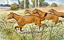
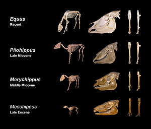
This image shows a representative sequence, but should not be construed to represent a "straight-line" evolution of the horse. Reconstruction, left forefoot skeleton (third digit emphasized yellow) and longitudinal section of molars of selected prehistoric horses

Skeletal evolution

The evolution of the horse occurred over a period of 50 million years, transforming the small, dog-sized,[1] forest-dwelling Eohippus into the modern horse. Paleozoologists have been able to piece together a more complete outline of the modern horse's evolutionary lineage than that of any other animal.

The horse belongs to the order Perissodactyla (odd-toed ungulates), the members of which all share hooved feet and an odd number of toes on each foot, as well as mobile upper lips and a similar tooth structure. This means that horses share a common ancestry with tapirs and rhinoceroses. The perissodactyls arose in the late Paleocene, less than 10 million years after the Cretaceous–Paleogene extinction event. This group of animals appears to have been originally specialized for life in tropical forests, but whereas tapirs and, to some extent, rhinoceroses, retained their jungle specializations, modern horses are adapted to life on drier land, in the much-harsher climatic conditions of the steppes. Other species of Equus are adapted to a variety of intermediate conditions.

The early ancestors of the modern horse walked on several spread-out toes, an accommodation to life spent walking on the soft, moist grounds of primeval forests. As grass species began to appear and flourish, the equids' diets shifted from foliage to grasses, leading to larger and more durable teeth. At the same time, as the steppes began to appear, the horse's predecessors needed to be capable of greater speeds to outrun predators. This was attained through the lengthening of limbs and the lifting of some toes from the ground in such a way that the weight of the body was gradually placed on one of the longest toes, the third.

History of research
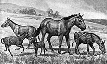

Extinct equids restored to scale. Left to right: Mesohippus, Neohipparion, Eohippus, Equus scotti and Hypohippus.

Indigenous modern horses died out in the New World at the end of the Pleistocene, about 12,000 years ago, and thus were absent until the Spanish brought domestic horses from Europe, beginning in 1493. Escaped horses quickly established large wild herds. In the 1760s, the early naturalist Buffon suggested this was an indication of inferiority of the New World fauna, but later reconsidered this idea.[2] William Clark's 1807 expedition to Big Bone Lick found "leg and foot bones of the Horses", which were included with other fossils sent to Thomas Jefferson and evaluated by the anatomist Caspar Wistar, but neither commented on the significance of this find.[3]

The first equid fossil was found in the gypsum quarries in Montmartre, Paris in the 1820s. The tooth was sent to the Paris Conservatory, where it was identified by Georges Cuvier, who identified it as a browsing equine related to the tapir.[4] His sketch of the entire animal matched later skeletons found at the site.[5]

During the Beagle survey expedition, the young naturalist Charles Darwin had remarkable success with fossil hunting in Patagonia. On 10 October 1833 at Santa Fe, Argentina, he was "filled with astonishment" when he found a horse's tooth in the same stratum as fossil giant armadillos, and wondered if it might have been washed down from a later layer, but concluded this was "not very probable".[6] After the expedition returned in 1836, the anatomist Richard Owen confirmed the tooth was from an extinct species, which he subsequently named Equus curvidens, and remarked, "This evidence of the former existence of a genus, which, as regards South America, had become extinct, and has a second time been introduced into that Continent, is not one of the least interesting fruits of Mr. Darwin's palæontological discoveries."[3][7]

In 1848, a study On the fossil horses of America by Joseph Leidy systematically examined Pleistocene horse fossils from various collections, including that of the Academy of Natural Sciences and concluded at least two ancient horse species had existed in North America: Equus curvidens and another, which he named Equus americanus. A decade later, however, he found the latter name had already been taken and renamed it Equus complicatus.[2] In the same year, he visited Europe and was introduced by Owen to Darwin.[8]
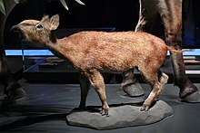
Restoration of Eurohippus parvulus, a mid- to late Eocene equid of Europe (Museum für Naturkunde, Berlin)

The original sequence of species believed to have evolved into the horse was based on fossils discovered in North America in the 1870s by paleontologist Othniel Charles Marsh. The sequence, from Eohippus to the modern horse (Equus), was popularized by Thomas Huxley and became one of the most widely known examples of a clear evolutionary progression. The horse's evolutionary lineage became a common feature of biology textbooks, and the sequence of transitional fossils was assembled by the American Museum of Natural History into an exhibit that emphasized the gradual, "straight-line" evolution of the horse.

Since then, as the number of equid fossils has increased, the actual evolutionary progression from Eohippus to Equus has been discovered to be much more complex and multibranched than was initially supposed. The straight, direct progression from the former to the latter has been replaced by a more elaborate model with numerous branches in different directions, of which the modern horse is only one of many. George Gaylord Simpson in 1951[9] first recognized the modern horse was not the "goal" of the entire lineage of equids,[10] it is simply the only genus of the many horse lineages to survive.

Detailed fossil information on the rate and distribution of new equid species has also revealed that the progression between species was not as smooth and consistent as was once believed. Although some transitions, such as that of Dinohippus to Equus, were indeed gradual progressions, a number of others, such as that of Epihippus to Mesohippus, were relatively abrupt in geologic time, taking place over only a few million years. Both anagenesis (gradual change in an entire population's gene frequency) and cladogenesis (a population "splitting" into two distinct evolutionary branches) occurred, and many species coexisted with "ancestor" species at various times. The change in equids' traits was also not always a "straight line" from Eohippus to Equus: some traits reversed themselves at various points in the evolution of new equid species, such as size and the presence of facial fossae, and only in retrospect can certain evolutionary trends be recognized.[11]

Before Odd-toed ungulates

Phenacodontidae


Restoration of Phenacodus

Phenacodontidae is the earliest family in the order condylarthra which is believed to be the ancestor to all odd-toed ungulates.[citation needed] It contains the genera Almogaver, Copecion, Ectocion, Eodesmatodon, Meniscotherium, Ordathspidotherium, Phenacodus and Pleuraspidotherium. The family lived from the Early Paleocene to the Middle Eocene in Europe and were about the size of a sheep, with tails making slightly less than half of the length of their bodies and unlike their ancestors, good running skills for eluding predators.[citation needed]

Eocene and Oligocene: early equids

Eohippus

Eohippus appeared in the Ypresian (early Eocene), about 52 mya (million years ago). It was an animal approximately the size of a fox (250–450 mm in height), with a relatively short head and neck and a springy, arched back. It had 44 low-crowned teeth, in the typical arrangement of an omnivorous, browsing mammal: three incisors, one canine, four premolars, and three molars on each side of the jaw. Its molars were uneven, dull, and bumpy, and used primarily for grinding foliage. The cusps of the molars were slightly connected in low crests. Eohippus browsed on soft foliage and fruit, probably scampering between thickets in the mode of a modern muntjac. It had a small brain, and possessed especially small frontal lobes.[11]

Eohippus, with left forefoot (third metacarpal colored) and tooth (a enamel; b dentin; c cement) detailed

Its limbs were decently long relative to its body, already showing the beginnings of adaptations for running. However, all of the major leg bones were unfused, leaving the legs flexible and rotatable. Its wrist and hock joints were low to the ground. The forelimbs had developed five toes, of which four were equipped with small proto-hooves; the large fifth "toe-thumb" was off the ground. The hind limbs had small hooves on three out of the five toes, while the vestigial first and fifth toes did not touch the ground. Its feet were padded, much like a dog's, but with the small hooves in place of claws.[12]

For a span of about 20 million years, Eohippus thrived with few significant evolutionary changes.[11] The most significant change was in the teeth, which began to adapt to its changing diet, as these early Equidae shifted from a mixed diet of fruits and foliage to one focused increasingly on browsing foods. During the Eocene, an Eohippus species (most likely Eohippus angustidens) branched out into various new types of Equidae. Thousands of complete, fossilized skeletons of these animals have been found in the Eocene layers of North American strata, mainly in the Wind River basin in Wyoming. Similar fossils have also been discovered in Europe, such as Propalaeotherium (which is not considered ancestral to the modern horse).[13]

Orohippus

Approximately 50 million years ago, in the early-to-middle Eocene, Eohippus smoothly transitioned into Orohippus through a gradual series of changes.[13] Although its name means "mountain horse", Orohippus was not a true horse and did not live in the mountains. It resembled Eohippus in size, but had a slimmer body, an elongated head, slimmer forelimbs, and longer hind legs, all of which are characteristics of a good jumper. Although Orohippus was still pad-footed, the vestigial outer toes of Eohippus were not present in the Orohippus; there were four toes on each fore leg, and three on each hind leg.

The most dramatic change between Eohippus and Orohippus was in the teeth: the first of the premolar teeth were dwarfed, the last premolar shifted in shape and function into a molar, and the crests on the teeth became more pronounced. Both of these factors gave the teeth of Orohippus greater grinding ability, suggesting Orohippus ate tougher plant material.

Epihippus

In the mid-Eocene, about 47 million years ago, Epihippus, a genus which continued the evolutionary trend of increasingly efficient grinding teeth, evolved from Orohippus. Epihippus had five grinding, low-crowned cheek teeth with well-formed crests. A late species of Epihippus, sometimes referred to as Duchesnehippus intermedius, had teeth similar to Oligocene equids, although slightly less developed. Whether Duchesnehippus was a subgenus of Epihippus or a distinct genus is disputed.[citation needed] Epihippus was only 2 feet tall.[citation needed]

Mesohippus

In the late Eocene and the early stages of the Oligocene epoch (32–24 mya), the climate of North America became drier, and the earliest grasses began to evolve. The forests were yielding to flatlands,[citation needed] home to grasses and various kinds of brush. In a few areas, these plains were covered in sand,[citation needed] creating the type of environment resembling the present-day prairies.

Restoration of Mesohippus

In response to the changing environment, the then-living species of Equidae also began to change. In the late Eocene, they began developing tougher teeth and becoming slightly larger and leggier, allowing for faster running speeds in open areas, and thus for evading predators in nonwooded areas[citation needed]. About 40 mya, Mesohippus ("middle horse") suddenly developed in response to strong new selective pressures to adapt, beginning with the species Mesohippus celer and soon followed by Mesohippus westoni.

In the early Oligocene, Mesohippus was one of the more widespread mammals in North America. It walked on three toes on each of its front and hind feet (the first and fifth toes remained, but were small and not used in walking). The third toe was stronger than the outer ones, and thus more weighted; the fourth front toe was diminished to a vestigial nub. Judging by its longer and slimmer limbs, Mesohippus was an agile animal.

Mesohippus was slightly larger than Epihippus, about 610 mm (24 in) at the shoulder. Its back was less arched, and its face, snout, and neck were somewhat longer. It had significantly larger cerebral hemispheres, and had a small, shallow depression on its skull called a fossa, which in modern horses is quite detailed. The fossa serves as a useful marker for identifying an equine fossil's species. Mesohippus had six grinding "cheek teeth", with a single premolar in front—a trait all descendant Equidae would retain. Mesohippus also had the sharp tooth crests of Epihippus, improving its ability to grind down tough vegetation.

Miohippus

Around 36 million years ago, soon after the development of Mesohippus, Miohippus ("lesser horse") emerged, the earliest species being Miohippus assiniboiensis. As with Mesohippus, the appearance of Miohippus was relatively abrupt, though a few transitional fossils linking the two genera have been found. Mesohippus was once believed to have anagenetically evolved into Miohippus by a gradual series of progressions, but new evidence has shown its evolution was cladogenetic: a Miohippus population split off from the main Mesohippus genus, coexisted with Mesohippus for around four million years, and then over time came to replace Mesohippus.[14]

Miohippus was significantly larger than its predecessors, and its ankle joints had subtly changed. Its facial fossa was larger and deeper, and it also began to show a variable extra crest in its upper cheek teeth, a trait that became a characteristic feature of equine teeth.

Miohippus ushered in a major new period of diversification in Equidae.[15] While Mesohippus died out in the mid-Oligocene, Miohippus continued to thrive, and in the early Miocene (24–5.3 mya), it began to rapidly diversify and speciate. It branched out into two major groups, one of which adjusted to the life in forests once again, while the other remained suited to life on the prairies.[citation needed]

Miocene and Pliocene: true equines

Kalobatippus


The forest-suited form was Kalobatippus (or Miohippus intermedius, depending on whether it was a new genus or species), whose second and fourth front toes were long, well-suited travel on the soft forest floors. Kalobatippus probably gave rise to Anchitherium, which travelled to Asia via the Bering Strait land bridge, and from there to Europe.[16] In both North America and Eurasia, larger-bodied genera evolved from Anchitherium: Sinohippus in Eurasia and Hypohippus and Megahippus in North America.[17] Hypohippus became extinct by the late Miocene.[18]

Parahippus

The Miohippus population that remained on the steppes is believed to be ancestral to Parahippus, a North American animal about the size of a small pony, with a prolonged skull and a facial structure resembling the horses of today. Its third toe was stronger and larger, and carried the main weight of the body. Its four premolars resembled the molar teeth and the first were small and almost nonexistent. The incisor teeth of Parahippus, like those of its predecessors, had a crown as humans do; however, the top incisors had a trace of a shallow crease marking the beginning of the core/cup.

Merychippus


Merychippus, an effective grazer and runner

In the middle of the Miocene epoch, the grazer Merychippus flourished. It had wider molars than its predecessors, which are believed to have been used for crunching the hard grasses of the steppes. The hind legs, which were relatively short, had side toes equipped with small hooves, but they probably only touched the ground when running.[15] Merychippus radiated into at least 19 additional grassland species.

Hipparion


Protohippus simus

Three lineages within Equidae are believed to be descended from the numerous varieties of Merychippus: Hipparion, Protohippus and Pliohippus. The most different from Merychippus was Hipparion, mainly in the structure of tooth enamel: in comparison with other Equidae, the inside, or tongue side, had a completely isolated parapet. A complete and well-preserved skeleton of the North American Hipparion shows an animal the size of a small pony. They were very slim, rather like antelopes, and were adapted to life on dry prairies. On its slim legs, Hipparion had three toes equipped with small hooves, but the side toes did not touch the ground.

In North America, Hipparion and its relatives (Cormohipparion, Nannippus, Neohipparion, and Pseudhipparion), proliferated into many kinds of equids, at least one of which managed to migrate to Asia and Europe during the Miocene epoch.[19] (European Hipparion differs from American Hipparion in its smaller body size – the best-known discovery of these fossils was near Athens.)

Pliohippus


Pliohippus pernix

Pliohippus arose from Callippus in the middle Miocene, around 12 mya. It was very similar in appearance to Equus, though it had two long extra toes on both sides of the hoof, externally barely visible as callused stubs. The long and slim limbs of Pliohippus reveal a quick-footed steppe animal.
Until recently, Pliohippus was believed to be the ancestor of present-day horses because of its many anatomical similarities. However, though Pliohippus was clearly a close relative of Equus, its skull had deep facial fossae, whereas Equus had no fossae at all. Additionally, its teeth were strongly curved, unlike the very straight teeth of modern horses. Consequently, it is unlikely to be the ancestor of the modern horse; instead, it is a likely candidate for the ancestor of Astrohippus.[20]

Dinohippus

Dinohippus was the most common species of Equidae in North America during the late Pliocene. It was originally thought to be monodactyl, but a 1981 fossil find in Nebraska shows some were tridactyl.

Plesippus


Mounted skeleton of Hagerman horse (Equus simplicidens)

Plesippus is often considered an intermediate stage between Dinohippus and the extant genus, Equus.
The famous fossils found near Hagerman, Idaho were originally thought to be a part of the genus Plesippus. Hagerman Fossil Beds (Idaho) is a Pliocene site, dating to about 3.5 mya. The fossilized remains were originally called Plesippus shoshonensis, but further study by paleontologists determined the fossils represented the oldest remains of the genus Equus.[21] Their estimated average weight was 425 kg, roughly the size of an Arabian horse.

At the end of the Pliocene, the climate in North America began to cool significantly and most of the animals were forced to move south. One population of Plesippus moved across the Bering land bridge into Eurasia around 2.5 mya.[22]

Modern horses

Equus
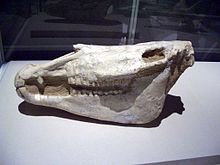
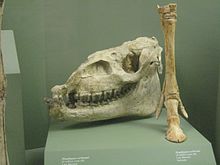
Skull of a giant extinct horse, Equus eisenmannae

The genus Equus, which includes all extant equines, is believed to have evolved from Dinohippus, via the intermediate form Plesippus. One of the oldest species is Equus simplicidens, described as zebra-like with a donkey-shaped head. The oldest material to date is ~3.5 million years old from Idaho, USA. The genus appears to have spread quickly into the Old World, with the similarly aged Equus livenzovensis documented from western Europe and Russia.[23]

Molecular phylogenies indicate the most recent common ancestor of all modern equids (members of the genus Equus) lived ~5.6 (3.9–7.8) mya. Direct paleogenomic sequencing of a 700,000 year-old middle Pleistocene horse metapodial bone from Canada implies a more recent 4.07 Myr before present date for the most recent common ancestor (MRCA) within the range of 4.0 to 4.5 Myr BP.[24] The oldest divergencies are the Asian hemiones (subgenus E. (Asinus)), including the kulan, onager, and kiang), followed by the African zebras (subgenera E. (Dolichohippus), and E. (Hippotigris)). All other modern forms including the domesticated horse (and many fossil Pliocene and Pleistocene forms) belong to the subgenus E. (Equus) which diverged ~4.8 (3.2–6.5) million years ago.[25]

Pleistocene horse fossils have been assigned to a multitude of species, with over 50 species of equines described from the Pleistocene of North America alone, although the taxonomic validity of most of these has been called into question.[26] Recent genetic work on fossils has found evidence for only three genetically divergent equid lineages in Pleistocene North and South America.[25] These results suggest all North American fossils of caballine-type horses (which also include the domesticated horse and Przewalski's horse of Europe and Asia), as well as South American fossils traditionally placed in the subgenus E. (Amerhippus)[27] belong to the same species: E. ferus. Remains attributed to a variety of species and lumped as New World stilt-legged horses (including E. francisci, E. tau, E. quinni and potentially North American Pleistocene fossils previously attributed to E. cf. hemiones, and E. (Asinus) cf. kiang) probably all belong to a second species endemic to North America, which despite a superficial resemblance to species in the subgenus E. (Asinus) (and hence occasionally referred to as North American ass) is closely related to E. ferus.[25] Surprisingly, the third species, endemic to South America, and traditionally referred to as Hippidion, originally believed to be descended from Pliohippus, was shown to be a third species in the genus Equus, closely related to the New World stilt-legged horse.[25] The temporal and regional variation in body size and morphological features within each lineage indicates extraordinary intraspecific plasticity.
Such environment-driven adaptative changes would explain why the taxonomic diversity of Pleistocene equids has been overestimated on morphoanatomical grounds.[27]

According to these results, it appears the genus Equus evolved from a Dinohippus-like ancestor ~4–7 mya. It rapidly spread into the Old World and there diversified into the various species of asses and zebras. A North American lineage of the subgenus E. (Equus) evolved into the New World stilt-legged horse (NWSLH). Subsequently, populations of this species entered South America as part of the Great American Interchange shortly after the formation of the Isthmus of Panama, and evolved into the form currently referred to as Hippidion ~2.5 million years ago. Hippidion is thus unrelated to the morphologically similar Pliohippus, which presumably went extinct during the Miocene. Both the NWSLH and Hippidium show adaptations to dry, barren ground, whereas the shortened legs of Hippidion may have been a response to sloped terrain.[27] In contrast, the geographic origin of the closely related modern E. ferus is not resolved. However, genetic results on extant and fossil material of Pleistocene age indicate two clades, potentially subspecies, one of which had a holarctic distribution spanning from Europe through Asia and across North America and would become the founding stock of the modern domesticated horse.[28][29] The other population appears to have been restricted to North America. One or more North American populations of E. ferus entered South America ~1.0–1.5 million years ago, leading to the forms currently known as E. (Amerhippus), which represent an extinct geographic variant or race of E. ferus, however.

Genome sequencing

In June 2013, a group of researchers announced that they had sequenced the DNA of a 560–780 thousand year old horse, using material extracted from a leg bone found buried in permafrost in Canada's Yukon territory.[30] Prior to this publication, the oldest nuclear genome that had been successfully sequenced was dated at 110–130 thousand years ago. For comparison, the researchers also sequenced the genomes of a 43,000 year old Pleistocene horse, a Przewalski's horse, five modern horse breeds, and a donkey.[31] Analysis of differences between these genomes indicated that the last common ancestor of modern horses, donkeys, and zebras existed 4 to 4.5 million years ago.[30] The results also indicated that Przewalski's horse diverged from other modern types of horse about 43,000 years ago, and had never in its evolutionary history been domesticated.[24]

Pleistocene extinctions

Digs in western Canada have unearthed clear evidence horses existed in North America until about 12,000 years ago.[32] However, all Equidae in North America ultimately became extinct. The causes of this extinction (simultaneous with the extinctions of a variety of other American megafauna) have been a matter of debate. Given the suddenness of the event and because these mammals had been flourishing for millions of years previously, something quite unusual must have happened. The first main hypothesis attributes extinction to climate change. For example, in Alaska, beginning approximately 12,500 years ago, the grasses characteristic of a steppe ecosystem gave way to shrub tundra, which was covered with unpalatable plants.[33][34] The other hypothesis suggests extinction was linked to overexploitation of naive prey by newly arrived humans. The extinctions were roughly simultaneous with the end of the most recent glacial advance and the appearance of the big game-hunting Clovis culture.[35][36] Several studies have indicated humans probably arrived in Alaska at the same time or shortly before the local extinction of horses.[36][37][38] Additionally, it has been proposed that the steppe-tundra vegetation transition in Beringia may have been a consequence, rather than a cause, of the extinction of megafaunal grazers.[39]

In Eurasia, horse fossils began occurring frequently again in archaeological sites in Kazakhstan and the southern Ukraine about 6,000 years ago.[28] From then on, domesticated horses, as well as the knowledge of capturing, taming, and rearing horses, probably spread relatively quickly, with wild mares from several wild populations being incorporated en route.[29][40]

Return to the Americas

Horses only returned to the Americas with Christopher Columbus in 1493. These were Iberian horses first brought to Hispaniola and later to Panama, Mexico, Brazil, Peru, Argentina, and, in 1538, Florida.[41] The first horses to return to the main continent were 16 specifically identified horses brought by Hernán Cortés. Subsequent explorers, such as Coronado and De Soto brought ever-larger numbers, some from Spain and others from breeding establishments set up by the Spanish in the Caribbean. Later, as Spanish missions were founded on the mainland, horses would eventually be lost or stolen, and proliferated into large herds of feral horses that became known as mustangs.[citation needed]

The indigenous peoples of the Americas did not have a specific word for horses, and came to refer to them in various languages as a type of dog or deer (in one case, "elk-dog", in other cases "big dog" or "seven dogs", referring to the weight each animal could pull).
Details

Toes 

The ancestors of the horse came to walk only on the end of the third toe and both side toes. Skeletal remnants show obvious wear on the back of both sides of metacarpal and metatarsal bones, commonly called the "splint bones". They are the remnants of the second and the fourth toe. Modern horses retain the splint bones; they are often believed to be useless attachments, but they in fact play an important role in supporting the carpal joints (front knees) and even the tarsal joints (hocks).[citation needed]

Teeth

Throughout the phylogenetic development, the teeth of the horse underwent significant changes. The type of the original omnivorous teeth with short, "bumpy" molars, with which the prime members of the evolutionary line distinguished themselves, gradually changed into the teeth common to herbivorous mammals. They became long (as much as 100 mm), roughly cubical molars equipped with flat grinding surfaces. In conjunction with the teeth, during the horse's evolution, the elongation of the facial part of the skull is apparent, and can also be observed in the backward-set eyeholes. In addition, the relatively short neck of the equine ancestors became longer, with equal elongation of the legs. Finally, the size of the body grew as well.[citation needed]

Coat color

The ancestral coat color of E. ferus was probably a uniform dun, consistent with modern populations of Przewalski's horses. Pre-domestication variants including black and spotted have been inferred from cave wall paintings and confirmed by genomic analysis.[42] Domestication may have also led to more varieties of coat colors.[43]

Molecular self-assembly


From Wikipedia, the free encyclopedia


An example of a molecular self-assembly through hydrogen bonds.[1]

Molecular self-assembly is the process by which molecules adopt a defined arrangement without guidance or management from an outside source. There are two types of self-assembly. These are intramolecular self-assembly and intermolecular self-assembly. Commonly, the term molecular self-assembly refers to intermolecular self-assembly, while the intramolecular analog is more commonly called folding.

Supramolecular systems

Molecular self-assembly is a key concept in supramolecular chemistry.[2][3][4] This is because assembly of molecules in such systems is directed through noncovalent interactions (e.g., hydrogen bonding, metal coordination, hydrophobic forces, van der Waals forces, π-π interactions, and/or electrostatic) as well as electromagnetic interactions. Common examples include the formation of micelles, vesicles, liquid crystal phases, and Langmuir monolayers by surfactant molecules.[5] Further examples of supramolecular assemblies demonstrate that a variety of different shapes and sizes can be obtained using molecular self-assembly.[6]

Molecular self-assembly allows the construction of challenging molecular topologies. One example is Borromean rings, interlocking rings wherein removal of one ring unlocks each of the other rings. DNA has been used to prepare a molecular analog of Borromean rings.[7] More recently, a similar structure has been prepared using non-biological building blocks.[8]

Biological systems

Molecular self-assembly underlies the construction of biologic macromolecular assemblies in living organisms, and so is crucial to the function of cells. It is exhibited in the self-assembly of lipids to form the membrane, the formation of double helical DNA through hydrogen bonding of the individual strands, and the assembly of proteins to form quaternary structures. Molecular self-assembly of incorrectly folded proteins into insoluble amyloid fibers is responsible for infectious prion-related neurodegenerative diseases. Molecular self-assembly of nanoscale structures plays a role in the growth of the remarkable β-keratin lamellae/setae/spatulae structures used to give geckos the ability to climb walls and adhere to ceilings and rock overhangs.[9][10]

Nanotechnology


The DNA structure at left (schematic shown) will self-assemble into the structure visualized by atomic force microscopy at right. Image from Strong.[11]

Molecular self-assembly is an important aspect of bottom-up approaches to nanotechnology. Using molecular self-assembly the final (desired) structure is programmed in the shape and functional groups of the molecules. Self-assembly is referred to as a 'bottom-up' manufacturing technique in contrast to a 'top-down' technique such as lithography where the desired final structure is carved from a larger block of matter. In the speculative vision of molecular nanotechnology, microchips of the future might be made by molecular self-assembly. An advantage to constructing nanostructure using molecular self-assembly for biological materials is that they will degrade back into individual molecules that can be broken down by the body.

DNA nanotechnology

DNA nanotechnology is an area of current research that uses the bottom-up, self-assembly approach for nanotechnological goals. DNA nanotechnology uses the unique molecular recognition properties of DNA and other nucleic acids to create self-assembling branched DNA complexes with useful properties.[12] DNA is thus used as a structural material rather than as a carrier of biological information, to make structures such as two-dimensional periodic lattices (both tile-based as well as using the "DNA origami" method) and three-dimensional structures in the shapes of polyhedra.[13] These DNA structures have also been used as templates in the assembly of other molecules such as gold nanoparticles[14] and streptavidin proteins.[15]

Two-dimensional monolayers

The spontaneous assembly of a single layer of molecules at interfaces is usually referred to as two-dimensional self-assembly. Early direct proofs showing that molecules can assembly into higher-order architectures at solid interfaces came with the development of scanning tunneling microscopy and shortly thereafter.[16] Eventually two strategies became popular for the self-assembly of 2D architectures, namely self-assembly following ultra-high-vacuum deposition and annealing and self-assembly at the solid-liquid interface.[17] The design of molecules and conditions leading to the formation of highly-crystalline architectures is considered today a form of 2D crystal engineering at the nanoscopic scale.

Tissue engineering


From Wikipedia, the free encyclopedia


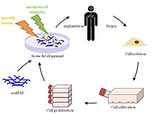
Principle of tissue engineering

Tissue engineering is the use of a combination of cells, engineering and materials methods, and suitable biochemical and physico-chemical factors to improve or replace biological functions. While it was once categorized as a sub-field of biomaterials, having grown in scope and importance it can be considered as a field in its own right.

While most definitions of tissue engineering cover a broad range of applications, in practice the term is closely associated with applications that repair or replace portions of or whole tissues (i.e., bone, cartilage, blood vessels, bladder, skin, muscle etc.). Often, the tissues involved require certain mechanical and structural properties for proper functioning. The term has also been applied to efforts to perform specific biochemical functions using cells within an artificially-created support system (e.g. an artificial pancreas, or a bio artificial liver). The term regenerative medicine is often used synonymously with tissue engineering, although those involved in regenerative medicine place more emphasis on the use of stem cells or progenitor cells to produce tissues.

Overview


Micro-mass cultures of C3H-10T1/2 cells at varied oxygen tensions stained with Alcian blue

 A commonly applied definition of tissue engineering, as stated by Langer[1] and Vacanti,[2] is "an interdisciplinary field that applies the principles of engineering and life sciences toward the development of biological substitutes that restore, maintain, or improve tissue function or a whole organ".[3] Tissue engineering has also been defined as "understanding the principles of tissue growth, and applying this to produce functional replacement tissue for clinical use."[4] A further description goes on to say that an "underlying supposition of tissue engineering is that the employment of natural biology of the system will allow for greater success in developing therapeutic strategies aimed at the replacement, repair, maintenance, and/or enhancement of tissue function."[4]

Powerful developments in the multidisciplinary field of tissue engineering have yielded a novel set of tissue replacement parts and implementation strategies. Scientific advances in biomaterials, stem cells, growth and differentiation factors, and biomimetic environments have created unique opportunities to fabricate tissues in the laboratory from combinations of engineered extracellular matrices ("scaffolds"), cells, and biologically active molecules. Among the major challenges now facing tissue engineering is the need for more complex functionality, as well as both functional and biomechanical stability in laboratory-grown tissues destined for transplantation. The continued success of tissue engineering, and the eventual development of true human replacement parts, will grow from the convergence of engineering and basic research advances in tissue, matrix, growth factor, stem cell, and developmental biology, as well as materials science and bio informatics.

In 2003, the NSF published a report entitled "The Emergence of Tissue Engineering as a Research Field", which gives a thorough description of the history of this field.[5]

Examples

Cells as building blocks


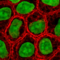
Stained cells in culture

Tissue engineering utilizes living cells as engineering materials. Examples include using living fibroblasts in skin replacement or repair, cartilage repaired with living chondrocytes, or other types of cells used in other ways.

Cells became available as engineering materials when scientists at Geron Corp. discovered how to extend telomeres in 1998, producing immortalized cell lines.[citation needed] Before this, laboratory cultures of healthy, noncancerous mammalian cells would only divide a fixed number of times, up to the Hayflick limit.

Extraction

From fluid tissues such as blood, cells are extracted by bulk methods, usually centrifugation or apheresis. From solid tissues, extraction is more difficult. Usually the tissue is minced, and then digested with the enzymes trypsin or collagenase to remove the extracellular matrix that holds the cells. After that, the cells are free floating, and extracted using centrifugation or apheresis.

Digestion with trypsin is very dependent on temperature. Higher temperatures digest the matrix faster, but create more damage. Collagenase is less temperature dependent, and damages fewer cells, but takes longer and is a more expensive reagent.

Types of cells


Mouse embryonic stem cells

Cells are often categorized by their source:
  • Autologous cells are obtained from the same individual to which they will be reimplanted. Autologous cells have the fewest problems with rejection and pathogen transmission, however in some cases might not be available. For example in genetic disease suitable autologous cells are not available. Also very ill or elderly persons, as well as patients suffering from severe burns, may not have sufficient quantities of autologous cells to establish useful cell lines. Moreover since this category of cells needs to be harvested from the patient, there are also some concerns related to the necessity of performing such surgical operations that might lead to donor site infection or chronic pain. Autologous cells also must be cultured from samples before they can be used: this takes time, so autologous solutions may not be very quick. Recently there has been a trend towards the use of mesenchymal stem cells from bone marrow and fat. These cells can differentiate into a variety of tissue types, including bone, cartilage, fat, and nerve. A large number of cells can be easily and quickly isolated from fat, thus opening the potential for large numbers of cells to be quickly and easily obtained.
  • Allogeneic cells come from the body of a donor of the same species. While there are some ethical constraints to the use of human cells for in vitro studies, the employment of dermal fibroblasts from human foreskin has been demonstrated to be immunologically safe and thus a viable choice for tissue engineering of skin.
  • Xenogenic cells are these isolated from individuals of another species. In particular animal cells have been used quite extensively in experiments aimed at the construction of cardiovascular implants.
  • Syngenic or isogenic cells are isolated from genetically identical organisms, such as twins, clones, or highly inbred research animal models.
  • Primary cells are from an organism.
  • Secondary cells are from a cell bank.
  • Stem cells are undifferentiated cells with the ability to divide in culture and give rise to different forms of specialized cells. According to their source stem cells are divided into "adult" and "embryonic" stem cells, the first class being multipotent and the latter mostly pluripotent; some cells are totipotent, in the earliest stages of the embryo. While there is still a large ethical debate related with the use of embryonic stem cells, it is thought that another alternative source - induced stem cells may be useful for the repair of diseased or damaged tissues, or may be used to grow new organs.

Scaffolds

Cells are often implanted or 'seeded' into an artificial structure capable of supporting three-dimensional tissue formation. These structures, typically called scaffolds, are often critical, both ex vivo as well as in vivo, to recapitulating the in vivo milieu and allowing cells to influence their own microenvironments. Scaffolds usually serve at least one of the following purposes:
  • Allow cell attachment and migration
  • Deliver and retain cells and biochemical factors
  • Enable diffusion of vital cell nutrients and expressed products
  • Exert certain mechanical and biological influences to modify the behaviour of the cell phase

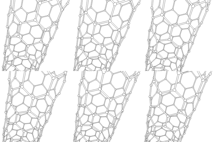
This animation of a rotating Carbon nanotube shows its 3D structure. Carbon nanotubes are among the numerous candidates for tissue engineering scaffolds since they are biocompatible, resistant to biodegradation and can be functionalized with biomolecules. However, the possibility of toxicity with non-biodegradable nano-materials is not fully understood.

To achieve the goal of tissue reconstruction, scaffolds must meet some specific requirements. A high porosity and an adequate pore size are necessary to facilitate cell seeding and diffusion throughout the whole structure of both cells and nutrients. Biodegradability is often an essential factor since scaffolds should preferably be absorbed by the surrounding tissues without the necessity of a surgical removal. The rate at which degradation occurs has to coincide as much as possible with the rate of tissue formation: this means that while cells are fabricating their own natural matrix structure around themselves, the scaffold is able to provide structural integrity within the body and eventually it will break down leaving the neotissue, newly formed tissue which will take over the mechanical load. Injectability is also important for clinical uses. Recent research on organ printing is showing how crucial a good control of the 3D environment is to ensure reproducibility of experiments and offer better results.

Materials

Many different materials (natural and synthetic, biodegradable and permanent) have been investigated. Most of these materials have been known in the medical field before the advent of tissue engineering as a research topic, being already employed as bioresorbable sutures. Examples of these materials are collagen and some polyesters.

New biomaterials have been engineered to have ideal properties and functional customization: injectability, synthetic manufacture, biocompatibility, non-immunogenicity, transparency, nano-scale fibers, low concentration, resorption rates, etc. PuraMatrix, originating from the MIT labs of Zhang, Rich, Grodzinsky and Langer is one of these new biomimetic scaffold families which has now been commercialized and is impacting clinical tissue engineering.

A commonly used synthetic material is PLA - polylactic acid. This is a polyester which degrades within the human body to form lactic acid, a naturally occurring chemical which is easily removed from the body. Similar materials are polyglycolic acid (PGA) and polycaprolactone (PCL): their degradation mechanism is similar to that of PLA, but they exhibit respectively a faster and a slower rate of degradation compared to PLA.

Scaffolds may also be constructed from natural materials: in particular different derivatives of the extracellular matrix have been studied to evaluate their ability to support cell growth. Proteic materials, such as collagen or fibrin, and polysaccharidic materials, like chitosan[16] or glycosaminoglycans (GAGs), have all proved suitable in terms of cell compatibility, but some issues with potential immunogenicity still remains. Among GAGs hyaluronic acid, possibly in combination with cross linking agents (e.g. glutaraldehyde, water soluble carbodiimide, etc...), is one of the possible choices as scaffold material. Functionalized groups of scaffolds may be useful in the delivery of small molecules (drugs) to specific tissues. Another form of scaffold under investigation is decellularised tissue extracts whereby the remaining cellular remnants/extracellular matrices act as the scaffold.

A 2009 study by Ratmir et al. aimed to improve in vivo-like conditions for 3D tissue via "stacking and de-stacking layers of paper impregnated with suspensions of cells in extracellular matrix hydrogel, making it possible to control oxygen and nutrient gradients in 3D, and to analyze molecular and genetic responses".[17] It is possible to manipulate gradients of soluble molecules, and to characterize cells in these complex gradients more effectively than conventional 3D cultures based on hydrogels, cell spheroids, or 3D perfusion reactors.[18] Different thicknesses of paper and types of medium can support a variety of experimental environments. Upon deconstruction, these sheets can be useful in cell-based high-throughput screening and drug discovery.[18]

Synthesis


Tissue engineered vascular graft

Tissue engineered heart valve

A number of different methods have been described in literature for preparing porous structures to be employed as tissue engineering scaffolds. Each of these techniques presents its own advantages, but none are free of drawbacks.
Nanofiber Self-Assembly
Molecular self-assembly is one of the few methods for creating biomaterials with properties similar in scale and chemistry to that of the natural in vivo extracellular matrix (ECM), a crucial step toward tissue engineering of complex tissues.[19] Moreover, these hydrogel scaffolds have shown superiority in in vivo toxicology and biocompatibility compared to traditional macroscaffolds and animal-derived materials.
Textile technologies
These techniques include all the approaches that have been successfully employed for the preparation of non-woven meshes of different polymers. In particular, non-woven polyglycolide structures have been tested for tissue engineering applications: such fibrous structures have been found useful to grow different types of cells. The principal drawbacks are related to the difficulties in obtaining high porosity and regular pore size.
Solvent Casting & Particulate Leaching (SCPL)
This approach allows for the preparation of structures with regular porosity, but with limited thickness. First, the polymer is dissolved into a suitable organic solvent (e.g. polylactic acid could be dissolved into dichloromethane), then the solution is cast into a mold filled with porogen particles. Such porogen can be an inorganic salt like sodium chloride, crystals of saccharose, gelatin spheres or paraffin spheres. The size of the porogen particles will affect the size of the scaffold pores, while the polymer to porogen ratio is directly correlated to the amount of porosity of the final structure. After the polymer solution has been cast the solvent is allowed to fully evaporate, then the composite structure in the mold is immersed in a bath of a liquid suitable for dissolving the porogen: water in the case of sodium chloride, saccharose and gelatin or an aliphatic solvent like hexane for use with paraffin. Once the porogen has been fully dissolved, a porous structure is obtained. Other than the small thickness range that can be obtained, another drawback of SCPL lies in its use of organic solvents which must be fully removed to avoid any possible damage to the cells seeded on the scaffold.
Gas Foaming
To overcome the need to use organic solvents and solid porogens, a technique using gas as a porogen has been developed. First, disc-shaped structures made of the desired polymer are prepared by means of compression molding using a heated mold. The discs are then placed in a chamber where they are exposed to high pressure CO2 for several days. The pressure inside the chamber is gradually restored to atmospheric levels. During this procedure the pores are formed by the carbon dioxide molecules that abandon the polymer, resulting in a sponge-like structure. The main problems resulting from such a technique are caused by the excessive heat used during compression molding (which prohibits the incorporation of any temperature labile material into the polymer matrix) and by the fact that the pores do not form an interconnected structure.
Emulsification/Freeze-drying
This technique does not require the use of a solid porogen like SCPL. First, a synthetic polymer is dissolved into a suitable solvent (e.g. polylactic acid in dichloromethane) then water is added to the polymeric solution and the two liquids are mixed in order to obtain an emulsion. Before the two phases can separate, the emulsion is cast into a mold and quickly frozen by means of immersion into liquid nitrogen. The frozen emulsion is subsequently freeze-dried to remove the dispersed water and the solvent, thus leaving a solidified, porous polymeric structure. While emulsification and freeze-drying allow for a faster preparation when compared to SCPL (since it does not require a time consuming leaching step), it still requires the use of solvents. Moreover, pore size is relatively small and porosity is often irregular. Freeze-drying by itself is also a commonly employed technique for the fabrication of scaffolds. In particular, it is used to prepare collagen sponges: collagen is dissolved into acidic solutions of acetic acid or hydrochloric acid that are cast into a mold, frozen with liquid nitrogen and then lyophilized.
Thermally Induced Phase Separation (TIPS)
Similar to the previous technique, this phase separation procedure requires the use of a solvent with a low melting point that is easy to sublime. For example dioxane could be used to dissolve polylactic acid, then phase separation is induced through the addition of a small quantity of water: a polymer-rich and a polymer-poor phase are formed. Following cooling below the solvent melting point and some days of vacuum-drying to sublime the solvent, a porous scaffold is obtained. Liquid-liquid phase separation presents the same drawbacks of emulsification/freeze-drying.
Electrospinning
A highly versatile technique that can be used to produce continuous fibers from submicrometer to nanometer diameters. In a typical electrospinning set-up, a solution is fed through a spinneret and a high voltage is applied to the tip. The buildup of electrostatic repulsion within the charged solution, causes it to eject a thin fibrous stream. A mounted collector plate or rod with an opposite or grounded charge draws in the continuous fibers, which arrive to form a highly porous network. The primary advantages of this technique are its simplicity and ease of variation. At a laboratory level, a typical electrospinning set-up only requires a high voltage power supply (up to 30 kV), a syringe, a flat tip needle and a conducting collector. For these reasons, electrospinning has become a common method of scaffold manufacture in many labs. By modifying variables such as the distance to collector, magnitude of applied voltage, or solution flow rate—researchers can dramatically change the overall scaffold architecture.
CAD/CAM Technologies
Because most of the above techniques are limited when it comes to the control of porosity and pore size, computer assisted design and manufacturing techniques have been introduced to tissue engineering. First, a three-dimensional structure is designed using CAD software. The porosity can be tailored using algorithms within the software.[20] The scaffold is then realized by using ink-jet printing of polymer powders or through Fused Deposition Modeling of a polymer melt.[21]
A 2011 study by El-Ayoubi et al. investigated "3D-plotting technique to produce (biocompatible and biodegradable) poly-L-Lactide macroporous scaffolds with two different pore sizes" via solid free-form fabrication (SSF) with computer-aided-design (CAD), to explore therapeutic articular cartilage replacement as an "alternative to conventional tissue repair".[22] The study found the smaller the pore size paired with mechanical stress in a bioreactor (to induce in vivo-like conditions), the higher the cell viability in potential therapeutic functionality via decreasing recovery time and increasing transplant effectiveness.[22]
Laser-assisted BioPrinting (LaBP)
In a 2012 study,[23] Koch et al. focused on whether Laser-assisted BioPrinting (LaBP) can be used to build multicellular 3D patterns in natural matrix, and whether the generated constructs are functioning and forming tissue. LaBP arranges small volumes of living cell suspensions in set high-resolution patterns.[23] The investigation was successful, the researchers foresee that "generated tissue constructs might be used for in vivo testing by implanting them into animal models" (14). As of this study, only human skin tissue has been synthesized, though researchers project that "by integrating further cell types (e.g. melanocytes, Schwann cells, hair follicle cells) into the printed cell construct, the behavior of these cells in a 3D in vitro microenvironment similar to their natural one can be analyzed", useful for drug discovery and toxicology studies.[23]

Assembly methods

One of the continuing, persistent problems with tissue engineering is mass transport limitations. Engineered tissues generally lack an initial blood supply, thus making it difficult for any implanted cells to obtain sufficient oxygen and nutrients to survive, and/or function properly.

Self-assembly

Self-assembly may play an important role here, both from the perspective of encapsulating cells and proteins, as well as creating scaffolds on the right physical scale for engineered tissue constructs and cellular ingrowth. The micromasonry is a prime technology to get cells grown in a lab to assemble into three-dimensional shapes. To break down tissue into single-cell building blocks, researchers have to dissolve the extracellular mortar that normally binds them together. But once that glue is removed, it's quite difficult to get cells to reassemble into the complex structures that make up our natural tissues. While cells aren't easily stackable, building blocks are. So the micromasonry starts with the encapsulation of living cells in polymer cubes. From there, the blocks self-assemble in any shape using templates.

Liquid-based template assembly

The air-liquid surface established by Faraday waves is explored as a template to assemble biological entities for bottom-up tissue engineering. This liquid-based template can be dynamically reconfigured in a few seconds, and the assembly on the template can be achieved in a scalable and parallel manner. Assembly of microscale hydrogels, cells, neuron-seeded micro-carrier beads, cell spheroids into various symmetrical and periodic structures was demonstrated with good cell viability. Formation of 3D neural network was achieved after 14-day tissue culture.[24]

Additive manufacturing

It might be possible to print organs, or possibly entire organisms using additive manufacturing techniques. A recent innovative method of construction uses an ink-jet mechanism to print precise layers of cells in a matrix of thermoreversible gel. Endothelial cells, the cells that line blood vessels, have been printed in a set of stacked rings. When incubated, these fused into a tube.[21][25]

The field of three-dimensional and highly accurate models of biological systems is pioneered by multiple projects and technologies including a rapid method for creating tissues and even whole organs involves a 3D printer that can print the scaffolding and cells layer by layer into a working tissue sample or organ. The device is presented in a TED talk by Dr. Anthony Atala, M.D. the Director of the Wake Forest Institute for Regenerative Medicine, and the W.H. Boyce Professor and Chair of the Department of Urology at Wake Forest University, in which a full bladder is printed on stage during the seminar and then presented to the crowd.[26][27] This specific technology has been used to print a bladder for a young man that would have otherwise died without the availability of a transplant, and is anticipated to be able to print kidneys and perhaps livers in the future for transplantation and theoretically for toxicology and other biological studies as well.

Scaffolding

In 2013, using a 3-d scaffolding of Matrigel in various configurations, substantial pancreatic organoids was produced in vitro. Clusters of small numbers of cells proliferated into 40,000 cells within one week. The clusters transform into cells that make either digestive enzymes or hormones like insulin, self-organizing into branched pancreatic organoids that resemble the pancreas.[28]
The cells are sensitive to the environment, such as gel stiffness and contact with other cells. Individual cells do not thrive; a minimum of four proximate cells was required for subsequent organoid development. Modifications to the medium composition produced either hollow spheres mainly composed of pancreatic progenitors, or complex organoids that spontaneously undergo pancreatic morphogenesis and differentiation. Maintenance and expansion of pancreatic progenitors require active Notch and FGF signaling, recapitulating in vivo niche signaling interactions.[29]

The organoids were seen as potentially offering mini-organs for drug testing and for spare insulin-producing cells.[28]

Tissue culture

In many cases, creation of functional tissues and biological structures in vitro requires extensive culturing to promote survival, growth and inducement of functionality. In general, the basic requirements of cells must be maintained in culture, which include oxygen, pH, humidity, temperature, nutrients and osmotic pressure maintenance.

Tissue engineered cultures also present additional problems in maintaining culture conditions. In standard cell culture, diffusion is often the sole means of nutrient and metabolite transport. However, as a culture becomes larger and more complex, such as the case with engineered organs and whole tissues, other mechanisms must be employed to maintain the culture, such as the creation of capillary networks within the tissue.

Bioreactor for cultivation of vascular grafts

Another issue with tissue culture is introducing the proper factors or stimuli required to induce functionality. In many cases, simple maintenance culture is not sufficient. Growth factors, hormones, specific metabolites or nutrients, chemical and physical stimuli are sometimes required. For example, certain cells respond to changes in oxygen tension as part of their normal development, such as chondrocytes, which must adapt to low oxygen conditions or hypoxia during skeletal development. Others, such as endothelial cells, respond to shear stress from fluid flow, which is encountered in blood vessels. Mechanical stimuli, such as pressure pulses seem to be beneficial to all kind of cardiovascular tissue such as heart valves, blood vessels or pericardium.

Bioreactors

A bioreactor in tissue engineering, as opposed to industrial bioreactors, is a device that attempts to simulate a physiological environment in order to promote cell or tissue growth in vivo. A physiological environment can consist of many different parameters such as temperature and oxygen or carbon dioxide concentration, but can extend to all kinds of biological, chemical or mechanical stimuli. Therefore, there are systems that may include the application of forces or stresses to the tissue or even of electrical current in two- or three-dimensional setups.
In academic and industry research facilities, it is typical for bioreactors to be developed to replicate the specific physiological environment of the tissue being grown (e.g., flex and fluid shearing for heart tissue growth).[30] Several general-use and application-specific bioreactors are also commercially available, and may provide static chemical stimulation or combination of chemical and mechanical stimulation.

The Bioreactors used for 3D cell cultures are small plastic cylindrical chambers with regulated internal humidity and moisture specifically engineered for the purpose of growing cells in three dimensions.[31] The bioreactor uses bioactive synthetic materials such as polyethylene terephthalate membranes to surround the spheroid cells in an environment that maintains high levels of nutrients.[18][32] They are easy to open and close, so that cell spheroids can be removed for testing, yet the chamber is able to maintain 100% humidity throughout.[33] This humidity is important to achieve maximum cell growth and function. The bioreactor chamber is part of a larger device that rotates to ensure equal cell growth in each direction across three dimensions.[33] MC2 Biotek has developed a bioreactor known as ProtoTissue[31] that uses gas exchange to maintain high oxygen levels within the cell chamber; improving upon previous bioreactors, because the higher oxygen levels help the cell grow and undergo normal cell respiration.[34]

Long fiber generation

In 2013, a group from the University of Tokyo developed cell laden fibers up to a meter in length and on the order of 100 µm in size.[35] These fibers were created using a microfluidic device that forms a double coaxial laminar flow. Each 'layer' of the microfluidic device (cells seeded in ECM, a hydrogel sheath, and finally a calcium chloride solution). The seeded cells culture within the hydrogel sheath for several days, and then the sheath is removed with viable cell fibers. Various cell types were inserted into the ECM core, including myocytes, endothelial cells, nerve cell fibers, and epithelial cell fibers. This group then showed that these fibers can be woven together to fabricate tissues or organs in a mechanism similar to textile weaving. Fibrous morphologies are advantageous in that they provide an alternative to traditional scaffold design, and many organs (such as muscle) are composed of fibrous cells.

Bioartificial organs

An artificial organ is a man-made device that is implanted or integrated into a human to replace a natural organ, for the purpose of restoring a specific function or a group of related functions so the patient may return to a normal life as soon as possible. The replaced function doesn't necessarily have to be related to life support, but often is. The ultimate goal of tissue engineering as a discipline is to allow both 'off the shelf' bioartificial organs and regeneration of injured tissue in the body. In order to successfully create bioartificial organs from a patients stem cells, researchers continue to make improvements in the generation of complex tissues by tissue engineering. For example, much research is aimed at understanding nanoscale cues present in a cell’s microenvironment.[19]

Declaration of the Rights of Man and of the Citizen

From Wikipedia, the free encyclopedia https://en.wikipedia.org/wiki/Declarati...