Search This Blog

Tuesday, June 20, 2023

Split-brain

From Wikipedia, the free encyclopedia

Split-brain or callosal syndrome is a type of disconnection syndrome when the corpus callosum connecting the two hemispheres of the brain is severed to some degree. It is an association of symptoms produced by disruption of, or interference with, the connection between the hemispheres of the brain. The surgical operation to produce this condition (corpus callosotomy) involves transection of the corpus callosum, and is usually a last resort to treat refractory epilepsy. Initially, partial callosotomies are performed; if this operation does not succeed, a complete callosotomy is performed to mitigate the risk of accidental physical injury by reducing the severity and violence of epileptic seizures. Before using callosotomies, epilepsy is instead treated through pharmaceutical means. After surgery, neuropsychological assessments are often performed.

After the right and left brain are separated, each hemisphere will have its own separate perception, concepts, and impulses to act. Having two "brains" in one body can create some interesting dilemmas. When one split-brain patient dressed himself, he sometimes pulled his pants up with one hand (that side of his brain wanted to get dressed) and down with the other (this side did not). He also reported to have grabbed his wife with his left hand and shaken her violently, at which point his right hand came to her aid and grabbed the aggressive left hand. However, such conflicts are very rare. If a conflict arises, one hemisphere usually overrides the other.

When split-brain patients are shown an image only in the left half of each eye's visual field, they cannot vocally name what they have seen. This is because the image seen in the left visual field is sent only to the right side of the brain (see optic tract), and most people's speech-control center is on the left side of the brain. Communication between the two sides is inhibited, so the patient cannot say out loud the name of that which the right side of the brain is seeing. A similar effect occurs if a split-brain patient touches an object with only the left hand while receiving no visual cues in the right visual field; the patient will be unable to name the object, as each cerebral hemisphere of the primary somatosensory cortex only contains a tactile representation of the opposite side of the body. If the speech-control center is on the right side of the brain, the same effect can be achieved by presenting the image or object to only the right visual field or hand.

The same effect occurs for visual pairs and reasoning. For example, a patient with split brain is shown a picture of a chicken foot and a snowy field in separate visual fields and asked to choose from a list of words the best association with the pictures. The patient would choose a chicken to associate with the chicken foot and a shovel to associate with the snow; however, when asked to reason why the patient chose the shovel, the response would relate to the chicken (e.g. "the shovel is for cleaning out the chicken coop").

History

Early anatomists, such as Galen (129 – c. 216 CE) and Vesalius (1514 – 1564 CE), identified the corpus callosum. They generally described its function as a structure holding together the two halves of the brain. In 1784, Félix Vicq-d'Azyr described the corpus callosum as allowing communication between the two halves of the brain. He proposed that eliminating the corpus callosum would divide the brain into two independent parts. In 1892, Joseph Jules Dejerine reported symptoms in a person who had destruction of part of the corpus callosum (along with damage to the visual cortex: inability to read while retaining the ability to write, now referred to as pure alexia or as Dejerine syndrome. In 1908, Hugo Liepmann observed left-sided apraxia (a motor disorder of motor planning to perform tasks or movements) and agraphia (loss of the ability to communicate through writing) following a lesion in the corpus callosum.

According to Vaddiparti et al. (2021), the first surgical cuts to the corpus callosum, partial corpus callosotomy, were made by neurosurgeon Walter Dandy in order to access and to remove tumors in the pineal gland. In 1936, Dandy described three cases in which he cut the corpus callosum from its posterior (towards the back of the head) across about two thirds of its width. He described these cuts as "bloodless" and that "no symptoms follow[ed] [the] .. division" of the corpus callosum. He concluded that his operations "dispose ... of the extravagant claims to function of the corpus callosum" (p. 40).

Prior to the 1960s, research on people with certain brain injuries led to the notion that there is a "language center" only in the left hemisphere of the brain. For example, people with lesions in two specific areas of the left hemisphere lost their ability to talk, to read, and to understand speech. Roger Sperry and his colleagues pioneered research showing that creating another lesion (done to relieve otherwise untreatable epilepsy), in the connections between the left and right hemispheres, revealed that the right hemisphere can allow people to read, to understand speech, and to say some simple words. Research over the next twenty years showed that the disconnected right hemisphere is superior to the disconnected left hemisphere in allowing people to understand spatial information (such as maps), music, and emotions, whereas the disconnected left hemisphere is superior in allowing analytical thinking, talking, reading, and understanding speech. This research led to a Nobel Prize in Physiology or Medicine for Sperry in 1981.

Sperry's initial colleagues included his Caltech PhD students, Michael Gazzaniga and Jerre Levy. Even though Sperry is considered the founder of split-brain research, Gazzaniga's clear summaries of their collaborative work are consistently cited in psychology texts. In Sperry and Gazzaniga's "The Split Brain in Man" experiment published in Scientific American in 1967 they attempted to explore the extent to which two halves of the human brain were able to function independently and whether or not they had separate and unique abilities. They wanted to examine how perceptual and intellectual skills were affected in someone with a split-brain. At Caltech, Gazzaniga worked with Sperry on the effects of split-brain surgery on perception, vision and other brain functions. The surgery, which was a treatment for severe epilepsy, involved severing the corpus callosum, which carries signals between the left-brain hemisphere, the seat of speech and analytical capacity, and the right-brain hemisphere, which helps recognize visual patterns. At the time this article was written, only ten patients had undergone the surgery to sever their corpus callosum (corpus callosotomy). Four of these patients had consented to participate in Sperry and Gazzaniga's research. After the corpus callosum severing, all four participants' personality, intelligence, and emotions appeared to be unaffected. However, the testing done by Sperry and Gazzaniga showed the subjects demonstrated unusual mental abilities. The researchers created different types of tests to analyze the range of cognitive capabilities of the split-brain subjects. These included tests of their visual stimulation abilities, a tactile stimulation situation, and a test that involved both visual and tactile information.

Visual test

The first test started with a board that had a horizontal row of lights. The subject was told to sit in front of the board and stare at a point in the middle of the lights, then the bulbs would flash across both the right and left visual fields. When the patients were asked to describe afterward what they saw, they said that only the lights on the right side of the board had lit up. Next, when Sperry and Gazzaniga flashed the lights on the right side of the board on the subjects left side of their visual field, they claimed not to have seen any lights at all. When the experimenters conducted the test again, they asked the subjects to point to the lights that lit up. Although subjects had only reported seeing the lights flash on the right, they actually pointed to all the lights in both visual fields. This showed that both brain hemispheres had seen the lights and were equally competent in visual perception. The subjects did not say they saw the lights when they flashed in the left visual field even though they did see them because the center for speech is located in the brain's left hemisphere. This test supports the idea that in order to say one has seen something, the region of the brain associated with speech must be able to communicate with areas of the brain that process the visual information.

Tactile test

In a second experiment, Sperry and Gazzaniga placed a small object in the subject's right or left hand, without being able to see (or hear) it. Placed in the right hand, the isolated left hemisphere perceived the object and could easily describe and name it. However, placed in the left hand, the isolated right hemisphere could not name or describe the object. Questioning this result, the researchers found that the subjects could later match it from several similar objects; tactile sensations limited to the right hemisphere were accurately perceived but could not be verbalized. This further demonstrated the apparent location (or lateralization) of language functions in the left hemisphere.

Combination of both tests

In the last test the experimenters combined both the tactile and visual test. They presented subjects with a picture of an object to only their right hemisphere, and subjects were unable to name it or describe it. There were no verbal responses to the picture at all. If the subject however was able to reach under the screen with their left hand to touch various objects, they were able to pick the one that had been shown in the picture. The subjects were also reported to be able to pick out objects that were related to the picture presented, if that object was not under the screen.

Sperry and Gazzaniga went on to conduct other tests to shed light on the language processing abilities of the right hemisphere as well as auditory and emotional reactions as well. The significance of the findings of these tests by Sperry and Gazzaniga were extremely telling and important to the psychology world. Their findings showed that the two halves of the brain have numerous functions and specialized skills. They concluded that each hemisphere really has its own functions. One's left hemisphere of the brain is thought to be better at writing, speaking, mathematical calculation, reading, and is the primary area for language. The right hemisphere is seen to possess capabilities for problem solving, recognizing faces, symbolic reasoning, art, and spatial relationships.

Roger Sperry continued this line of research up until his death in 1994. Michael Gazzaniga continues to research the split-brain. Their findings have been rarely critiqued and disputed, however, a popular belief that some people are more "right-brained" or "left-brained" has developed. In the mid-1980s Jarre Levy, a psychobiologist at the University of Chicago, had set out and been in the forefront of scientists who wanted to dispel the notion we have two functioning brains. She believes that because each hemisphere has separate functions that they must integrate their abilities instead of separating them. Levy also claims that no human activity uses only one side of the brain. In 1998 a French study by Hommet and Billiard was published that questioned Sperry and Gazzaniga's study that severing the corpus callosum actually divides the hemispheres of the brain. They found that children born without a corpus callosum demonstrated that information was being transmitted between hemispheres, and concluded that subcortical connections must be present in these children with this rare brain malformation. They are unclear about whether these connections are present in split-brain patients though. Another study by Parsons, Gabrieli, Phelps, and Gazzaniga in 1998 demonstrated that split-brain patients may commonly perceive the world differently from the rest of us. Their study suggested that communication between brain hemispheres is necessary for imaging or simulating in your mind the movements of others. Morin's research on inner speech in 2001 suggested that an alternative for interpretation of commissurotomy according to which split-brain patients exhibit two uneven streams of self-awareness: a "complete" one in the left hemisphere and a "primitive" one in the right hemisphere.

Hemispheric specialization

The two hemispheres of the cerebral cortex are linked by the corpus callosum, through which they communicate and coordinate actions and decisions. Communication and coordination between the two hemispheres is essential because each hemisphere has some separate functions. The right hemisphere of the cortex excels at nonverbal and spatial tasks, whereas the left hemisphere is more dominant in verbal tasks, such as speaking and writing. The right hemisphere controls the primary sensory functions of the left side of the body. In a cognitive sense the right hemisphere is responsible for recognizing objects and timing, and in an emotional sense it is responsible for empathy, humour and depression. On the other hand, the left hemisphere controls the primary sensory functions of the right side of the body and is responsible for scientific and maths skills, and logic. The extent of specialised brain function by an area remains under investigation. It is claimed that the difference between the two hemispheres is that the left hemisphere is "analytic" or "logical" while the right hemisphere is "holistic" or "intuitive." Many simple tasks, especially comprehension of inputs, require functions that are specific to both the right and left hemispheres and together form a one direction systematised way of creating an output through the communication and coordination that occurs between hemispheres.

Role of the corpus callosum

The corpus callosum is a structure in the brain along the longitudinal fissure that facilitates much of the communication between the two hemispheres. This structure is composed of white matter: millions of axons that have their dendrites and terminal boutons projecting in both the right and left hemisphere. However, there is evidence that the corpus callosum may also have some inhibitory functions. Post-mortem research on human and monkey brains shows that the corpus callosum is functionally organised. It proves that the right hemisphere is superior for detecting faces. This organisation results in modality-specific regions of the corpus callosum that are responsible for the transfer of different types of information. Research has revealed that the anterior midbody transfers motor information, the posterior midbody transfers somatosensory information, the isthmus transfers auditory information and the splenium transfers visual information. Although much of the interhemispheric transfer occurs at the corpus callosum, there are trace amounts of transfer via subcortical pathways.

Studies of the effects on the visual pathway on split-brained patients has revealed that there is a redundancy gain (the ability of target detection to benefit from multiple copies of the target) in simple reaction time. In a simple response to visual stimuli, split-brained patients experience a faster reaction time to bilateral stimuli than predicted by model. A model proposed by Iacoboni et al. suggests split-brained patients experience asynchronous activity that causes a stronger signal, and thus a decreased reaction time. Iacoboni also suggests there exists dual attention in split-brained patients, which is implying that each cerebral hemisphere has its own attentional system. An alternative approach taken by Reuter-Lorenz et al. suggests that enhanced redundancy gain in the split brain is primarily due to a slowing of responses to unilateral stimuli, rather than a speeding of responses to bilateral ones. It is important to note that the simple reaction time in split-brained patients, even with enhanced redundancy gain, is slower than the reaction time of normal adults.

Functional plasticity

Following a stroke or other injury to the brain, functional deficiencies are common. The deficits are expected to be in areas related to the part of the brain that has been damaged; if a stroke has occurred in the motor cortex, deficits may include paralysis, abnormal posture, or abnormal movement synergies. Significant recovery occurs during the first several weeks after the injury. However, recovery is generally thought not to continue past 6 months. If a specific region of the brain is injured or destroyed, its functions can sometimes be transferred and taken over by a neighbouring region. There is little functional plasticity observed in partial and complete callosotomies; however, much more plasticity can be seen in infant patients receiving a hemispherectomy, which suggests that the opposite hemisphere can adapt some functions typically performed by its opposite pair. In a study done by Anderson, it proved a correlation between the severity of the injury, the age of the individual and their cognitive performance. It was evident that there was more neuroplasticity in older children, even if their injury was extremely severe, than infants who suffered moderate brain injury. In some incidents of any moderate to severe brain injury, it mostly causes developmental impairments and in some of the most severe injuries it can cause a profound impact on their development that can lead to long-term cognitive effects. In the aging brain, it is extremely uncommon for neuroplasticity to occur; "olfactory bulb and hippocampus are two regions of the mammalian brain in which mutations preventing adult neurogenesis were never beneficial, or simply never occurred" (Anderson, 2005).

Corpus callosotomy

Corpus callosotomy is a surgical procedure that sections the corpus callosum, resulting in either the partial or complete disconnection between the two hemispheres. It is typically used as a last resort measure in treatment of intractable epilepsy. The modern procedure typically involves only the anterior third of the corpus callosum; however, if the epileptic seizures continue, the following third is lesioned prior to the remaining third if the seizures persist. This results in a complete callosotomy in which most of the information transfer between hemispheres is lost.

Due to the functional mapping of the corpus callosum, a partial callosotomy has less detrimental effects because it leaves parts of the corpus callosum intact. There is little functional plasticity observed in partial and complete callosotomies on adults, the most neuroplasticity is seen in young children but not in infants.

It is known that when the corpus callosum is severed during an experimental procedure, the experimenter can ask each side of the brain the same question and receive two different answers. When the experimenter asks the right visual field/left hemisphere what they see the participant will respond verbally, whereas if the experimenter asks the left visual field/right hemisphere what they see the participant will not be able to respond verbally but will pick up the appropriate object with their left hand.

Memory

It is known that the right and the left hemisphere have different functions when it comes to memory. The right hemisphere is better at recognizing objects and faces, recalling knowledge that the individual has already learned, or recalling images already seen. The left hemisphere is better at mental manipulation, language production, and semantic priming but was more susceptible to memory confusion than the right hemisphere. The main issue for individuals that have undergone a callosotomy is that because the function of memory is split into two major systems, the individual is more likely to become confused between knowledge they already know and information that they have only inferred.

In tests, memory in either hemisphere of split-brained patients is generally lower than normal, though better than in patients with amnesia, suggesting that the forebrain commissures are important for the formation of some kinds of memory. This suggests that posterior callosal sections that include the hippocampal commissures cause a mild memory deficit (in standardised free-field testing) involving recognition.

Control

In general, split-brained patients behave in a coordinated, purposeful and consistent manner, despite the independent, parallel, usually different and occasionally conflicting processing of the same information from the environment by the two disconnected hemispheres. When two hemispheres receive competing stimuli at the same time, the response mode tends to determine which hemisphere controls behaviour.

Often, split-brained patients are indistinguishable from normal adults. This is due to the compensatory phenomena; split-brained patients progressively acquire a variety of strategies to get around their interhemispheric transfer deficits. One issue that can happen with their body control is that one side of the body is doing the opposite of the other side called the intermanual effect.

Attention

Experiments on covert orienting of spatial attention using the Posner paradigm confirm the existence of two different attentional systems in the two hemispheres. The right hemisphere was found superior to the left hemisphere on modified versions of spatial relations tests and in locations testing, whereas the left hemisphere was more object based. The components of mental imagery are differentially specialised: the right hemisphere was found superior for mental rotation, the left hemisphere superior for image generation. It was also found that the right hemisphere paid more attention to landmarks and scenes whereas the left hemisphere paid more attention to exemplars of categories.

Case studies of split-brain patients

Patient WJ

Patient WJ was the first patient to undergo a full corpus callosotomy in 1962, after experiencing fifteen years of convulsions resulting from grand mal seizures. He was a World War II paratrooper who was injured at 30 years old during a bombing raid jump over the Netherlands, and again in a prison camp following his first injury. After returning home, he began to suffer from blackouts in which he would not remember what he was doing or where, and how or when he got there. At age 37, he suffered his first generalised convulsion. One of his worst episodes occurred in 1953, when he suffered a series of convulsions lasting for many days. During these convulsions, his left side would go numb and he would recover quickly, but after the series of convulsions, he never regained complete feeling on his left side.

Before his surgery, both hemispheres functioned and interacted normally, his sensory and motor functions were normal aside from slight hypoesthesia, and he could correctly identify and understand visual stimuli presented to both sides of his visual field. During his surgery in 1962, his surgeons determined that no massa intermedia had developed, and he had undergone atrophy in the part of the right frontal lobe exposed during the procedure. His operation was a success, in that it led to decreases in the frequency and intensity of his seizures.

Patient JW

Funnell et al. (2007) tested patient JW some time before June 2006. They described JW as

a right-handed male who was 47 years old at the time of testing. He successfully completed high school and has no reported learning disabilities. He had his first seizure at the age of 16 and the age of 25, he underwent a two-stage resection of the corpus callosum for relief of intractable epilepsy. Complete sectioning of the corpus callosum has been confirmed by MRI. Post-surgical MRI also revealed no evidence of other neurological damage.

Funnell et al.'s (2007) experiments were to determine each of JW's hemisphere's ability to perform simple addition, subtraction, multiplication and division. For example, in one experiment, on each trial, they presented an arithmetic problem in the center of the screen for 1 second, followed by a central cross hair JW was to look at. After 1 more second, Funnell et al. presented a number to one or the other hemisphere/visual field for 150 ms—too fast for JW to move his eyes. Randomly in half the trials, the number was the correct answer; in the other half of the trials it was the incorrect answer. With the hand on the same side as the number, JW pressed one key if the number was correct and another key if the number was incorrect.

Funnell et al.'s results were that performance of the left hemisphere was highly accurate (around 95%)—much better than performance of the right hemisphere, which was at chance for subtraction, multiplication, and division. Nevertheless the right hemisphere showed better than chance performance for addition (around 58%).

Turk et al. (2002) tested hemispheric differences in JW's recognition of himself and of familiar faces. They used faces that were composites of JW's face and Dr. Michael Gazzaniga's face. Composites ranged from 100% JW, through 50% JW and 50% Gazzaniga, to 100% Gazzaniga. JW pressed keys to say whether a presented face looked like him or Gazzaniga. Turk et al.concluded there are cortical networks in the left hemisphere that play an important role in self-recognition.

Patient VP

Patient VP is a woman who underwent a two-stage callosotomy in 1979 at the age of 27. Although the callosotomy was reported to be complete, follow-up MRI in 1984 revealed spared fibers in the rostrum and splenium. The spared rostral fibers constituted approximately 1.8% of the total cross-sectional area of the corpus callosum and the spared splenial fibers constituted approximately 1% of the area. VP's postsurgery intelligence and memory quotients were within normal limits.

One of the experiments involving VP attempted to investigate systematically the types of visual information that could be transferred via VP's spared splenial fibers. The first experiment was designed to assess VP's ability to make between-field perceptual judgements about simultaneously presented pairs of stimuli. The stimuli were presented in varying positions with respect to the horizontal and vertical midline with VP's vision fixated on a central crosshair. The judgements were based on differences in colour, shape or size. The testing procedure was the same for all three types of stimuli; after presentation of each pair, VP verbally responded "yes" if the two items in the pair were identical and "no" if they were not. The results show that there was no perceptual transfer for colour, size or shape with binomial tests showing that VP's accuracy was not greater than chance.

A second experiment involving VP attempted to investigate what aspects of words transferred between the two hemispheres. The set up was similar to the previous experiment, with VP's vision fixated on a central cross hair. A word pair was presented with one word on each side of the cross-hair for 150 ms. The words presented were in one of four categories: words that looked and sounded like rhymes (e.g. tire and fire), words that looked as if they should rhyme but did not (e.g. cough and dough), words that did not look as if they should rhyme but did (e.g. bake and ache), and words that neither looked nor sounded like rhymes (e.g. keys and fort). After presentation of each word pair, VP responded "yes" if the two words rhymed and "no" if they did not. VP's performance was above chance and she was able to distinguish among the different conditions. When the word pairs did not sound like rhymes, VP was able to say accurately that the words did not rhyme, regardless of whether or not they looked as if they should rhyme. When the words did rhyme, VP was more likely to say they rhymed, particularly if the words also looked as if they should rhyme.

Although VP showed no evidence for transfer of colour, shape or size, there was evidence for transfer of word information. This is consistent with the speculation that the transfer of word information involves fibres in the ventroposterior region of the splenium—the same region in which V.P. had callosal sparing. V.P. is able to integrate words presented to both visual fields, creating a concept that is not suggested by either word. For example, she combines "head" and "stone" to form the integrated concept of a tombstone.

Kim Peek

Kim Peek was arguably the most well-known savant. He was born on November 11, 1951 with an enlarged head, sac-like protrusions of the brain and the membranes that cover it through openings in the skull, a malformed cerebellum, and without a corpus callosum, an anterior commissure, or a posterior commissure. He was able to memorize over 9,000 books, and information from approximately 15 subject areas. These include: world/American history, sports, movies, geography, actors and actresses, the Bible, church history, literature, classical music, area codes/zip codes of the United States, television stations serving these areas, and step by step directions within any major U.S. city. Despite these abilities, he had an IQ of 87, was diagnosed as autistic, was unable to button his shirt, and had difficulties performing everyday tasks. The missing structures of his brain have yet to be linked to his increased abilities, but they can be linked to his ability to read pages of a book in 8–10 seconds. He was able to view the left page of a book with his left visual field and the right page of a book with his right visual fields so he could read both pages simultaneously. He also had developed language areas in both hemispheres, something very uncommon in split-brain patients. Language is processed in areas of the left temporal lobe, and involves a contralateral transfer of information before the brain can process what is being read. In Peek's case, there was no transfer ability—this is what led to his development of language centers in each hemisphere. Many believe this is the reason behind his extremely fast reading capabilities.

Although Peek did not undergo corpus callosotomy, he is considered a natural split-brain patient and is critical to understanding the importance of the corpus callosum. Kim Peek died in 2009.

Reticular formation

From Wikipedia, the free encyclopedia
 
Reticular formation
Gray701.png
Coronal section of the pons, at its upper part. (Formatio reticularis labeled at left.)
 
Gray694.png
Traverse section of the medulla oblongata at about the middle of the olive. (Formatio reticularis grisea and formatio reticularis alba labeled at left.)
 
Details
LocationBrainstem
Identifiers
Latinformatio reticularis
MeSHD012154
NeuroNames1223
NeuroLex IDnlx_143558
TA98A14.1.00.021
A14.1.05.403
A14.1.06.327
TA25367
FMA77719

The reticular formation is a set of interconnected nuclei that are located throughout the brainstem. It is not anatomically well defined, because it includes neurons located in different parts of the brain. The neurons of the reticular formation make up a complex set of networks in the core of the brainstem that extend from the upper part of the midbrain to the lower part of the medulla oblongata. The reticular formation includes ascending pathways to the cortex in the ascending reticular activating system (ARAS) and descending pathways to the spinal cord via the reticulospinal tracts.

Neurons of the reticular formation, particularly those of the ascending reticular activating system, play a crucial role in maintaining behavioral arousal and consciousness. The overall functions of the reticular formation are modulatory and premotor, involving somatic motor control, cardiovascular control, pain modulation, sleep and consciousness, and habituation. The modulatory functions are primarily found in the rostral sector of the reticular formation and the premotor functions are localized in the neurons in more caudal regions.

The reticular formation is divided into three columns: raphe nuclei (median), gigantocellular reticular nuclei (medial zone), and parvocellular reticular nuclei (lateral zone). The raphe nuclei are the place of synthesis of the neurotransmitter serotonin, which plays an important role in mood regulation. The gigantocellular nuclei are involved in motor coordination. The parvocellular nuclei regulate exhalation.

The reticular formation is essential for governing some of the basic functions of higher organisms and is one of the phylogenetically oldest portions of the brain.

Structure

A cross section of the lower part of the pons showing the pontine reticular formation labeled as #9

The human reticular formation is composed of almost 100 brain nuclei and contains many projections into the forebrain, brainstem, and cerebellum, among other regions. It includes the reticular nuclei, reticulothalamic projection fibers, diffuse thalamocortical projections, ascending cholinergic projections, descending non-cholinergic projections, and descending reticulospinal projections. The reticular formation also contains two major neural subsystems, the ascending reticular activating system and descending reticulospinal tracts, which mediate distinct cognitive and physiological processes. It has been functionally cleaved both sagittally and coronally.

Traditionally the reticular nuclei are divided into three columns:

  • In the median column – the raphe nuclei
  • In the medial column – gigantocellular nuclei (because of larger size of the cells)
  • In the lateral column – parvocellular nuclei (because of smaller size of the cells)

The original functional differentiation was a division of caudal and rostral. This was based upon the observation that the lesioning of the rostral reticular formation induces a hypersomnia in the cat brain. In contrast, lesioning of the more caudal portion of the reticular formation produces insomnia in cats. This study has led to the idea that the caudal portion inhibits the rostral portion of the reticular formation.

Sagittal division reveals more morphological distinctions. The raphe nuclei form a ridge in the middle of the reticular formation, and, directly to its periphery, there is a division called the medial reticular formation. The medial RF is large and has long ascending and descending fibers, and is surrounded by the lateral reticular formation. The lateral RF is close to the motor nuclei of the cranial nerves, and mostly mediates their function.

Medial and lateral reticular formation

The medial reticular formation and lateral reticular formation are two columns of nuclei with ill-defined boundaries that send projections through the medulla and into the midbrain. The nuclei can be differentiated by function, cell type, and projections of efferent or afferent nerves. Moving caudally from the rostral midbrain, at the site of the rostral pons and the midbrain, the medial RF becomes less prominent, and the lateral RF becomes more prominent.

Existing on the sides of the medial reticular formation is its lateral cousin, which is particularly pronounced in the rostral medulla and caudal pons. Out from this area spring the cranial nerves, including the very important vagus nerve. The lateral RF is known for its ganglions and areas of interneurons around the cranial nerves, which serve to mediate their characteristic reflexes and functions.

Function

The reticular formation consists of more than 100 small neural networks, with varied functions including the following:

  1. Somatic motor control – Some motor neurons send their axons to the reticular formation nuclei, giving rise to the reticulospinal tracts of the spinal cord. These tracts function in maintaining tone, balance, and posture – especially during body movements. The reticular formation also relays eye and ear signals to the cerebellum so that the cerebellum can integrate visual, auditory, and vestibular stimuli in motor coordination. Other motor nuclei include gaze centers, which enable the eyes to track and fixate objects, and central pattern generators, which produce rhythmic signals of breathing and swallowing.
  2. Cardiovascular control – The reticular formation includes the cardiac and vasomotor centers of the medulla oblongata.
  3. Pain modulation – The reticular formation is one means by which pain signals from the lower body reach the cerebral cortex. It is also the origin of the descending analgesic pathways. The nerve fibers in these pathways act in the spinal cord to block the transmission of some pain signals to the brain.
  4. Sleep and consciousness – The reticular formation has projections to the thalamus and cerebral cortex that allow it to exert some control over which sensory signals reach the cerebrum and come to our conscious attention. It plays a central role in states of consciousness like alertness and sleep. Injury to the reticular formation can result in irreversible coma.
  5. Habituation – This is a process in which the brain learns to ignore repetitive, meaningless stimuli while remaining sensitive to others. A good example of this is a person who can sleep through loud traffic in a large city, but is awakened promptly due to the sound of an alarm or crying baby. Reticular formation nuclei that modulate activity of the cerebral cortex are part of the ascending reticular activating system.

Major subsystems

Ascending reticular activating system

Ascending reticular activating system. Reticular formation labeled near center.

The ascending reticular activating system (ARAS), also known as the extrathalamic control modulatory system or simply the reticular activating system (RAS), is a set of connected nuclei in the brains of vertebrates that is responsible for regulating wakefulness and sleep-wake transitions. The ARAS is a part of the reticular formation and is mostly composed of various nuclei in the thalamus and a number of dopaminergic, noradrenergic, serotonergic, histaminergic, cholinergic, and glutamatergic brain nuclei.

Structure of the ARAS

The ARAS is composed of several neural circuits connecting the dorsal part of the posterior midbrain and anterior pons to the cerebral cortex via distinct pathways that project through the thalamus and hypothalamus. The ARAS is a collection of different nuclei – more than 20 on each side in the upper brainstem, the pons, medulla, and posterior hypothalamus. The neurotransmitters that these neurons release include dopamine, norepinephrine, serotonin, histamine, acetylcholine, and glutamate. They exert cortical influence through direct axonal projections and indirect projections through thalamic relays.

The thalamic pathway consists primarily of cholinergic neurons in the pontine tegmentum, whereas the hypothalamic pathway is composed primarily of neurons that release monoamine neurotransmitters, namely dopamine, norepinephrine, serotonin, and histamine. The glutamate-releasing neurons in the ARAS were identified much more recently relative to the monoaminergic and cholinergic nuclei; the glutamatergic component of the ARAS includes one nucleus in the hypothalamus and various brainstem nuclei. The orexin neurons of the lateral hypothalamus innervate every component of the ascending reticular activating system and coordinate activity within the entire system.

The ARAS consists of evolutionarily ancient areas of the brain, which are crucial to the animal's survival and protected during adverse periods, such as during inhibitory periods of Totsellreflex, aka, "animal hypnosis". The ascending reticular activating system which sends neuromodulatory projections to the cortex - mainly connects to the prefrontal cortex. There seems to be low connectivity to the motor areas of the cortex.

Functions of the ARAS

Consciousness

The ascending reticular activating system is an important enabling factor for the state of consciousness. The ascending system is seen to contribute to wakefulness as characterised by cortical and behavioural arousal.

Regulating sleep-wake transitions

The main function of the ARAS is to modify and potentiate thalamic and cortical function such that electroencephalogram (EEG) desynchronization ensues. There are distinct differences in the brain's electrical activity during periods of wakefulness and sleep: Low voltage fast burst brain waves (EEG desynchronization) are associated with wakefulness and REM sleep (which are electrophysiologically similar); high voltage slow waves are found during non-REM sleep. Generally speaking, when thalamic relay neurons are in burst mode the EEG is synchronized and when they are in tonic mode it is desynchronized. Stimulation of the ARAS produces EEG desynchronization by suppressing slow cortical waves (0.3–1 Hz), delta waves (1–4 Hz), and spindle wave oscillations (11–14 Hz) and by promoting gamma band (20–40 Hz) oscillations.

The physiological change from a state of deep sleep to wakefulness is reversible and mediated by the ARAS. The ventrolateral preoptic nucleus (VLPO) of the hypothalamus inhibits the neural circuits responsible for the awake state, and VLPO activation contributes to the sleep onset. During sleep, neurons in the ARAS will have a much lower firing rate; conversely, they will have a higher activity level during the waking state. In order that the brain may sleep, there must be a reduction in ascending afferent activity reaching the cortex by suppression of the ARAS.

Attention

The ARAS also helps mediate transitions from relaxed wakefulness to periods of high attention. There is increased regional blood flow (presumably indicating an increased measure of neuronal activity) in the midbrain reticular formation (MRF) and thalamic intralaminar nuclei during tasks requiring increased alertness and attention.

Clinical significance of the ARAS

Mass lesions in brainstem ARAS nuclei can cause severe alterations in level of consciousness (e.g., coma). Bilateral damage to the reticular formation of the midbrain may lead to coma or death.

Direct electrical stimulation of the ARAS produces pain responses in cats and elicits verbal reports of pain in humans. Ascending reticular activation in cats can produce mydriasis, which can result from prolonged pain. These results suggest some relationship between ARAS circuits and physiological pain pathways.

Pathologies

Some pathologies of the ARAS may be attributed to age, as there appears to be a general decline in reactivity of the ARAS with advancing years. Changes in electrical coupling have been suggested to account for some changes in ARAS activity: if coupling were down-regulated, there would be a corresponding decrease in higher-frequency synchronization (gamma band). Conversely, up-regulated electrical coupling would increase synchronization of fast rhythms that could lead to increased arousal and REM sleep drive. Specifically, disruption of the ARAS has been implicated in the following disorders:

  • Narcolepsy: Lesions along the pedunculopontine (PPT/PPN) / laterodorsal tegmental (LDT) nuclei are associated with narcolepsy. There is a significant down-regulation of PPN output and a loss of orexin peptides, promoting the excessive daytime sleepiness that is characteristic of this disorder.
  • Progressive supranuclear palsy (PSP) : Dysfunction of nitrous oxide signaling has been implicated in the development of PSP.
  • Parkinson's disease: REM sleep disturbances are common in Parkinson's. It is mainly a dopaminergic disease, but cholinergic nuclei are depleted as well. Degeneration in the ARAS begins early in the disease process.
Developmental influences

There are several potential factors that may adversely influence the development of the ascending reticular activating system:

Descending reticulospinal tracts

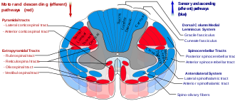
Spinal cord tracts - reticulospinal tract labeled in red, near-center at left in figure

The reticulospinal tracts, also known as the descending or anterior reticulospinal tracts, are extrapyramidal motor tracts that descend from the reticular formation in two tracts to act on the motor neurons supplying the trunk and proximal limb flexors and extensors. The reticulospinal tracts are involved mainly in locomotion and postural control, although they do have other functions as well. The descending reticulospinal tracts are one of four major cortical pathways to the spinal cord for musculoskeletal activity. The reticulospinal tracts works with the other three pathways to give a coordinated control of movement, including delicate manipulations. The four pathways can be grouped into two main system pathways – a medial system and a lateral system. The medial system includes the reticulospinal pathway and the vestibulospinal pathway, and this system provides control of posture. The corticospinal and the rubrospinal tract pathways belong to the lateral system which provides fine control of movement.

Medial and lateral tracts

This descending tract is divided into two parts, the medial (or pontine) and lateral (or medullary) reticulospinal tracts (MRST and LRST).

  • The medial reticulospinal tract is responsible for exciting anti-gravity, extensor muscles. The fibers of this tract arise from the caudal pontine reticular nucleus and the oral pontine reticular nucleus and project to lamina VII and lamina VIII of the spinal cord.
  • The lateral reticulospinal tract is responsible for inhibiting excitatory axial extensor muscles of movement. It is also responsible for automatic breathing. The fibers of this tract arise from the medullary reticular formation, mostly from the gigantocellular nucleus, and descend the length of the spinal cord in the anterior part of the lateral column. The tract terminates in lamina VII mostly with some fibers terminating in lamina IX of the spinal cord.

The ascending sensory tract conveying information in the opposite direction is known as the spinoreticular tract.

Functions of the reticulospinal tracts

  1. Integrates information from the motor systems to coordinate automatic movements of locomotion and posture
  2. Facilitates and inhibits voluntary movement; influences muscle tone
  3. Mediates autonomic functions
  4. Modulates pain impulses
  5. Influences blood flow to lateral geniculate nucleus of the thalamus.

Clinical significance of the reticulospinal tracts

The reticulospinal tracts provide a pathway by which the hypothalamus can control sympathetic thoracolumbar outflow and parasympathetic sacral outflow.

Two major descending systems carrying signals from the brainstem and cerebellum to the spinal cord can trigger automatic postural response for balance and orientation: vestibulospinal tracts from the vestibular nuclei and reticulospinal tracts from the pons and medulla. Lesions of these tracts result in profound ataxia and postural instability.

Physical or vascular damage to the brainstem disconnecting the red nucleus (midbrain) and the vestibular nuclei (pons) may cause decerebrate rigidity, which has the neurological sign of increased muscle tone and hyperactive stretch reflexes. Responding to a startling or painful stimulus, both arms and legs extend and turn internally. The cause is the tonic activity of lateral vestibulospinal and reticulospinal tracts stimulating extensor motoneurons without the inhibitions from rubrospinal tract.

Brainstem damage above the red nucleus level may cause decorticate rigidity. Responding to a startling or painful stimulus, the arms flex and the legs extend. The cause is the red nucleus, via the rubrospinal tract, counteracting the extensor motorneuron's excitation from the lateral vestibulospinal and reticulospinal tracts. Because the rubrospinal tract only extends to the cervical spinal cord, it mostly acts on the arms by exciting the flexor muscles and inhibiting the extensors, rather than the legs.

Damage to the medulla below the vestibular nuclei may cause flaccid paralysis, hypotonia, loss of respiratory drive, and quadriplegia. There are no reflexes resembling early stages of spinal shock because of complete loss of activity in the motorneurons, as there is no longer any tonic activity arising from the lateral vestibulospinal and reticulospinal tracts.

History

The term "reticular formation" was coined in the late 19th century by Otto Deiters, coinciding with Ramon y Cajal's neuron doctrine. Allan Hobson states in his book The Reticular Formation Revisited that the name is an etymological vestige from the fallen era of the aggregate field theory in the neural sciences. The term "reticulum" means "netlike structure", which is what the reticular formation resembles at first glance. It has been described as being either too complex to study or an undifferentiated part of the brain with no organization at all. Eric Kandel describes the reticular formation as being organized in a similar manner to the intermediate gray matter of the spinal cord. This chaotic, loose, and intricate form of organization is what has turned off many researchers from looking farther into this particular area of the brain. The cells lack clear ganglionic boundaries, but do have clear functional organization and distinct cell types. The term "reticular formation" is seldom used anymore except to speak in generalities. Modern scientists usually refer to the individual nuclei that compose the reticular formation.

Moruzzi and Magoun first investigated the neural components regulating the brain's sleep-wake mechanisms in 1949. Physiologists had proposed that some structure deep within the brain controlled mental wakefulness and alertness. It had been thought that wakefulness depended only on the direct reception of afferent (sensory) stimuli at the cerebral cortex.

As direct electrical stimulation of the brain could simulate electrocortical relays, Magoun used this principle to demonstrate, on two separate areas of the brainstem of a cat, how to produce wakefulness from sleep. He first stimulated the ascending somatic and auditory paths; second, a series of "ascending relays from the reticular formation of the lower brain stem through the midbrain tegmentum, subthalamus and hypothalamus to the internal capsule." The latter was of particular interest, as this series of relays did not c.[orrespond to any known anatomical pathways for the wakefulness signal transduction and was coined the ascending reticular activating system (ARAS).

Next, the significance of this newly identified relay system was evaluated by placing lesions in the medial and lateral portions of the front of the midbrain. Cats with mesencephalic interruptions to the ARAS entered into a deep sleep and displayed corresponding brain waves. In alternative fashion, cats with similarly placed interruptions to ascending auditory and somatic pathways exhibited normal sleeping and wakefulness, and could be awakened with physical stimuli. Because these external stimuli would be blocked on their way to the cortex by the interruptions, this indicated that the ascending transmission must travel through the newly discovered ARAS.

Finally, Magoun recorded potentials within the medial portion of the brain stem and discovered that auditory stimuli directly fired portions of the reticular activating system. Furthermore, single-shock stimulation of the sciatic nerve also activated the medial reticular formation, hypothalamus, and thalamus. Excitation of the ARAS did not depend on further signal propagation through the cerebellar circuits, as the same results were obtained following decerebellation and decortication. The researchers proposed that a column of cells surrounding the midbrain reticular formation received input from all the ascending tracts of the brain stem and relayed these afferents to the cortex and therefore regulated wakefulness.

Hallucination

From Wikipedia, the free encyclopedia

Hallucination
August Natterer Meine Augen zur Zeit der Erscheinungen.jpg
My eyes at the moment of the apparitions by August Natterer, a German artist who created many drawings of his hallucinations 
SpecialtyPsychiatry
SymptomsVisual, Auditory, Gustatory, Olfactory, and Tactile Hallucinations.
DurationIn people with Brief Psychotic Disorder, it lasts for less than one month. Schizophrenic Hallucinations may be lifelong in the absence of treatment.
DeathsUnknown. (Mostly caused by Auditory Hallucinations commanding the affected person to act out actions that are harmful to oneself or others).

A hallucination is a perception in the absence of an external stimulus that has the qualities of a real perception. Hallucinations are vivid, substantial, and are perceived to be located in external objective space. Hallucination is a combination of two conscious states of brain wakefulness and REM sleep. They are distinguishable from several related phenomena, such as dreaming (REM sleep), which does not involve wakefulness; pseudohallucination, which does not mimic real perception, and is accurately perceived as unreal; illusion, which involves distorted or misinterpreted real perception; and mental imagery, which does not mimic real perception, and is under voluntary control. Hallucinations also differ from "delusional perceptions", in which a correctly sensed and interpreted stimulus (i.e., a real perception) is given some additional significance. Many hallucinations happen also during sleep paralysis.

Hallucinations can occur in any sensory modalityvisual, auditory, olfactory, gustatory, tactile, proprioceptive, equilibrioceptive, nociceptive, thermoceptive and chronoceptive. Hallucinations are referred to as multimodal if multiple sensory modalities occur.

A mild form of hallucination is known as a disturbance, and can occur in most of the senses above. These may be things like seeing movement in peripheral vision, or hearing faint noises or voices. Auditory hallucinations are very common in schizophrenia. They may be benevolent (telling the subject good things about themselves) or malicious, cursing the subject. 55% of auditory hallucinations are malicious in content, for example, people talking about the subject, not speaking to them directly. Like auditory hallucinations, the source of the visual counterpart can also be behind the subject. This can produce a feeling of being looked or stared at, usually with malicious intent. Frequently, auditory hallucinations and their visual counterpart are experienced by the subject together.

Hypnagogic hallucinations and hypnopompic hallucinations are considered normal phenomena. Hypnagogic hallucinations can occur as one is falling asleep and hypnopompic hallucinations occur when one is waking up. Hallucinations can be associated with drug use (particularly deliriants), sleep deprivation, psychosis, neurological disorders, and delirium tremens.

The word "hallucination" itself was introduced into the English language by the 17th-century physician Sir Thomas Browne in 1646 from the derivation of the Latin word alucinari meaning to wander in the mind. For Browne, hallucination means a sort of vision that is "depraved and receive[s] its objects erroneously".

Classification

Hallucinations may be manifested in a variety of forms. Various forms of hallucinations affect different senses, sometimes occurring simultaneously, creating multiple sensory hallucinations for those experiencing them.

Auditory

Auditory hallucinations (also known as paracusia) are the perception of sound without outside stimulus. Auditory hallucinations can be divided into elementary and complex, along with verbal and nonverbal. These hallucinations are the most common type of hallucination, with auditory verbal hallucinations being more common than nonverbal. Elementary hallucinations are the perception of sounds such as hissing, whistling, an extended tone, and more. In many cases, tinnitus is an elementary auditory hallucination. However, some people who experience certain types of tinnitus, especially pulsatile tinnitus, are actually hearing the blood rushing through vessels near the ear. Because the auditory stimulus is present in this situation, it does not qualify it as a hallucination.

Complex hallucinations are those of voices, music, or other sounds that may or may not be clear, may or may not be familiar, and may be friendly, aggressive, or among other possibilities. A hallucination of a single individual person of one or more talking voices is particularly associated with psychotic disorders such as schizophrenia, and hold special significance in diagnosing these conditions.

In schizophrenia voices are normally perceived coming from outside the person but in dissociative disorders they are perceived as originating from within the person, commenting in their head instead of behind their back. Differential diagnosis between schizophrenia and dissociative disorders is challenging due to many overlapping symptoms, especially Schneiderian first rank symptoms such as hallucinations. However, many people who do not have a diagnosable mental illness may sometimes hear voices as well. One important example to consider when forming a differential diagnosis for a patient with paracusia is lateral temporal lobe epilepsy. Despite the tendency to associate hearing voices, or otherwise hallucinating, and psychosis with schizophrenia or other psychiatric illnesses, it is crucial to take into consideration that, even if a person does exhibit psychotic features, they do not necessarily have a psychiatric disorder on its own. Disorders such as Wilson's disease, various endocrine diseases, numerous metabolic disturbances, multiple sclerosis, systemic lupus erythematosus, porphyria, sarcoidosis, and many others can present with psychosis.

Musical hallucinations are also relatively common in terms of complex auditory hallucinations and may be the result of a wide range of causes ranging from hearing-loss (such as in musical ear syndrome, the auditory version of Charles Bonnet syndrome), lateral temporal lobe epilepsy, arteriovenous malformation, stroke, lesion, abscess, or tumor.

The Hearing Voices Movement is a support and advocacy group for people who hallucinate voices, but do not otherwise show signs of mental illness or impairment.

High caffeine consumption has been linked to an increase in likelihood of one experiencing auditory hallucinations. A study conducted by the La Trobe University School of Psychological Sciences revealed that as few as five cups of coffee a day (approximately 500 mg of caffeine) could trigger the phenomenon.

Visual

A visual hallucination is "the perception of an external visual stimulus where none exists". A separate but related phenomenon is a visual illusion, which is a distortion of a real external stimulus. Visual hallucinations are classified as simple or complex:

  • Simple visual hallucinations (SVH) are also referred to as non-formed visual hallucinations and elementary visual hallucinations. These terms refer to lights, colors, geometric shapes, and indiscrete objects. These can be further subdivided into phosphenes which are SVH without structure, and photopsias which are SVH with geometric structures.
  • Complex visual hallucinations (CVH) are also referred to as formed visual hallucinations. CVHs are clear, lifelike images or scenes such as people, animals, objects, places, etc.

For example, one may report hallucinating a giraffe. A simple visual hallucination is an amorphous figure that may have a similar shape or color to a giraffe (looks like a giraffe), while a complex visual hallucination is a discrete, lifelike image that is, unmistakably, a giraffe.

Command

Command hallucinations are hallucinations in the form of commands; they appear to be from an external source, or can appear coming from the subject's head. The contents of the hallucinations can range from the innocuous to commands to cause harm to the self or others. Command hallucinations are often associated with schizophrenia. People experiencing command hallucinations may or may not comply with the hallucinated commands, depending on the circumstances. Compliance is more common for non-violent commands.

Command hallucinations are sometimes used to defend a crime that has been committed, often homicides. In essence, it is a voice that one hears and it tells the listener what to do. Sometimes the commands are quite benign directives such as "Stand up" or "Shut the door." Whether it is a command for something simple or something that is a threat, it is still considered a "command hallucination." Some helpful questions that can assist one in determining if they may have this includes: "What are the voices telling you to do?", "When did your voices first start telling you to do things?", "Do you recognize the person who is telling you to harm yourself (or others)?", "Do you think you can resist doing what the voices are telling you to do?"

Olfactory

Phantosmia (olfactory hallucinations), smelling an odor that is not actually there, and parosmia (olfactory illusions), inhaling a real odor but perceiving it as different scent than remembered, are distortions to the sense of smell (olfactory system), and in most cases, are not caused by anything serious and will usually go away on their own in time. It can result from a range of conditions such as nasal infections, nasal polyps, dental problems, migraines, head injuries, seizures, strokes, or brain tumors. Environmental exposures can sometimes cause it as well, such as smoking, exposure to certain types of chemicals (e.g., insecticides or solvents), or radiation treatment for head or neck cancer. It can also be a symptom of certain mental disorders such as depression, bipolar disorder, intoxication, substance withdrawal, or psychotic disorders (e.g., schizophrenia). The perceived odors are usually unpleasant and commonly described as smelling burned, foul, spoiled, or rotten.

Tactile

Tactile hallucinations are the illusion of tactile sensory input, simulating various types of pressure to the skin or other organs. One subtype of tactile hallucination, formication, is the sensation of insects crawling underneath the skin and is frequently associated with prolonged cocaine use. However, formication may also be the result of normal hormonal changes such as menopause, or disorders such as peripheral neuropathy, high fevers, Lyme disease, skin cancer, and more.

Gustatory

This type of hallucination is the perception of taste without a stimulus. These hallucinations, which are typically strange or unpleasant, are relatively common among individuals who have certain types of focal epilepsy, especially temporal lobe epilepsy. The regions of the brain responsible for gustatory hallucination in this case are the insula and the superior bank of the sylvian fissure.

General somatic sensations

General somatic sensations of a hallucinatory nature are experienced when an individual feels that their body is being mutilated, i.e. twisted, torn, or disemboweled. Other reported cases are invasion by animals in the person's internal organs, such as snakes in the stomach or frogs in the rectum. The general feeling that one's flesh is decomposing is also classified under this type of this hallucination.

Multimodal

A hallucination involving sensory modalities is called multimodal, analogous to unimodal hallucinations which have only one sensory modality. The multiple sensory modalities can occur at the same time (simultaneously) or with a delay (serial), be related or unrelated to each other, and be consistent with reality (congruent) or not (incongruent). For example, a person talking in a hallucination would be congruent with reality, but a cat talking would not be.

Multimodal hallucinations are correlated to poorer mental health outcomes, and are often experienced as feeling more real.

Cause

Hallucinations can be caused by a number of factors.

Hypnagogic hallucination

These hallucinations occur just before falling asleep and affect a high proportion of the population: in one survey 37% of the respondents experienced them twice a week. The hallucinations can last from seconds to minutes; all the while, the subject usually remains aware of the true nature of the images. These may be associated with narcolepsy. Hypnagogic hallucinations are sometimes associated with brainstem abnormalities, but this is rare.

Peduncular hallucinosis

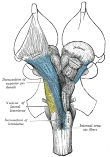
Peduncular means pertaining to the peduncle, which is a neural tract running to and from the pons on the brain stem. These hallucinations usually occur in the evenings, but not during drowsiness, as in the case of hypnagogic hallucination. The subject is usually fully conscious and then can interact with the hallucinatory characters for extended periods of time. As in the case of hypnagogic hallucinations, insight into the nature of the images remains intact. The false images can occur in any part of the visual field, and are rarely polymodal.

Delirium tremens

One of the more enigmatic forms of visual hallucination is the highly variable, possibly polymodal delirium tremens. Individuals with delirium tremens may be agitated and confused, especially in the later stages of this disease. Insight is gradually reduced with the progression of this disorder. Sleep is disturbed and occurs for a shorter period of time, with rapid eye movement sleep.

Parkinson's disease and Lewy body dementia

Parkinson's disease is linked with Lewy body dementia for their similar hallucinatory symptoms. The symptoms strike during the evening in any part of the visual field, and are rarely polymodal. The segue into hallucination may begin with illusions where sensory perception is greatly distorted, but no novel sensory information is present. These typically last for several minutes, during which time the subject may be either conscious and normal or drowsy/inaccessible. Insight into these hallucinations is usually preserved and REM sleep is usually reduced. Parkinson's disease is usually associated with a degraded substantia nigra pars compacta, but recent evidence suggests that PD affects a number of sites in the brain. Some places of noted degradation include the median raphe nuclei, the noradrenergic parts of the locus coeruleus, and the cholinergic neurons in the parabrachial area and pedunculopontine nuclei of the tegmentum.

Migraine coma

This type of hallucination is usually experienced during the recovery from a comatose state. The migraine coma can last for up to two days, and a state of depression is sometimes comorbid. The hallucinations occur during states of full consciousness, and insight into the hallucinatory nature of the images is preserved. It has been noted that ataxic lesions accompany the migraine coma.

Charles Bonnet syndrome

Charles Bonnet syndrome is the name given to visual hallucinations experienced by a partially or severely sight impaired person. The hallucinations can occur at any time and can distress people of any age, as they may not initially be aware that they are hallucinating. They may fear for their own mental health initially, which may delay them sharing with carers until they start to understand it themselves. The hallucinations can frighten and disconcert as to what is real and what is not. The hallucinations can sometimes be dispersed by eye movements, or by reasoned logic such as, "I can see fire but there is no smoke and there is no heat from it" or perhaps, "We have an infestation of rats but they have pink ribbons with a bell tied on their necks." Over elapsed months and years, the hallucinations may become more or less frequent with changes in ability to see. The length of time that the sight impaired person can have these hallucinations varies according to the underlying speed of eye deterioration. A differential diagnosis are ophthalmopathic hallucinations.

Focal epilepsy

Visual hallucinations due to focal seizures differ depending on the region of the brain where the seizure occurs. For example, visual hallucinations during occipital lobe seizures are typically visions of brightly colored, geometric shapes that may move across the visual field, multiply, or form concentric rings and generally persist from a few seconds to a few minutes. They are usually unilateral and localized to one part of the visual field on the contralateral side of the seizure focus, typically the temporal field. However, unilateral visions moving horizontally across the visual field begin on the contralateral side and move toward the ipsilateral side.

Temporal lobe seizures, on the other hand, can produce complex visual hallucinations of people, scenes, animals, and more as well as distortions of visual perception. Complex hallucinations may appear to be real or unreal, may or may not be distorted with respect to size, and may seem disturbing or affable, among other variables. One rare but notable type of hallucination is heautoscopy, a hallucination of a mirror image of one's self. These "other selves" may be perfectly still or performing complex tasks, may be an image of a younger self or the present self, and tend to be briefly present. Complex hallucinations are a relatively uncommon finding in temporal lobe epilepsy patients. Rarely, they may occur during occipital focal seizures or in parietal lobe seizures.

Distortions in visual perception during a temporal lobe seizure may include size distortion (micropsia or macropsia), distorted perception of movement (where moving objects may appear to be moving very slowly or to be perfectly still), a sense that surfaces such as ceilings and even entire horizons are moving farther away in a fashion similar to the dolly zoom effect, and other illusions. Even when consciousness is impaired, insight into the hallucination or illusion is typically preserved.

Drug-induced hallucination

Drug-induced hallucinations are caused by hallucinogens, dissociatives, and deliriants, including many drugs with anticholinergic actions and certain stimulants, which are known to cause visual and auditory hallucinations. Some psychedelics such as lysergic acid diethylamide (LSD) and psilocybin can cause hallucinations that range in the spectrum of mild to intense.

Hallucinations, pseudohallucinations, or intensification of pareidolia, particularly auditory, are known side effects of opioids to different degrees—it may be associated with the absolute degree of agonism or antagonism of especially the kappa opioid receptor, sigma receptors, delta opioid receptor and the NMDA receptors or the overall receptor activation profile as synthetic opioids like those of the pentazocine, levorphanol, fentanyl, pethidine, methadone and some other families are more associated with this side effect than natural opioids like morphine and codeine and semi-synthetics like hydromorphone, amongst which there also appears to be a stronger correlation with the relative analgesic strength. Three opioids, Cyclazocine (a benzormorphan opioid/pentazocine relative) and two levorphanol-related morphinan opioids, Cyclorphan and Dextrorphan are classified as hallucinogens, and Dextromethorphan as a dissociative. These drugs also can induce sleep (relating to hypnagogic hallucinations) and especially the pethidines have atropine-like anticholinergic activity, which was possibly also a limiting factor in the use, the psychotomimetic side effects of potentiating morphine, oxycodone, and other opioids with scopolamine (respectively in the Twilight Sleep technique and the combination drug Skophedal, which was eukodal (oxycodone), scopolamine and ephedrine, called the "wonder drug of the 1930s" after its invention in Germany in 1928, but only rarely specially compounded today) (q.q.v.).

Sensory deprivation hallucination

Hallucinations can be caused by sensory deprivation when it occurs for prolonged periods of time, and almost always occurs in the modality being deprived (visual for blindfolded/darkness, auditory for muffled conditions, etc.) 

Experimentally-induced hallucinations

Anomalous experiences, such as so-called benign hallucinations, may occur in a person in a state of good mental and physical health, even in the apparent absence of a transient trigger factor such as fatigue, intoxication or sensory deprivation.

The evidence for this statement has been accumulating for more than a century. Studies of benign hallucinatory experiences go back to 1886 and the early work of the Society for Psychical Research, which suggested approximately 10% of the population had experienced at least one hallucinatory episode in the course of their life. More recent studies have validated these findings; the precise incidence found varies with the nature of the episode and the criteria of "hallucination" adopted, but the basic finding is now well-supported.

Non-celiac gluten sensitivity

There is tentative evidence of a relationship with non-celiac gluten sensitivity, the so-called "gluten psychosis".

Pathophysiology

Dopaminergic and serotonergic hallucinations

It has been reported that in serotonergic hallucinations, the person maintains an awareness that they are hallucinating, unlike dopaminergic hallucinations.

Neuroanatomy

Hallucinations are associated with structural and functional abnormalities in primary and secondary sensory cortices. Reduced grey matter in regions of the superior temporal gyrus/middle temporal gyrus, including Broca's area, is associated with auditory hallucinations as a trait, while acute hallucinations are associated with increased activity in the same regions along with the hippocampus, parahippocampus, and the right hemispheric homologue of Broca's area in the inferior frontal gyrus. Grey and white matter abnormalities in visual regions are associated with visual hallucinations in diseases such as Alzheimer's disease, further supporting the notion of dysfunction in sensory regions underlying hallucinations.

One proposed model of hallucinations posits that over-activity in sensory regions, which is normally attributed to internal sources via feedforward networks to the inferior frontal gyrus, is interpreted as originating externally due to abnormal connectivity or functionality of the feedforward network. This is supported by cognitive studies of those with hallucinations, who have demonstrated abnormal attribution of self generated stimuli.

Disruptions in thalamocortical circuitry may underlie the observed top down and bottom up dysfunction. Thalamocortical circuits, composed of projections between thalamic and cortical neurons and adjacent interneurons, underlie certain electrophysical characteristics (gamma oscillations) that are underlie sensory processing. Cortical inputs to thalamic neurons enable attentional modulation of sensory neurons. Dysfunction in sensory afferents, and abnormal cortical input may result in pre-existing expectations modulating sensory experience, potentially resulting in the generation of hallucinations. Hallucinations are associated with less accurate sensory processing, and more intense stimuli with less interference are necessary for accurate processing and the appearance of gamma oscillations (called "gamma synchrony"). Hallucinations are also associated with the absence of reduction in P50 amplitude in response to the presentation of a second stimuli after an initial stimulus; this is thought to represent failure to gate sensory stimuli, and can be exacerbated by dopamine release agents.

Abnormal assignment of salience to stimuli may be one mechanism of hallucinations. Dysfunctional dopamine signaling may lead to abnormal top down regulation of sensory processing, allowing expectations to distort sensory input.

Treatments

There are few treatments for many types of hallucinations. However, for those hallucinations caused by mental disease, a psychologist or psychiatrist should be consulted, and treatment will be based on the observations of those doctors. Antipsychotic and atypical antipsychotic medication may also be utilized to treat the illness if the symptoms are severe and cause significant distress. For other causes of hallucinations there is no factual evidence to support any one treatment is scientifically tested and proven. However, abstaining from hallucinogenic drugs, stimulant drugs, managing stress levels, living healthily, and getting plenty of sleep can help reduce the prevalence of hallucinations. In all cases of hallucinations, medical attention should be sought out and informed of one's specific symptoms. Meta-analyses show that cognitive behavioral therapy and metacognitive training can also reduce the severity of hallucinations.

Epidemiology

Prevalence of hallucinations varies depending on underlying medical conditions, which sensory modalities are affected, age and culture. As of 2022, auditory hallucinations are the most well studied and most common sensory modality of hallucinations, with an estimated lifetime prevalence of 9.6%. Children and adolescents have been found to experience similar rates (12.7% and 12.4% respectively) which occur mostly during late childhood and adolescence. This is compared with adults and those over 60 (with rates of 5.8% and 4.8% respectively). For those with schizophrenia, the lifetime prevalence of hallucinations is 80% and the estimated prevalence of visual hallucinations is 27%, compared to 79% for auditory hallucinations. A 2019 study suggested 16.2% of adults with hearing impairment experience hallucinations, with prevalence rising to 24% in the most hearing impaired group.

A risk factor for multimodal hallucinations is prior experience of unimodal hallucinations. In 90% cases of psychosis, a visual hallucination occurs in combination with another sensory modality, most often being auditory or somatic. In schizophrenia, multimodal hallucinations are twice as common as unimodal ones.

A 2015 review of 55 publications from 1962 to 2014 found 16–28.6% of those experiencing hallucinations report at least some religious content in them, along with 20–60% reporting some religious content in delusions. There is some evidence for delusions being a risk factor for religious hallucinations, with and 61.7% of people having experienced any delusion and 75.9% of those having experienced a religious delusion found to also experience hallucinations.

Fearmongering

From Wikipedia, the free encyclopedia https://en.wikipedia.org/wiki/Fearmongering Fearmongering ,...