Search This Blog

Sunday, September 8, 2024

Lactose intolerance

From Wikipedia, the free encyclopedia
Lactose intolerance
Other namesLactase deficiency, hypolactasia, alactasia
Lactose is made up of two simple sugars
SpecialtyGastroenterology
SymptomsAbdominal pain, bloating, diarrhea, flatulence, nausea
ComplicationsDoes not cause damage to the GI tract
Usual onset30–120 minutes after consuming dairy products
CausesNon-increased ability to digest lactose (genetic, small intestine injury)
Differential diagnosisIrritable bowel syndrome, celiac disease, inflammatory bowel disease, milk allergy
TreatmentDecreasing lactose in the diet, lactase supplements, treat the underlying cause
MedicationLactase
Frequency~65% of people worldwide (less common in Europeans and East Africans)

Lactose intolerance is caused by a lessened ability or a complete inability to digest lactose, a sugar found in dairy products. Humans vary in the amount of lactose they can tolerate before symptoms develop. Symptoms may include abdominal pain, bloating, diarrhea, flatulence, and nausea. These symptoms typically start thirty minutes to two hours after eating or drinking something containing lactose, with the severity typically depending on the amount consumed. Lactose intolerance does not cause damage to the gastrointestinal tract.

Lactose intolerance is due to the lack of the enzyme lactase in the small intestines to break lactose down into glucose and galactose. There are four types: primary, secondary, developmental, and congenital. Primary lactose intolerance occurs as the amount of lactase declines as people grow up. Secondary lactose intolerance is due to injury to the small intestine. Such injury could be the result of infection, celiac disease, inflammatory bowel disease, or other diseases. Developmental lactose intolerance may occur in premature babies and usually improves over a short period of time. Congenital lactose intolerance is an extremely rare genetic disorder in which little or no lactase is made from birth. The reduction of lactase production starts typically in late childhood or early adulthood, but prevalence increases with age.

Diagnosis may be confirmed if symptoms resolve following eliminating lactose from the diet. Other supporting tests include a hydrogen breath test and a stool acidity test. Other conditions that may produce similar symptoms include irritable bowel syndrome, celiac disease, and inflammatory bowel disease. Lactose intolerance is different from a milk allergy. Management is typically by decreasing the amount of lactose in the diet, taking lactase supplements, or treating the underlying disease. People are typically able to drink at least one cup of milk without developing symptoms, with greater amounts tolerated if drunk with a meal or throughout the day.

Worldwide, around 65% of adults are affected by lactose malabsorption. Other mammals usually lose the ability to digest lactose after weaning. Lactose intolerance is the ancestral state of all humans before the recent evolution of lactase persistence in some cultures, which extends lactose tolerance into adulthood. Lactase persistence evolved in several populations independently, probably as an adaptation to the domestication of dairy animals around 10,000 years ago. Today the prevalence of lactose tolerance varies widely between regions and ethnic groups. The ability to digest lactose is most common in people of European descent, and to a lesser extent in some parts of the Middle East and Africa. Lactose intolerance is most common among people of East Asian descent, with 90% lactose intolerance, and Jewish descent, as well as in many African countries and Arab countries. Traditional food cultures reflect local variations in tolerance and historically many societies have adapted to low levels of tolerance by making dairy products that contain less lactose than fresh milk. The medicalization of lactose intolerance as a disorder has been attributed to biases in research history, since most early studies were conducted amongst populations which are normally tolerant, as well as the cultural and economic importance and impact of milk in countries such as the United States.

Terminology

Lactose intolerance primarily refers to a syndrome with one or more symptoms upon the consumption of food substances containing lactose sugar. Individuals may be lactose intolerant to varying degrees, depending on the severity of these symptoms.

Hypolactasia is the term specifically for the small intestine producing little or no lactase enzyme. If a person with hypolactasia consumes lactose sugar, it results in lactose malabsorption. The digestive system is unable to process the lactose sugar, and the unprocessed sugars in the gut produce the symptoms of lactose intolerance.

Lactose intolerance is not an allergy, because it is not an immune response, but rather a sensitivity to dairy caused by a deficiency of lactase enzyme. Milk allergy, occurring in about 2% of the population, is a separate condition, with distinct symptoms that occur when the presence of milk proteins trigger an immune reaction.

Signs and symptoms

The principal manifestation of lactose intolerance is an adverse reaction to products containing lactose (primarily milk), including abdominal bloating and cramps, flatulence, diarrhea, nausea, borborygmi, and vomiting (particularly in adolescents). These appear one-half to two hours after consumption. The severity of these signs and symptoms typically increases with the amount of lactose consumed; most lactose-intolerant people can tolerate a certain level of lactose in their diets without ill effects.

Because lactose intolerance is not an allergy, it does not produce allergy symptoms (such as itching, hives, or anaphylaxis).

Causes

Lactose intolerance is a consequence of lactase deficiency, which may be genetic (primary hypolactasia and primary congenital alactasia) or environmentally induced (secondary or acquired hypolactasia). In either case, symptoms are caused by insufficient levels of lactase in the lining of the duodenum. Lactose, a disaccharide molecule found in milk and dairy products, cannot be directly absorbed through the wall of the small intestine into the bloodstream, so, in the absence of lactase, passes intact into the colon. Bacteria in the colon can metabolise lactose, and the resulting fermentation produces copious amounts of gas (a mixture of hydrogen, carbon dioxide, and methane) that causes the various abdominal symptoms. The unabsorbed sugars and fermentation products also raise the osmotic pressure of the colon, causing an increased flow of water into the bowels (diarrhea).

Lactose intolerance in infants (congenital lactase deficiency) is caused by mutations in the LCT gene. The LCT gene provides the instructions for making lactase. Mutations are believed to interfere with the function of lactase, causing affected infants to have a severely impaired ability to digest lactose in breast milk or formula. Lactose intolerance in adulthood is a result of gradually decreasing activity (expression) of the LCT gene after infancy, which occurs in most humans. The specific DNA sequence in the MCM6 gene helps control whether the LCT gene is turned on or off. At least several thousand years ago, some humans developed a mutation in the MCM6 gene that keeps the LCT gene turned on even after breast feeding is stopped. Populations that are lactose intolerant lack this mutation. The LCT and MCM6 genes are both located on the long arm (q) of chromosome 2 in region 21. The locus can be expressed as 2q21. The lactase deficiency also could be linked to certain heritages and varies widely. A 2016 study of over 60,000 participants from 89 countries found regional prevalence of lactose malabsorption was "64% (54–74) in Asia (except Middle East), 47% (33–61) in eastern Europe, Russia, and former Soviet Republics, 38% (CI 18–57) in Latin America, 70% (57–83) in the Middle East, 66% (45–88) in northern Africa, 42% (13–71) in northern America, 45% (19–71) in Oceania, 63% (54–72) in sub-Saharan Africa, and 28% (19–37) in northern, southern and western Europe." According to Johns Hopkins Medicine, lactose intolerance is more common in Asian Americans, African Americans, Mexican Americans, and Native Americans. Analysis of the DNA of 94 ancient skeletons in Europe and Russia concluded that the mutation for lactose tolerance appeared about 4,300 years ago and spread throughout the European population.

Some human populations have developed lactase persistence, in which lactase production continues into adulthood probably as a response to the benefits of being able to digest milk from farm animals. Some have argued that this links intolerance to natural selection favoring lactase-persistent individuals, but it is also consistent with a physiological response to decrease lactase production when it is not needed in cultures in which dairy products are not an available food source. Although populations in Europe, India, Arabia, and Africa were first thought to have high rates of lactase persistence because of a single mutation, lactase persistence has been traced to a number of mutations that occurred independently. Different alleles for lactase persistence have developed at least three times in East African populations, with persistence extending from 26% in Tanzania to 88% in the Beja pastoralist population in Sudan.

The accumulation of epigenetic factors, primarily DNA methylation, in the extended LCT region, including the gene enhancer located in the MCM6 gene near C/T-13910 SNP, may also contribute to the onset of lactose intolerance in adults. Age-dependent expression of LCT in mice intestinal epithelium has been DNA methylation in the gene enhancer.

Lactose intolerance is classified according to its causes as:

Primary hypolactasia
Primary hypolactasia, or primary lactase deficiency, is genetic, develops in childhood at various ages, and is caused by the absence of a lactase persistence allele. In individuals without the lactase persistence allele, less lactase is produced by the body over time, leading to hypolactasia in adulthood. The frequency of lactase persistence, which allows lactose tolerance, varies enormously worldwide, with the highest prevalence in Northwestern Europe, declines across southern Europe and the Middle East and is low in Asia and most of Africa, although it is common in pastoralist populations from Africa.
Secondary hypolactasia
Secondary hypolactasia or secondary lactase deficiency, also called acquired hypolactasia or acquired lactase deficiency, is caused by an injury to the small intestine. This form of lactose intolerance can occur in both infants and lactase persistent adults and is generally reversible. It may be caused by acute gastroenteritis, coeliac disease, Crohn's disease, ulcerative colitis, chemotherapy, intestinal parasites (such as giardia), or other environmental causes.
Primary congenital alactasia
Primary congenital alactasia, also called congenital lactase deficiency, is an extremely rare, autosomal recessive enzyme defect that prevents lactase expression from birth. People with congenital lactase deficiency cannot digest lactose from birth, so cannot digest breast milk. This genetic defect is characterized by a complete lack of lactase (alactasia). About 40 cases have been reported worldwide, mainly limited to Finland. Before the 20th century, babies born with congenital lactase deficiency often did not survive, but death rates decreased with soybean-derived infant formulas and manufactured lactose-free dairy products.

Diagnosis

In order to assess lactose intolerance, intestinal function is challenged by ingesting more dairy products than can be readily digested. Clinical symptoms typically appear within 30 minutes, but may take up to two hours, depending on other foods and activities. Substantial variability in response (symptoms of nausea, cramping, bloating, diarrhea, and flatulence) is to be expected, as the extent and severity of lactose intolerance varies among individuals.

The next step is to determine whether it is due to primary lactase deficiency or an underlying disease that causes secondary lactase deficiency. Physicians should investigate the presence of undiagnosed coeliac disease, Crohn's disease, or other enteropathies when secondary lactase deficiency is suspected and infectious gastroenteritis has been ruled out.

Lactose intolerance is distinct from milk allergy, an immune response to cow's milk proteins. They may be distinguished in diagnosis by giving lactose-free milk, producing no symptoms in the case of lactose intolerance, but the same reaction as to normal milk in the presence of a milk allergy. A person can have both conditions. If positive confirmation is necessary, four tests are available.

Hydrogen breath test

In a hydrogen breath test, the most accurate lactose intolerance test, after an overnight fast, 25 grams of lactose (in a solution with water) are swallowed. If the lactose cannot be digested, enteric bacteria metabolize it and produce hydrogen, which, along with methane, if produced, can be detected on the patient's breath by a clinical gas chromatograph or compact solid-state detector. The test takes about 2.5 hours to complete. If the hydrogen levels in the patient's breath are high, they may have lactose intolerance. This test is not usually done on babies and very young children, because it can cause severe diarrhea.

Lactose tolerance test

In conjunction, measuring blood glucose level every 10 to 15 minutes after ingestion will show a "flat curve" in individuals with lactose malabsorption, while the lactase persistent will have a significant "top", with a typical elevation of 50% to 100%, within one to two hours. However, due to the need for frequent blood sampling, this approach has been largely replaced by breath testing.

After an overnight fast, blood is drawn and then 50 grams of lactose (in aqueous solution) are swallowed. Blood is then drawn again at the 30-minute, 1-hour, 2-hour, and 3-hour marks. If the lactose cannot be digested, blood glucose levels will rise by less than 20 mg/dl.

Stool acidity test

This test can be used to diagnose lactose intolerance in infants, for whom other forms of testing are risky or impractical. The infant is given lactose to drink. If the individual is tolerant, the lactose is digested and absorbed in the small intestine; otherwise, it is not digested and absorbed, and it reaches the colon. The bacteria in the colon, mixed with the lactose, cause acidity in stools. Stools passed after the ingestion of the lactose are tested for level of acidity. If the stools are acidic, the infant is intolerant to lactose. Stool pH in lactose intolerance is less than 5.5.

Intestinal biopsy

An intestinal biopsy must confirm lactase deficiency following discovery of elevated hydrogen in the hydrogen breath test. Modern techniques have enabled a bedside test, identifying presence of lactase enzyme on upper gastrointestinal endoscopy instruments. However, for research applications such as mRNA measurements, a specialist laboratory is required.

Stool sugar chromatography

Chromatography can be used to separate and identify undigested sugars present in faeces. Although lactose may be detected in the faeces of people with lactose intolerance, this test is not considered reliable enough to conclusively diagnose or exclude lactose intolerance.

Genetic diagnostic

Genetic tests may be useful in assessing whether a person has primary lactose intolerance. Lactase activity persistence in adults is associated with two polymorphisms: C/T 13910 and G/A 22018 located in the MCM6 gene. These polymorphisms may be detected by molecular biology techniques at the DNA extracted from blood or saliva samples; genetic kits specific for this diagnosis are available. The procedure consists of extracting and amplifying DNA from the sample, following with a hybridation protocol in a strip. Colored bands are obtained as result, and depending on the different combinations, it would be possible to determine whether the patient is lactose intolerant. This test allows a noninvasive definitive diagnostic.

Frequency

Lactose intolerance is most common in people of East Asian descent, with 70 to 100 percent of people affected in these communities. Lactose intolerance is also more common in people of West African, Arab, and Jewish descent, while only about 5 percent of people of northern European descent are lactose intolerant.

Management

When lactose intolerance is due to secondary lactase deficiency, treatment of the underlying disease may allow lactase activity to return to normal levels. In people with celiac disease, lactose intolerance normally reverts or improves several months after starting a gluten-free diet, but temporary dietary restriction of lactose may be needed.

People with primary lactase deficiency cannot modify their body's ability to produce lactase. In societies where lactose intolerance is the norm, it is not considered a condition that requires treatment. However, where dairy is a larger component of the normal diet, a number of efforts may be useful. There are four general principles in dealing with lactose intolerance: avoidance of dietary lactose, substitution to maintain nutrient intake, regulation of calcium intake, and use of enzyme substitute. Regular consumption of dairy food by lactase deficient individuals may also reduce symptoms of intolerance by promoting colonic bacteria adaptation.

Dietary avoidance

The primary way of managing the symptoms of lactose intolerance is to limit the intake of lactose to a level that can be tolerated. Lactase deficient individuals vary in the amount of lactose they can tolerate, and some report that their tolerance varies over time, depending on health status and pregnancy. However, as a rule of thumb, people with primary lactase deficiency and no small intestine injury are usually able to consume at least 12 grams of lactose per sitting without symptoms, or with only mild symptoms, with greater amounts tolerated if consumed with a meal or throughout the day.

Typical lactose levels in dairy products
Dairy product Serving
size
Lactose
content
Fraction
Milk, regular 250 ml(g) 12 g 4.8%
Milk, reduced fat 250 ml(g) 13 g 5.2%
Yogurt, plain, regular 200 g 9 g 4.5%
Yogurt, plain, low-fat 200 g 12 g 6.0%
Cheddar cheese 30 g 0.02 g 0.07%
Cottage cheese 30 g 0.1 g 0.33%
Butter 5 g 0.03 g 0.6%
Ice cream 50 g 3 g 6.0%

Lactose is found primarily in dairy products, which vary in the amount of lactose they contain:

  • Milk – unprocessed cow's milk is about 4.7% lactose; goat's milk 4.7%; sheep's milk 4.7%; buffalo milk 4.86%; and yak milk 4.93%.
  • Sour cream and buttermilk – if made in the traditional way, this may be tolerable, but most modern brands add milk solids.
  • Yogurtlactobacilli used in the production of yogurt metabolize lactose to varying degrees, depending on the type of yogurt. Some bacteria found in yogurt also produce their own lactase, which facilitates digestion in the intestines of lactose intolerant individuals.
  • Cheese – The curdling of cheese concentrates most of the lactose from milk into the whey: fresh cottage cheese contains 7% of the lactose found in an equivalent mass of milk. Further fermentation and aging converts the remaining lactose into lactic acid; traditionally made hard cheeses, which have a long ripening period, contain virtually no lactose: cheddar contains less than 1.5% of the lactose found in an equivalent mass of milk. However, manufactured cheeses may be produced using processes that do not have the same lactose-reducing properties.

There used to be a lack of standardization on how lactose is measured and reported in food. The different molecular weights of anhydrous lactose or lactose monohydrate result in up to 5% difference. One source recommends using the "carbohydrates" or "sugars" part of the nutritional label as surrogate for lactose content, but such "lactose by difference" values are not assured to correspond to real lactose content. The stated dairy content of a product also varies according to manufacturing processes and labelling practices, and commercial terminology varies between languages and regions. As a result, absolute figures for the amount of lactose consumed (by weight) may not be very reliable.

Kosher products labeled pareve or fleishig are free of milk. However, if a "D" (for "dairy") is present next to the circled "K", "U", or other hechsher, the food product likely contains milk solids, although it may also simply indicate the product was produced on equipment shared with other products containing milk derivatives.

Lactose is also a commercial food additive used for its texture, flavor, and adhesive qualities. It is found in additives labelled as casein, caseinate, whey, lactoserum, milk solids, modified milk ingredients, etc. As such, lactose is found in foods such as processed meats (sausages/hot dogs, sliced meats, pâtés), gravy stock powder, margarines, sliced breads, breakfast cereals, potato chips, processed foods, medications, prepared meals, meal replacements (powders and bars), protein supplements (powders and bars), and even beers in the milk stout style. Some barbecue sauces and liquid cheeses used in fast-food restaurants may also contain lactose. When dining out, carrying lactose intolerance cards that explain dietary restrictions in the local language can help communicate needs to restaurant staff. Lactose is often used as the primary filler (main ingredient) in most prescription and non-prescription solid pill form medications, though product labeling seldom mentions the presence of 'lactose' or 'milk', and neither do product monograms provided to pharmacists, and most pharmacists are unaware of the very wide scale yet common use of lactose in such medications until they contact the supplier or manufacturer for verification.

Milk substitutes

Plant-based milks and derivatives such as soy milk, rice milk, almond milk, coconut milk, hazelnut milk, oat milk, hemp milk, macadamia nut milk, and peanut milk are inherently lactose-free. Low-lactose and lactose-free versions of foods are often available to replace dairy-based foods for those with lactose intolerance.

Lactase supplements

When lactose avoidance is not possible, or on occasions when a person chooses to consume such items, then enzymatic lactase supplements may be used.

Lactase enzymes similar to those produced in the small intestines of humans are produced industrially by fungi of the genus Aspergillus. The enzyme, β-galactosidase, is available in tablet form in a variety of doses, in many countries without a prescription. It functions well only in high-acid environments, such as that found in the human gut due to the addition of gastric juices from the stomach. Unfortunately, too much acid can denature it, so it should not be taken on an empty stomach. Also, the enzyme is ineffective if it does not reach the small intestine by the time the problematic food does. Lactose-sensitive individuals can experiment with both timing and dosage to fit their particular needs.

While essentially the same process as normal intestinal lactose digestion, direct treatment of milk employs a different variety of industrially produced lactase. This enzyme, produced by yeast from the genus Kluyveromyces, takes much longer to act, must be thoroughly mixed throughout the product, and is destroyed by even mildly acidic environments. Its main use is in producing the lactose-free or lactose-reduced dairy products sold in supermarkets.

Rehabituation to dairy products

Regular consumption of dairy foods containing lactose can promote a colonic bacteria adaptation, enhancing a favorable microbiome, which allows people with primary lactase deficiency to diminish their intolerance and to consume more dairy foods. The way to induce tolerance is based on progressive exposure, consuming smaller amounts frequently, distributed throughout the day. Lactose intolerance can also be managed by ingesting live yogurt cultures containing lactobacilli that are able to digest the lactose in other dairy products.

Epidemiology

An estimate of the percentage of adults that can digest lactose in the indigenous population of the Old World

Worldwide, about 65% of people experience some form of lactose intolerance as they age past infancy, but there are significant differences between populations and regions. As few as 5% of northern Europeans are lactose intolerant, while as many as 90% of adults in parts of Asia are lactose intolerant.

In northern European countries, early adoption of dairy farming conferred a selective evolutionary advantage to individuals that could tolerate lactose. This led to higher frequencies of lactose tolerance in these countries. For example, almost 100% of Irish people are predicted to be lactose tolerant. Conversely, regions of the south, such as Africa, did not adopt dairy farming as early and tolerance from milk consumption did not occur the same way as in northern Europe. Lactose intolerance is common among people of Jewish descent, as well as from West Africa, the Arab countries, Greece, and Italy. Different populations will present certain gene constructs depending on the evolutionary and cultural pre-settings of the geographical region.

History

Greater lactose tolerance has come about in two ways. Some populations have developed genetic changes to allow the digestion of lactose: lactase persistence. Other populations developed cooking methods like milk fermentation.

Lactase persistence in humans evolved relatively recently (in the last 10,000 years) among some populations. Around 8,000 years ago in modern-day Turkey, humans became reliant on newly-domesticated animals that could be milked; such as cows, sheep, and goats. This resulted in higher frequency of lactase persistence. Lactase persistence became high in regions such as Europe, Scandinavia, the Middle East and Northwestern India. However, most people worldwide remain lactase non-persistent. Populations that raised animals not used for milk tend to have 90–100 percent of a lactose intolerant rate. For this reason, lactase persistence is of some interest to the fields of anthropology, human genetics, and archaeology, which typically use the genetically derived persistence/non-persistence terminology.

The rise of dairy and producing dairy related products from cow milk alone, varies across different regions of the world, aside from genetic predisposition. The process of turning milk into cheese dates back earlier than 5200 BC.

DNA analysis in February 2012 revealed that Ötzi was lactose intolerant, supporting the theory that lactose intolerance was still common at that time, despite the increasing spread of agriculture and dairying.

Genetic analysis shows lactase persistence has developed several times in different places independently in an example of convergent evolution.

History of research

It was not until relatively recently that medicine recognised the worldwide prevalence of lactose intolerance and its genetic causes. Its symptoms were described as early as Hippocrates (460–370 BC), but until the 1960s, the prevailing assumption was that tolerance was the norm. Intolerance was explained as the result of a milk allergy, intestinal pathogens, or as being psychosomatic – it being recognised that some cultures did not practice dairying, and people from those cultures often reacted badly to consuming milk. Two reasons have been given for this misconception. One was that early research was conducted solely on European-descended populations, which have an unusually low incidence of lactose intolerance and an extensive cultural history of dairying. As a result, researchers wrongly concluded that tolerance was the global norm. Another reason is that lactose intolerance tends to be under-reported: lactose intolerant individuals can tolerate at least some lactose before they show symptoms, and their symptoms differ in severity. The large majority of people are able to digest some quantity of milk, for example in tea or coffee, without developing any adverse effects. Fermented dairy products, such as cheese, also contain significantly less lactose than plain milk. Therefore, in societies where tolerance is the norm, many lactose intolerant people who consume only small amounts of dairy, or have only mild symptoms, may be unaware that they cannot digest lactose.

Eventually, in the 1960s, it was recognised that lactose intolerance was correlated with race in the United States. Subsequent research revealed that lactose intolerance was more common globally than tolerance, and that the variation was due to genetic differences, not an adaptation to cultural practices.

Other animals

Most mammals normally cease to produce lactase and become lactose intolerant after weaning. The downregulation of lactase expression in mice could be attributed to the accumulation of DNA methylation in the Lct gene and the adjacent Mcm6 gene.

Food intolerance

From Wikipedia, the free encyclopedia
https://en.wikipedia.org/wiki/Food_intolerance

Food intolerance is a detrimental reaction, often delayed, to a food, beverage, food additive, or compound found in foods that produces symptoms in one or more body organs and systems, but generally refers to reactions other than food allergy. Food hypersensitivity is used to refer broadly to both food intolerances and food allergies.

Food allergies are immune reactions, typically an IgE reaction caused by the release of histamine but also encompassing non-IgE immune responses. This mechanism causes allergies to typically give immediate reaction (a few minutes to a few hours) to foods.

Food intolerances can be classified according to their mechanism. Intolerance can result from the absence of specific chemicals or enzymes needed to digest a food substance, as in hereditary fructose intolerance. It may be a result of an abnormality in the body's ability to absorb nutrients, as occurs in fructose malabsorption. Food intolerance reactions can occur to naturally occurring chemicals in foods, as in salicylate sensitivity. Drugs sourced from plants, such as aspirin, can also cause these kinds of reactions.

Definitions

Food hypersensitivity is used to refer broadly to both food intolerances and food allergies. There are a variety of earlier terms which are no longer in use such as "pseudo-allergy".

Food intolerance reactions can include pharmacologic, metabolic, and gastro-intestinal responses to foods or food compounds. Food intolerance does not include either psychological responses or foodborne illness.

A non-allergic food hypersensitivity is an abnormal physiological response. It can be difficult to determine the poorly tolerated substance as reactions can be delayed, dose-dependent, and a particular reaction-causing compound may be found in many foods.

  • Metabolic food reactions are due to inborn or acquired errors of metabolism of nutrients, such as in lactase deficiency, phenylketonuria and favism.
  • Pharmacological reactions are generally due to low-molecular-weight chemicals which occur either as natural compounds, such as salicylates, amines and glutamates or to food additives, such as preservatives, colouring, emulsifiers and flavour enhancers. These chemicals are capable of causing drug-like (biochemical) side effects in susceptible individuals.
  • Gastro-intestinal (GI) reactions can be due to malabsorption or other GI tract abnormalities.
  • Immunological responses are mediated by non-IgE immunoglobulins, where the immune system recognises a particular food as a foreign body.
  • Toxins may either be present naturally in food, be released by bacteria, or be due to contamination of food products. Toxic food reactions are caused by the direct action of a food or substance without immune involvement.
  • Psychological reactions involve manifestation of clinical symptoms caused not by food but by emotions associated with food. These symptoms do not occur when the food is given in an unrecognisable form.

Elimination diets are useful to assist in the diagnosis of food intolerance. There are specific diagnostic tests for certain food intolerances.

Signs and symptoms

Food intolerance is more chronic, less acute, less obvious in its presentation, and often more difficult to diagnose than a food allergy. Symptoms of food intolerance vary greatly, and can be mistaken for the symptoms of a food allergy. While true allergies are associated with fast-acting immunoglobulin IgE responses, it can be difficult to determine the offending food causing a food intolerance because the response generally takes place over a prolonged period of time. Thus, the causative agent and the response are separated in time, and may not be obviously related. Food intolerance symptoms usually begin about half an hour after eating or drinking the food in question, but sometimes symptoms may be delayed by up to 48 hours.

Food intolerance can present with symptoms affecting the skin, respiratory tract, gastrointestinal tract (GIT) either individually or in combination. On the skin may include skin rashes, urticaria (hives), angioedema, dermatitis, and eczema. Respiratory tract symptoms can include nasal congestion, sinusitis, pharyngeal irritations, asthma and an unproductive cough. GIT symptoms include mouth ulcers, abdominal cramp, nausea, gas, intermittent diarrhea, constipation, irritable bowel syndrome (IBS), and may include anaphylaxis.

Food intolerance has been found associated with irritable bowel syndrome and inflammatory bowel disease, chronic constipation, chronic hepatitis C infection, eczema, NSAID intolerance, respiratory complaints, including asthma, rhinitis and headache, functional dyspepsia, eosinophilic esophagitis[9] and ear, nose and throat (ENT) illnesses.

Causes

Reactions to chemical components of the diet may be more common than true food allergies, although there is no evidence to support this. They are caused by various organic chemicals occurring naturally in a wide variety of foods, animal and vegetable, more often than to food additives, preservatives, colourings and flavourings, such as sulfites or dyes. Both natural and artificial ingredients may cause adverse reactions in sensitive people if consumed in sufficient amounts, the degree of sensitivity varying between individuals.

Pharmacological responses to naturally occurring compounds in food, or chemical intolerance, can occur in individuals from both allergic and non-allergic family backgrounds. Symptoms may begin at any age, and may develop quickly or slowly. Triggers may range from a viral infection or illness to environmental chemical exposure. Chemical intolerance occurs more commonly in women, which may be because of hormone differences, as many food chemicals mimic hormones.

A deficiency in digestive enzymes can also cause some types of food intolerances. Lactose intolerance is a result of the body not producing sufficient lactase to digest the lactose in milk; dairy foods which are lower in lactose, such as cheese, are less likely to trigger a reaction in this case. Another carbohydrate intolerance caused by enzyme deficiency is hereditary fructose intolerance.

Celiac disease, an autoimmune disorder caused by an immune response to the protein gluten, results in gluten intolerance and can lead to temporary lactose intolerance.

The most widely distributed naturally occurring food chemical capable of provoking reactions is salicylate, although tartrazine and benzoic acid are well recognised in susceptible individuals. Benzoates and salicylates occur naturally in many foods, including fruits, juices, vegetables, spices, herbs, nuts, tea, wines, and coffee. Salicylate sensitivity causes reactions to aspirin and other NSAIDs, and also in foods which naturally contain salicylates, such as cherries.

Other natural chemicals which commonly cause reactions and cross reactivity include amines, nitrates, sulphites and some antioxidants. Chemicals involved in aroma and flavour are often suspect.

The classification or avoidance of foods based on botanical families bears no relationship to their chemical content and is not relevant in the management of food intolerance.

Salicylate-containing foods include apples, citrus fruits, strawberries, tomatoes, and wine, while reactions to chocolate, cheese, bananas, avocado, tomato or wine point to amines as the likely food chemical. Thus, exclusion of single foods does not necessarily identify the chemical responsible as several chemicals can be present in a food, the patient may be sensitive to multiple food chemicals and reaction more likely to occur when foods containing the triggering substance are eaten in a combined quantity that exceeds the patient's sensitivity thresholds. People with food sensitivities have different sensitivity thresholds, and so more sensitive people will react to much smaller amounts of the substance.

Pathogenesis

Food intolerance are all other adverse reactions to food. Subgroups include enzymatic (e.g. lactose intolerance due to lactase deficiency), pharmacological (e.g. reactions against biogenic amines, histamine intolerance), and undefined food intolerance (e.g. against some food additives).

Food intolerances can be caused by enzymatic defects in the digestive system, can also result from pharmacological effects of vasoactive amines present in foods (e.g. histamine), among other metabolic, pharmacological and digestive abnormalities.

Allergies and intolerances to a food group may coexist with separate pathologies; for example, cow's milk allergy (CMA) and lactose intolerance are two distinct pathologies.

Diagnosis

Diagnosis of food intolerance can include hydrogen breath testing for lactose intolerance and fructose malabsorption, professionally supervised elimination diets, and ELISA testing for IgG-mediated immune responses to specific foods. It is important to be able to distinguish between food allergy, food intolerance, and autoimmune disease in the management of these disorders. Non-IgE-mediated intolerance is more chronic, less acute, less obvious in its clinical presentation, and often more difficult to diagnose than allergy, as skin tests and standard immunological studies are not helpful. Elimination diets must remove all poorly tolerated foods, or all foods containing offending compounds. Clinical investigation is generally undertaken only for more serious cases, as for minor complaints which do not significantly limit the person's lifestyle the cure may be more inconvenient than the problem.

Immunoglobulin (IgG) tests measure the types of food-specific antibodies present. There are four types of IgG, IgG1 makes up 60-70% of the total IgG, followed by IgG2 (20-30%), IgG3 (5-8%), and IgG4 (1-4%). Most commercially available tests only test for IgG4 antibodies, however some companies such as YorkTest Laboratories test for all four types.

IgG4 only tests are debatably invalid; IgG4 presence indicates that the person has been repeatedly exposed to food proteins recognized as foreign by the immune system which is a normal physiological response of the immune system after exposure to food components. Although elimination of foods based on IgG-4 testing in IBS patients resulted in an improvement in symptoms, the positive effects of food elimination were more likely due to wheat and milk elimination than IgG-4 test-determined factors. The IgG-4 test specificity is questionable as healthy individuals with no symptoms of food intolerance also test positive for IgG-4 to several foods.

Diagnosis is made using medical history and cutaneous and serological tests to exclude other causes, but to obtain final confirmation a double blind controlled food challenge must be performed. Treatment can involve long-term avoidance, or if possible re-establishing a level of tolerance.

The antigen leukocyte cellular antibody test (ALCAT) has been commercially promoted as an alternative, but has not been reliably shown to be of clinical value.

Prevention

There is emerging evidence from studies of cord blood that both sensitization and the acquisition of tolerance can begin in pregnancy, however, the window of main danger for sensitization to foods extends prenatally, remaining most critical during early infancy when the immune system and intestinal tract are still maturing. There is no conclusive evidence to support the restriction of dairy intake in the maternal diet during pregnancy, and this is generally not recommended since the drawbacks in terms of loss of nutrition can out-weigh the benefits. However, further randomised, controlled trials are required to examine if dietary exclusion by lactating mothers can truly minimize risk to a significant degree and if any reduction in risk is out-weighed by deleterious impacts on maternal nutrition.

A Cochrane review has concluded feeding with a soy formula cannot be recommended for prevention of allergy or food intolerance in infants. Further research may be warranted to determine the role of soy formulas for prevention of allergy or food intolerance in infants unable to be breast fed with a strong family history of allergy or cow's milk protein intolerance. In the case of allergy and celiac disease others recommend a dietary regimen that is effective in the prevention of allergic diseases in high-risk infants, particularly in early infancy. The most effective dietary regimen is exclusive breastfeeding for at least 4–6 months or, in absence of breast milk, formulas with documented reduced allergenicity for at least the first 4 months, combined with avoidance of solid food and cow's milk for the first 4 months.

Management

Individuals can try minor changes of diet to exclude foods causing obvious reactions, and for many this may be adequate without the need for professional assistance. For reasons mentioned above foods causing problems may not be so obvious since food sensitivities may not be noticed for hours or even days after one has digested food. Persons unable to isolate foods and those more sensitive or with disabling symptoms should seek expert medical and dietitian help. The dietetic department of a teaching hospital is a good start.

Guidance can also be given to your general practitioner to assist in diagnosis and management. Food elimination diets have been designed to exclude food compounds likely to cause reactions and foods commonly causing true allergies and those foods where enzyme deficiency cause symptoms. These elimination diets are not everyday diets but intended to isolate problem foods and chemicals.

It takes around five days of total abstinence to unmask a food or chemical, during the first week on an elimination diet withdrawal symptoms can occur but it takes at least two weeks to remove residual traces. If symptoms have not subsided after six weeks, food intolerance is unlikely to be involved and a normal diet should be restarted. Withdrawals are often associated with a lowering of the threshold for sensitivity which assists in challenge testing, but in this period individuals can be ultra-sensitive even to food smells so care must be taken to avoid all exposures.

After two or more weeks if the symptoms have reduced considerably or gone for at least five days then challenge testing can begin. This can be carried out with selected foods containing only one food chemical, to isolate it if reactions occur. In Australia, purified food chemicals in capsule form are available to doctors for patient testing. These are often combined with placebo capsules for control purposes. This type of challenge is more definitive. New challenges should only be given after 48 hours if no reactions occur or after five days of no symptoms if reactions occur.

Once all food chemical sensitivities are identified a dietitian can prescribe an appropriate diet for the individual to avoid foods with those chemicals. Lists of suitable foods are available from various hospitals and patient support groups can give local food brand advice. A dietitian will ensure adequate nutrition is achieved with safe foods and supplements if need be.

Over a period of time it is possible for individuals avoiding food chemicals to build up a level of resistance by regular exposure to small amounts in a controlled way, but care must be taken, the aim being to build up a varied diet with adequate composition.

Prognosis

The prognosis of children diagnosed with intolerance to milk is good: patients respond to diet which excludes cow's milk protein and the majority of patients succeed in forming tolerance. Children with non-IgE-mediated cows milk intolerance have a good prognosis, whereas children with IgE-mediated cows milk allergy in early childhood have a significantly increased risk for persistent allergy, development of other food allergies, asthma and rhinoconjunctivitis.

A study has demonstrated that identifying and appropriately addressing food sensitivity in IBS patients not previously responding to standard therapy results in a sustained clinical improvement and increased overall well-being and quality of life.

Epidemiology

Estimates of the prevalence of food intolerance vary widely from 2% to over 20% of the population. So far only three prevalence studies in Dutch and English adults have been based on double-blind, placebo-controlled food challenges. The reported prevalences of food allergy/intolerance (by questionnaires) were 12% to 19%, whereas the confirmed prevalences varied from 0.8% to 2.4%. For intolerance to food additives the prevalence varied between 0.01 and 0.23%.

Food intolerance rates were found to be similar in the population in Norway. Out of 4,622 subjects with adequately filled-in questionnaires, 84 were included in the study (1.8%) Perceived food intolerance is a common problem with significant nutritional consequences in a population with IBS. Of these 59 (70%) had symptoms related to intake of food, 62% limited or excluded food items from the diet. Tests were performed for food allergy and malabsorption, but not for intolerance. There were no associations between the tests for food allergy and malabsorption and perceived food intolerance, among those with IBS. Perceived food intolerance was unrelated to musculoskeletal pain and mood disorders.

According to the RACP working group, "Though not considered a "cause" of CFS, some patients with chronic fatigue report food intolerances that can exacerbate symptoms."

History

In 1978 Australian researchers published details of an 'exclusion diet' to exclude specific food chemicals from the diet of patients. This provided a basis for challenge with these additives and natural chemicals. Using this approach the role played by dietary chemical factors in the pathogenesis of chronic idiopathic urticaria (CIU) was first established and set the stage for future DBPCT trials of such substances in food intolerance studies.

In 1995 the European Academy of Allergology and Clinical Immunology suggested a classification on the basis of the responsible pathogenetic mechanism; according to this classification, non-toxic reactions can be divided into 'food allergies' when they recognize immunological mechanisms, and 'food intolerances' when there are no immunological implications. Reactions secondary to food ingestion are defined generally as 'adverse reactions to food'.

In 2003 the Nomenclature Review Committee of the World Allergy Organization issued a report of revised nomenclature for global use on food allergy and food intolerance, that has had general acceptance. Food intolerance is described as a 'non-allergic hypersensitivity' to food.

Society and culture

In the UK, scepticism about food intolerance as a specific condition influenced doctors' perceptions of patients and of the patients' underlying problems. However, rather than risk damaging the doctor-patient relationship, general practitioners (GPs) chose - despite their scepticism and guided by an element of awareness of the limitations of modern medicine - to negotiate mutually acceptable ground with patients and with patients' beliefs. As a result, whether due to a placebo effect, a secondary benefit, or a biophysical result of excluding a food from the diet, the GPs acknowledge both personal and therapeutic benefits.

In the Netherlands, patients and their doctors (GPs) have different perceptions of the efficacy of diagnostic and dietary interventions in IBS. Patients consider food intolerance and GPs regard lack of fibre as the main etiologic dietary factor. It has been suggested that Dutch GPs explore the patients' expectations and potentially incorporate these in their approach to IBS patients.

New food labeling regulations were introduced into the US and Europe in 2006, which are said to benefit people with intolerances. In general, food-allergic consumers were not satisfied with the current labelling practices. In the USA food companies propose distinguishing between food allergy and food intolerance and use a mechanism-based (i.e., immunoglobulin-E-mediated), acute life-threatening anaphylaxis that is standardized and measurable and reflects the severity of health risk, as the principal inclusion criterion for food allergen labeling. Symptoms due to, or exacerbated by, food additives usually involve non-IgE-mediated mechanisms (food intolerance) and are usually less severe than those induced by food allergy, but can include anaphylaxis.

Research directions

FODMAPs are fermentable oligo-, di-, monosaccharides and polyols, which are poorly absorbed in the small intestine and subsequently fermented by the bacteria in the distal small and proximal large intestine. This is a normal phenomenon, common to everyone. The resultant production of gas potentially results in bloating and flatulence. Although FODMAPs can produce certain digestive discomfort in some people, not only do they not cause intestinal inflammation, but they avoid it, because they produce beneficial alterations in the intestinal flora that contribute to maintain the good health of the colon. FODMAPs are not the cause of irritable bowel syndrome nor other functional gastrointestinal disorders, but rather a person develops symptoms when the underlying bowel response is exaggerated or abnormal. A low-FODMAP diet might help to improve short-term digestive symptoms in adults with irritable bowel syndrome, but its long-term follow-up can have negative effects because it causes a detrimental impact on the gut microbiota and metabolome. It should only be used for short periods of time and under the advice of a specialist. More studies are needed to assess the true impact of this diet on health.

Also, when a low FODMAP diet is used without a previous complete medical evaluation can cause serious health risks. It can ameliorate and mask the digestive symptoms of serious diseases, such as celiac disease, inflammatory bowel disease and colon cancer, avoiding their correct diagnosis and therapy. This is especially relevant in the case of celiac disease. Since the consumption of gluten is suppressed or reduced with a low-FODMAP diet, the improvement of the digestive symptoms with this diet may not be related to the withdrawal of the FODMAPs, but of gluten, indicating the presence of an unrecognized celiac disease, avoiding its diagnosis and correct treatment, with the consequent risk of several serious health complications, including various types of cancer.

A three-month randomized, blinded, controlled trial on people with irritable bowel syndrome found that those who withdrew from the diet the foods to which they had shown an increased IgG antibody response experienced an improvement in their symptoms. In individuals with Crohn's disease and ulcerative colitis food-specific-IgG-based elimination diets have been shown to be effective at reducing symptoms.

Increased intestinal permeability, so called leaky gut, has been linked to food allergies and some food intolerances. Research is currently focussing on specific conditions and effects of certain food constituents. At present there are a number of ways to limit the increased permeability, but additional studies are required to assess if this approach reduces the prevalence and severity of specific conditions.

Colonoscopy

From Wikipedia, the free encyclopedia
Colonoscopy
Colonoscopy being performed

Colonoscopy (/ˌkɒləˈnɒskəpi/) or coloscopy (/kəˈlɒskəpi/) is a medical procedure involving the endoscopic examination of the large bowel (colon) and the distal portion of the small bowel. This examination is performed using either a CCD camera or a fiber optic camera, which is mounted on a flexible tube and passed through the anus.

The purpose of a colonoscopy is to provide a visual diagnosis via inspection of the internal lining of the colon wall, which may include identifying issues such as ulceration or precancerous polyps, and to enable the opportunity for biopsy or the removal of suspected colorectal cancer lesions.

Colonoscopy is similar in principle to sigmoidoscopy, with the primary distinction being the specific parts of the colon that each procedure can examine. The same instrument used for sigmoidoscopy performs the colonoscopy. A colonoscopy permits a comprehensive examination of the entire colon, which is typically around 1,200 to 1,500 millimeters in length.

In contrast, a sigmoidoscopy allows for the examination of only the distal portion of the colon, which spans approximately 600 millimeters. This distinction is medically significant because the benefits of colonoscopy in terms of improving cancer survival have primarily been associated with the detection of lesions in the distal portion of the colon.

Routine use of colonoscopy screening varies globally. In the US, colonoscopy is a commonly recommended and widely utilized screening method for colorectal cancer, often beginning at age 45 or 50, depending on risk factors and guidelines from organizations like the American Cancer Society. However, screening practices differ worldwide. For example, in the European Union, several countries primarily employ fecal occult blood testing (FOBT) or sigmoidoscopy for population-based screening. These variations stem from differences in healthcare systems, policies, and cultural factors. Recent studies have stressed the need for screening strategies and awareness campaigns to combat colorectal cancer - on a global scale.

Medical uses

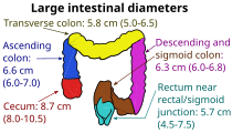
Inner diameters of colon sections

Conditions that call for colonoscopies include gastrointestinal hemorrhage, unexplained changes in bowel habit and suspicion of malignancy. Colonoscopies are often used to diagnose colon polyp and colon cancer, but are also frequently used to diagnose inflammatory bowel disease.

Another common indication for colonoscopy is the investigation of iron deficiency with or without anaemia. The examination of the colon, to rule out a lesion contributing to blood loss, along with an upper gastrointestinal endoscopy (gastroscopy) to rule out oesophageal, stomach, and proximal duodenal sources of blood loss.

Fecal occult blood is a quick test which can be done to test for microscopic traces of blood in the stool. A positive test is almost always an indication to do a colonoscopy. In most cases the positive result is just due to hemorrhoids; however, it can also be due to diverticulosis, inflammatory bowel disease (Crohn's disease, ulcerative colitis), colon cancer, or polyps. Colonic polypectomy has become a routine part of colonoscopy, allowing quick and simple removal of polyps during the procedure, without invasive surgery.

With regard to blood in the stool either visible or occult, it is worthy of note, that occasional rectal bleeding may have multiple non-serious potential causes.

Colon cancer screening

Colonoscopy is one of the colorectal cancer screening tests available to people in the US who are 45 years of age and older. The other screening tests include flexible sigmoidoscopy, double-contrast barium enema, computed tomographic (CT) colonography (virtual colonoscopy), guaiac-based fecal occult blood test (gFOBT), fecal immunochemical test (FIT), and multitarget stool DNA screening test (Cologuard).

Subsequent rescreenings are then scheduled based on the initial results found, with a five- or ten-year recall being common for colonoscopies that produce normal results. People with a family history of colon cancer are often first screened during their teenage years. Among people who have had an initial colonoscopy that found no polyps, the risk of developing colorectal cancer within five years is extremely low. Therefore, there is no need for those people to have another colonoscopy sooner than five years after the first screening.

Some medical societies in the US recommend a screening colonoscopy every 10 years beginning at age 50 for adults without increased risk for colorectal cancer. Research shows that the risk of cancer is low for 10 years if a high-quality colonoscopy does not detect cancer, so tests for this purpose are indicated every ten years.

Colonoscopy screening is associated with approximately two-thirds fewer deaths due to colorectal cancers on the left side of the colon, and is not associated with a significant reduction in deaths from right-sided disease. It is speculated that colonoscopy might reduce rates of death from colon cancer by detecting some colon polyps and cancers on the left side of the colon early enough that they may be treated, and a smaller number on the right side.

Since polyps often take 10 to 15 years to transform into cancer in someone at average risk of colorectal cancer, guidelines recommend 10 years after a normal screening colonoscopy before the next colonoscopy. (This interval does not apply to people at high risk of colorectal cancer or those who experience symptoms of the disease.)

The large randomized pragmatic clinical trial NordICC was the first published trial on the use of colonoscopy as a screening test to prevent colorectal cancer, related death, and death from any cause. It included 84,585 healthy men and women aged 55 to 64 years in Poland, Norway, and Sweden, who were randomized to either receive an invitation to undergo a single screening colonoscopy (invited group) or to receive no invitation or screening (usual-care group). Of the 28,220 people in the invited group, 11,843 (42.0%) underwent screening. A total of 15 people who underwent colonoscopy (0.13%) had major bleeding after polyp removal.

None of the participants experienced a colon perforation due to colonoscopy. After 10 years, an intention-to-screen analysis showed a significant relative risk reduction of 18% in the risk of colorectal cancer (0.98% in the invited group vs. 1.20% in the usual-care group). The analysis showed no significant change in the risk of death from colorectal cancer (0.28% vs. 0.31%) or in the risk of death from any cause (11.03% vs. 11.04%). To prevent one case of colorectal cancer, 455 invitations to colonoscopy were required.

As of 2023, the CONFIRM trial, a randomized trial evaluating colonoscopy vs. FIT is currently ongoing.

In 2021, the US spent $43 billion on cancer screening to prevent five cancers, with colonoscopies accounting for 55% of the total. According to expert Dr. H. Gilbert Welch, the death rate from colon cancer has been on a linear decline for 40 years, falling by nearly 50 percent from the 1980s (when few were screened) to 2024; however, the increase in screening did not accelerate the decline. Therefore, resources devoted to cancer screening would be better directed toward ensuring widespread access to effective cancer treatment.

Recommendations

The American Cancer Society issues recommendations on colorectal cancer screening guidelines. These guidelines often change and are updated as new studies and technologies have become available.

Many other national organizations also issue such guidance, such as the UK's NHS and various European agencies, guidance can vary between such agencies.

Medicare coverage

In the United States, Medicare insurance covers a number of colorectal-cancer screening tests.

Procedural risks

The American Society for Gastrointestinal Endoscopy estimates around three in 1,000 colonoscopies lead to serious complications.

Perforation

The most serious complication is generally gastrointestinal perforation, which is life-threatening and requires immediate surgical intervention.

The key to managing a colonoscopic perforation is diagnosis at the time. The reasons are because, typically, the bowel prep done to facilitate the examination acts to reduce the potential for contamination resulting in a higher likelihood of conservative management. In addition, recognised at the time allows the proceduralist to deploy strategies to seal the colon, or mark it should the patient require an operation.

Issues from general anesthesia

As with any procedure involving anaesthesia, complications can occur, such as:

  • allergic reactions,
  • cardiovascular issues,
  • paradoxical agitation,
  • aspiration,
  • dental injury.

Colon preparation electrolyte issues

Electrolyte imbalances caused by bowel preparation solutions is a possibility, but current bowel cleansing laxatives are formulated to account for electrolyte balance and it is a very rare event.

Other

During colonoscopies when a polyp is removed (a polypectomy), the complication risk is higher. One of the most serious complications is postpolypectomy coagulation syndrome, occurring in 1 in 1000 procedures. It results from a burn injury to the wall of the colon causing abdominal pain, fever, elevated white blood cell count and elevated serum C-reactive protein. Treatment consists of intravenous fluids, antibiotics, and avoiding oral intake of food, water, etc. until symptoms improve. Risk factors include right colon polypectomy, large polyp size (>2 cm), non-polypoid lesions (laterally spreading lesions), and hypertension.

Although rare, infections of the colon are a potential colonoscopy risk. The colon is not a sterile environment and infections can occur during biopsies by creating essentially a 'small shallow cut' during the biopsy, enabling bacterial intrusion into lower parts of the colon wall. In cases where the lining of the colon is perforated, bacteria can infiltrate the abdominal cavity. Infection may also be introduced if the endoscope is not cleaned and sterilized appropriately between procedures.

Minor colonoscopy risks may include nausea, vomiting or allergies to the sedatives that may have been used. If medication is given intravenously, the vein may become irritated or mild phlebitis.

Technique

Preparation

The colon must be free of solid matter for the test to be performed properly. For one to three days, the patient is required to follow a low fiber or clear-liquid-only diet. Examples of clear fluids are apple juice, chicken and/or beef broth or bouillon, lemon-lime soda, lemonade, sports drink, and water. It is important that the patient remains hydrated. Sports drinks contain electrolytes which are depleted during the purging of the bowel. Drinks containing fiber such as prune and orange juice should not be consumed, nor should liquids dyed red, purple, orange, or sometimes brown; however, cola is allowed. In most cases, tea or coffee taken without milk are allowed.

The day before the colonoscopy (or colorectal surgery), the patient is either given a laxative preparation (such as bisacodyl, phospho soda, sodium picosulfate, or sodium phosphate and/or magnesium citrate) and large quantities of fluid, or whole bowel irrigation is performed using a solution of polyethylene glycol and electrolytes. The procedure may involve both a pill-form laxative and a bowel irrigation preparation with the polyethylene glycol powder dissolved into any clear liquid, such as a sports drink that contains electrolytes.

A container of PEG (polyethylene glycol) with electrolyte used to clean out the intestines before certain bowel exam procedures such as colonoscopy.
A container of PEG (polyethylene glycol or macrogol) with electrolyte used to clean out the intestines before certain bowel exam procedures such as a colonoscopy.

The patient may be asked not to take aspirin or similar products such as salicylate, ibuprofen, etc. for up to ten days before the procedure to avoid the risk of bleeding if a polypectomy is performed during the procedure. A blood test may be performed before the procedure.

Procedure

Schematic overview of colonoscopy procedure

During the procedure, the patient is often given sedation intravenously, employing agents such as fentanyl or midazolam. Although meperidine (Demerol) may be used as an alternative to fentanyl, the concern of seizures has relegated this agent to second choice for sedation behind the combination of fentanyl and midazolam. The average person will receive a combination of these two drugs, usually between 25 and 100 μg IV fentanyl and 1–4 mg IV midazolam. Sedation practices vary between practitioners and nations; in some clinics in Norway, sedation is rarely administered.

The first step is usually a digital rectal examination (DRE), to examine the tone of the anal sphincter and to determine if preparation has been adequate. A DRE is also useful in detecting anal neoplasms and the clinician may note issues with the prostate gland in men undergoing this procedure. The endoscope is then passed through the anus up the rectum, the colon (sigmoid, descending, transverse and ascending colon, the cecum), and ultimately the terminal ileum. The endoscope has a movable tip and multiple channels for instrumentation, air, suction and light. The bowel is occasionally insufflated with air to maximize visibility (a procedure that gives the patient the false sensation of needing to take a bowel movement). Biopsies are frequently taken for histology. Additionally in a procedure known as chromoendoscopy, a contrast-dye (such as indigo carmine) may be sprayed through the endoscope onto the bowel wall to help visualize any abnormalities in the mucosal morphology. A Cochrane review updated in 2016 found strong evidence that chromoscopy enhances the detection of cancerous tumors in the colon and rectum.

In most experienced hands, the endoscope is advanced to the junction of where the colon and small bowel join up (cecum) in under 10 minutes in 95% of cases. Due to tight turns and redundancy in areas of the colon that are not "fixed", loops may form in which advancement of the endoscope creates a "bowing" effect that causes the tip to actually retract. These loops often result in discomfort due to stretching of the colon and its associated mesentery. Manoeuvres to "reduce" or remove the loop include pulling the endoscope backwards while twisting it. Alternatively, body position changes and abdominal support from external hand pressure can often "straighten" the endoscope to allow the scope to move forward. In a minority of patients, looping is often cited as a cause for an incomplete examination. Usage of alternative instruments leading to completion of the examination has been investigated, including use of pediatric colonoscope, push enteroscope and upper GI endoscope variants.

Lawsuits over missed cancerous lesions have recently prompted some institutions to better document endoscope examination times, as rapid examination times may be a source of potential medical legal liability. This is often a real concern in clinical settings where high caseloads could provide financial incentive to complete colonoscopies as quickly as possible.

Patient comfort and pain management

The pain associated with the procedure is not caused by the insertion of the scope but rather by the inflation of the colon in order to do the inspection. The scope itself is essentially a long, flexible tube about a centimeter in diameter — that is, as big around as the little finger, which is less than the diameter of an average stool.

The colon is wrinkled and corrugated, somewhat like an accordion or a clothes-dryer exhaust tube, which gives it the large surface area needed for nutrition and water absorption. In order to inspect this surface thoroughly, the physician blows it up like a balloon, using air from a compressor or carbon dioxide from a gas bottle (CO2 is absorbed into the bloodstream through the mucosal lining of the colon much faster than air and then exhaled through the lungs which is associated with less post procedural pain), in order to get the creases out.

The colon has sensors that can tell when there is unexpected gas pushing the colon walls out—which may cause mild discomfort. Usually, total anesthesia or a partial twilight sedative are used to reduce the patient's awareness of pain or discomfort, or just the unusual sensations of the procedure. Once the colon has been inflated, the doctor inspects it with the scope as it is slowly pulled backward. If any polyps are found they are then cut out for later biopsy.

Colonoscopy can be carried out without any sedation and a number of studies have been performed evaluating colonoscopy outcomes without sedation. Though in the US and EU the procedure is usually carried out with some form of sedation.

Economics

Researchers have found that older patients with three or more significant health problems (i.e., dementia or heart failure) had higher rates of repeat colonoscopies without medical indications. These patients are less likely to live long enough to develop colon cancer.

History

In the 1960s, Dr. Niwa and Dr. Yamagata at Tokyo University developed the fibre-optic endoscopy device. After 1968, Dr. William Wolff and Dr. Hiromi Shinya pioneered the development of the colonoscope. Their invention, in 1969 in Japan, was a significant advance over the barium enema and the flexible sigmoidoscope because it allowed for the visualization and removal of polyps from the entire colon. Wolff and Shinya advocated for their invention and published much of the early evidence needed to overcome skepticism about the device's safety and efficacy.

Some of the leading medical device companies in the colonoscopy market as of 2023 include: Fujifilm, Karl Storz SE, Pro Scope Systems, Olympus Corporation, Medtronic Plc, Steris and Pentax Medical.

Etymology

The terms colonoscopy or coloscopy are derived from the ancient Greek noun κόλον, same as English colon, and the verb σκοπεῖν, look (in)to, examine. The term colonoscopy is however ill-constructed, as this form supposes that the first part of the compound consists of a possible root κολων- or κολον-, with the connecting vowel -o, instead of the root κόλ- of κόλον. A compound such as κολωνοειδής, like a hill, (with the additional -on-) is derived from the ancient Greek word κολώνη or κολωνός, hill. Similarly, colonoscopy (with the additional -on-) can literally be translated as examination of the hill, instead of the examination of the colon.

In English, multiple words exist that are derived from κόλον, such as colectomy, colocentesis, colopathy, and colostomy among many others, that actually lack the incorrect additional -on-. A few compound words such as colonopathy have doublets with -on- inserted.

Society and culture

The procedure of colonoscopy gained national attention in the United States in 1985 when President Ronald Reagan underwent a life-saving colonoscopy.

A survey on colonoscopy shows a poor understanding of its protective value and widespread misconceptions. The public has perceptual gaps around the purpose of colonoscopies, the subjective experience of the colonoscopy procedure, and the quantity of bowel preparation needed.

Actors Ryan Reynolds and Rob McElhenney have used their social media platform to raise awareness about the importance of colonoscopy as a procedure for colon cancer screening. They filmed their own colonoscopies as part of a campaign called "Lead From Behind", demonstrating that the procedure can be both easy and lifesaving.

Kounis syndrome

From Wikipedia, the free encyclopedia
https://en.wikipedia.org/wiki/Kounis_syndrome
 
Kounis syndrome
Other namesAllergic acute coronary syndrome
SpecialtyCardiology

Kounis syndrome is defined as acute coronary syndrome (symptoms such as chest pain relating to reduced blood flow to the heart) caused by an allergic reaction or a strong immune reaction to a drug or other substance. It is a rare syndrome with authentic cases reported in 130 males and 45 females, as reviewed in 2017; however, the disorder is suspected of being commonly overlooked and therefore much more prevalent. Mast cell activation and release of inflammatory cytokines as well as other inflammatory agents from the reaction leads to spasm of the arteries leading to the heart muscle or a plaque breaking free and blocking one or more of those arteries.

The Kounis syndrome is distinguished from two other causes of coronary artery spasms and symptoms viz., the far more common, non-allergic syndrome, Prinzmetal's angina and eosinophilic coronary periarteritis, an extremely rare disorder caused by extensive eosinophilic infiltration of the adventitia and periadventitia, i.e. the soft tissues, surrounding the coronary arteries.

Epidemiology

Through various case observations, Kounis syndrome was noted in many different races and geographical areas. However, most cases have been found in southern Europe including Turkey, Greece, Italy, and Spain. A wide age range is observed from pediatric patients to the elderly including the ages from 2 to 90. Commonly seen comorbidities include hyperlipidemia, diabetes, smoking, hypertension, and prior allergic reactions to a precipitating factor. The exact prevalence is difficult to determine given that this diagnosis is missed or under-diagnosed. There is a possibility for gene-environment interactions as a study reported all patients admitted following emergency department evaluation had a heterozygous E148Q mutation.

Etiology

Many causes have been discovered to precipitate this syndrome including drugs, various health conditions, food, and environmental exposures. Any of these precipitating factors that cause IgE antibody production can contribute to this syndrome. Drugs that have been found previously include analgesics such as aspirin and dipyrone, anesthetics, multiple antibiotics, anticoagulants such as heparin and Lepirudin, thrombolytics such as TPA, anti-platelet therapy including Clopidogrel, anti-neoplastics, glucocorticoids, nonsteroidal anti-inflammatory drugs, proton pump inhibitors, and skin disinfectants. Additionally, sympathomimetics, volume expanders, antifungals, antivirals, and oral contraceptives can also trigger this syndrome. Other specific common medications include Allopurinol, Enalapril, Losartan, insulin, and many more. Conditions that incriminate Kounis syndrome include bronchial asthma, Churg–Strauss syndrome, serum sickness, scombroid syndrome, angioedema, hay fever, anaphylaxis (exercise induced or idiopathic), and anisakiasis. Coronary stenting, a common procedure used in coronary artery disease patients has also been found to be a cause. Environmental exposure to poison ivy, grass, latex, and nicotine have been found to be contributory. Bites from creatures that can precipitate Kounis syndrome include spiders, snakes, scorpions, fire ants and jellyfish. Miscellaneous triggers include contrast media. Reactions to various foods that cause an allergic and inflammatory response can lead to acute coronary syndrome.

Signs and symptoms

Allergic ACS is a syndrome involving two components. One component is immune mediated resulting in hypersensitivity, allergy, and an anaphylactic or anaphylactoid reaction. The second component involves cardiac signs and symptoms seen with acute coronary syndrome (ACS). Cardiac symptoms vary depending on the type of variant the patient presents with. Acute coronary syndrome is usually associated with a constrictive pain in the chest, characteristically with radiation to the neck or the left arm and often associated with pallor, sweatiness, nausea and breathlessness. Cardiac signs on exam also include cold extremities, bradycardia, tachycardia, hypotension, possible cardiorespiratory arrest, or sudden death. Just as in an allergic reaction can vary from a mild and localized reaction to something that is widespread and life-threatening, the allergic component of allergic ACS presents the same way. In allergic ACS there may also be specific symptoms relating to the underlying allergic reaction, such as swelling of the face and tongue, wheeze, hives and potentially very low blood pressure (anaphylactic shock). Additional findings can include stridor, drowsiness, syncope, abdominal pain, diarrhea, vomiting, and acute pulmonary edema if severe.

Myocardial infarction, acute cardiac failure, and sudden cardiac death may also be seen. As high as 13% of adult onset  sudden cardiac deaths are coupled with mast cell degranulation concluding that Kounis syndrome can involve a silent allergic reaction.

Pathophysiology

In allergy, mast cells release inflammatory substances such as histamine, neutral proteases, arachidonic acid derivatives, platelet activating factor and a variety of cytokines and chemokines. These mediators can precipitate coronary artery spasm and accelerate the rupture of atheromatous plaques of the coronary arteries. This interferes with the blood flow to the heart muscle and causes symptoms otherwise indistinguishable from unstable angina.

It is possible that even in people without direct evidence of allergy, the allergic response may be playing a role in acute coronary syndrome: markers of mast cell activation are found in people with ACS.

The main marker of mast cell activation is inducible macrophage protein 1a (MIP-1α), which binds to mast cells when they are in close proximity to each other. After allergen exposure, MIP-1α transcription and expression is induced by resident mononuclear cells in the substantia propria, which consist of CD68+ macrophages and monocytes.

Diagnosis

Kounis syndrome is often missed or underdiagnosed so understanding the disease process and clinical presentation while having a high suspicion for the problem is compulsory.  It is important to focus on the duration of time between the trigger exposure and the onset of symptoms. Majority of the cases had a duration of under one hour while some had a duration of 6 hours. EKG, chest x-ray, echocardiography, and angiography are needed if suspicion for myocardial ischemia or infarction is present.

Patients with systemic allergic reactions associated with clinical, electrocardiographic, angiographic, echocardiographic and laboratory findings of acute myocardial ischemia should be diagnosed as having Kounis syndrome. EKG changes can be consistent with infarction most commonly in the inferior leads, ischemia, sinus bradycardia or tachycardia, heart block, atrial fibrillation, ventricular fibrillation, ventricular ectopic beats, QRS and QT prolongation, and findings similar to digoxin toxicity. Echocardiography assists with finding atherosclerotic stenosis and thrombosis.

Serum tryptase, histamine, immunoglobulins (IgE), cardiac enzymes, cardiac troponins are helpful to confirm the diagnosis. In Kounis syndrome, the newer techniques such as thallium-201 single-photon emission computer tomography (SPECT) and 125I-15-(p-iodophenyl)-3-(R,S) methylpentadecanoic acid (BMIPP) SPECT have revealed severe myocardial ischemia while coronary angiography showed normal coronary arteries. Furthermore, with cardiac magnetic resonance imaging (MRI), the delayed contrast-enhanced images show normal washout in the subendocardial lesion area in patients with Kounis syndrome type I variant.

Other similar presentations to rule out include Takotsubo and hypersensitivity myocarditis.

Classification

Three variants of Kounis syndrome are recognised:

  • Type I is also known as allergic vasospastic angina due to endothelial dysfunction. It occurs in people without underlying coronary artery disease or predisposing factors who have allergic ACS secondary to coronary artery spasm. Inflammatory mediators during an allergic reaction can cause arterial spasms with normal troponins. However, this may lead to myocardial infarction which will elevate troponins. MINOCA, which stands for myocardial infarction with non-obstructive coronary arteries is new clinical presentation that includes endothelial dysfunction and can be caused by this type I variant.
  • Type II occurs in people with underlying asymptomatic coronary artery disease where an allergic reaction leads to either coronary artery spasm or plaque erosion. A myocardial infarction can also be seen here, in which case troponins would be elevated.
  • Type III occurs in the setting of coronary thrombosis (including stent thrombosis) where aspirated thrombus stained with hematoxylin-eosin and Giemsa demonstrate the presence of eosinophils and mast cells respectively. It also includes those with people who have died suddenly after previous coronary stent insertion, where evidence of an allergic reaction to the stent is found on post-mortem examination. Type III is subdivided now to stent thrombosis (subtype a) and stent restenosis (subtype b).

Management

The management of these patients may be challenging for clinicians. Although beta blockers can be beneficial in ACS, they are contraindicated in Kounis syndrome. In allergic ACS, blocking beta receptors while giving epinephrine (which is the basis of treatment of anaphylaxis) can lead to an unopposed activity of α-adrenergic receptors which would aggravate the coronary spasm. Also opioids, indicated to relieve chest pain, may induce massive mast cell degranulation which in turn will worsen the anaphylaxis. They should hence be given carefully in such patients

Type I variant

Type I variant is treated based on its clinical presentation and how severe the allergic reaction is. If it is a mild reaction, then antihistamines and corticosteroids can help control the symptoms. If the patient's presentation involves anaphylaxis, intramuscular adrenaline should be given.

Treatment of the allergic event alone can abolish type I variant. Giving vasodilators such as nitroglycerin or calcium channel blockers is recommended. Consequences include hypotension and worsening of anaphylaxis. Antihistamine and mast cell stabilizers e.g. cromoglicate or nedocromil can be also considered.

Type II variant

Acute coronary event protocol is applied and type II can be treated similarly to type I for cardiac symptom control. Glucagon may be a better option than adrenaline for acute anaphylaxis in patients with prior use of chronic beta-blockers. In addition, beta-blockers can increase coronary vasospasm and ischemia. Opiates should be used with caution.

Type III variant

In addition to the application of the acute coronary syndrome protocol, thrombus aspiration, and placing a new stent is needed. The use of mast cell stabilizers in association with steroids and antihistamines are recommended. Harvesting of intrastent thrombus together with histological examination of aspirated material and staining for eosinophils and mast cells should be undertaken. When allergic symptoms are present following stent implantation, desensitization measures should be applied.

History

While there are several older reports associating, clinically, allergy and the heart with names such as morphologic cardiac reactions, acute carditis or lesions with basic characteristics of rheumatic carditis, the first full description of allergy-mediated acute coronary syndrome is attributed to the Greek cardiologist Nicholas Kounis, who in 1991 reported on the possible role of allergy in cases of coronary artery spasm (now, type I variant). Braunwald recognized the allergy induced coronary artery occlusion mediated by these spasms. Three variants of Kounis syndrome were found and a study concluded that type 1 variant was most commonly seen followed by type 2 and 3 respectively.

Renaissance philosophy

From Wikipedia, the free encyclopedia   Renaissance The School of Athens (15...