Search This Blog

Friday, June 21, 2019

Hematopoietic stem cell transplantation

From Wikipedia, the free encyclopedia

Hematopoietic stem cell transplantation
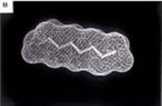
KM Transplantat.JPEG
Bone marrow transplant
ICD-9-CM41.0
MeSHD018380
MedlinePlus003009

Hematopoietic stem cell transplantation (HSCT) is the transplantation of multipotent hematopoietic stem cells, usually derived from bone marrow, peripheral blood, or umbilical cord blood. It may be autologous (the patient's own stem cells are used), allogeneic (the stem cells come from a donor) or syngeneic (from an identical twin).

It is most often performed for patients with certain cancers of the blood or bone marrow, such as multiple myeloma or leukemia. In these cases, the recipient's immune system is usually destroyed with radiation or chemotherapy before the transplantation. Infection and graft-versus-host disease are major complications of allogeneic HSCT.

Hematopoietic stem cell transplantation remains a dangerous procedure with many possible complications; it is reserved for patients with life-threatening diseases. As survival following the procedure has increased, its use has expanded beyond cancer to autoimmune diseases and hereditary skeletal dysplasias; notably malignant infantile osteopetrosis and mucopolysaccharidosis.

Medical uses

Indications

Indications for stem cell transplantation are as follows:

Malignant (cancerous)

Non-malignant (non-cancerous)

Many recipients of HSCTs are multiple myeloma or leukemia patients who would not benefit from prolonged treatment with, or are already resistant to, chemotherapy. Candidates for HSCTs include pediatric cases where the patient has an inborn defect such as severe combined immunodeficiency or congenital neutropenia with defective stem cells, and also children or adults with aplastic anemia who have lost their stem cells after birth. Other conditions treated with stem cell transplants include sickle-cell disease, myelodysplastic syndrome, neuroblastoma, lymphoma, Ewing's sarcoma, desmoplastic small round cell tumor, chronic granulomatous disease, Hodgkin's disease and Wiskott–Aldrich syndrome. More recently non-myeloablative, ""mini transplant (microtransplantation)," procedures have been developed that require smaller doses of preparative chemo and radiation. This has allowed HSCT to be conducted in the elderly and other patients who would otherwise be considered too weak to withstand a conventional treatment regimen.

Number of procedures

In 2006 a total of 50,417 first hematopoietic stem cell transplants were reported as taking place worldwide, according to a global survey of 1327 centers in 71 countries conducted by the Worldwide Network for Blood and Marrow Transplantation. Of these, 28,901 (57 percent) were autologous and 21,516 (43 percent) were allogeneic (11,928 from family donors and 9,588 from unrelated donors). The main indications for transplant were lymphoproliferative disorders (55 percent) and leukemias (34 percent), and the majority took place in either Europe (48 percent) or the Americas (36 percent).

The Worldwide Network for Blood and Marrow Transplantation reported the millionth transplant to have been undertaken in December 2012.

In 2014, according to the World Marrow Donor Association (WMDA), stem cell products provided for unrelated transplantation worldwide had increased to 20,604 (4,149 bone marrow donations, 12,506 peripheral blood stem cell donations, and 3,949 cord blood units).

Graft types

Autologous

Autologous HSCT requires the extraction (apheresis) of hematopoietic stem cells (HSC) from the patient and storage of the harvested cells in a freezer. The patient is then treated with high-dose chemotherapy with or without radiotherapy with the intention of eradicating the patient's malignant cell population at the cost of partial or complete bone marrow ablation (destruction of patient's bone marrow's ability to grow new blood cells). The patient's own stored stem cells are then transfused into his/her bloodstream, where they replace destroyed tissue and resume the patient's normal blood cell production. Autologous transplants have the advantage of lower risk of infection during the immune-compromised portion of the treatment since the recovery of immune function is rapid. Also, the incidence of patients experiencing rejection (and graft-versus-host disease is impossible) is very rare due to the donor and recipient being the same individual. These advantages have established autologous HSCT as one of the standard second-line treatments for such diseases as lymphoma.

However, for other cancers such as acute myeloid leukemia, the reduced mortality of the autogenous relative to allogeneic HSCT may be outweighed by an increased likelihood of cancer relapse and related mortality, and therefore the allogeneic treatment may be preferred for those conditions.

Researchers have conducted small studies using non-myeloablative hematopoietic stem cell transplantation as a possible treatment for type I (insulin dependent) diabetes in children and adults. Results have been promising; however, as of 2009 it was premature to speculate whether these experiments will lead to effective treatments for diabetes.

Allogeneic

Allogeneic HSCT involves two people: the (healthy) donor and the (patient) recipient. Allogeneic HSC donors must have a tissue (HLA) type that matches the recipient. Matching is performed on the basis of variability at three or more loci of the HLA gene, and a perfect match at these loci is preferred. Even if there is a good match at these critical alleles, the recipient will require immunosuppressive medications to mitigate graft-versus-host disease. Allogeneic transplant donors may be related (usually a closely HLA matched sibling), syngeneic (a monozygotic or 'identical' twin of the patient – necessarily extremely rare since few patients have an identical twin, but offering a source of perfectly HLA matched stem cells) or unrelated (donor who is not related and found to have very close degree of HLA matching). Unrelated donors may be found through a registry of bone marrow donors such as the National Marrow Donor Program. People who would like to be tested for a specific family member or friend without joining any of the bone marrow registry data banks may contact a private HLA testing laboratory and be tested with a mouth swab to see if they are a potential match. A "savior sibling" may be intentionally selected by preimplantation genetic diagnosis in order to match a child both regarding HLA type and being free of any obvious inheritable disorder. Allogeneic transplants are also performed using umbilical cord blood as the source of stem cells. In general, by transfusing healthy stem cells to the recipient's bloodstream to reform a healthy immune system, allogeneic HSCTs appear to improve chances for cure or long-term remission once the immediate transplant-related complications are resolved.

A compatible donor is found by doing additional HLA-testing from the blood of potential donors. The HLA genes fall in two categories (Type I and Type II). In general, mismatches of the Type-I genes (i.e. HLA-A, HLA-B, or HLA-C) increase the risk of graft rejection. A mismatch of an HLA Type II gene (i.e. HLA-DR, or HLA-DQB1) increases the risk of graft-versus-host disease. In addition, a genetic mismatch as small as a single DNA base pair is significant so perfect matches require knowledge of the exact DNA sequence of these genes for both donor and recipient. Leading transplant centers currently perform testing for all five of these HLA genes before declaring that a donor and recipient are HLA-identical. 

Race and ethnicity are known to play a major role in donor recruitment drives, as members of the same ethnic group are more likely to have matching genes, including the genes for HLA.

As of 2013, there were at least two commercialized allogeneic cell therapies, Prochymal and Cartistem.

Sources and storage of cells

To limit the risks of transplanted stem cell rejection or of severe graft-versus-host disease in allogeneic HSCT, the donor should preferably have the same human leukocyte antigens (HLA) as the recipient. About 25 to 30 percent of allogeneic HSCT recipients have an HLA-identical sibling. Even so-called "perfect matches" may have mismatched minor alleles that contribute to graft-versus-host disease.

Bone marrow

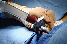
Bone marrow harvest.
 
In the case of a bone marrow transplant, the HSC are removed from a large bone of the donor, typically the pelvis, through a large needle that reaches the center of the bone. The technique is referred to as a bone marrow harvest and is performed under general anesthesia.

Peripheral blood stem cells

Peripheral blood stem cells
 
Peripheral blood stem cells are now the most common source of stem cells for HSCT. They are collected from the blood through a process known as apheresis. The donor's blood is withdrawn through a sterile needle in one arm and passed through a machine that removes white blood cells. The red blood cells are returned to the donor. The peripheral stem cell yield is boosted with daily subcutaneous injections of granulocyte-colony stimulating factor, serving to mobilize stem cells from the donor's bone marrow into the peripheral circulation.

Amniotic fluid

It is also possible to extract stem cells from amniotic fluid for both autologous or heterologous use at the time of childbirth.

Umbilical cord blood

Umbilical cord blood is obtained when a mother donates her infant's umbilical cord and placenta after birth. Cord blood has a higher concentration of HSC than is normally found in adult blood. However, the small quantity of blood obtained from an umbilical cord (typically about 50 mL) makes it more suitable for transplantation into small children than into adults. Newer techniques using ex-vivo expansion of cord blood units or the use of two cord blood units from different donors allow cord blood transplants to be used in adults. 

Cord blood can be harvested from the umbilical cord of a child being born after preimplantation genetic diagnosis (PGD) for human leukocyte antigen (HLA) matching (see PGD for HLA matching) in order to donate to an ill sibling requiring HSCT.

Storage of HSC

Unlike other organs, bone marrow cells can be frozen (cryopreserved) for prolonged periods without damaging too many cells. This is a necessity with autologous HSC because the cells must be harvested from the recipient months in advance of the transplant treatment. In the case of allogeneic transplants, fresh HSC are preferred in order to avoid cell loss that might occur during the freezing and thawing process. Allogeneic cord blood is stored frozen at a cord blood bank because it is only obtainable at the time of childbirth. To cryopreserve HSC, a preservative, DMSO, must be added, and the cells must be cooled very slowly in a controlled-rate freezer to prevent osmotic cellular injury during ice crystal formation. HSC may be stored for years in a cryofreezer, which typically uses liquid nitrogen.

Conditioning regimens

Myeloablative

The chemotherapy or irradiation given immediately prior to a transplant is called the conditioning regimen, the purpose of which is to help eradicate the patient's disease prior to the infusion of HSC and to suppress immune reactions. The bone marrow can be ablated (destroyed) with dose-levels that cause minimal injury to other tissues. In allogeneic transplants a combination of cyclophosphamide with total body irradiation is conventionally employed. This treatment also has an immunosuppressive effect that prevents rejection of the HSC by the recipient's immune system. The post-transplant prognosis often includes acute and chronic graft-versus-host disease that may be life-threatening. However, in certain leukemias this can coincide with protection against cancer relapse owing to the graft-versus-tumor effect. Autologous transplants may also use similar conditioning regimens, but many other chemotherapy combinations can be used depending on the type of disease.

Non-myeloablative

A newer treatment approach, non-myeloablative allogeneic transplantation, also termed reduced-intensity conditioning (RIC), uses doses of chemotherapy and radiation too low to eradicate all the bone marrow cells of the recipient. Instead, non-myeloablative transplants run lower risks of serious infections and transplant-related mortality while relying upon the graft versus tumor effect to resist the inherent increased risk of cancer relapse. Also significantly, while requiring high doses of immunosuppressive agents in the early stages of treatment, these doses are less than for conventional transplants. This leads to a state of mixed chimerism early after transplant where both recipient and donor HSC coexist in the bone marrow space. 

Decreasing doses of immunosuppressive therapy then allow donor T-cells to eradicate the remaining recipient HSC and to induce the graft-versus-tumor effect. This effect is often accompanied by mild graft-versus-host disease, the appearance of which is often a surrogate marker for the emergence of the desirable graft versus tumor effect, and also serves as a signal to establish an appropriate dosage level for sustained treatment with low levels of immunosuppressive agents

Because of their gentler conditioning regimens, these transplants are associated with a lower risk of transplant-related mortality and therefore allow patients who are considered too high-risk for conventional allogeneic HSCT to undergo potentially curative therapy for their disease. The optimal conditioning strategy for each disease and recipient has not been fully established, but RIC can be used in elderly patients unfit for myeloablative regimens, for whom a higher risk of cancer relapse may be acceptable.

Engraftment

After several weeks of growth in the bone marrow, expansion of HSC and their progeny is sufficient to normalize the blood cell counts and re-initiate the immune system. The offspring of donor-derived hematopoietic stem cells have been documented to populate many different organs of the recipient, including the heart, liver, and muscle, and these cells had been suggested to have the abilities of regenerating injured tissue in these organs. However, recent research has shown that such lineage infidelity does not occur as a normal phenomenon
.
Chimerism monitoring is a method to monitor the balance between the patient's own stem cells and the new stem cells from a donor. In case the patient's own stem cells are increasing in number post-treatment, this might be a sign the treatment did not work as intended.

Complications

HSCT is associated with a high treatment-related mortality in the recipient, which limits its use to conditions that are themselves life-threatening. (The one-year survival rate has been estimated to be roughly 60%, although this figure includes deaths from the underlying disease as well as from the transplant procedure.) Major complications are veno-occlusive disease, mucositis, infections (sepsis), graft-versus-host disease and the development of new malignancies.

Infection

Bone marrow transplantation usually requires that the recipient's own bone marrow be destroyed (myeloablation). Prior to the administration of new cells (engraftment) patients may go for several weeks without appreciable numbers of white blood cells to help fight infection. This puts a patient at high risk of infections, sepsis and septic shock, despite prophylactic antibiotics. However, antiviral medications, such as acyclovir and valacyclovir, are quite effective in prevention of HSCT-related outbreak of herpetic infection in seropositive patients. The immunosuppressive agents employed in allogeneic transplants for the prevention or treatment of graft-versus-host disease further increase the risk of opportunistic infection. Immunosuppressive drugs are given for a minimum of 6-months after a transplantation, or much longer if required for the treatment of graft-versus-host disease. Transplant patients lose their acquired immunity, for example immunity to childhood diseases such as measles or polio. For this reason transplant patients must be re-vaccinated with childhood vaccines once they are off immunosuppressive medications.

Veno-occlusive disease

Severe liver injury can result from hepatic veno-occlusive disease (VOD). Elevated levels of bilirubin, hepatomegaly and fluid retention are clinical hallmarks of this condition. There is now a greater appreciation of the generalized cellular injury and obstruction in hepatic vein sinuses, and hepatic VOD has lately been referred to as sinusoidal obstruction syndrome (SOS). Severe cases of SOS are associated with a high mortality rate. Anticoagulants or defibrotide may be effective in reducing the severity of VOD but may also increase bleeding complications. Ursodiol has been shown to help prevent VOD, presumably by facilitating the flow of bile.

Mucositis

The injury of the mucosal lining of the mouth and throat is a common regimen-related toxicity following ablative HSCT regimens. It is usually not life-threatening but is very painful, and prevents eating and drinking. Mucositis is treated with pain medications plus intravenous infusions to prevent dehydration and malnutrition.

Hemorrhagic cystitis

The mucosal lining of the bladder could also be involved in approximately 5 percent of the children undergoing hematopoietic stem cell transplantation. This causes hematuria, frequency, abdominal pain and thrombocytopnea.

Graft-versus-host disease

Graft-versus-host disease (GVHD) is an inflammatory disease that is unique to allogeneic transplantation. It is an attack by the "new" bone marrow's immune cells against the recipient's tissues. This can occur even if the donor and recipient are HLA-identical because the immune system can still recognize other differences between their tissues. It is aptly named graft-versus-host disease because bone marrow transplantation is the only transplant procedure in which the transplanted cells must accept the body rather than the body accepting the new cells.

Acute graft-versus-host disease typically occurs in the first 3 months after transplantation and may involve the skin, intestine, or the liver. High-dose corticosteroids such as prednisone are a standard treatment; however this immuno-suppressive treatment often leads to deadly infections. Chronic graft-versus-host disease may also develop after allogeneic transplant. It is the major source of late treatment-related complications, although it less often results in death. In addition to inflammation, chronic graft-versus-host disease may lead to the development of fibrosis, or scar tissue, similar to scleroderma; it may cause functional disability and require prolonged immunosuppressive therapy. Graft-versus-host disease is usually mediated by T cells, which react to foreign peptides presented on the MHC of the host.

Graft-versus-tumor effect

Graft-versus-tumor effect (GVT) or "graft versus leukemia" effect is the beneficial aspect of the Graft-versus-Host phenomenon. For example, HSCT patients with either acute, or in particular chronic, graft-versus-host disease after an allogeneic transplant tend to have a lower risk of cancer relapse. This is due to a therapeutic immune reaction of the grafted donor T lymphocytes against the diseased bone marrow of the recipient. This lower rate of relapse accounts for the increased success rate of allogeneic transplants, compared to transplants from identical twins, and indicates that allogeneic HSCT is a form of immunotherapy. GVT is the major benefit of transplants that do not employ the highest immuno-suppressive regimens. 

Graft versus tumor is mainly beneficial in diseases with slow progress, e.g. chronic leukemia, low-grade lymphoma, and in some cases multiple myeloma. However, it is less effective in rapidly growing acute leukemias.

If cancer relapses after HSCT, another transplant can be performed, infusing the patient with a greater quantity of donor white blood cells (Donor lymphocyte infusion).

Oral carcinoma

Patients after HSCT are at a higher risk for oral carcinoma. Post-HSCT oral cancer may have more aggressive behavior with poorer prognosis, when compared to oral cancer in non-HSCT patients.

Prognosis

Prognosis in HSCT varies widely dependent upon disease type, stage, stem cell source, HLA-matched status (for allogeneic HSCT) and conditioning regimen. A transplant offers a chance for cure or long-term remission if the inherent complications of graft versus host disease, immuno-suppressive treatments and the spectrum of opportunistic infections can be survived. In recent years, survival rates have been gradually improving across almost all populations and sub-populations receiving transplants.

Mortality for allogeneic stem cell transplantation can be estimated using the prediction model created by Sorror et al., using the Hematopoietic Cell Transplantation-Specific Comorbidity Index (HCT-CI). The HCT-CI was derived and validated by investigators at the Fred Hutchinson Cancer Research Center (Seattle, WA). The HCT-CI modifies and adds to a well-validated comorbidity index, the Charlson Comorbidity Index (CCI) (Charlson et al.) The CCI was previously applied to patients undergoing allogeneic HCT but appears to provide less survival prediction and discrimination than the HCT-CI scoring system.

Risks to donor

The risks of a complication depend on patient characteristics, health care providers and the apheresis procedure, and the colony-stimulating factor used (G-CSF). G-CSF drugs include filgrastim (Neupogen, Neulasta), and lenograstim (Graslopin).

Drug risks

Filgrastim is typically dosed in the 10 microgram/kg level for 4–5 days during the harvesting of stem cells. The documented adverse effects of filgrastim include splenic rupture (indicated by left upper abdominal or shoulder pain, risk 1 in 40000), Acute respiratory distress syndrome (ARDS), alveolar hemorrhage, and allergic reactions (usually expressed in first 30 minutes, risk 1 in 300). In addition, platelet and hemoglobin levels dip post-procedure, not returning to normal until after one month.

The question of whether geriatrics (patients over 65) react the same as patients under 65 has not been sufficiently examined. Coagulation issues and inflammation of atherosclerotic plaques are known to occur as a result of G-CSF injection. G-CSF has also been described to induce genetic changes in mononuclear cells of normal donors. There is no statistically significant evidence either for or against the hypothesis that myelodysplasia (MDS) or acute myeloid leukaemia (AML) can be induced by GCSF in susceptible individuals.

Access risks

Blood was drawn peripherally in a majority of patients, but a central line to jugular/subclavian/femoral veins may be used in 16 percent of women and 4 percent of men. Adverse reactions during apheresis were experienced in 20 percent of women and 8 percent of men, these adverse events primarily consisted of numbness/tingling, multiple line attempts, and nausea.

Clinical observations

A study involving 2408 donors (18–60 years) indicated that bone pain (primarily back and hips) as a result of filgrastim treatment is observed in 80 percent of donors by day 4 post-injection. This pain responded to acetaminophen or ibuprofen in 65 percent of donors and was characterized as mild to moderate in 80 percent of donors and severe in 10 percent. Bone pain receded post-donation to 26 percent of patients 2 days post-donation, 6 percent of patients one week post-donation, and less than 2 percent 1 year post-donation. Donation is not recommended for those with a history of back pain. Other symptoms observed in more than 40 percent of donors include myalgia, headache, fatigue, and insomnia. These symptoms all returned to baseline 1 month post-donation, except for some cases of persistent fatigue in 3 percent of donors.

In one metastudy that incorporated data from 377 donors, 44 percent of patients reported having adverse side effects after peripheral blood HSCT. Side effects included pain prior to the collection procedure as a result of GCSF injections, post-procedural generalized skeletal pain, fatigue and reduced energy.

Severe reactions

A study that surveyed 2408 donors found that serious adverse events (requiring prolonged hospitalization) occurred in 15 donors (at a rate of 0.6 percent), although none of these events were fatal. Donors were not observed to have higher than normal rates of cancer with up to 4–8 years of follow up. One study based on a survey of medical teams covered approximately 24,000 peripheral blood HSCT cases between 1993 and 2005, and found a serious cardiovascular adverse reaction rate of about 1 in 1500. This study reported a cardiovascular-related fatality risk within the first 30 days HSCT of about 2 in 10000. For this same group, severe cardiovascular events were observed with a rate of about 1 in 1500. The most common severe adverse reactions were pulmonary edema/deep vein thrombosis, splenic rupture, and myocardial infarction. Haematological malignancy induction was comparable to that observed in the general population, with only 15 reported cases within 4 years.

History

Georges Mathé, a French oncologist, performed the first European bone marrow transplant in November 1958 on five Yugoslavian nuclear workers whose own marrow had been damaged by irradiation caused by a criticality accident at the Vinča Nuclear Institute, but all of these transplants were rejected. Fortunately, the five treated were able to ultimately recover, perhaps in part due to the transplants. Mathé later pioneered the use of bone marrow transplants in the treatment of leukemia.

Stem cell transplantation was pioneered using bone-marrow-derived stem cells by a team at the Fred Hutchinson Cancer Research Center from the 1950s through the 1970s led by E. Donnall Thomas, whose work was later recognized with a Nobel Prize in Physiology or Medicine. Thomas' work showed that bone marrow cells infused intravenously could repopulate the bone marrow and produce new blood cells. His work also reduced the likelihood of developing a life-threatening complication called graft-versus-host disease. Collaborating with University of Washington Professor Eloise Giblett, he discovered genetic markers that could confirm donor matches. 

The first physician to perform a successful human bone marrow transplant on a disease other than cancer was Robert A. Good at the University of Minnesota in 1968. In 1975, John Kersey, M.D., also of the University of Minnesota, performed the first successful bone marrow transplant to cure lymphoma. His patient, a 16-year-old-boy, is today the longest-living lymphoma transplant survivor.

Donor registration and recruitment

At the end of 2012, 20.2 million people had registered their willingness to be a bone marrow donor with one of the 67 registries from 49 countries participating in Bone Marrow Donors Worldwide. 17.9 million of these registered donors had been ABDR typed, allowing easy matching. A further 561,000 cord blood units had been received by one of 46 cord blood banks from 30 countries participating. The highest total number of bone marrow donors registered were those from the US (8.0 million), and the highest number per capita were those from Cyprus (15.4 percent of the population).

Within the United States, racial minority groups are the least likely to be registered and therefore the least likely to find a potentially life-saving match. In 1990, only six African-Americans were able to find a bone marrow match, and all six had common European genetic signatures.

Africans are more genetically diverse than people of European descent, which means that more registrations are needed to find a match. Bone marrow and cord blood banks exist in South Africa, and a new program is beginning in Nigeria. Many people belonging to different races are requested to donate as there is a shortage of donors in African, Mixed race, Latino, Aboriginal, and many other communities. 

Two registries in the United States recruit unrelated allogeneic donors: NMDP/Be the Match and the Gift of Life Marrow Registry.

Research

HIV

In 2007, a team of doctors in Berlin, Germany, including Gero Hütter, performed a stem cell transplant for leukemia patient Timothy Ray Brown, who was also HIV-positive. From 60 matching donors, they selected a [CCR5]-Δ32 homozygous individual with two genetic copies of a rare variant of a cell surface receptor. This genetic trait confers resistance to HIV infection by blocking attachment of HIV to the cell. Roughly one in 1000 people of European ancestry have this inherited mutation, but it is rarer in other populations. The transplant was repeated a year later after a leukemia relapse. Over three years after the initial transplant, and despite discontinuing antiretroviral therapy, researchers cannot detect HIV in the transplant recipient's blood or in various biopsies of his tissues. Levels of HIV-specific antibodies have also declined, leading to speculation that the patient may have been functionally cured of HIV. However, scientists emphasise that this is an unusual case. Potentially fatal transplant complications (the "Berlin patient" suffered from graft-versus-host disease and leukoencephalopathy) mean that the procedure could not be performed in others with HIV, even if sufficient numbers of suitable donors were found.

In 2012, Daniel Kuritzkes reported results of two stem cell transplants in patients with HIV. They did not, however, use donors with the Δ32 deletion. After their transplant procedures, both were put on antiretroviral therapies, during which neither showed traces of HIV in their blood plasma and purified CD4 T cells using a sensitive culture method (less than 3 copies/mL). However, the virus was once again detected in both patients some time after the discontinuation of therapy.

In 2019, a British man became the second to be cleared of HIV after receiving a bone marrow transplant from a virus-resistant (Δ32) donor. This patient is being called "the London patient" (a reference to the famous Berlin patient).

Multiple sclerosis

Since McAllister's 1997 report on a patient with multiple sclerosis (MS) who received a bone marrow transplant for CML, over 600 reports have been published describing HSCTs performed primarily for MS. These have been shown to "reduce or eliminate ongoing clinical relapses, halt further progression, and reduce the burden of disability in some patients" that have aggressive highly active MS, "in the absence of chronic treatment with disease-modifying agents".
Thursday, June 20, 2019

Bone marrow

From Wikipedia, the free encyclopedia

Bone marrow
Details
SystemImmune system
Identifiers
LatinMedulla ossium
MeSHD001853
TAA13.1.01.001
FMA9608

Bone marrow is a semi-solid tissue which may be found within the spongy or cancellous portions of bones. In birds and mammals, bone marrow is the primary site of new blood cell production or hematopoiesis. It is composed of hematopoietic cells, marrow adipose tissue, and supportive stromal cells. In adult humans, bone marrow is primarily located in the ribs, vertebrae, sternum, and bones of the pelvis. On average, bone marrow constitutes 4% of the total body mass of humans; in an adult having 65 kilograms of mass (143 lb), bone marrow typically accounts for approximately 2.6 kilograms (5.7 lb).

Human marrow produces approximately 500 billion blood cells per day, which join the systemic circulation via permeable vasculature sinusoids within the medullary cavity. All types of hematopoietic cells, including both myeloid and lymphoid lineages, are created in bone marrow; however, lymphoid cells must migrate to other lymphoid organs (e.g. thymus) in order to complete maturation.

Bone marrow transplants can be conducted to treat severe diseases of the bone marrow, including certain forms of cancer such as leukemia. Additionally, bone marrow stem cells have been successfully transformed into functional neural cells, and can also potentially be used to treat illnesses such as inflammatory bowel disease.

Structure

The composition of marrow is dynamic, as the mixture of cellular and non-cellular components (connective tissue) shifts with age and in response to systemic factors. In humans, marrow is colloquially characterized as "red" or "yellow" marrow (Latin: medulla ossium rubra, Latin: medulla ossium flava, respectively) depending on the prevalence of hematopoetic cells vs fat cells. While the precise mechanisms underlying marrow regulation are not understood, compositional changes occur according to stereotypical patterns. For example, a newborn baby's bones exclusively contain hematopoietically active "red" marrow, and there is a progressive conversion towards "yellow" marrow with age. In adults, red marrow is found mainly in the central skeleton, such as the pelvis, sternum, cranium, ribs, vertebrae and scapulae, and variably found in the proximal epiphyseal ends of long bones such as the femur and humerus. In circumstances of chronic hypoxia, the body can convert yellow marrow back to red marrow to increase blood cell production.

Hematopoietic components

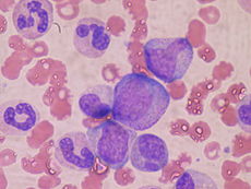
Hematopoietic precursor cells: promyelocyte in the center, two metamyelocytes next to it and band cells from a bone marrow aspirate.
 
At the cellular level, the main functional component of bone marrow includes the progenitor cells which are destined to mature into blood and lymphoid cells. Marrow contains hematopoietic stem cells which give rise to the three classes of blood cells that are found in circulation: white blood cells (leukocytes), red blood cells (erythrocytes), and platelets (thrombocytes).

Cellular constitution of the red bone marrow parenchyma
Group Cell type Average
fraction
Reference
range
Myelopoietic
cells
Myeloblasts 0.9% 0.2–1.5
Promyelocytes 3.3% 2.1–4.1
Neutrophilic myelocytes 12.7% 8.2–15.7
Eosinophilic myelocytes 0.8% 0.2–1.3
Neutrophilic metamyelocytes 15.9% 9.6–24.6
Eosinophilic metamyelocytes 1.2% 0.4–2.2
Neutrophilic band cells 12.4% 9.5–15.3
Eosinophilic band cells 0.9% 0.2–2.4
Segmented neutrophils 7.4% 6.0–12.0
Segmented eosinophils 0.5% 0.0–1.3
Segmented basophils and mast cells 0.1% 0.0–0.2
Erythropoietic
cells
Pronormoblasts 0.6% 0.2–1.3
Basophilic normoblasts 1.4% 0.5–2.4
Polychromatic normoblasts 21.6% 17.9–29.2
Orthochromatic normoblast 2.0% 0.4–4.6
Other cell
types
Megakaryocytes < 0.1% 0.0-0.4
Plasma cells 1.3% 0.4-3.9
Reticular cells 0.3% 0.0-0.9
Lymphocytes 16.2% 11.1-23.2
Monocytes 0.3% 0.0-0.8

Stroma

The stroma of the bone marrow includes all tissue not directly involved in the marrow's primary function of hematopoiesis. Stromal cells may be indirectly involved in hematopoiesis, providing a microenvironment that influences the function and differentiation of hematopoeietic cells. For instance, they generate colony stimulating factors, which have a significant effect on hematopoiesis. Cell types that constitute the bone marrow stroma include:

Function

Mesenchymal stem cells

The bone marrow stroma contains mesenchymal stem cells (MSCs), also known as marrow stromal cells. These are multipotent stem cells that can differentiate into a variety of cell types. MSCs have been shown to differentiate, in vitro or in vivo, into osteoblasts, chondrocytes, myocytes, marrow adipocytes and beta-pancreatic islets cells.

Bone marrow barrier

The blood vessels of the bone marrow constitute a barrier, inhibiting immature blood cells from leaving the marrow. Only mature blood cells contain the membrane proteins, such as aquaporin and glycophorin, that are required to attach to and pass the blood vessel endothelium. Hematopoietic stem cells may also cross the bone marrow barrier, and may thus be harvested from blood.

Lymphatic role

The red bone marrow is a key element of the lymphatic system, being one of the primary lymphoid organs that generate lymphocytes from immature hematopoietic progenitor cells. The bone marrow and thymus constitute the primary lymphoid tissues involved in the production and early selection of lymphocytes. Furthermore, bone marrow performs a valve-like function to prevent the backflow of lymphatic fluid in the lymphatic system.

Compartmentalization

Biological compartmentalization is evident within the bone marrow, in that certain cell types tend to aggregate in specific areas. For instance, erythrocytes, macrophages, and their precursors tend to gather around blood vessels, while granulocytes gather at the borders of the bone marrow.

As food

Animal bone marrow has been used in cuisine worldwide for millennia, such as the famed Milanese Ossobuco.

Clinical significance

Disease

The normal bone marrow architecture can be damaged or displaced by aplastic anemia, malignancies such as multiple myeloma, or infections such as tuberculosis, leading to a decrease in the production of blood cells and blood platelets. The bone marrow can also be affected by various forms of leukemia, which attacks its hematologic progenitor cells. Furthermore, exposure to radiation or chemotherapy will kill many of the rapidly dividing cells of the bone marrow, and will therefore result in a depressed immune system. Many of the symptoms of radiation poisoning are due to damage sustained by the bone marrow cells. 

To diagnose diseases involving the bone marrow, a bone marrow aspiration is sometimes performed. This typically involves using a hollow needle to acquire a sample of red bone marrow from the crest of the ilium under general or local anesthesia.

Application of stem cells in therapeutics

Bone marrow derived stem cells have a wide array of application in regenerative medicine.

Imaging

Medical imaging may provide a limited amount of information regarding bone marrow. Plain film x-rays pass through soft tissues such as marrow and do not provide visualization, although any changes in the structure of the associated bone may be detected. CT imaging has somewhat better capacity for assessing the marrow cavity of bones, although with low sensitivity and specificity. For example, normal fatty "yellow" marrow in adult long bones is of low density (-30 to -100 Hounsfield units), between subcutaneous fat and soft tissue. Tissue with increased cellular composition, such as normal "red" marrow or cancer cells within the medullary cavity will measure variably higher in density.

MRI is more sensitive and specific for assessing bone composition. MRI enables assessment of the average molecular composition of soft tissues, and thus provides information regarding the relative fat content of marrow. In adult humans, "yellow" fatty marrow is the dominant tissue in bones, particularly in the (peripheral) appendicular skeleton. Because fat molecules have a high T1-relaxivity, T1-weighted imaging sequences show "yellow" fatty marrow as bright (hyperintense). Furthermore, normal fatty marrow loses signal on fat-saturation sequences, in a similar pattern to subcutaneous fat. 

When "yellow" fatty marrow becomes replaced by tissue with more cellular composition, this change is apparent as decreased brightness on T1-weighted sequences. Both normal "red" marrow and pathologic marrow lesions (such as cancer) are darker than "yellow" marrow on T1-weight sequences, although can often be distinguished by comparison with the MR signal intensity of adjacent soft tissues. Normal "red" marrow is typically equivalent or brighter than skeletal muscle or intervertebral disc on T1-weighted sequences.

Fatty marrow change, the inverse of red marrow hyperplasia, can occur with normal aging, though it can also be seen with certain treatments such as radiation therapy. Diffuse marrow T1 hypointensity without contrast enhancement or cortical discontinuity suggests red marrow conversion or myelofibrosis. Falsely normal marrow on T1 can be seen with diffuse multiple myeloma or leukemic infiltration when the water to fat ratio is not sufficiently altered, as may be seen with lower grade tumors or earlier in the disease process.

Histology

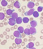
A Wright's-stained bone marrow aspirate smear from a patient with leukemia.
 
Bone marrow examination is the pathologic analysis of samples of bone marrow obtained via biopsy and bone marrow aspiration. Bone marrow examination is used in the diagnosis of a number of conditions, including leukemia, multiple myeloma, anemia, and pancytopenia. The bone marrow produces the cellular elements of the blood, including platelets, red blood cells and white blood cells. While much information can be gleaned by testing the blood itself (drawn from a vein by phlebotomy), it is sometimes necessary to examine the source of the blood cells in the bone marrow to obtain more information on hematopoiesis; this is the role of bone marrow aspiration and biopsy. 

The ratio between myeloid series and erythroid cells is relevant to bone marrow function, and also to diseases of the bone marrow and peripheral blood, such as leukemia and anemia. The normal myeloid-to-erythroid ratio is around 3:1; this ratio may increase in myelogenous leukemias, decrease in polycythemias, and reverse in cases of thalassemia.

Donation and transplantation

A bone marrow harvest in progress.
 
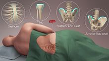
The preferred sites for the procedure
 
In a bone marrow transplant, hematopoietic stem cells are removed from a person and infused into another person (allogenic) or into the same person at a later time (autologous). If the donor and recipient are compatible, these infused cells will then travel to the bone marrow and initiate blood cell production. Transplantation from one person to another is conducted for the treatment of severe bone marrow diseases, such as congenital defects, autoimmune diseases or malignancies. The patient's own marrow is first killed off with drugs or radiation, and then the new stem cells are introduced. Before radiation therapy or chemotherapy in cases of cancer, some of the patient's hematopoietic stem cells are sometimes harvested and later infused back when the therapy is finished to restore the immune system.

Bone marrow stem cells can be induced to become neural cells to treat neurological illnesses, and can also potentially be used for the treatment of other illnesses, such as inflammatory bowel disease. In 2013, following a clinical trial, scientists proposed that bone marrow transplantation could be used to treat HIV in conjunction with antiretroviral drugs; however, it was later found that HIV remained in the bodies of the test subjects.

Harvesting

The stem cells are typically harvested directly from the red marrow in the iliac crest, often under general anesthesia. The procedure is minimally invasive and does not require stitches afterwards. Depending on the donor's health and reaction to the procedure, the actual harvesting can be an outpatient procedure, or can require 1–2 days of recovery in the hospital.

Another option is to administer certain drugs that stimulate the release of stem cells from the bone marrow into circulating blood. An intravenous catheter is inserted into the donor's arm, and the stem cells are then filtered out of the blood. This procedure is similar to that used in blood or platelet donation. In adults, bone marrow may also be taken from the sternum, while the tibia is often used when taking samples from infants. In newborns, stem cells may be retrieved from the umbilical cord.

Fossil record

Bone marrow may have first evolved in Eusthenopteron, a species of prehistoric fish with close links to early tetrapods.
 
The earliest fossilised evidence of bone marrow was discovered in 2014 in Eusthenopteron, a lobe-finned fish which lived during the Devonian period approximately 370 million years ago. Scientists from Uppsala University and the European Synchrotron Radiation Facility used X-ray synchrotron microtomography to study the fossilised interior of the skeleton's humerus, finding organised tubular structures akin to modern vertebrate bone marrow. Eusthenopteron is closely related to the early tetrapods, which ultimately evolved into the land-dwelling mammals and lizards of the present day.

Liquid breathing

From Wikipedia, the free encyclopedia

Liquid breathing is a form of respiration in which a normally air-breathing organism breathes an oxygen-rich liquid (such as a perfluorocarbon), rather than breathing air

This requires certain physical properties such as respiratory gas solubility, density, viscosity, vapor pressure, and lipid solubility which some, but not all, perfluorochemicals (perfluorocarbon) have. Thus, it is critical to choose the appropriate PFC for a specific biomedical application, such as liquid ventilation, drug delivery or blood substitutes. The physical properties of PFC liquids vary substantially; however, the one common property is their high solubility for respiratory gases. In fact, these liquids carry more oxygen and carbon dioxide than blood.

In theory, liquid breathing could assist in the treatment of patients with severe pulmonary or cardiac trauma, especially in pediatric cases. Liquid breathing has also been proposed for use in deep diving and space travel. Despite some recent advances in liquid ventilation, a standard mode of application has not yet been established.

Approaches

Physicochemical properties (37 °C at 1 atm) of 18 perfluorochemical liquids used for biomedical applications. This table characterizes the most significant physical properties related to systemic physiology and their range of properties.
Gas solubility
Oxygen 33–66 mL / 100 mL PFC
Carbon dioxide 140–166 mL / 100 mL PFC
Vapor pressure 0.2–400 torr
Density 1.58–2.0 g/mL
Viscosity 0.8–8.0 cS

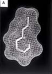
Computer models of three perfluorochemical molecules used for biomedical applications and for liquid ventilation studies: a) FC-75, b) perflubron, and c) perfluorodecalin.
 
Because liquid breathing is still a highly experimental technique, there are several proposed approaches.

Total liquid ventilation

Although total liquid ventilation (TLV) with completely liquid-filled lungs can be beneficial, the complex liquid-filled tube system required is a disadvantage compared to gas ventilation—the system must incorporate a membrane oxygenator, heater, and pumps to deliver to, and remove from the lungs tidal volume aliquots of conditioned perfluorocarbon (PFC). One research group led by Thomas H. Shaffer has maintained that with the use of microprocessors and new technology, it is possible to maintain better control of respiratory variables such as liquid functional residual capacity and tidal volume during TLV than with gas ventilation. Consequently, the total liquid ventilation necessitates a dedicated liquid ventilator similar to a medical ventilator except that it uses a breathable liquid. Many prototypes are used for animal experimentation, but experts recommend continued development of a liquid ventilator toward clinical applications. Specific preclinical liquid ventilator (Inolivent) is currently under joint development in Canada and France. The main application of this liquid ventilator is the ultra-fast induction of therapeutic hypothermia after cardiac arrest. This has been demonstrated to be more protective than slower cooling method after experimental cardiac arrest.

Partial liquid ventilation

In contrast, partial liquid ventilation (PLV) is a technique in which a PFC is instilled into the lung to a volume approximating functional residual capacity (approximately 40% of total lung capacity). Conventional mechanical ventilation delivers tidal volume breaths on top of it. This mode of liquid ventilation currently seems technologically more feasible than total liquid ventilation, because PLV could utilise technology currently in place in many neonatal intensive-care units (NICU) worldwide.

The influence of PLV on oxygenation, carbon dioxide removal and lung mechanics has been investigated in several animal studies using different models of lung injury. Clinical applications of PLV have been reported in patients with acute respiratory distress syndrome (ARDS), meconium aspiration syndrome, congenital diaphragmatic hernia and respiratory distress syndrome (RDS) of neonates. In order to correctly and effectively conduct PLV, it is essential to
  1. properly dose a patient to a specific lung volume (10–15 ml/kg) to recruit alveolar volume
  2. redose the lung with PFC liquid (1–2 ml/kg/h) to oppose PFC evaporation from the lung.
If PFC liquid is not maintained in the lung, PLV can not effectively protect the lung from biophysical forces associated with the gas ventilator. 

New application modes for PFC have been developed.

Partial liquid ventilation (PLV) involves filling the lungs with a fluid. This fluid is perfluorocarbon, also called Liquivent or Perflubron. The liquid has some unique properties. It has a very low surface tension, similar to surfactant, a substance that is produced in the lungs to prevent the alveoli from collapsing and sticking together during exhalation. It also has a high density, oxygen readily diffuses through it, and it may have some anti-inflammatory properties. In PLV, the lungs are filled with the liquid, the patient is then ventilated with a conventional ventilator using a protective lung ventilation strategy. This is called partial liquid ventilation. The hope is that the liquid will help the transport of oxygen to parts of the lung that are flooded and filled with debris, help remove this debris and open up more alveoli improving lung function. The study of PLV involves comparison to protocolized ventilator strategy designed to minimize lung damage.

PFC vapor

Vaporization of perfluorohexane with two anesthetic vaporizers calibrated for perfluorohexane has been shown to improve gas exchange in oleic acid-induced lung injury in sheep.

Predominantly PFCs with high vapor pressure are suitable for vaporization.

Aerosol-PFC

With aerosolized perfluorooctane, significant improvement of oxygenation and pulmonary mechanics was shown in adult sheep with oleic acid-induced lung injury. 

In surfactant-depleted piglets, persistent improvement of gas exchange and lung mechanics was demonstrated with Aerosol-PFC. The aerosol device is of decisive importance for the efficacy of PFC aerosolization, as aerosolization of PF5080 (a less purified FC77) has been shown to be ineffective using a different aerosol device in surfactant-depleted rabbits. Partial liquid ventilation and Aerosol-PFC reduced pulmonary inflammatory response.

Proposed uses

Diving

Gas pressure increases with depth, rising 1 bar (14.5 psi (100 kPa)) every 10 meters to over 1,000 bar at the bottom of the Mariana Trench. Diving becomes more dangerous as depth increases, and deep diving presents many hazards. All surface-breathing animals are subject to decompression sickness, including aquatic mammals and free-diving humans. Breathing at depth can cause nitrogen narcosis and oxygen toxicity. Holding the breath while ascending after breathing at depth can cause air embolisms, burst lung, and collapsed lung

Special breathing gas mixes such as trimix or heliox ameliorate the risk of decompression illness but do not eliminate it. Heliox further eliminates the risk of nitrogen narcosis but introduces the risk of helium tremors below about 500 feet (150 m). Atmospheric diving suits maintain body and breathing pressure at 1 bar, eliminating most of the hazards of descending, ascending, and breathing at depth. However, the rigid suits are bulky, clumsy, and very expensive. 

Liquid breathing offers a third option, promising the mobility available with flexible dive suits and the reduced risks of rigid suits. With liquid in the lungs, the pressure within the diver's lungs could accommodate changes in the pressure of the surrounding water without the huge gas partial pressure exposures required when the lungs are filled with gas. Liquid breathing would not result in the saturation of body tissues with high pressure nitrogen or helium that occurs with the use of non-liquids, thus would reduce or remove the need for slow decompression

A significant problem, however, arises from the high viscosity of the liquid and the corresponding reduction in its ability to remove CO2. All uses of liquid breathing for diving must involve total liquid ventilation (see above). Total liquid ventilation, however, has difficulty moving enough liquid to carry away CO2, because no matter how great the total pressure is, the amount of partial CO2 gas pressure available to dissolve CO2 into the breathing liquid can never be much more than the pressure at which CO2 exists in the blood (about 40 mm of mercury (Torr)).

At these pressures, most fluorocarbon liquids require about 70 mL/kg minute-ventilation volumes of liquid (about 5 L/min for a 70 kg adult) to remove enough CO2 for normal resting metabolism. This is a great deal of fluid to move, particularly as liquids are more viscous and denser than gases, (for example water is about 850 times the density of air). Any increase in the diver's metabolic activity also increases CO2 production and the breathing rate, which is already at the limits of realistic flow rates in liquid breathing. It seems unlikely that a person would move 10 liters/min of fluorocarbon liquid without assistance from a mechanical ventilator, so "free breathing" may be unlikely. However, it has been suggested that a liquid breathing system could be combined with a CO2 scrubber connected to the diver's blood supply; a US patent has been filed for such a method.

Medical treatment

Computer-generated model of perflubron and gentamicin molecules in liquid suspension for pulmonary administration
 
The most promising area for the use of liquid ventilation is in the field of pediatric medicine. The first medical use of liquid breathing was treatment of premature babies and adults with acute respiratory distress syndrome (ARDS) in the 1990s. Liquid breathing was used in clinical trials after the development by Alliance Pharmaceuticals of the fluorochemical perfluorooctyl bromide, or perflubron for short. Current methods of positive-pressure ventilation can contribute to the development of lung disease in pre-term neonates, leading to diseases such as bronchopulmonary dysplasia. Liquid ventilation removes many of the high pressure gradients responsible for this damage. Furthermore, perfluorocarbons have been demonstrated to reduce lung inflammation, improve ventilation-perfusion mismatch and to provide a novel route for the pulmonary administration of drugs.

In order to explore drug delivery techniques that would be useful for both partial and total liquid ventilation, more recent studies have focused on PFC drug delivery using a nanocrystal suspension. The first image is a computer model of a PFC liquid (perflubron) combined with gentamicin molecules. 

The second image shows experimental results comparing both plasma and tissue levels of gentamicin after an intratracheal (IT) and intravenous (IV) dose of 5 mg/kg in a newborn lamb during gas ventilation. Note that the plasma levels of the IV dose greatly exceed the levels of the IT dose over the 4 hour study period; whereas, the lung tissue levels of gentamicin when delivered by an intratracheal (IT) suspension, uniformly exceed the intravenous (IV) delivery approach after 4 hours. Thus, the IT approach allows more effective delivery of the drug to the target organ while maintaining a safer level systemically. Both images represent the in-vivo time course over 4 hours. Numerous studies have now demonstrated the effectiveness of PFC liquids as a delivery vehicle to the lungs.

Comparison of IT and IV administration of gentamicin.
 
Clinical trials with premature infants, children and adults were conducted. Since the safety of the procedure and the effectiveness were apparent from an early stage, the US Food and Drug Administration (FDA) gave the product "fast track" status (meaning an accelerated review of the product, designed to get it to the public as quickly as is safely possible) due to its life-saving potential. Clinical trials showed that using perflubron with ordinary ventilators improved outcomes as much as using high frequency oscillating ventilation (HFOV). But because perflubron was not better than HFOV, the FDA did not approve perflubron, and Alliance is no longer pursuing the partial liquid ventilation application. Whether perflubron would improve outcomes when used with HFOV or has fewer long-term consequences than HFOV remains an open question. 

In 1996 Mike Darwin and Steven B. Harris proposed using cold liquid ventilation with perfluorocarbon to quickly lower the body temperature of victims of cardiac arrest and other brain trauma to allow the brain to better recover. The technology came to be called gas/liquid ventilation (GLV), and was shown able to achieve a cooling rate of 0.5 °C per minute in large animals. It has not yet been tried in humans. 

Most recently, hypothermic brain protection has been associated with rapid brain cooling. In this regard, a new therapeutic approach is the use of intranasal perfluorochemical spray for preferential brain cooling. The nasopharyngeal (NP) approach is unique for brain cooling due to anatomic proximity to the cerebral circulation and arteries. Based on preclinical studies in adult sheep, it was shown that independent of region, brain cooling was faster during NP-perfluorochemical versus conventional whole body cooling with cooling blankets. To date, there have been four human studies including a completed randomized intra-arrest study (200 patients). Results clearly demonstrated that prehospital intra-arrest transnasal cooling is safe, feasible and is associated with an improvement in cooling time.

Space travel

Liquid immersion provides a way to reduce the physical stress of G forces. Forces applied to fluids are distributed as omnidirectional pressures. Because liquids cannot be practically compressed, they do not change density under high acceleration such as performed in aerial maneuvers or space travel. A person immersed in liquid of the same density as tissue has acceleration forces distributed around the body, rather than applied at a single point such as a seat or harness straps. This principle is used in a new type of G-suit called the Libelle G-suit, which allows aircraft pilots to remain conscious and functioning at more than 10 G acceleration by surrounding them with water in a rigid suit. 

Acceleration protection by liquid immersion is limited by the differential density of body tissues and immersion fluid, limiting the utility of this method to about 15 to 20 G. Extending acceleration protection beyond 20 G requires filling the lungs with fluid of density similar to water. An astronaut totally immersed in liquid, with liquid inside all body cavities, will feel little effect from extreme G forces because the forces on a liquid are distributed equally, and in all directions simultaneously. However effects will be felt because of density differences between different body tissues, so an upper acceleration limit still exists. 

Liquid breathing for acceleration protection may never be practical because of the difficulty of finding a suitable breathing medium of similar density to water that is compatible with lung tissue. Perfluorocarbon fluids are twice as dense as water, hence unsuitable for this application.

Examples in fiction

Literary works

  • Alexander Beliaev's 1928 science fiction novel Amphibian Man is based on a scientist and a maverick surgeon, who makes his son, Ichthyander (etymology: "fish" + "man") a life-saving transplant – a set of shark gills. There is a film based on the novel.
  • L. Sprague de Camp's 1938 short story "The Merman" hinges on an experimental process to make lungs function as gills, thus allowing a human being to "breathe" under water.
  • Hal Clement's 1973 novel Ocean on Top portrays a small underwater civilization living in a 'bubble' of oxygenated fluid denser than seawater.
  • Joe Haldeman's 1975 novel The Forever War describes liquid immersion and breathing in great detail as a key technology to allow space travel and combat with acceleration up to 50 G.
  • In the Star Trek: The Next Generation novel The Children of Hamlin (1988) the crew of the Enterprise-D encounter an alien race whose ships contain a breathable liquid environment.
  • Peter Benchley's 1994 novel White Shark centers around a Nazi scientist's experimental attempts to create an amphibious human, whose lungs are surgically modified to breathe underwater, and trained to reflexively do so after being flooded with a fluorocarbon solution.
  • Ben Bova's novel Jupiter (2000) features a craft in which the crew are suspended in a breathable liquid that allows them to survive in the high-pressure environment of Jupiter's atmosphere.
  • In Scott Westerfeld's sci-fi novel The Risen Empire (2003), the lungs of soldiers performing insertion from orbit are filled with an oxygen-rich polymer gel with embedded pseudo-alveoli and a rudimentary artificial intelligence.
  • The novel Mechanicum (2008) by Graham McNeill, Book 9 in the Horus Heresy book series, describes physically crippled Titan (gigantic war machine) pilots encased in nutrient fluid tanks. This allows them to continue operating beyond the limits normally imposed by the body.
  • In the 2009 novel The Lost Symbol by Dan Brown, Robert Langdon (the protagonist) is completely submerged in breathable liquid mixed with hallucinogenic chemicals and sedatives as a torture and interrogation technique by Mal'akh (the antagonist). He goes through a near death experience when he inhales the liquid and blacks out, losing control over his body, but is soon revived.
  • In Greg van Eekhout's 2014 novel California Bones, two characters are put into tanks filled with liquid: "They were given no breathing apparatus, but the water in the tank was rich with perfluorocarbon, which carried more oxygen than blood."
  • In author A.L. Mengel's science fiction novel The Wandering Star (2016), several characters breathe oxygenated fluid during a dive to explore an underwater city. They submerge in high pressure "bubbles" filled with the perfluorocarbon fluid.

Films and television

  • The aliens in the Gerry Anderson UFO series (1970-1971) use liquid-breathing spacesuits.
  • The 1989 film The Abyss by James Cameron features a character using liquid breathing to dive thousands of feet without compressing. The Abyss also features a scene with a rat submerged in and breathing fluorocarbon liquid, filmed in real life.
  • In the 1995 anime Neon Genesis Evangelion, the cockpits of the titular mecha are filled with a fictional oxygenated liquid called LCL which is required for the pilot to mentally sync with an Evangelion, as well as providing direct oxygenation of their blood, and dampening the impacts from battle. Once the cockpit is flooded the LCL is ionized, bringing its density, opacity, and viscosity close to that of air.
  • In the movies Event Horizon (1997) and Mission to Mars (2000), a character is depicted as being immersed in apparent breathable fluid before a high-acceleration launch.
  • In season 1, episode 13 of Seven Days (1998-2001) chrononaut Frank Parker is seen breathing a hyper-oxygenated perfluorocarbon liquid that is pumped through a sealed full body suit that he is wearing. This suit and liquid combination allow him to board a Russian submarine through open ocean at a depth of almost 1000 feet. Upon boarding the submarine he removes his helmet, expels the liquid from his lungs and is able to breathe air again.
  • In an episode of the Adult Swim cartoon series Metalocalypse (2006-2013), the other members of the band submerge guitarist Toki in a "liquid oxygen isolation chamber" while recording an album in the Mariana Trench.
  • In an episode of the Syfy Channel show Eureka (2006-2012), Sheriff Jack Carter is submerged in a tank of "oxygen rich plasma" to be cured of the effects of a scientific accident.
  • In the anime series Aldnoah.Zero (2014-2015), episode 5 shows that Slaine Troyard was in a liquid-filled capsule when he crashed. Princess Asseylum witnessed the crash, helped him to get out of the capsule, then used CPR on him to draw out the liquid from his lungs.

Video games

Memory and trauma

From Wikipedia, the free encyclopedia https://en.wikipedia.org/wiki/Memory_and_trauma ...