Search This Blog

Friday, May 11, 2018

Molecular neuroscience

From Wikipedia, the free encyclopedia
 
Molecular neuroscience is a branch of neuroscience that observes concepts in molecular biology applied to the nervous systems of animals. The scope of this subject covers topics such as molecular neuroanatomy, mechanisms of molecular signaling in the nervous system, the effects of genetics and epigenetics on neuronal development, and the molecular basis for neuroplasticity and neurodegenerative diseases.[1] As with molecular biology, molecular neuroscience is a relatively new field that is considerably dynamic.

Locating neurotransmitters

In molecular biology, communication between neurons typically occurs by chemical transmission across gaps between the cells called synapses. The transmitted chemicals, known as neurotransmitters, regulate a significant fraction of vital body functions.[2] It is possible to anatomically locate neurotransmitters by labeling techniques. It is possible to chemically identify certain neurotransmitters such as catecholamines by fixing neural tissue sections with formaldehyde. This can give rise to formaldehyde-induced fluorescence when exposed to ultraviolet light. Dopamine, a catecholamine, was identified in the nematode C. elegans by using this technique.[3]  Immunocytochemistry, which involves raising antibodies against targeted chemical or biological entities, includes a few other techniques of interest. A targeted neurotransmitter could be specifically tagged by primary and secondary antibodies with radioactive labeling in order to identify the neurotransmitter by autoradiography. The presence of neurotransmitters (though not necessarily the location) can be observed in enzyme-linked immunocytochemistry or enzyme--linked immunosorbent assays (ELISA) in which substrate-binding in the enzymatic assays can induce precipitates, fluorophores, or chemiluminescence. In the event that neurotransmitters cannot be histochemically identified, an alternative method is to locate them by their neural uptake mechanisms.[1]

Voltage-gated ion channels

Structure of eukaryotic voltage-gated potassium ion channels

Excitable cells in living organisms have voltage-gated ion channels. These can be observed throughout the nervous system in neurons. The first ion channels to be characterized were the sodium and potassium ion channels by A.L. Hodgkin and A.F. Huxley in the 1950s upon studying the giant axon of the squid genus Loligo. Their research demonstrated the selective permeability of cellular membranes, dependent on physiological conditions, and the electrical effects that result from these permeabilities to produce action potentials.[4]

Sodium ion channels

Sodium channels were the first voltage-gated ion channels to be isolated in 1984 from the eel Electrophorus electricus by Shosaku Numa. The pufferfish toxin tetrodotoxin (TTX), a sodium channel blocker, was used to isolate the sodium channel protein by binding it using the column chromatography technique for chemical separation. The amino acid sequence of the protein was analyzed by Edman degradation and then used to construct a cDNA library which could be used to clone the channel protein. Cloning the channel itself allowed for applications such as identifying the same channels in other animals.[1] Sodium channels are known for working in concert with potassium channels during the development of graded potentials and action potentials. Sodium channels allow an influx of Na+ ions into a neuron, resulting in a depolarization from the resting membrane potential of a neuron to lead to a graded potential or action potential, depending on the degree of depolarization.[5]

Potassium ion channels

Potassium channels come in a variety of forms, are present in most eukaryotic cells, and typically tend to stabilize the cell membrane at the potassium equilibrium potential. As with sodium ions, graded potentials and action potentials are also dependent on potassium channels. While influx of Na+ ions into a neuron induce cellular depolarization, efflux of influx of K+ ions out of a neuron causes a cell to repolarize to resting membrane potential. The activation of potassium ion channels themselves are dependent on the depolarization resulting from Na+ influx during an action potential.[1] As with sodium channels, the potassium channels have their own toxins that block channel protein action. An example of such a toxin is the large cation, tetraethylammonium (TEA), but it is notable that the toxin does not have the same mechanism of action on all potassium channels, given the variety of channel types across species. The presence of potassium channels was first identified in Drosophila melanogaster mutant flies that shook uncontrollably upon anesthesia due to problems in cellular repolarization that led to abnormal neuron and muscle electrophysiology. Potassium channels were first identified by manipulating molecular genetics (of the flies) instead of performing channel protein purification because there were no known high-affinity ligands for potassium channels (such as TEA) at the time of discovery.[1][6]

Calcium ion channels

Calcium channels are important for certain cell-signaling cascades as well as neurotransmitter release at axon terminals. A variety of different types of calcium ion channels are found in excitable cells. As with sodium ion channels, calcium ion channels have been isolated and cloned by chromatographic purification techniques. It is notable, as with the case of neurotransmitter release, that calcium channels can interact with intracelluar proteins and plays a strong role in signaling, especially in locations such as the sarcoplasmic reticulum of muscle cells.[1]

Receptors

Various types of receptors can be used for cell signaling and communication and can include ionotropic receptors and metabotropic receptors. These cell surface receptor types are differentiated by the mechanism and duration of action with ionotropic receptors being associated with fast signal transmission and metabotropic receptors being associated with slow signal transmission. Metabotropic receptors happen to cover a wide variety of cell-surface receptors with notably different signaling cascades.[1][5]

Ionotropic receptors

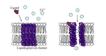
Prototypical depiction of ionotropic receptor in the case of Ca2+ ion flow

Ionotropic receptors, otherwise known as ligand-gated ion channels, are fast acting receptors that mediate neural and physiological function by ion channel flow with ligand-binding. Nicotinic, GABA, and Glutamate receptors are among some of the cell surface receptors regulated by ligand-gated ion channel flow. GABA is the brain's main inhibitory neurotransmitter and glutamate is the brain's main excitatory neurotransmitter.[1]

GABA receptors

GABAA and GABAC receptors are known to be ionotropic, while the GABAB receptor is metabotropic. GABAA receptors mediate fast inhibitory responses in the central nervous system (CNS) and are found on neurons, glial cells, and adrenal medulla cells. It is responsible for inducing Cl ion influx into cells, thereby reducing the probability that membrane depolarization will occur upon the arrival of a graded potential or an action potential. GABA receptors can also interact with non-endogenous ligands to influence activity. For example, the compound diazepam (marketed as Valium) is an allosteric agonist which increases the affinity of the receptor for GABA. The increased physiological inhibitory effects resulting from increased GABA binding make diazepam a useful tranquilizer or anticonvulsant (antiepileptic drugs). On the other hand, GABA receptors can also be targeted by decreasing Cl cellular influx with the effect of convulsants like picrotoxin. The antagonistic mechanism of action for this compound is not directly on the GABA receptor, but there are other compounds that are capable of allosteric inactivation, including T-butylbicyclophorothionate (TBPS) and pentylenetetrazole (PZT). Compared with GABAA, GABAC receptors have a higher affinity for GABA, they are likely to be longer-lasting in activity, and their responses are likely to be generated by lower GABA concentrations.[1]

Glutamate receptors

Ionotropic glutamate receptors can include NMDA, AMPA, and kainate receptors. These receptors are named after agonists that facilitate glutamate activity. NMDA receptors are notable for their excitatory mechanisms to affect neuronal plasticity in learning and memory, as well as neuropathologies such as stroke and epilepsy. NDMA receptors have multiple binding sites just like ionotropic GABA receptors and can be influenced by co-agonists such the glycine neurotransmitter or phencyclidine (PCP). The NMDA receptors carry a current by Ca2+ ions and can be blocked by extracellular Mg2+ ions depending on voltage and membrane potential. This Ca2+ influx is increased by excitatory postsynaptic potentials (EPSPs) produced by NMDA receptors, activating Ca2+-based signaling cascades (such as neurotransmitter release). AMPA generate shorter and larger excitatory postsynaptic currents than other ionotropic glutamate receptors.[5]

Nicotinic ACh receptors

Nicotinic receptors bind the acetylcholine (ACh) neurotransmitter to produce non-selective cation channel flow that generates excitatory postsynaptic responses. Receptor activity, which can be influenced by nicotine consumption, produces feelings of euphoria, relaxation, and inevitably addiction in high levels.[5]

Metabotropic receptors

G-protein-linked receptor signaling cascade

Metabotropic receptors, are slow response receptors in postsynaptic cells. Typically these slow responses are characterized by more elaborate intracellular changes in biochemistry. Responses of neurotransmitter uptake by metabotropic receptors can result in the activation of intracellualar enzymes and cascades involving second messengers, as is the case with G protein-linked receptors. Various metabotropic receptors can include certain glutamate receptors, muscarinic ACh receptors, GABAB receptors, and receptor tyrosine kinases.

G protein-linked receptors

The G protein-linked signaling cascade can significantly amplify the signal of a particular neurotransmitter to produce hundreds to thousands of second messengers in a cell. The mechanism of action by which G protein-linked receptors cause a signaling cascade is as follows:
  1. Neurotransmitter binds to the receptor
  2. The receptor undergoes a conformational change to allow G-protein complex binding
  3. GDP is exchanged with GTP upon G protein complex binding to the receptor
  4. The α-subunit of the G protein complex is bound to GTP and separates to bind with a target protein such as adenylate cyclase
  5. The binding to the target protein either increases or decreases the rate of second messenger (such as cyclic AMP) production
  6. GTPase hydrolyzes the α-subunit so that is bound to GDP and the α-subunit returns to the G protein complex inactive

Neurotransmitter release

Structure of a synapse where neurotransmitter release and uptake occurs

Neurotransmitters are released in discrete packets known as quanta from the axon terminal of one neuron to the dendrites of another across a synapse. These quanta have been identified by electron microscopy as synaptic vesicles. Two types of vesicles are small synaptic vessicles (SSVs), which are about 40-60nm in diameter, and large dense-core vesicles (LDCVs), electron-dense vesicles approximately 120-200nm in diameter.[1] The former is derived from endosomes and houses neurotransmitters such as acetylcholine, glutamate, GABA, and glycine. The latter is derived from the Golgi apparatus and houses larger neurotransmitters such as catecholamines and other peptide neurotransmitters.[7] Neurotransmitters are released from an axon terminal and bind to postsynaptic dendrites in the following procession:[5]
  1. Mobilization/recruitment of synaptic vesicle from cytoskeleton
  2. Docking of vesicle (binding) to presynaptic membrane
  3. Priming of vesicle by ATP (relatively slow step)
  4. Fusion of primed vesicle with presynaptic membrane and exocytosis of the housed neurotransmitter
  5. Uptake of neurotransmitters in receptors of a postsynaptic cell
  6. Initiation or inhibition of action potential in postsynaptic cell depending on whether the neurotransmitters are excitatory or inhibitory (excitatory will result in depolarization of the postsynaptic membrane)

Neurotransmitter release is calcium-dependent

Neurotransmitter release is dependent on an external supply of Ca2+ ions which enter axon terminals via voltage-gated calcium channels. Vesicular fusion with the terminal membrane and release of the neurotransmitter is caused by the generation of Ca2+ gradients induced by incoming action potentials. The Ca2+ ions cause the mobilization of newly synthesized vesicles from a reserve pool to undergo this membrane fusion. This mechanism of action was discovered in squid giant axons.[8] Lowering intracellular Ca2+ ions provides a direct inhibitory effect on neurotransmitter release.[1] After release of the neurotransmitter occurs, vesicular membranes are recycled to their origins of production. Calcium ion channels can vary depending on the location of incidence. For example, the channels at an axon terminal differ from the typical calcium channels of a cell body (whether neural or not). Even at axon terminals, calcium ion channel types can vary, as is the case with P-type calcium channels located at the neuromuscular junction.[1]

Neuronal gene expression

Sex differences

Differences in sex determination are controlled by sex chromosomes. Sex hormonal releases have a significant effect on sexual dimorphisms (phenotypic differentiation of sexual characteristics) of the brain. Recent studies seem to suggest that regulating these dimorphisms has implications for understanding normal and abnormal brain function. Sexual dimorphisms may be significantly influenced by sex-based brain gene expression which varies from species to species.

Animal models such as rodents, Drosophila melanogaster, and Caenorhabditis elegans, have been used to observe the origins and/or extent of sex bias in the brain versus the hormone-producing gonads of an animal. With the rodents, studies on genetic manipulation of sex chromosomes resulted in an effect on one sex that was completely opposite of the effect in the other sex. For example, a knockout of a particular gene only resulted in anxiety-like effects in males. With studies on D. menlanogaster it was found that a large brain sex bias of expression occurred even after the gonads were removed, suggesting that sex bias could be independent of hormonal control in certain aspects.[9]

Observing sex-biased genes has the potential for clinical significance in observing brain physiology and the potential for related (whether directly or indirectly) neurological disorders. Examples of diseases with sex biases in development include Huntington's disease, cerebral ischemia, and Alzheimer's disease.[9]

Epigenetics of the brain

Many brain functions can be influenced at the cellular and molecular level by variations and changes in gene expression, without altering the sequence of DNA in an organism. This is otherwise known as epigenetic regulation. Examples of epigenetic mechanisms include histone modifications and DNA methylation. Such changes have been found to be strongly influential in the incidence of brain disease, mental illness, and addiction.[10] Epigenetic control has been shown to be involved in high levels of plasticity in early development, thereby defining its importance in the critical period of an organism.[11] Examples of how epigenetic changes can affect the human brain are as follows:
  • Higher methylation levels in rRNA genes in the hippocampus of the brain results in a lower production of proteins and thus limited hippocampal function can result in learning and memory impairment and resultant suicidal tendencies.[12]
  • In a study comparing genetic differences between healthy people and psychiatric patients 60 different epigenetic markers associated with brain cell signaling were found.[12]
  • Environmental factors such as child abuse appears to cause the expression of an epigenetic tag on glucocorticoid receptors (associated with stress responses) that was not found in suicide victims.[12] This is an example of experience-dependent plasticity.
  • Environmental enrichment in individuals is associated with increased hippocampal gene histone acetylation and thus improved memory consolidation (notably spatial memory).[11]

Molecular mechanisms of neurodegenerative diseases

Excitotoxicity and glutamate receptors

Excitotoxicity is phenomenon in which glutamate receptors are inappropriately activated. It can be caused by prolonged excitatory synaptic transmission in which high levels of glutamate neurotransmitter cause excessive activation in a postsynaptic neuron that can result in the death of the postsynaptic neuron. Following brain injury (such as from ischemia), it has been found that excitotoxicity is a significant cause of neuronal damage. This can be understandable in the case where sudden perfusion of blood after reduced blood flow to the brain can result in excessive synaptic activity caused by the presence of increased glutamate and aspartate during the period of ischemia.[5][13]

Alzheimer's disease

Alzheimer's disease is the most common neurodegenerative disease and is the most common form of dementia in the elderly. The disorder is characterized by progressive loss of memory and various cognitive functions. It is hypothesized that the deposition of amyloid-β peptide (40-42 amino acid residues) in the brain is integral in the incidence of Alzheimer's disease. Accumulation is purported to block hippocampal long-term potentiation. It is also possible that a receptor for amyloid-β oligomers could be a prion protein.[14]

Parkinson's disease

Parkinson's disease is the second most common neurodegenerative disease after Alzheimer's disease. It is a hypokinetic movement basal ganglia disease caused by the loss of dopaminergic neurons in the substantia nigra of the human brain. The inhibitory outflow of the basal ganglia is thus not decreased, and so upper motor neurons, mediated by the thalamus, are not activated in a timely manner. Specific symptoms include rigidity, postural problems, slow movements, and tremors. Blocking GABA receptor input from medium spiny neurons to reticulata cells, causes inhibition of upper motor neurons similar to the inhibition that occurs in Parkinson's disease.[5]

Huntington's disease

Huntington's disease is a hyperkinetic movement basal ganglia disease caused by lack of normal inhibitory inputs from medium spiny neurons of the basal ganglia. This poses the opposite effects of those associated with Parkinson's disease, including inappropriate activation of upper motor neurons. As with the GABAergic mechanisms observed in relation to Parkinson's disease, a GABA agonist injected into the substantia nigra pars reticulata decreases inhibition of upper motor neurons, resulting in ballistic involuntary motor movements, similar to symptoms of Huntington's disease.[5]

Neuroscience and intelligence

From Wikipedia, the free encyclopedia
Neuroscience and intelligence refers to the various neurological factors that are partly responsible for the variation of intelligence within a species or between different species. A large amount of research in this area has been focused on the neural basis of human intelligence. Historic approaches to study the neuroscience of intelligence consisted of correlating external head parameters, for example head circumference, to intelligence.[1] Post-mortem measures of brain weight and brain volume have also been used.[1] More recent methodologies focus on examining correlates of intelligence within the living brain using techniques such as magnetic resonance imaging (MRI), functional MRI (fMRI), electroencephalography (EEG), positron emission tomography and other non-invasive measures of brain structure and activity.[1]

Researchers have been able to identify correlates of intelligence within the brain and its functioning. These include overall brain volume,[2] grey matter volume,[3] white matter volume,[4] white matter integrity,[5] cortical thickness[3] and neural efficiency.[6] Although the evidence base for our understanding of the neural basis of human intelligence has increased greatly over the past 30 years, even more research is needed to fully understand it.[1]

The neural basis of intelligence has also been examined in animals such as primates, cetaceans and rodents.[7]

Humans

Brain volume

One of the main methods used to establish a relationship between intelligence and the brain is to use measures of Brain volume.[1] The earliest attempts at estimating brain volume were done using measures of external head parameters, such as head circumference as a proxy for brain size.[1] More recent methodologies employed to study this relationship include post-mortem measures of brain weight and volume. These have their own limitations and strengths.[8] The advent of MRI as a non-invasive highly-accurate measure of living brain structure and function (using fMRI) made this the pre-dominant and preferred method for measuring brain volume.[1]

Overall, larger brain size and volume is associated with better cognitive functioning and higher intelligence.[1] The specific regions that show the most robust correlation between volume and intelligence are the frontal, temporal and parietal lobes of the brain.[9][10][11] A large number of studies have been conducted with uniformly positive correlations, leading to the generally safe conclusion that larger brains predict greater intelligence.[12][13] In healthy adults, the correlation of total brain volume and IQ is ~ 0.4 [14]

Less is known about variation on scales less than total brain volume. A meta-analytic review by McDaniel found that the correlation between Intelligence and in vivo brain size was larger for females (0.40) than for males (0.25).[15] The same study also found that the correlation between brain size and Intelligence increased with age, with children showing smaller correlations.[15] It has been suggested that the link between larger brain volumes and higher intelligence is related to variation in specific brain regions: a whole-brain measure would under-estimate these links.[9] For functions more specific than general intelligence, regional effects may be more important. For instance evidence suggests that in adolescents learning new words, vocabulary growth is associated with gray matter density in bilateral posterior supramarginal gyri.[16] Small studies have shown transient changes in gray-matter associated with developing a new physical skill (juggling) occipito-temporal cortex. [17]

Brain volume is not a perfect account of intelligence: the relationship explains a modest amount of variance in intelligence – 12% to 36% of the variance.[8][9] The amount of variance explained by brain volume may also depend on the type of intelligence measured.[8] Up to 36% of variance in verbal intelligence can be explained by brain volume, while only approximately 10% of variance in visuospatial intelligence can be explained by brain volume.[8] A 2015 study by researcher Stuart J Ritchie found that brain size explained 12% of the variance in intelligence among individuals.[18] These caveats imply that there are other major factors influencing how intelligent an individual is apart from brain size.[1] In a large meta-analysis consisting of 88 studies Pietschnig et al. (2015) estimated the correlation between brain volume and intelligence to be about correlation coefficient of 0.24 which equates to 6% variance.[19] Taking into account measurement quality, and sample type and IQ-range, the meta-analytic association of brain volume in appears to be ~ .4 in normal adults.[14] Researcher Jakob Pietschnig argued that the strength of the positive association of brain volume and IQ remains robust, but has been overestimated in the literature. He has stated that "It is tempting to interpret this association in the context of human cognitive evolution and species differences in brain size and cognitive ability, we show that it is not warranted to interpret brain size as an isomorphic proxy of human intelligence differences".[19]

Grey matter

Grey matter has been examined as a potential biological foundation for differences in intelligence. Similarly to brain volume, global grey matter volume is positively associated with intelligence.[1] More specifically, higher intelligence has been associated with larger cortical grey matter in the prefrontal and posterior temporal cortex in adults.[3] Furthermore, both verbal and nonverbal intelligence have been shown to be positively correlated with grey matter volume across the parietal, temporal and occipital lobes in young healthy adults, implying that intelligence is associated with a wide variety of structures within the brain.[20]

There appear to be sex differences between the relationship of grey matter to intelligence between men and women.[21] Men appear to show more intelligence to grey matter correlations in the frontal and parietal lobes, while the strongest correlations between intelligence and grey matter in women can be found in the frontal lobes and Broca's area.[21] However, these differences do not seem to impact overall Intelligence, implying that the same cognitive ability levels can be attained in different ways.[21]

One specific methodology used to study grey matter correlates of intelligence in areas of the brain is known as voxel-based morphometry (VBM). VBM allows researchers to specify areas of interest with great spatial resolution, allowing the examination of grey matter areas correlated with intelligence with greater special resolution. VBM has been used to correlate grey matter positively with intelligence in the frontal, temporal, parietal, and occipital lobes in healthy adults.[22] VBM has also been used to show that grey matter volume in the medial region of the prefrontal cortex and the dorsomedial prefrontal cortex correlate positively with intelligence in a group of 55 healthy adults.[23] VBM has also been successfully used to establish a positive correlation between grey matter volumes in the anterior cingulate and intelligence in children aged 5 to 18 years old.[24]

Grey matter has also been shown to positively correlate with intelligence in children.[24][25][26] Reis and colleagues[26] have found that grey matter in the prefrontal cortex contributes most robustly to variance in Intelligence in children between 5 and 17, while subcortical grey matter is related to intelligence to a lesser extent. Frangou and colleagues[25] examined the relationship between grey matter and intelligence in children and young adults aged between 12 and 21, and found that grey matter in the orbitofrontal cortex, cingulate gyrus, cerebellum and thalamus was positively correlated to intelligence, while grey matter in the caudate nucleus is negatively correlated with intelligence. However, the relationship between grey matter volume and intelligence only develops over time, as no significant positive relationship can be found between grey matter volume and intelligence in children under 11.[24]

An underlying caveat to research into the relationship of grey matter volume and intelligence is demonstrated by the hypothesis of neural efficiency.[6][27] The findings that more intelligent individuals are more efficient at using their neurons might indicate that the correlation of grey matter to intelligence reflects selective elimination of unused synapses, and thus a better brain circuitry.[28]

White matter

Similar to grey matter, white matter has been shown to correlate positively with intelligence in humans.[1][4] White matter consists mainly of myelinated neuronal axons, responsible for delivering signals between neurons. The pinkish-white color of white matter is actually a result of these myelin sheaths that electrically insulate neurons that are transmitting signals to other neurons. White matter connects different regions of grey matter in the cerebrum together. These interconnections make transport more seamless and allow us to perform tasks easier. Significant correlations between intelligence and the corpus callosum have been found, as larger callosal areas have been positively correlated with cognitive performance.[1] However, there appear to be differences in importance for white matter between verbal and nonverbal intelligence, as although both verbal and nonverbal measures of intelligence correlate positively with the size of the corpus callosum, the correlation for intelligence and corpus callosum size was larger (.47) for nonverbal measures than that for verbal measures (.18).[29] Anatomical mesh-based geometrical modelling[30][31][32] has also shown positive correlations between the thickness of the corpus callosum and Intelligence in healthy adults.[33]

White matter integrity has also been found to be related to Intelligence.[5] White matter tract integrity is important for information processing speed, and therefore reduced white matter integrity is related to lower intelligence.[5] The effect of white matter integrity is mediate entirely through information processing speed.[5] These findings indicate that the brain is structurally interconnected and that axonal fibres are integrally important for fast information process, and thus general intelligence.[5]

Contradicting the findings described above, VBM failed to find a relationship between the corpus callosum and intelligence in healthy adults.[22] This contradiction can be viewed to signify that the relationship between white matter volume and intelligence is not as robust as that of grey matter and intelligence.[1]

Cortical thickness

Cortical thickness has also been found to correlate positively with intelligence in humans.[3] However, the rate of growth of cortical thickness is also related to intelligence.[28] In early childhood, cortical thickness displays a negative correlation with intelligence, while by late childhood this correlation has shifted to a positive one.[28] More intelligent children were found to develop cortical thickness more steadily and over longer periods of time than less bright children.[28] Studies have found cortical thickness to explain 5% in the variance of intelligence among individuals.[18] In a study conducted to find associations between cortical thickness and general intelligence between different groups of people, sex did not play a role in intelligence.[34] Although it is hard to pin intelligence on age based on cortical thickness due to different socioeconomic circumstances and education levels, older subjects (17 - 24) tended to have less variances in terms of intelligence than when compared to younger subjects (19 - 17).[34][dubious ]

Cortical convolution

Cortical convolution has increased the folding of the brain’s surface over the course of human evolution. It has been hypothesized that the high degree of cortical convolution may be a neurological substrate that supports some of the human brain's most distinctive cognitive abilities. Consequently, individual intelligence within the human species might be modulated by the degree of cortical convolution.[35]

Neural efficiency

The neural efficiency hypothesis postulates that more intelligent individuals display less activation in the brain during cognitive tasks, as measured by Glucose metabolism.[6] A small sample of participants (N=8) displayed negative correlations between intelligence and absolute regional metabolic rates ranging from -0.48 to -0.84, as measured by PET scans, indicating that brighter individuals were more effective processors of information, as they use less energy.[6] According to an extensive review by Neubauer & Fink[36] a large number of studies (N=27) have confirmed this finding using methods such as PET scans,[37] EEG[38] and fMRI.[39]

fMRI and EEG studies have revealed that task difficulty is an important factor affecting neural efficiency.[36] More intelligent individuals display neural efficiency only when faced with tasks of subjectively easy to moderate difficulty, while no neural efficiency can be found during difficult tasks.[40] In fact, more able individuals appear to invest more cortical resources in tasks of high difficulty.[36] This appears to be especially true for the Prefrontal Cortex, as individuals with higher intelligence displayed increased activation of this area during difficult tasks compared to individuals with lower intelligence.[41][42] It has been proposed that the main reason for the neural efficiency phenomenon could be that individuals with high intelligence are better at blocking out interfering information than individuals with low intelligence.[43]

Further research

Some scientists prefer to look at more qualitative variables to relate to the size of measurable regions of known function, for example relating the size of the primary visual cortex to its corresponding functions, that of visual performance.[44][45]

In a study of the head growth of 633 term-born children from the Avon Longitudinal Study of Parents and Children cohort, it was shown that prenatal growth and growth during infancy were associated with subsequent IQ. The study’s conclusion was that the brain volume a child achieves by the age of 1 year helps determine later intelligence. Growth in brain volume after infancy may not compensate for poorer earlier growth.[46]

There is an association between IQ and myopia. One suggested explanation is that one or several pleiotropic gene(s) affect the size of the neocortex part of the brain and eyes simultaneously.[47]

Parieto-frontal integration theory

In 2007, Behavioral and Brain Sciences published a target article that put forth a biological model of intelligence based on 37 peer-reviewed neuroimaging studies (Jung & Haier, 2007). Their review of a wealth of data from functional imaging (functional magnetic resonance imaging and positron emission tomography) and structural imaging (diffusion MRI, voxel-based morphometry, in vivo magnetic resonance spectroscopy) argues that human intelligence arises from a distributed and integrated neural network comprising brain regions in the frontal and parietal lobes.[48]

A recent lesion mapping study conducted by Barbey and colleagues provides evidence to support the P-FIT theory of intelligence.[49][50][51]

Brain injuries at an early age isolated to one side of the brain typically results in relatively spared intellectual function and with IQ in the normal range.[52]

Primates

Brain size

Another theory of brain size in vertebrates is that it may relate to social rather than mechanical skill. Cortical size relates directly to a pairbonding life style and among primates cerebral cortex size varies directly with the demands of living in a large complex social network. Compared to other mammals, primates have significantly larger brain size. Additionally, most primates are found to be polygynandrous, having many social relationships with others. Although inconclusive, some studies have shown that this polygnandrous statue correlates to brain size.[53]

Health

Several environmental factors related to health can lead to significant cognitive impairment, particularly if they occur during pregnancy and childhood when the brain is growing and the blood–brain barrier is less effective. Developed nations have implemented several health policies regarding nutrients and toxins known to influence cognitive function. These include laws requiring fortification of certain food products and laws establishing safe levels of pollutants (e.g. lead, mercury, and organochlorides). Comprehensive policy recommendations targeting reduction of cognitive impairment in children have been proposed.[54]

Evolution of human intelligence

From Wikipedia, the free encyclopedia
The evolution of human intelligence is closely tied to the evolution of the human brain and to the origin of language. The timeline of human evolution spans approximately 7 million years,[1] from the separation of the genus Pan until the emergence of behavioral modernity by 50,000 years ago. The first 3 million years of this timeline concern Sahelanthropus, the following 2 million concern Australopithecus and the final 2 million span the history of the genus Homo in the Paleolithic era.

Many traits of human intelligence, such as empathy, theory of mind, mourning, ritual, and the use of symbols and tools, are apparent in great apes although in less sophisticated forms than found in humans, such as great ape language.

History

Hominidae

The great apes (hominidae) show considerable cognitive and empathic abilities. Chimpanzees make tools and use them to acquire foods and for social displays; they have sophisticated hunting strategies requiring cooperation, influence and rank; they are status conscious, manipulative and capable of deception; they can learn to use symbols and understand aspects of human language including some relational syntax, concepts of number and numerical sequence.[2]

In one study, young chimpanzees outperformed human college students in tasks requiring remembering numbers.[3] This claim was refuted in a later study after it was noted that the chimpanzees had received extensive practice with the task while the students were evaluated on their first attempt. When human subjects were given time to practice, they substantially outperformed the young chimps.[4]

Homininae


Chimpanzee mother and baby

Around 10 million years ago, the Earth's climate entered a cooler and drier phase, which led eventually to the Quaternary glaciation beginning some 2.6 million years ago. One consequence of this was that the north African tropical forest began to retreat, being replaced first by open grasslands and eventually by desert (the modern Sahara). As their environment changed from continuous forest to patches of forest separated by expanses of grassland, some primates adapted to a partly or fully ground-dwelling life. Here they were exposed to predators, such as the big cats, from whom they had previously been safe.

These environmental pressures caused selection to favor bipedalism: walking on hind legs. This gave the Homininae's eyes greater elevation, the ability to see approaching danger further off, and a more efficient means of locomotion.[citation needed] It also freed the arms from the task of walking and made the hands available for tasks such as gathering food. At some point the bipedal primates developed handedness, giving them the ability to pick up sticks, bones and stones and use them as weapons, or as tools for tasks such as killing smaller animals, cracking nuts, or cutting up carcasses. In other words, these primates developed the use of primitive technology. Bipedal tool-using primates form the Hominina subtribe, of which the earliest species, such as Sahelanthropus tchadensis, date to about 7 to 5 million years ago.

From about 5 million years ago, the hominin brain began to develop rapidly in both size and differentiation of function.

There has been a gradual increase in brain volume as humans progressed along the timeline of evolution (see Homininae), starting from about 600 cm3 in Homo habilis up to 1500 cm3 in Homo neanderthalensis. Thus, in general there's a correlation between brain volume and intelligence.[citation needed] However, modern Homo sapiens have a brain volume slightly smaller (1250 cm3) than neanderthals, and the Flores hominids (Homo floresiensis), nicknamed hobbits, had a cranial capacity of about 380 cm3 (considered small for a chimpanzee) about a third of that of H. erectus. It is proposed that they evolved from H. erectus as a case of insular dwarfism. With their three times smaller brain the Flores hominids apparently used fire and made tools as sophisticated as those of their ancestor H.erectus. In this case, it seems that for intelligence, the structure of the brain is more important than its volume.

Homo

By 2.4 million years ago Homo habilis had appeared in East Africa: the first known human species, and the first known to make stone tools, yet the disputed findings of signs of tool use from even earlier age and from the vicinity as multiple Australopithecus fossils may put this to question its "greater intelligence when compared to earlier and more primitive Australopithecus genus".

The use of tools conferred a crucial evolutionary advantage, and required a larger and more sophisticated brain to co-ordinate the fine hand movements required for this task.[5] Our knowledge of the complexity of behaviour of Homo habilis is not limited to stone culture, they also had habitual therapic use of toothpicks.[6] The evolution of a larger brain created a problem for early humans, however. A larger brain requires a larger skull, and thus requires the female to have a wider birth canal for the newborn's larger skull to pass through. But if the female's birth canal grew too wide, her pelvis would be so wide that she would lose the ability to run, which was a necessary skill 2 million years ago.[citation needed]

The solution to this was to give birth at an early stage of fetal development, before the skull grew too large to pass through the birth canal. This adaptation enabled the human brain to continue to grow, but it imposed a new discipline. The need to care for helpless infants for long periods of time forced humans to become less mobile[citation needed]. Human bands increasingly stayed in one place for long periods, so that females could care for infants, while males hunted food and fought with other bands that competed for food sources[citation needed]. As a result, humans became even more dependent on tool-making to compete with other animals and other humans, and relied less on body size and strength[citation needed].

About 200,000 years ago Europe and the Middle East were colonized by Neanderthal man, extinct by 39,000 years ago following the appearance of modern humans in the region from 40,000–45,000 years ago.

Homo sapiens


"The Lion-man", found in the Hohlenstein-Stadel cave of Germany's Swabian Alb and dated to 40,000 years ago, is associated with the Aurignacian culture and is the oldest known anthropomorphic animal figurine in the world.
Quaternary extinction event Quaternary extinction event Holocene extinction Holocene extinction Yellowstone Caldera Yellowstone Caldera Toba catastrophe theory Homo heidelbergensis Homo neanderthalensis Homo antecessor Homo sapiens Homo habilis Homo georgicus Homo ergaster Homo erectus Homo Homo
Dates approximate, consult articles for details
(From 2000000 BC till 2013 AD in (partial) exponential notation)
See also: Java Man (−1.75e+06), Yuanmou Man (−1.75e+06 : -0.73e+06),
Lantian Man (−1.7e+06), Nanjing Man (- 0.6e+06), Tautavel Man (- 0.5e+06),
Peking Man (- 0.4e+06), Solo Man (- 0.4e+06), and Peștera cu Oase (- 0.378e+05)

Homo sapiens intelligence

Around 200,000 years ago, Homo sapiens first appeared in East Africa. It is unclear to what extent these early modern humans had developed language, music, religion etc. They spread throughout Africa over the following approximately 50,000 years.[citation needed]

According to proponents of the Toba catastrophe theory, the climate in non-tropical regions of the earth experienced a sudden freezing about 70,000 years ago, because of a huge explosion of the Toba volcano that filled the atmosphere with volcanic ash for several years. This reduced the human population to less than 10,000 breeding pairs in equatorial Africa, from which all modern humans are descended. Being unprepared for the sudden change in climate, the survivors were those intelligent enough to invent new tools and ways of keeping warm and finding new sources of food (for example, adapting to ocean fishing based on prior fishing skills used in lakes and streams that became frozen).[citation needed]

Around 80–100,000 years ago, three main lines of Homo sapiens diverged, bearers of mitochondrial haplogroup L1 (mtDNA) / A (Y-DNA) colonizing Southern Africa (the ancestors of the Khoisan/Capoid peoples), bearers of haplogroup L2 (mtDNA) / B (Y-DNA) settling Central and West Africa (the ancestors of Niger–Congo and Nilo-Saharan speaking peoples), while the bearers of haplogroup L3 remained in East Africa.[citation needed]

The "Great Leap Forward" leading to full behavioral modernity sets in only after this separation. Rapidly increasing sophistication in tool-making and behaviour is apparent from about 80,000 years ago, and the migration out of Africa follows towards the very end of the Middle Paleolithic, some 60,000 years ago. Fully modern behaviour, including figurative art, music, self-ornamentation, trade, burial rites etc. is evident by 30,000 years ago. The oldest unequivocal examples of prehistoric art date to this period, the Aurignacian and the Gravettian periods of prehistoric Europe, such as the Venus figurines and cave painting (Chauvet Cave) and the earliest musical instruments (the bone pipe of Geissenklösterle, Germany, dated to about 36,000 years ago).[7]

Models

Social brain hypothesis

The social brain hypothesis was proposed by British anthropologist Robin Dunbar, who argues that human intelligence did not evolve primarily as a means to solve ecological problems, but rather as a means of surviving and reproducing in large and complex social groups.[8][9] Some of the behaviors associated with living in large groups include reciprocal altruism, deception and coalition formation. These group dynamics relate to Theory of Mind or the ability to understand the thoughts and emotions of others, though Dunbar himself admits in the same book that it is not the flocking itself that causes intelligence to evolve (as shown by ruminants).[8]

Dunbar argues that when the size of a social group increases, the number of different relationships in the group may increase by orders of magnitude. Chimpanzees live in groups of about 50 individuals whereas humans typically have a social circle of about 150 people, which is also the typical size of social communities in small societies and personal social networks;[10] this number is now referred to as Dunbar's number. In addition, there is evidence to suggest that the success of groups is dependent on their size at foundation, with groupings of around 150 being particularly successful, potentially reflecting the fact that communities of this size strike a balance between the minimum size of effective functionality and the maximum size for creating a sense of commitment to the community.[11] According to the social brain hypothesis, when hominids started living in large groups, selection favored greater intelligence. As evidence, Dunbar cites a relationship between neocortex size and group size of various mammals.[8]

Criticism

Phylogenetic studies of brain sizes in primates show that while diet predicts primate brain size, sociality does not predict brain size when corrections are made for cases in which diet affects both brain size and sociality. The exceptions to the predictions of the social intelligence hypothesis, which that hypothesis has no predictive model for, are successfully predicted by diets that are either nutritious but scarce or abundant but poor in nutrients.[12]

Meerkats have far more social relationships than their small brain capacity would suggest. Another hypothesis is that it is actually intelligence that causes social relationships to become more complex, because intelligent individuals are more difficult to learn to know.[13]

There are also studies that show that Dunbar's number is not the upper limit of the number of social relationships in humans either.[14][15]

The hypothesis that it is brain capacity that sets the upper limit for the number of social relationships is also contradicted by computer simulations that show simple unintelligent reactions to be sufficient to emulate "ape politics"[16] and by the fact that some social insects such as the paper wasp do have hierarchies in which each individual has its place (as opposed to herding without social structure) and maintains their hierarchies in groups of approximately 80 individuals with their brains smaller than that of any mammal.[17]

Social exchange theory

Other studies suggest that social exchange between individuals is a vital adaptation to the human brain, going as far to say that the human mind could be equipped with a neurocognitive system specialized for reasoning about social change. Social Exchange is a vital adaptation that evolved in social species and has become exceptionally specialized in humans.This adaption will develop by natural selection when two parties can make themselves better off than they were before by exchanging things one party values less for things the other party values for more. However, selection will only pressure social exchange when both parties are receiving mutual benefits from their relative situation; if one party cheats the other by receiving a benefit while the other is harmed, then selection will stop. Consequently, the existence of cheaters—those who fail to deliver fair benefits—threatens the evolution of exchange. Using evolutionary game theory, it has been shown that adaptations for social exchange can be favored and stably maintained by natural selection, but only if they include design features that enable them to detect cheaters, and cause them to channel future exchanges to reciprocators and away from cheaters. Thus, humans use social contracts to lay the benefits and losses each party will be receiving (if you accept benefit B from me, then you must satisfy my requirement R). Humans have evolved an advanced cheater detection system, equipped with proprietary problem-solving strategies that evolved to match the recurrent features of their corresponding problem domains. Not only do humans need to determine that the contract was violated, but also if the violation was intentionally done. Therefore, systems are specialized to detect contract violations that imply intentional cheating.[18]

One problem with the hypothesis that specific punishment for intentional deception could coevolve with intelligence is the fact that selective punishment of individuals with certain characteristics selects against the characteristics in question. For example, if only individuals capable of remembering what they had agreed to were punished for breaking agreements, evolution would have selected against the ability to remember what one had agreed to.[19][20][21]

Sexual selection

This model, which invokes sexual selection, is proposed by Geoffrey Miller who argues that human intelligence is unnecessarily sophisticated for the needs of hunter-gatherers to survive. He argues that the manifestations of intelligence such as language, music and art did not evolve because of their utilitarian value to the survival of ancient hominids. Rather, intelligence may have been a fitness indicator. Hominids would have been chosen for greater intelligence as an indicator of healthy genes and a Fisherian runaway positive feedback loop of sexual selection would have led to the evolution of human intelligence in a relatively short period.[22]

In many species, only males have impressive secondary sexual characteristics such as ornaments and show-off behavior, but sexual selection is also thought to be able to act on females as well in at least partially monogamous species.[23] With complete monogamy, there is assortative mating for sexually selected traits. This means that less attractive individuals will find other less attractive individuals to mate with. If attractive traits are good fitness indicators, this means that sexual selection increases the genetic load of the offspring of unattractive individuals. Without sexual selection, an unattractive individual might find a superior mate with few deleterious mutations, and have healthy children that are likely to survive. With sexual selection, an unattractive individual is more likely to have access only to an inferior mate who is likely to pass on many deleterious mutations to their joint offspring, who are then less likely to survive.[22]

Sexual selection is often thought to be a likely explanation for other female-specific human traits, for example breasts and buttocks far larger in proportion to total body size than those found in related species of ape.[22] It is often assumed that if breasts and buttocks of such large size were necessary for functions such as suckling infants, they would be found in other species. That human female breasts ( typical mammalian breast tissue is small)[24] are found sexually attractive by many men is in agreement with sexual selection acting on human females secondary sexual characteristics.

Sexual selection for intelligence and judging ability can act on indicators of success, such as highly visible displays of wealth. Growing human brains require more nutrition than brains of related species of ape. It is possible that for females to successfully judge male intelligence, they must be intelligent themselves. This could explain why despite the absence of clear differences in intelligence between males and females on average, there are clear differences between male and female propensities to display their intelligence in ostentatious forms.[22]

Critique

The sexual selection by the disability principle/fitness display model of the evolution of human intelligence is criticized by certain researchers for issues of timing of the costs relative to reproductive age. While sexually selected ornaments such as peacock feathers and moose antlers develop either during or after puberty, timing their costs to a sexually mature age, human brains expend large amounts of nutrients building myelin and other brain mechanisms for efficient communication between the neurons early in life. These costs early in life build facilitators that reduce the cost of neuron firing later in life, and as a result the peaks of the brain's costs and the peak of the brain's performance are timed on opposite sides of puberty with the costs peaking at a sexually immature age while performance peaks at a sexually mature age. Critical researchers argue that this means that the costs that intelligence is a signal of reduce the chances of surviving to reproductive age, does not signal fitness of sexually mature individuals and, since the disability principle is about selection for disabilities in sexually immature individuals that evolutionarily increase the offspring's chance of surviving to reproductive age, would be selected against and not for by its mechanisms. These critics argue that human intelligence evolved by natural selection citing that unlike sexual selection, natural selection have produced many traits that cost the most nutrients before puberty including immune systems and accumulation and modification for increased toxicity of poisons in the body as a protective measure against predators.[25][26]

Intelligence as a disease-resistance sign

A 2008 study argues that human cleverness is simply selected within the context of sexual selection as an honest signal of genetic resistance against parasites and pathogens.[27][unreliable medical source?] The number of people with severe cognitive impairment caused by childhood viral infections like meningitis, protists like Toxoplasma and Plasmodium, and animal parasites like intestinal worms and schistosomes is estimated to be in the hundreds of millions.[28] Even more people live with moderate mental damages, such as inability to complete difficult tasks, that are not classified as ‘diseases’ by medical standards, may still be considered as inferior mates by potential sexual partners.

Thus, widespread, virulent, and archaic infections are greatly involved in natural selection for cognitive abilities. People infected with parasites may have brain damage and obvious maladaptive behavior in addition to visible signs of disease. Smarter people can more skillfully learn to distinguish safe non-polluted water and food from unsafe kinds and learn to distinguish mosquito infested areas from safe areas. Smarter people can more skillfully find and develop safe food sources and living environments. Given this situation, preference for smarter child-bearing/rearing partners increases the chance that their descendants will inherit the best resistance alleles, not only for immune system resistance to disease, but also smarter brains for learning skills in avoiding disease and selecting nutritious food. When people search for mates based on their success, wealth, reputation, disease-free body appearance, or psychological traits such as benevolence or confidence; the effect is to select for superior intelligence that results in superior disease resistance.

Ecological dominance-social competition model

A predominant model describing the evolution of human intelligence is ecological dominance-social competition (EDSC),[29] explained by Mark V. Flinn, David C. Geary and Carol V. Ward based mainly on work by Richard D. Alexander. According to the model, human intelligence was able to evolve to significant levels because of the combination of increasing domination over habitat and increasing importance of social interactions. As a result, the primary selective pressure for increasing human intelligence shifted from learning to master the natural world to competition for dominance among members or groups of its own species.

As advancement, survival and reproduction within an increasing complex social structure favored ever more advanced social skills, communication of concepts through increasingly complex language patterns ensued. Since competition had shifted bit by bit from controlling "nature" to influencing other humans, it became of relevance to outmaneuver other members of the group seeking leadership or acceptance, by means of more advanced social skills. A more social and communicative person would be more easily selected.

Intelligence dependent on brain size

Human intelligence is developed to an extreme level that is not necessarily adaptive in an evolutionary sense. Firstly, larger-headed babies are more difficult to give birth to and large brains are costly in terms of nutrient and oxygen requirements.[30] Thus the direct adaptive benefit of human intelligence is questionable at least in modern societies, while it is difficult to study in prehistoric societies. Since 2005, scientists have been evaluating genomic data on gene variants thought to influence head size, and have found no evidence that those genes are under strong selective pressure in current human populations.[31] The trait of head size has become generally fixed in modern human beings.[32]

Group selection

Group selection theory contends that organism characteristics that provide benefits to a group (clan, tribe, or larger population) can evolve despite individual disadvantages such as those cited above. The group benefits of intelligence (including language, the ability to communicate between individuals, the ability to teach others, and other cooperative aspects) have apparent utility in increasing the survival potential of a group.

Nutritional status

Higher cognitive functioning develops better in an environment with adequate nutrition,[33] and diets deficient in iron, zinc, protein, iodine, B vitamins, omega 3 fatty acids, magnesium and other nutrients can result in lower intelligence[34][35] either in the mother during pregnancy or in the child during development. While these inputs did not have an effect on the evolution of intelligence they do govern its expression. A higher intelligence could be a signal that an individual comes from and lives in a physical and social environment where nutrition levels are high, whereas a lower intelligence could imply a child, its mother, or both, come from a physical and social environment where nutritional levels are low. Previc emphasizes the contribution of nutritional factors, especially meat and shellfish consumption, to elevations of dopaminergic activity in the brain, which may have been responsible for the evolution of human intelligence since dopamine is crucial to working memory, cognitive shifting, abstract, distant concepts, and other hallmarks of advanced intelligence.[36]

Entropy (information theory)

From Wikipedia, the free encyclopedia https://en.wikipedia.org/wiki/Entropy_(information_theory) In info...