Search This Blog

Friday, November 16, 2018

Memory and aging

From Wikipedia, the free encyclopedia

Woman suffering from senile dementia

Age-related memory loss, sometimes described as "normal aging", is qualitatively different from memory loss associated with dementias such as Alzheimer's disease, and is believed to have a different brain mechanism.

Mild cognitive impairment

Mild cognitive impairment (MCI) is a condition in which people face memory problems more often than that of the average person their age. These symptoms, however, do not prevent them from carrying out normal activities and are not as severe as the symptoms for Alzheimer's disease (AD). Symptoms often include misplacing items, forgetting events or appointments, and having trouble finding words.

According to recent research, MCI is seen as the transitional state between cognitive changes of normal aging and Alzheimer's disease. Several studies have indicated that individuals with MCI are at an increased risk for developing AD, ranging from 1% to 25% per year; in one study 24% of MCI patients progressed to AD in 2 years and 20% more over 3 years, whereas another study indicated that the progression of MCI subjects was 55% in 4.5 years. Some patients with MCI, however, never progress to AD.

Studies have also indicated patterns that are found in both MCI and AD. Much like patients with Alzheimer's disease, those suffering from mild cognitive impairment have difficulty accurately defining words and using them appropriately in sentences when asked. While MCI patients had a lower performance in this task than the control group, AD patients performed worse overall. The abilities of MCI patients stood out, however, due to the ability to provide examples to make up for their difficulties. AD patients failed to use any compensatory strategies and therefore exhibited the difference in use of episodic memory and executive functioning.

Normal aging

Normal aging is associated with a decline in various memory abilities in many cognitive tasks; the phenomenon is known as age-related memory impairment (AMI) or age-associated memory impairment (AAMI). The ability to encode new memories of events or facts and working memory shows decline in both cross-sectional and longitudinal studies. Studies comparing the effects of aging on episodic memory, semantic memory, short-term memory and priming find that episodic memory is especially impaired in normal aging; some types of short-term memory are also impaired. The deficits may be related to impairments seen in the ability to refresh recently processed information.

Source information is one type of episodic memory that suffers with old age; this kind of knowledge includes where and when the person learned the information. Knowing the source and context of information can be extremely important in daily decision-making, so this is one way in which memory decline can affect the lives of the elderly. Therefore, reliance on political stereotypes is one way to use their knowledge about the sources when making judgments, and the use of metacognitive knowledge gains importance. This deficit may be related to declines in the ability to bind information together in memory during encoding and retrieve those associations at a later time.

Episodic memory is supported by networks spanning frontal, temporal, and parietal lobes. The interconnections in the lobes are presumed to enable distinct aspects of memory, whereas the effects of gray matter lesions have been extensively studied, less is known about the interconnecting fiber tracts. In aging, degradation of white matter structure has emerged as an important general factor, further focusing attention on the critical white matter connections.

Exercise affects many people young and old. For the young if exercise is introduced it can form a constructive habit that can be instilled throughout adulthood. For the elderly, especially those with Alzheimer’s or other diseases that affect the memory, when the brain is introduced to exercise the hippocampus part of the brain can regain in size and improve memory.

In particular, associative learning, which is another type of episodic memory, is vulnerable to the effects of aging, and this has been demonstrated across various study paradigms. This has been explained by the Associative Deficit Hypothesis (ADH), which states that aging is associated with a deficiency in creating and retrieving links between single units of information. This can include knowledge about context, events or items. The ability to bind pieces of information together with their episodic context in a coherent whole has been reduced in the elderly population. Furthermore, the older adults’ performances in free recall involved temporal contiguity to a lesser extent than for younger people, indicating that associations regarding contiguity become weaker with age.

Several reasons have been speculated as to why older adults use less effective encoding and retrieval strategies as they age. The first is the “disuse” view, which states that memory strategies are used less by older adults as they move further away from the educational system. Second is the “diminished attentional capacity” hypothesis, which means that older people engage less in self-initiated encoding due to reduced attentional capacity. The third reason is the “memory self-efficacy,” which indicates that older people do not have confidence in their own memory performances, leading to poor consequences. It is known that patients with Alzheimer’s disease and patients with semantic dementia both exhibit difficulty in tasks that involve picture naming and category fluency. This is tied to damage to their semantic network, which stores knowledge of meanings and understandings.

One phenomenon, known as "Senior Moments", is a memory deficit that appears to have a biological cause. When an older adult is interrupted while completing a task, it is likely that the original task at hand can be forgotten. Studies have shown that the brain of an older adult does not have the ability to re-engage after an interruption and continues to focus on the particular interruption unlike that of a younger brain. This inability to multi-task is normal with aging and is expected to become more apparent with the increase of older generations remaining in the work field.

A biological explanation for memory deficits in aging includes a postmortem examination of five brains of elderly people with better memory than average. These people are called the "super aged,” and it was found that these individuals had fewer fiber-like tangles of tau protein than in typical elderly brains. However, a similar amount of amyloid plaque was found.

More recent research has extended established findings of age related decline in executive functioning, by examining related cognitive processes that underlie healthy older adults’ sequential performance. Sequential performance refers to the execution of a series steps needed to complete a routine, such as the steps required to make a cup of coffee or drive a car. An important part of healthy aging involves older adults’ use of memory and inhibitory processes to carry out daily activities in a fixed order without forgetting the sequence of steps that were just completed while remembering the next step in the sequence. A recent study examined how young and older adults differ in the underlying representation of a sequence of tasks and their efficiency at retrieving the information needed to complete their routine. Findings from this study revealed that when older and young adults had to remember a sequence of 8 animal images arranged in a fixed order, both age groups spontaneously used the organizational strategy of chunking to facilitate retrieval of information. However, older adults were slower at accessing each chunk compared to younger adults, and were better able to benefit from the use of memory aids, such as verbal rehearsal to remember the order of the fixed sequence. Results from this study suggest that there are age differences in memory and inhibitory processes that affect people’s sequence of actions and the use of memory aids could facilitate the retrieval of information in older age.

Causes

Memory lapses can be both aggravating and frustrating but they are due to the overwhelming amount of information that is being taken in by the brain. Issues in memory can also be linked to several common physical and psychological causes, such as: anxiety, dehydration, depression, infections, medication side effects, poor nutrition, vitamin B12 deficiency, psychological stress, substance abuse, chronic alcoholism, thyroid imbalances, and blood clots in the brain. Taking care of your body and mind with appropriate medication, doctoral check-ups, and daily mental and physical exercise can prevent some of these memory issues.

Some memory issues are due to stress, anxiety, or depression. A traumatic life event, such as the death of a spouse, can lead to changes in lifestyle and can leave an elderly person feeling unsure of themselves, sad, and lonely. Dealing with such drastic life changes can therefore leave some people confused or forgetful. While in some cases these feelings may fade, it is important to take these emotional problems seriously. By emotionally supporting a struggling relative and seeking help from a doctor or counselor, the forgetfulness can be improved.

Theories

Tests and data show that as people age, the contiguity effect weakens. This is supported by the associative deficit theory of memory, which asserts old people's poor memory performance is attributed to their difficulty in creating and retaining cohesive episodes. The supporting research in this test, after controlling for sex, education, and other health-related issues, show that greater age was associated with lower hit and greater false alarm rates, and also a more liberal bias response on recognition tests.

Older people have a higher tendency to make outside intrusions during a memory test. This can be attributed to the inhibition effect. Inhibition caused participants to take longer time in recalling or recognizing an item, and also subjected the participants to make more frequent errors. For instance, in a study using metaphors as the test subject, older participants rejected correct metaphors more often than literally false statements.

Working memory, which as previously stated is a memory system that stores and manipulates information as we complete cognitive tasks, demonstrates great declines during the aging process. There have been various theories offered to explain why these changes may occur, which include fewer attentional resources, slower speed of processing, less capacity to hold information, and lack of inhibitory control. All of these theories offer strong arguments, and it is likely that the decline in working memory is due to the problems cited in all of these areas.

Some theorists argue that the capacity of working memory decreases as we age, and we are able to hold less information. In this theory, declines in working memory are described as the result of limiting the amount of information an individual can simultaneously keep active, so that a higher degree of integration and manipulation of information is not possible because the products of earlier memory processing are forgotten before the subsequent products.

Another theory that is being examined to explain age related declines in working memory is that there is a limit in attentional resources seen as we age. This means that older individuals are less capable of dividing their attention between two tasks, and thus tasks with higher attentional demands are more difficult to complete due to a reduction in mental energy. Tasks that are simple and more automatic, however, see fewer declines as we age. Working memory tasks often involve divided attention, thus they are more likely to strain the limited resources of aging individuals.

Speed of processing is another theory that has been raised to explain working memory deficits. As a result of various studies he has completed examining this topic, Salthouse argues that as we age our speed of processing information decreases significantly. It is this decrease in processing speed that is then responsible for our inability to use working memory efficiently as we age. The younger persons brain is able to obtain and process information at a quicker rate which allows for subsequent integration and manipulation needed to complete the cognitive task at hand. As this processing slows, cognitive tasks that rely on quick processing speed then become more difficult.

Finally, the theory of inhibitory control has been offered to account for decline seen in working memory. This theory examines the idea that older adults are unable to suppress irrelevant information in working memory, and thus the capacity for relevant information is subsequently limited. Less space for new stimuli due may attribute to the declines seen in an individual's working memory as they age.

As we age, deficits are seen in the ability to integrate, manipulate, and reorganize the contents of working memory in order to complete higher level cognitive tasks such as problem solving, decision making, goal setting, and planning. More research must be completed in order to determine what the exact cause of these age-related deficits in working memory are. It is likely that attention, processing speed, capacity reduction, and inhibitory control may all play a role in these age-related deficits. The brain regions that are active during working memory tasks are also being evaluated, and research has shown that different parts of the brain are activated during working memory in younger adults as compared to older adults. This suggests that younger and older adults are performing these tasks differently.

Mechanism research

A deficiency of the RbAp48 protein has been associated with age-related memory loss.

In 2010, experiments that have tested for the significance of under-performance of memory for an older adult group as compared to a young adult group, hypothesized that the deficit in associate memory due to age can be linked with a physical deficit. This deficit can be explained by the inefficient processing in the medial-temporal regions. This region is important in episodic memory, which is one of the two types of long-term human memory, and it contains the hippocampi, which are crucial in creating memorial association between items.

Age-related memory loss is believed to originate in the dentate gyrus, whereas Alzheimer's is believed to originate in the entorhinal cortex.

Prevention and treatment

Various actions have been suggested to prevent memory loss or even improve memory.

The Mayo Clinic has suggested seven steps: stay mentally active, socialize regularly, get organized, eat a healthy diet, include physical activity in your daily routine, and manage chronic conditions. Because some of the causes of memory loss include medications, stress, depression, heart disease, alcohol abuse, thyroid problems, vitamin B12 deficiency, not drinking enough water, and not eating nutritiously, fixing those problems could be a simple, effective way to slow down dementia. Some say that exercise is the best way to prevent memory problems, because that would increase blood flow to the brain and perhaps help new brain cells grow.

The treatment will depend on the cause of memory loss, but various drugs to treat Alzheimer’s disease have been suggested in recent years. There are four drugs currently approved by the FDA for the treatment of Alzheimer’s, and they all act on the cholinergic system: Donepezil, Galantamine, Rivastigmine, and Tacrine. Although these medications are not the cure for Alzheimer’s, symptoms may be reduced for up to eighteen months for mild or moderate dementia. These drugs do not forestall the ultimate decline to full Alzheimer's.

Also, modality is important in determining the strength of the memory. For instance, auditory creates stronger memory abilities than visual. This is shown by the higher recency and primacy effects of an auditory recall test compared to that of a visual test. Research has shown that auditory training, through instrumental musical activity or practice, can help preserve memory abilities as one ages. Specifically, in Hanna-Pladdy and McKay's experiment, they tested and found that the number of years of musical training, all things equal, leads to a better performance in non-verbal memory and increases the life span on cognition abilities in one's advanced years.

Caregiving

By keeping the patient active, focusing on their positive abilities, and avoiding stress, these tasks can easily be accomplished. Routines for bathing and dressing must be organized in a way so that the patient still feels a sense of independence. Simple approaches such as finding clothes with large buttons, elastic waist bands, or Velcro straps can ease the struggles of getting dressed in the morning. Further, finances must be managed. Changing passwords to prevent over-use and involving a trusted family member or friend in managing accounts can prevent financial issues. When household chores begin to pile up, find ways to break down large tasks into small, manageable steps that can be rewarded. Finally, talking with and visiting a family member or friend with memory issues is very important. Using a respectful and simple approach, talking one-on-one can ease the pain of social isolation and bring much mental stimulation.

Domains of memory spared vs. affected

In contrast, implicit, or procedural memory, typically shows no decline with age. Other types of short-term memory show little decline, and semantic knowledge (e.g. vocabulary) actually improves with age. In addition, the enhancement seen in memory for emotional events is also maintained with age.

Losing working memory has been cited as being the primary reason for a decline in a variety of cognitive tasks due to aging. These tasks include long-term memory, problem solving, decision making, and language. Working memory involves the manipulation of information that is being obtained, and then using this information to complete a task. For example, the ability of one to recite numbers they have just been given backwards requires working memory, rather than just simple rehearsal of the numbers which would require only short-term memory. One's ability to tap into one's working memory declines as the aging process progresses. It has been seen that the more complex a task is, the more difficulty the aging person has with completing this task. Active reorganization and manipulation of information becomes increasingly harder as adults age. When an older individual is completing a task, such as having a conversation or doing work, they are using their working memory to help them complete this task. As they age, their ability to multi-task seems to decline; thus after an interruption it is often more difficult for an aging individual to successfully finish the task at hand. Additionally, working memory plays a role in the comprehension and production of speech. There is often a decline in sentence comprehension and sentence production as individuals age. Rather than linking this decline directly to deficits in linguistic ability, it is actually deficits in working memory that contribute to these decreasing language skills.

Qualitative changes

Most research on memory and aging has focused on how older adults perform worse at a particular memory task. However, researchers have also discovered that simply saying that older adults are doing the same thing, only less of it, is not always accurate. In some cases, older adults seem to be using different strategies than younger adults. For example, brain imaging studies have revealed that older adults are more likely to use both hemispheres when completing memory tasks than younger adults. In addition, older adults sometimes show a positivity effect when remembering information, which seems to be a result of the increased focus on regulating emotion seen with age. For instance, eye tracking reveals that older adults showed preferential looking toward happy faces and away from sad faces.

Thursday, November 15, 2018

Acetylcholine

From Wikipedia, the free encyclopedia

Acetylcholine
Acetylcholine.svg
Clinical data
SynonymsACh
Physiological data
Source tissuesmotor neurons, parasympathetic nervous system, brain
Target tissuesskeletal muscles, brain, many other organs
Receptorsnicotinic, muscarinic
Agonistsnicotine, muscarine, cholinesterase inhibitors
Antagoniststubocurarine, atropine
Precursorcholine, acetyl-CoA
Biosynthesischoline acetyltransferase
Metabolismacetylcholinesterase

Acetylcholine (ACh) is an organic chemical that functions in the brain and body of many types of animals, including humans, as a neurotransmitter—a chemical message released by nerve cells to send signals to other cells [neurons, muscle cells, and gland cells]. Its name is derived from its chemical structure: it is an ester of acetic acid and choline. Parts in the body that use or are affected by acetylcholine are referred to as cholinergic. Substances that interfere with acetylcholine activity are called anticholinergics. Acetylcholine is the neurotransmitter used at the neuromuscular junction—in other words, it is the chemical that motor neurons of the nervous system release in order to activate muscles. This property means that drugs that affect cholinergic systems can have very dangerous effects ranging from paralysis to convulsions. Acetylcholine is also a neurotransmitter in the autonomic nervous system, both as an internal transmitter for the sympathetic nervous system and as the final product released by the parasympathetic nervous system.

The Acetylcholine (ACh), has also been traced in cells of non-neural origins and microbes. Recently, enzymes related to its synthesis, degradation and cellular uptake have been traced back to early origins of unicellular eukaryotes. The protist pathogen Acanthamoeba spp. has shown the presence of ACh, which provides growth and proliferative signals via a membrane located M1-muscarinic receptor homolog. In the brain, acetylcholine functions as a neurotransmitter and as a neuromodulator. The brain contains a number of cholinergic areas, each with distinct functions; such as playing an important role in arousal, attention, memory and motivation.

Partly because of its muscle-activating function, but also because of its functions in the autonomic nervous system and brain, a large number of important drugs exert their effects by altering cholinergic transmission. Numerous venoms and toxins produced by plants, animals, and bacteria, as well as chemical nerve agents such as Sarin, cause harm by inactivating or hyperactivating muscles via their influences on the neuromuscular junction. Drugs that act on muscarinic acetylcholine receptors, such as atropine, can be poisonous in large quantities, but in smaller doses they are commonly used to treat certain heart conditions and eye problems. Scopolamine, which acts mainly on muscarinic receptors in the brain, can cause delirium and amnesia. The addictive qualities of nicotine are derived from its effects on nicotinic acetylcholine receptors in the brain.

Chemistry

Acetylcholine is a choline molecule that has been acetylated at the oxygen atom. Because of the presence of a highly polar, charged ammonium group, acetylcholine does not penetrate lipid membranes. Because of this, when the drug is introduced externally, it remains in the extracellular space and does not pass through the blood–brain barrier. A synonym of this drug is miochol.

Biochemistry

Acetylcholine is synthesized in certain neurons by the enzyme choline acetyltransferase from the compounds choline and acetyl-CoA. Cholinergic neurons are capable of producing ACh. An example of a central cholinergic area is the nucleus basalis of Meynert in the basal forebrain.

The enzyme acetylcholinesterase converts acetylcholine into the inactive metabolites choline and acetate. This enzyme is abundant in the synaptic cleft, and its role in rapidly clearing free acetylcholine from the synapse is essential for proper muscle function. Certain neurotoxins work by inhibiting acetylcholinesterase, thus leading to excess acetylcholine at the neuromuscular junction, causing paralysis of the muscles needed for breathing and stopping the beating of the heart.

Functions

Acetylcholine pathway.

Acetylcholine functions in both the central nervous system (CNS) and the peripheral nervous system (PNS). In the CNS, cholinergic projections from the basal forebrain to the cerebral cortex and hippocampus support the cognitive functions of those target areas. In the PNS, acetylcholine activates muscles and is a major neurotransmitter in the autonomic nervous system.

Cellular effects

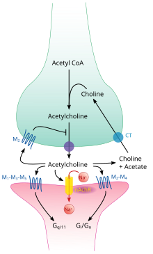
Acetylcholine processing in a synapse. After release acetylcholine is broken down by the enzyme acetylcholinesterase.

Like many other biologically active substances, acetylcholine exerts its effects by binding to and activating receptors located on the surface of cells. There are two main classes of acetylcholine receptor, nicotinic and muscarinic. They are named for chemicals that can selectively activate each type of receptor without activating the other: muscarine is a compound found in the mushroom Amanita muscaria; nicotine is found in tobacco.

Nicotinic acetylcholine receptors are ligand-gated ion channels permeable to sodium, potassium, and calcium ions. In other words, they are ion channels embedded in cell membranes, capable of switching from a closed to an open state when acetylcholine binds to them; in the open state they allow ions to pass through. Nicotinic receptors come in two main types, known as muscle-type and neuronal-type. The muscle-type can be selectively blocked by curare, the neuronal-type by hexamethonium. The main location of muscle-type receptors is on muscle cells, as described in more detail below. Neuronal-type receptors are located in autonomic ganglia (both sympathetic and parasympathetic), and in the central nervous system.

Muscarinic acetylcholine receptors have a more complex mechanism, and affect target cells over a longer time frame. In mammals, five subtypes of muscarinic receptors have been identified, labeled M1 through M5. All of them function as G protein-coupled receptors, meaning that they exert their effects via a second messenger system. The M1, M3, and M5 subtypes are Gq-coupled; they increase intracellular levels of IP3 and calcium by activating phospholipase C. Their effect on target cells is usually excitatory. The M2 and M4 subtypes are Gi/Go-coupled; they decrease intracellular levels of cAMP by inhibiting adenylate cyclase. Their effect on target cells is usually inhibitory. Muscarinic acetylcholine receptors are found in both the central nervous system and the peripheral nervous system of the heart, lungs, upper gastrointestinal tract, and sweat glands.

Neuromuscular junction

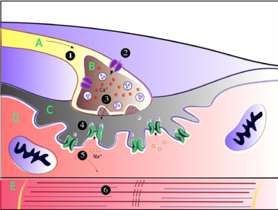
Muscles contract when they receive signals from motor neurons. The neuromuscular junction is the site of the signal exchange. The steps of this process in vertebrates occur as follows: (1) The action potential reaches the axon terminal. (2) Calcium ions flow into the axon terminal. (3) Acetylcholine is released into the synaptic cleft. (4) Acetylcholine binds to postsynaptic receptors. (5) This binding causes ion channels to open and allows sodium ions to flow into the muscle cell. (6) The flow of sodium ions across the membrane into the muscle cell generates an action potential which induces muscle contraction. Labels: A: Motor neuron axon B: Axon terminal C: Synaptic cleft D: Muscle cell E: Part of a Myofibril

Acetylcholine is the substance the nervous system uses to activate skeletal muscles, a kind of striated muscle. These are the muscles used for all types of voluntary movement, in contrast to smooth muscle tissue, which is involved in a range of involuntary activities such as movement of food through the gastrointestinal tract and constriction of blood vessels. Skeletal muscles are directly controlled by motor neurons located in the spinal cord or, in a few cases, the brainstem. These motor neurons send their axons through motor nerves, from which they emerge to connect to muscle fibers at a special type of synapse called the neuromuscular junction.

When a motor neuron generates an action potential, it travels rapidly along the nerve until it reaches the neuromuscular junction, where it initiates an electrochemical process that causes acetylcholine to be released into the space between the presynaptic terminal and the muscle fiber. The acetylcholine molecules then bind to nicotinic ion-channel receptors on the muscle cell membrane, causing the ion channels to open. Calcium ions then flow into the muscle cell, initiating a sequence of steps that finally produce muscle contraction.

Factors that decrease release of acetylcholine (and thereby affecting P-type calcium channels):

1) Antibiotics (clindamycin, polymyxin)
2) Magnesium:  antagonizes P-type calcium channels
3) Hypocalcemia
4) Anticonvulsants
5) Diuretics (furosemide)
6) Eaton-Lambert syndrome:  inhibits P-type calcium channels
7) Botulinum toxin:  inhibits SNARE proteins

Calcium channel blockers (nifedipine, diltiazem) do not affect P-channels.  These drugs affect L-type calcium channels.

Autonomic nervous system

Components and connections of the parasympathetic nervous system

The autonomic nervous system controls a wide range of involuntary and unconscious body functions. Its main branches are the sympathetic nervous system and parasympathetic nervous system. Broadly speaking, the function of the sympathetic nervous system is to mobilize the body for action; the phrase often invoked to describe it is fight-or-flight. The function of the parasympathetic nervous system is to put the body in a state conducive to rest, regeneration, digestion, and reproduction; the phrase often invoked to describe it is "rest and digest" or "feed and breed". Both of these aforementioned systems use acetylcholine, but in different ways.

At a schematic level, the sympathetic and parasympathetic nervous systems are both organized in essentially the same way: preganglionic neurons in the central nervous system send projections to neurons located in autonomic ganglia, which send output projections to virtually every tissue of the body. In both branches the internal connections, the projections from the central nervous system to the autonomic ganglia, use acetylcholine as a neurotransmitter to innervate (or excite) cholinergic neurons (neurons expressing nicotinic acetylcholine receptors). In the parasympathetic nervous system the output connections, the projections from ganglion neurons to tissues that don't belong to the nervous system, also release acetylcholine but act on muscarinic receptors. In the sympathetic nervous system the output connections mainly release noradrenaline, although acetylcholine is released at a few points, such as the sudomotor innervation of the sweat glands.

Direct vascular effects

Acetylcholine in the serum exerts a direct effect on vascular tone by binding to muscarinic receptors present on vascular endothelium. These cells respond by increasing production of nitric oxide, which signals the surrounding smooth muscle to relax, leading to vasodilation.

Central nervous system

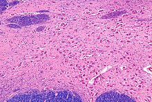
Micrograph of the nucleus basalis (of Meynert), which produces acetylcholine in the CNS. LFB-HE stain

In the central nervous system, ACh has a variety of effects on plasticity, arousal and reward. ACh has an important role in the enhancement of alertness when we wake up, in sustaining attention and in learning and memory.

Damage to the cholinergic (acetylcholine-producing) system in the brain has been shown to be associated with the memory deficits associated with Alzheimer's disease. ACh has also been shown to promote REM sleep.

In the brainstem acetylcholine originates from the Pedunculopontine nucleus and laterodorsal tegmental nucleus collectively known as the mesopontine tegmentum area or pontomesencephalotegmental complex. In the basal forebrain, it originates from the basal nucleus of Meynert and medial septal nucleus:
In addition, ACh acts as an important internal transmitter in the striatum, which is part of the basal ganglia. It is released by cholinergic interneurons. In humans, non-human primates and rodents, these interneurons respond to salient environmental stimuli with responses that are temporally aligned with the responses of dopaminergic neurons of the substantia nigra.

Memory

Acetylcholine has been implicated in learning and memory in several ways. The anticholinergic drug, scopolamine, impairs acquisition of new information in humans and animals. In animals, disruption of the supply of acetylcholine to the neocortex impairs the learning of simple discrimination tasks, comparable to the acquisition of factual information and disruption of the supply of acetylcholine to the hippocampus and adjacent cortical areas produces forgetting comparable to anterograde amnesia in humans.

Diseases and disorders

Myasthenia gravis

The disease myasthenia gravis, characterized by muscle weakness and fatigue, occurs when the body inappropriately produces antibodies against acetylcholine nicotinic receptors, and thus inhibits proper acetylcholine signal transmission. Over time, the motor end plate is destroyed. Drugs that competitively inhibit acetylcholinesterase (e.g., neostigmine, physostigmine, or primarily pyridostigmine) are effective in treating this disorder. They allow endogenously released acetylcholine more time to interact with its respective receptor before being inactivated by acetylcholinesterase in the synaptic cleft (the space between nerve and muscle).

Pharmacology

Blocking, hindering or mimicking the action of acetylcholine has many uses in medicine. Drugs acting on the acetylcholine system are either agonists to the receptors, stimulating the system, or antagonists, inhibiting it. Acetylcholine receptor agonists and antagonists can either have an effect directly on the receptors or exert their effects indirectly, e.g., by affecting the enzyme acetylcholinesterase, which degrades the receptor ligand. Agonists increase the level of receptor activation, antagonists reduce it.

Acetylcholine itself does not have therapeutic value as a drug for intravenous administration because of its multi-faceted action(non-selective) and rapid inactivation by cholinesterase. However, it is used in the form of eye drops to cause constriction of the pupil during cataract surgery, which facilitates quick post-operational recovery.

Nicotine

Nicotine binds to and activates nicotinic acetylcholine receptors, mimicking the effect of acetylcholine at these receptors. When ACh interacts with a nicotinic ACh receptor, it opens a Na+ channel and Na+ ions flow into the membrane. This causes a depolarization, and results in an excitatory post-synaptic potential. Thus, ACh is excitatory on skeletal muscle; the electrical response is fast and short-lived.

Atropine

Atropine is a non-selective competitive antagonist with Acetylcholine at muscarinic receptors.

Cholinesterase inhibitors

Many ACh receptor agonists work indirectly by inhibiting the enzyme acetylcholinesterase. The resulting accumulation of acetylcholine causes continuous stimulation of the muscles, glands, and central nervous system, which can result in fatal convulsions if the dose is high.
They are examples of enzyme inhibitors, and increase the action of acetylcholine by delaying its degradation; some have been used as nerve agents (Sarin and VX nerve gas) or pesticides (organophosphates and the carbamates). Many toxins and venoms produced by plants and animals also contain cholinesterase inhibitors. In clinical use, they are administered in low doses to reverse the action of muscle relaxants, to treat myasthenia gravis, and to treat symptoms of Alzheimer's disease (rivastigmine, which increases cholinergic activity in the brain).

Synthesis inhibitors

Organic mercurial compounds, such as methylmercury, have a high affinity for sulfhydryl groups, which causes dysfunction of the enzyme choline acetyltransferase. This inhibition may lead to acetylcholine deficiency, and can have consequences on motor function.

Release inhibitors

Botulinum toxin (Botox) acts by suppressing the release of acetylcholine, whereas the venom from a black widow spider (alpha-latrotoxin) has the reverse effect. ACh inhibition causes paralysis. When bitten by a black widow spider, one experiences the wastage of ACh supplies and the muscles begin to contract. If and when the supply is depleted, paralysis occurs.

Comparative biology and evolution

Acetylcholine is used by organisms in all domains of life for a variety of purposes. It is believed that choline, a precursor to acetylcholine, was used by single celled organisms billions of years ago for synthesizing cell membrane phospholipids. Following the evolution of choline transporters, the abundance of intracellular choline paved the way for choline to become incorporated into other synthetic pathways, including acetylcholine production. Acetylcholine is used by bacteria, fungi, and a variety of other animals. Many of the uses of acetylcholine rely on its action on ion channels via GPCRs like membrane proteins.

The two major types of acetylcholine receptors, muscarinic and nicotinic receptors, have convergently evolved to be responsive to acetylcholine. This means that rather than having evolved from a common homolog, these receptors evolved from separate receptor families. It is estimated that the nicotinic receptor family dates back longer than 2.5 billion years. Likewise, muscarinic receptors are thought to have diverged from other GPCRs at least 0.5 billion years ago. Both of these receptor groups have evolved numerous subtypes with unique ligand affinities and signaling mechanisms. The diversity of the receptor types enables acetylcholine to creating varying responses depending on which receptor types are activated, and allow for acetylcholine to dynamically regulate physiological processes.

History

Acetylcholine (ACh) was first identified in 1915 by Henry Hallett Dale for its actions on heart tissue. It was confirmed as a neurotransmitter by Otto Loewi, who initially gave it the name Vagusstoff because it was released from the vagus nerve. Both received the 1936 Nobel Prize in Physiology or Medicine for their work. Acetylcholine was also the first neurotransmitter to be identified.

Neurolinguistics

From Wikipedia, the free encyclopedia
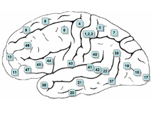
Surface of the human brain, with Brodmann areas numbered
 
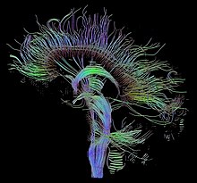
An image of neural pathways in the brain taken using diffusion tensor imaging
 
Neurolinguistics is the study of the neural mechanisms in the human brain that control the comprehension, production, and acquisition of language. As an interdisciplinary field, neurolinguistics draws methods and theories from fields such as neuroscience, linguistics, cognitive science, communication disorders and neuropsychology. Researchers are drawn to the field from a variety of backgrounds, bringing along a variety of experimental techniques as well as widely varying theoretical perspectives. Much work in neurolinguistics is informed by models in psycholinguistics and theoretical linguistics, and is focused on investigating how the brain can implement the processes that theoretical and psycholinguistics propose are necessary in producing and comprehending language. Neurolinguists study the physiological mechanisms by which the brain processes information related to language, and evaluate linguistic and psycholinguistic theories, using aphasiology, brain imaging, electrophysiology, and computer modeling.

History


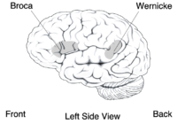
Neurolinguistics is historically rooted in the development in the 19th century of aphasiology, the study of linguistic deficits (aphasias) occurring as the result of brain damage. Aphasiology attempts to correlate structure to function by analyzing the effect of brain injuries on language processing. One of the first people to draw a connection between a particular brain area and language processing was Paul Broca, a French surgeon who conducted autopsies on numerous individuals who had speaking deficiencies, and found that most of them had brain damage (or lesions) on the left frontal lobe, in an area now known as Broca's area. Phrenologists had made the claim in the early 19th century that different brain regions carried out different functions and that language was mostly controlled by the frontal regions of the brain, but Broca's research was possibly the first to offer empirical evidence for such a relationship, and has been described as "epoch-making" and "pivotal" to the fields of neurolinguistics and cognitive science. Later, Carl Wernicke, after whom Wernicke's area is named, proposed that different areas of the brain were specialized for different linguistic tasks, with Broca's area handling the motor production of speech, and Wernicke's area handling auditory speech comprehension. The work of Broca and Wernicke established the field of aphasiology and the idea that language can be studied through examining physical characteristics of the brain. Early work in aphasiology also benefited from the early twentieth-century work of Korbinian Brodmann, who "mapped" the surface of the brain, dividing it up into numbered areas based on each area's cytoarchitecture (cell structure) and function; these areas, known as Brodmann areas, are still widely used in neuroscience today.

The coining of the term "neurolinguistics" is attributed to Edith Crowell Trager, Henri Hecaen and Alexandr Luria, in the late 1940s and 1950s; Luria's book "Problems in Neurolinguistics" is likely the first book with Neurolinguistics in the title. Harry Whitaker popularized neurolinguistics in the United States in the 1970s, founding the journal "Brain and Language" in 1974.

Although aphasiology is the historical core of neurolinguistics, in recent years the field has broadened considerably, thanks in part to the emergence of new brain imaging technologies (such as PET and fMRI) and time-sensitive electrophysiological techniques (EEG and MEG), which can highlight patterns of brain activation as people engage in various language tasks; electrophysiological techniques, in particular, emerged as a viable method for the study of language in 1980 with the discovery of the N400, a brain response shown to be sensitive to semantic issues in language comprehension. The N400 was the first language-relevant event-related potential to be identified, and since its discovery EEG and MEG have become increasingly widely used for conducting language research.

Discipline

Interaction with other fields

Neurolinguistics is closely related to the field of psycholinguistics, which seeks to elucidate the cognitive mechanisms of language by employing the traditional techniques of experimental psychology; today, psycholinguistic and neurolinguistic theories often inform one another, and there is much collaboration between the two fields.

Much work in neurolinguistics involves testing and evaluating theories put forth by psycholinguists and theoretical linguists. In general, theoretical linguists propose models to explain the structure of language and how language information is organized, psycholinguists propose models and algorithms to explain how language information is processed in the mind, and neurolinguists analyze brain activity to infer how biological structures (populations and networks of neurons) carry out those psycholinguistic processing algorithms. For example, experiments in sentence processing have used the ELAN, N400, and P600 brain responses to examine how physiological brain responses reflect the different predictions of sentence processing models put forth by psycholinguists, such as Janet Fodor and Lyn Frazier's "serial" model, and Theo Vosse and Gerard Kempen's "unification model". Neurolinguists can also make new predictions about the structure and organization of language based on insights about the physiology of the brain, by "generalizing from the knowledge of neurological structures to language structure".

Neurolinguistics research is carried out in all the major areas of linguistics; the main linguistic subfields, and how neurolinguistics addresses them, are given in the table below.

SubfieldDescriptionResearch questions in neurolinguistics
Phonetics the study of speech sounds how the brain extracts speech sounds from an acoustic signal, how the brain separates speech sounds from background noise
Phonology the study of how sounds are organized in a language how the phonological system of a particular language is represented in the brain
Morphology and lexicology the study of how words are structured and stored in the mental lexicon how the brain stores and accesses words that a person knows
Syntax the study of how multiple-word utterances are constructed how the brain combines words into constituents and sentences; how structural and semantic information is used in understanding sentences
Semantics the study of how meaning is encoded in language

Topics considered

Neurolinguistics research investigates several topics, including where language information is processed, how language processing unfolds over time, how brain structures are related to language acquisition and learning, and how neurophysiology can contribute to speech and language pathology.

Localizations of language processes

Much work in neurolinguistics has, like Broca's and Wernicke's early studies, investigated the locations of specific language "modules" within the brain. Research questions include what course language information follows through the brain as it is processed, whether or not particular areas specialize in processing particular sorts of information, how different brain regions interact with one another in language processing, and how the locations of brain activation differs when a subject is producing or perceiving a language other than his or her first language.

Time course of language processes

Another area of neurolinguistics literature involves the use of electrophysiological techniques to analyze the rapid processing of language in time. The temporal ordering of specific patterns of in brain activity may reflect discrete computational processes that the brain undergoes during language processing; for example, one neurolinguistic theory of sentence parsing proposes that three brain responses (the ELAN, N400, and P600) are products of three different steps in syntactic and semantic processing.

Language acquisition

Another topic is the relationship between brain structures and language acquisition. Research in first language acquisition has already established that infants from all linguistic environments go through similar and predictable stages (such as babbling), and some neurolinguistics research attempts to find correlations between stages of language development and stages of brain development, while other research investigates the physical changes (known as neuroplasticity) that the brain undergoes during second language acquisition, when adults learn a new language. Neuroplasticity is observed when both Second Language acquisition and Language Learning experience are induced, the result of this language exposure concludes that an increase of gray and white matter could be found in children, young adults and the elderly.

Language pathology

Neurolinguistic techniques are also used to study disorders and breakdowns in language, such as aphasia and dyslexia, and how they relate to physical characteristics of the brain.

Technology used

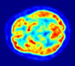
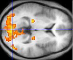
Images of the brain recorded with PET (top) and fMRI (bottom). In the PET image, the red areas are the most active. In the fMRI image, the yellowest areas are the areas that show the greatest difference in activation between two tasks (watching a moving stimulus, versus watching a black screen).

Since one of the focuses of this field is the testing of linguistic and psycholinguistic models, the technology used for experiments is highly relevant to the study of neurolinguistics. Modern brain imaging techniques have contributed greatly to a growing understanding of the anatomical organization of linguistic functions. Brain imaging methods used in neurolinguistics may be classified into hemodynamic methods, electrophysiological methods, and methods that stimulate the cortex directly.

Hemodynamic

Hemodynamic techniques take advantage of the fact that when an area of the brain works at a task, blood is sent to supply that area with oxygen (in what is known as the Blood Oxygen Level-Dependent, or BOLD, response). Such techniques include PET and fMRI. These techniques provide high spatial resolution, allowing researchers to pinpoint the location of activity within the brain; temporal resolution (or information about the timing of brain activity), on the other hand, is poor, since the BOLD response happens much more slowly than language processing. In addition to demonstrating which parts of the brain may subserve specific language tasks or computations, hemodynamic methods have also been used to demonstrate how the structure of the brain's language architecture and the distribution of language-related activation may change over time, as a function of linguistic exposure.

In addition to PET and fMRI, which show which areas of the brain are activated by certain tasks, researchers also use diffusion tensor imaging (DTI), which shows the neural pathways that connect different brain areas, thus providing insight into how different areas interact. Functional near-infrared spectroscopy (fNIRS) is another hemodynamic method used in language tasks.

Electrophysiological

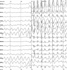
Brain waves recorded using EEG

Electrophysiological techniques take advantage of the fact that when a group of neurons in the brain fire together, they create an electric dipole or current. The technique of EEG measures this electric current using sensors on the scalp, while MEG measures the magnetic fields that are generated by these currents. In addition to these non-invasive methods, electrocorticography has also been used to study language processing. These techniques are able to measure brain activity from one millisecond to the next, providing excellent temporal resolution, which is important in studying processes that take place as quickly as language comprehension and production. On the other hand, the location of brain activity can be difficult to identify in EEG; consequently, this technique is used primarily to how language processes are carried out, rather than where. Research using EEG and MEG generally focuses on event-related potentials (ERPs), which are distinct brain responses (generally realized as negative or positive peaks on a graph of neural activity) elicited in response to a particular stimulus. Studies using ERP may focus on each ERP's latency (how long after the stimulus the ERP begins or peaks), amplitude (how high or low the peak is), or topography (where on the scalp the ERP response is picked up by sensors). Some important and common ERP components include the N400 (a negativity occurring at a latency of about 400 milliseconds), the mismatch negativity, the early left anterior negativity (a negativity occurring at an early latency and a front-left topography), the P600, and the lateralized readiness potential.

Experimental design

Experimental techniques

Neurolinguists employ a variety of experimental techniques in order to use brain imaging to draw conclusions about how language is represented and processed in the brain. These techniques include the subtraction paradigm, mismatch design, violation-based studies, various forms of priming, and direct stimulation of the brain.

Subtraction

Many language studies, particularly in fMRI, use the subtraction paradigm, in which brain activation in a task thought to involve some aspect of language processing is compared against activation in a baseline task thought to involve similar non-linguistic processes but not to involve the linguistic process. For example, activations while participants read words may be compared to baseline activations while participants read strings of random letters (in attempt to isolate activation related to lexical processing—the processing of real words), or activations while participants read syntactically complex sentences may be compared to baseline activations while participants read simpler sentences.

Mismatch paradigm

The mismatch negativity (MMN) is a rigorously documented ERP component frequently used in neurolinguistic experiments. It is an electrophysiological response that occurs in the brain when a subject hears a "deviant" stimulus in a set of perceptually identical "standards" (as in the sequence s s s s s s s d d s s s s s s d s s s s s d). Since the MMN is elicited only in response to a rare "oddball" stimulus in a set of other stimuli that are perceived to be the same, it has been used to test how speakers perceive sounds and organize stimuli categorically. For example, a landmark study by Colin Phillips and colleagues used the mismatch negativity as evidence that subjects, when presented with a series of speech sounds with acoustic parameters, perceived all the sounds as either /t/ or /d/ in spite of the acoustic variability, suggesting that the human brain has representations of abstract phonemes—in other words, the subjects were "hearing" not the specific acoustic features, but only the abstract phonemes. In addition, the mismatch negativity has been used to study syntactic processing and the recognition of word category.

Violation-based


Many studies in neurolinguistics take advantage of anomalies or violations of syntactic or semantic rules in experimental stimuli, and analyzing the brain responses elicited when a subject encounters these violations. For example, sentences beginning with phrases such as *the garden was on the worked, which violates an English phrase structure rule, often elicit a brain response called the early left anterior negativity (ELAN). Violation techniques have been in use since at least 1980, when Kutas and Hillyard first reported ERP evidence that semantic violations elicited an N400 effect. Using similar methods, in 1992, Lee Osterhout first reported the P600 response to syntactic anomalies. Violation designs have also been used for hemodynamic studies (fMRI and PET): Embick and colleagues, for example, used grammatical and spelling violations to investigate the location of syntactic processing in the brain using fMRI. Another common use of violation designs is to combine two kinds of violations in the same sentence and thus make predictions about how different language processes interact with one another; this type of crossing-violation study has been used extensively to investigate how syntactic and semantic processes interact while people read or hear sentences.

Priming

In psycholinguistics and neurolinguistics, priming refers to the phenomenon whereby a subject can recognize a word more quickly if he or she has recently been presented with a word that is similar in meaning or morphological makeup (i.e., composed of similar parts). If a subject is presented with a "prime" word such as doctor and then a "target" word such as nurse, if the subject has a faster-than-usual response time to nurse then the experimenter may assume that word nurse in the brain had already been accessed when the word doctor was accessed. Priming is used to investigate a wide variety of questions about how words are stored and retrieved in the brain and how structurally complex sentences are processed.

Stimulation

Transcranial magnetic stimulation (TMS), a new noninvasive technique for studying brain activity, uses powerful magnetic fields that are applied to the brain from outside the head. It is a method of exciting or interrupting brain activity in a specific and controlled location, and thus is able to imitate aphasic symptoms while giving the researcher more control over exactly which parts of the brain will be examined. As such, it is a less invasive alternative to direct cortical stimulation, which can be used for similar types of research but requires that the subject's scalp be removed, and is thus only used on individuals who are already undergoing a major brain operation (such as individuals undergoing surgery for epilepsy). The logic behind TMS and direct cortical stimulation is similar to the logic behind aphasiology: if a particular language function is impaired when a specific region of the brain is knocked out, then that region must be somehow implicated in that language function. Few neurolinguistic studies to date have used TMS; direct cortical stimulation and cortical recording (recording brain activity using electrodes placed directly on the brain) have been used with macaque monkeys to make predictions about the behavior of human brains.

Subject tasks

In many neurolinguistics experiments, subjects do not simply sit and listen to or watch stimuli, but also are instructed to perform some sort of task in response to the stimuli. Subjects perform these tasks while recordings (electrophysiological or hemodynamic) are being taken, usually in order to ensure that they are paying attention to the stimuli. At least one study has suggested that the task the subject does has an effect on the brain responses and the results of the experiment.

Lexical decision

The lexical decision task involves subjects seeing or hearing an isolated word and answering whether or not it is a real word. It is frequently used in priming studies, since subjects are known to make a lexical decision more quickly if a word has been primed by a related word (as in "doctor" priming "nurse").

Grammaticality judgment, acceptability judgment

Many studies, especially violation-based studies, have subjects make a decision about the "acceptability" (usually grammatical acceptability or semantic acceptability) of stimuli. Such a task is often used to "ensure that subjects [are] reading the sentences attentively and that they [distinguish] acceptable from unacceptable sentences in the way the [experimenter] expect[s] them to do."

Experimental evidence has shown that the instructions given to subjects in an acceptability judgment task can influence the subjects' brain responses to stimuli. One experiment showed that when subjects were instructed to judge the "acceptability" of sentences they did not show an N400 brain response (a response commonly associated with semantic processing), but that they did show that response when instructed to ignore grammatical acceptability and only judge whether or not the sentences "made sense".

Probe verification

Some studies use a "probe verification" task rather than an overt acceptability judgment; in this paradigm, each experimental sentence is followed by a "probe word", and subjects must answer whether or not the probe word had appeared in the sentence. This task, like the acceptability judgment task, ensures that subjects are reading or listening attentively, but may avoid some of the additional processing demands of acceptability judgments, and may be used no matter what type of violation is being presented in the study.

Truth-value judgment

Subjects may be instructed not to judge whether or not the sentence is grammatically acceptable or logical, but whether the proposition expressed by the sentence is true or false. This task is commonly used in psycholinguistic studies of child language.

Active distraction and double-task

Some experiments give subjects a "distractor" task to ensure that subjects are not consciously paying attention to the experimental stimuli; this may be done to test whether a certain computation in the brain is carried out automatically, regardless of whether the subject devotes attentional resources to it. For example, one study had subjects listen to non-linguistic tones (long beeps and buzzes) in one ear and speech in the other ear, and instructed subjects to press a button when they perceived a change in the tone; this supposedly caused subjects not to pay explicit attention to grammatical violations in the speech stimuli. The subjects showed a mismatch response (MMN) anyway, suggesting that the processing of the grammatical errors was happening automatically, regardless of attention—or at least that subjects were unable to consciously separate their attention from the speech stimuli.

Another related form of experiment is the double-task experiment, in which a subject must perform an extra task (such as sequential finger-tapping or articulating nonsense syllables) while responding to linguistic stimuli; this kind of experiment has been used to investigate the use of working memory in language processing.

Introduction to entropy

From Wikipedia, the free encyclopedia https://en.wikipedia.org/wiki/Introduct...