Search This Blog

Saturday, October 26, 2019

Sympathetic nervous system

From Wikipedia, the free encyclopedia
 
Sympathetic nervous system
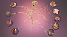
1501 Connections of the Sympathetic Nervous System.jpg
Schematic illustration showing the sympathetic nervous system with sympathetic cord and target organs.
Details
Identifiers
Latinpars sympathica divisionis autonomici systematis nervosi
Acronym(s)SNS
MeSHD013564
TAA14.3.01.001
FMA9906

The sympathetic nervous system (SNS) is one of the two main divisions of the autonomic nervous system, the other being the parasympathetic nervous system. (The enteric nervous system (ENS) is now usually referred to as separate from the autonomic nervous system since it has its own independent reflex activity.)

The autonomic nervous system functions to regulate the body's unconscious actions. The sympathetic nervous system's primary process is to stimulate the body's fight-flight-or-freeze response. It is, however, constantly active at a basic level to maintain homeostasis homeodynamics. The sympathetic nervous system is described as being antagonistic to the parasympathetic nervous system which stimulates the body to "feed and breed" and to (then) "rest-and-digest".

Structure

There are two kinds of neurons involved in the transmission of any signal through the sympathetic system: pre-ganglionic and post-ganglionic. The shorter preganglionic neurons originate in the thoracolumbar division of the spinal cord specifically at T1 to L2~L3, and travel to a ganglion, often one of the paravertebral ganglia, where they synapse with a postganglionic neuron. From there, the long postganglionic neurons extend across most of the body.

At the synapses within the ganglia, preganglionic neurons release acetylcholine, a neurotransmitter that activates nicotinic acetylcholine receptors on postganglionic neurons. In response to this stimulus, the postganglionic neurons release norepinephrine, which activates adrenergic receptors that are present on the peripheral target tissues. The activation of target tissue receptors causes the effects associated with the sympathetic system. However, there are three important exceptions:
  1. Postganglionic neurons of sweat glands release acetylcholine for the activation of muscarinic receptors, except for areas of thick skin, the palms and the plantar surfaces of the feet, where norepinephrine is released and acts on adrenergic receptors.
  2. Chromaffin cells of the adrenal medulla are analogous to post-ganglionic neurons; the adrenal medulla develops in tandem with the sympathetic nervous system and acts as a modified sympathetic ganglion. Within this endocrine gland, pre-ganglionic neurons synapse with chromaffin cells, triggering the release of two transmitters: a small proportion of norepinephrine, and more substantially, epinephrine. The synthesis and release of epinephrine as opposed to norepinephrine is another distinguishing feature of chromaffin cells compared to postganglionic sympathetic neurons.
  3. Postganglionic sympathetic nerves terminating in the kidney release dopamine, which acts on dopamine D1 receptors of blood vessels to control how much blood the kidney filters. Dopamine is the immediate metabolic precursor to norepinephrine, but is nonetheless a distinct signaling molecule.

Organization

The sympathetic nervous system extends from the thoracic to lumbar vertebrae and has connections with the thoracic, abdominal, and pelvic plexuses.
 
Sympathetic nerves arise from near the middle of the spinal cord in the intermediolateral nucleus of the lateral grey column, beginning at the first thoracic vertebra of the vertebral column and are thought to extend to the second or third lumbar vertebra. Because its cells begin in the thoracolumbar division – the thoracic and lumbar regions of the spinal cord, the sympathetic nervous system is said to have a thoracolumbar outflow. Axons of these nerves leave the spinal cord through the anterior root. They pass near the spinal (sensory) ganglion, where they enter the anterior rami of the spinal nerves. However, unlike somatic innervation, they quickly separate out through white rami connectors (so called from the shiny white sheaths of myelin around each axon) that connect to either the paravertebral (which lie near the vertebral column) or prevertebral (which lie near the aortic bifurcation) ganglia extending alongside the spinal column. 

To reach target organs and glands, the axons must travel long distances in the body, and, to accomplish this, many axons relay their message to a second cell through synaptic transmission. The ends of the axons link across a space, the synapse, to the dendrites of the second cell. The first cell (the presynaptic cell) sends a neurotransmitter across the synaptic cleft where it activates the second cell (the postsynaptic cell). The message is then carried to the final destination. 

Presynaptic nerves' axons terminate in either the paravertebral ganglia or prevertebral ganglia. There are four different paths an axon can take before reaching its terminal. In all cases, the axon enters the paravertebral ganglion at the level of its originating spinal nerve. After this, it can then either synapse in this ganglion, ascend to a more superior or descend to a more inferior paravertebral ganglion and synapse there, or it can descend to a prevertebral ganglion and synapse there with the postsynaptic cell. 

The postsynaptic cell then goes on to innervate the targeted end effector (i.e. gland, smooth muscle, etc.). Because paravertebral and prevertebral ganglia are relatively close to the spinal cord, presynaptic neurons are generally much shorter than their postsynaptic counterparts, which must extend throughout the body to reach their destinations.

A notable exception to the routes mentioned above is the sympathetic innervation of the suprarenal (adrenal) medulla. In this case, presynaptic neurons pass through paravertebral ganglia, on through prevertebral ganglia and then synapse directly with suprarenal tissue. This tissue consists of cells that have pseudo-neuron like qualities in that when activated by the presynaptic neuron, they will release their neurotransmitter (epinephrine) directly into the bloodstream.

In the sympathetic nervous system and other components of the peripheral nervous system, these synapses are made at sites called ganglia. The cell that sends its fiber is called a preganglionic cell, while the cell whose fiber leaves the ganglion is called a postganglionic cell. As mentioned previously, the preganglionic cells of the sympathetic nervous system are located between the first thoracic segment and third lumbar segments of the spinal cord. Postganglionic cells have their cell bodies in the ganglia and send their axons to target organs or glands.

The ganglia include not just the sympathetic trunks but also the cervical ganglia (superior, middle and inferior), which send sympathetic nerve fibers to the head and thorax organs, and the celiac and mesenteric ganglia, which send sympathetic fibers to the gut.

Autonomic nervous supply to organs in the human body
Organ Nerves Spinal column origin
stomach T5, T6, T7, T8, T9, sometimes T10
duodenum T5, T6, T7, T8, T9, sometimes T10
jejunum and ileum T5, T6, T7, T8, T9
spleen T6, T7, T8
gallbladder and liver T6, T7, T8, T9
colon
pancreatic head T8, T9
appendix T10
kidneys and ureters T11, T12

Information transmission

Sympathetic Nervous System - Information transmits through it affecting various organs.
 
Messages travel through the sympathetic nervous system in a bi-directional flow. Efferent messages can trigger changes in different parts of the body simultaneously. For example, the sympathetic nervous system can accelerate heart rate; widen bronchial passages; decrease motility (movement) of the large intestine; constrict blood vessels; increase peristalsis in the oesophagus; cause pupillary dilation, piloerection (goose bumps) and perspiration (sweating); and raise blood pressure. One exception is with certain blood vessels such as those in the cerebral and coronary arteries, which dilate (rather than constrict) with an increase in sympathetic tone. This is because of a proportional increase in the presence of β2 adrenergic receptors rather than α1 receptors. β2 receptors promote vessel dilation instead of constriction like α1 receptors. An alternative explanation is that the primary (and direct) effect of sympathetic stimulation on coronary arteries is vasoconstriction followed by a secondary vasodilation caused by the release of vasodilatory metabolites due to the sympathetically increased cardiac inotropy and heart rate. This secondary vasodilation caused by the primary vasoconstriction is termed functional sympatholysis, the overall effect of which on coronary arteries is dilation.

The target synapse of the postganglionic neuron is mediated by adrenergic receptors and is activated by either norepinephrine (noradrenaline) or epinephrine (adrenaline).

Function

Examples of sympathetic system action on various organs except where otherwise indicated.
Organ Effect
Eye Dilates
Heart Increases rate and force of contraction
Lungs Dilates bronchioles via circulating adrenaline
Blood vessels Dilate in skeletal muscle
Digestive system Constricts in gastrointestinal organs
Sweat glands Activates sweat secretion
Digestive tract Inhibits peristalsis
Kidney Increases renin secretion
Penis Inhibits tumescence
Ductus deferens Promotes emission prior to ejaculation

The sympathetic nervous system is responsible for up- and down-regulating many homeostatic mechanisms in living organisms. Fibers from the SNS innervate tissues in almost every organ system, providing at least some regulation of functions as diverse as pupil diameter, gut motility, and urinary system output and function. It is perhaps best known for mediating the neuronal and hormonal stress response commonly known as the fight-or-flight response. This response is also known as sympatho-adrenal response of the body, as the preganglionic sympathetic fibers that end in the adrenal medulla (but also all other sympathetic fibers) secrete acetylcholine, which activates the great secretion of adrenaline (epinephrine) and to a lesser extent noradrenaline (norepinephrine) from it. Therefore, this response that acts primarily on the cardiovascular system is mediated directly via impulses transmitted through the sympathetic nervous system and indirectly via catecholamines secreted from the adrenal medulla. 

The sympathetic nervous system is responsible for priming the body for action, particularly in situations threatening survival. One example of this priming is in the moments before waking, in which sympathetic outflow spontaneously increases in preparation for action. 

Sympathetic nervous system stimulation causes vasoconstriction of most blood vessels, including many of those in the skin, the digestive tract, and the kidneys. This occurs as a result of activation of alpha-1 adrenergic receptors by norepinephrine released by post-ganglionic sympathetic neurons. These receptors exist throughout the vasculature of the body but are inhibited and counterbalanced by beta-2 adrenergic receptors (stimulated by epinephrine release from the adrenal glands) in the skeletal muscles, the heart, the lungs, and the brain during a sympathoadrenal response. The net effect of this is a shunting of blood away from the organs not necessary to the immediate survival of the organism and an increase in blood flow to those organs involved in intense physical activity.

Sensation

The afferent fibers of the autonomic nervous system, which transmit sensory information from the internal organs of the body back to the central nervous system (or CNS), are not divided into parasympathetic and sympathetic fibers as the efferent fibers are. Instead, autonomic sensory information is conducted by general visceral afferent fibers

General visceral afferent sensations are mostly unconscious visceral motor reflex sensations from hollow organs and glands that are transmitted to the CNS. While the unconscious reflex arcs normally are undetectable, in certain instances they may send pain sensations to the CNS masked as referred pain. If the peritoneal cavity becomes inflamed or if the bowel is suddenly distended, the body will interpret the afferent pain stimulus as somatic in origin. This pain is usually non-localized. The pain is also usually referred to dermatomes that are at the same spinal nerve level as the visceral afferent synapse.

Relationship with the parasympathetic nervous system

Together with the other component of the autonomic nervous system, the parasympathetic nervous system, the sympathetic nervous system aids in the control of most of the body's internal organs. Reaction to stress—as in the flight-or-fight response—is thought to counteract the parasympathetic system, which generally works to promote maintenance of the body at rest. The comprehensive functions of both the parasympathetic and sympathetic nervous systems are not so straightforward, but this is a useful rule of thumb.

Disorders

In heart failure, the sympathetic nervous system increases its activity, leading to increased force of muscular contractions that in turn increases the stroke volume, as well as peripheral vasoconstriction to maintain blood pressure. However, these effects accelerate disease progression, eventually increasing mortality in heart failure.

Sympathicotonia is a stimulated condition of the sympathetic nervous system, marked by vascular spasm, elevated blood pressure, and goose bumps. A recent study has shown the expansion of Foxp3+ natural Treg in the bone marrow of mice after brain ischemia and this myeloid Treg expansion is related to sympathetic stress signaling after brain ischemia.

History and etymology

The name of this system can be traced to the concept of sympathy, in the sense of "connection between parts", first used medically by Galen. In the 18th century, Jacob B. Winslow applied the term specifically to nerves.

Electrical muscle stimulation

From Wikipedia, the free encyclopedia
 
Athlete squatting with four-channel, electrical muscle stimulation machine for training, attached through self-adhesive pads to her quadriceps.
 
Electrical muscle stimulation (EMS), also known as neuromuscular electrical stimulation (NMES) or electromyostimulation, is the elicitation of muscle contraction using electric impulses. EMS has received an increasing amount of attention in the last few years for many reasons: it can be utilized as a strength training tool for healthy subjects and athletes; it could be used as a rehabilitation and preventive tool for partially or totally immobilized patients; it could be utilized as a testing tool for evaluating the neural and/or muscular function in vivo; it could be used as a post-exercise recovery tool for athletes. The impulses are generated by a device and are delivered through electrodes on the skin near to the muscles being stimulated. The electrodes are generally pads that adhere to the skin. The impulses mimic the action potential that comes from the central nervous system, causing the muscles to contract. The use of EMS has been cited by sports scientists as a complementary technique for sports training, and published research is available on the results obtained. In the United States, EMS devices are regulated by the U.S. Food and Drug Administration (FDA).

A number of reviews have looked at the devices.

Uses

Active recovery session
Athlete recovering with four-channel, electrical muscle stimulation machine attached through self-adhesive pads to her hamstrings
 
Electrical muscle stimulation can be used as a training, therapeutic, or cosmetic tool.

Physical therapy

In medicine, EMS is used for rehabilitation purposes, for instance in physical therapy in the prevention muscle atrophy due to inactivity or neuromuscular imbalance, which can occur for example after musculoskeletal injuries (damage to bones, joints, muscles, ligaments and tendons). This is distinct from transcutaneous electrical nerve stimulation (TENS), in which an electric current is used for pain therapy. 

In EMS training few muscular groups are targeted at the same time, for specific training goals.

Weight loss

The FDA rejects certification of devices that claim weight reduction. EMS devices cause a calorie burning that is marginal at best: calories are burnt in significant amount only when most of the body is involved in physical exercise: several muscles, the heart and the respiratory system are all engaged at once. However, some authors imply that EMS can lead to exercise, since people toning their muscles with electrical stimulation are more likely afterwards to participate in sporting activities as the body becomes ready, fit, willing and able to take on physical activity.

Effects

"Strength training by NMES does promote neural and muscular adaptations that are complementary to the well-known effects of voluntary resistance training". This statement is part of the editorial summary of a 2010 world congress of researchers on the subject. Additional studies on practical applications, which came after that congress, pointed out important factors that make the difference between effective and ineffective EMS. This in retrospect explains why in the past some researchers and practitioners obtained results that others could not reproduce. Also, as published by reputable universities, EMS causes adaptation, i.e. training, of muscle fibers. Because of the characteristics of skeletal muscle fibers, different types of fibers can be activated to differing degrees by different types of EMS, and the modifications induced depend on the pattern of EMS activity. These patterns, referred to as protocols or programs, will cause a different response from contraction of different fiber types. Some programs will improve fatigue resistance, i.e. endurance, others will increase force production.

History

Luigi Galvani (1761) provided the first scientific evidence that current can activate muscle. During the 19th and 20th centuries, researchers studied and documented the exact electrical properties that generate muscle movement. It was discovered that the body functions induced by electrical stimulation caused long-term changes in the muscles. In the 1960s, Soviet sport scientists applied EMS in the training of elite athletes, claiming 40% force gains. In the 1970s, these studies were shared during conferences with the Western sport establishments. However, results were conflicting, perhaps because the mechanisms in which EMS acted were poorly understood. Recent medical physiology research pinpointed the mechanisms by which electrical stimulation causes adaptation of cells of muscles, blood vessels and nerves.

Society and culture

United States regulation

The U.S. Food and Drug Administration (FDA) certifies and releases EMS devices into two broad categories: over-the counter devices (OTC), and prescription devices. OTC devices are marketable only for muscle toning; prescription devices can be purchased only with a medical prescription for therapy. Prescription devices should be used under supervision of an authorized practitioner, for the following uses:
  • Relaxation of muscle spasms;
  • Prevention or retardation of disuse atrophy;
  • Increasing local blood circulation;
  • Muscle re-education;
  • Immediate post-surgical stimulation of calf muscles to prevent venous thrombosis;
  • Maintaining or increasing range of motion.
The FDA mandates that manuals prominently display contraindication, warnings, precautions and adverse reactions, including: no use for wearer of pacemaker; no use on vital parts, such as carotid sinus nerves, across the chest, or across the brain; caution in the use during pregnancy, menstruation, and other particular conditions that may be affected by muscle contractions; potential adverse effects include skin irritations and burns.

Only FDA-certified devices can be lawfully sold in the US without medical prescription. These can be found at the corresponding FDA webpage for certified devices. The FTC has cracked down on consumer EMS devices that made unsubstantiated claims; many have been removed from the market, some have obtained FDA certification.

Devices

Non-professional devices target home-market consumers with wearable units in which EMS circuitry is contained in belt-like garments (ab toning belts) or other clothing items.

The Relax-A-Cizor was one brand of device manufactured by the U.S. company Relaxacizor, Inc.

From the 1950s, the company marketed the device for use in weight loss and fitness. Electrodes from the device were attached to the skin and caused muscle contractions by way of electrical currents. The device caused 40 muscular contractions per minute in the muscles affected by the motor nerve points in the area of each pad. The directions for use recommended use of the device at least 30 minutes daily for each figure placement area, and suggested that the user might use it for longer periods if they wished. The device was offered in a number of different models which were powered either by battery or household current.

Relax-A-Cizors had from 1 to 6 channels. Two pads (or electrodes) were connected by wires to each channel. The user applied from 2 to 12 pads to various parts of their body. For each channel there was a dial which purported to control the intensity of the electrical current flowing into the user's body between the two pads connected to that channel.

As of 1970, the device was manufactured in Chicago, Illinois, by Eastwood Industries, Inc., a wholly owned subsidiary of Relaxacizor, Inc., and was then distributed throughout the country at the direction of Relaxacizor, Inc., or Relaxacizor Sales, Inc.

The device was banned by the United States Food and Drug Administration in 1970 as it was deemed to be potentially unhealthy and dangerous to the users. The case went to court, and the United States District Court for the Central District of California held that the Relax-A-Cizor was a "device" within the meaning of 21 U.S.C. § 321 (h) because it was intended to affect the structure and functions of the body as a girth reducer and exerciser, and upheld the FDA's assertions that the device was potentially hazardous to health.

The FDA informed owners of Relax-A-Cizors that second-hand sale of Relax-A-Cizors was illegal, and recommended that they should destroy the devices or render them inoperable.

Slendertone is another brand name. As of 2015 the company's Slendertone Flex product had been approved by the U.S. Food and Drug Administration for over-the-counter sale for toning, strengthening and firming abdominal muscles.

Motor neuron

From Wikipedia, the free encyclopedia
 
Motor neuron
Medulla oblongata - posterior - cn xii - very high mag.jpg
Micrograph of the hypoglossal nucleus showing motor neurons with their characteristic coarse Nissl substance ("tigroid" cytoplasm). H&E-LFB stain.
Details
LocationVentral horn of the spinal cord, some cranial nerve nuclei
ShapeProjection neuron
FunctionExcitatory projection (to NMJ)
NeurotransmitterUMN to LMN: glutamate; LMN to NMJ: ACh
Presynaptic connectionsPrimary motor cortex via the Corticospinal tract
Postsynaptic connectionsMuscle fibers and other neurons
Identifiers
MeSHD009046
NeuroLex IDnifext_103
TAA14.2.00.021
FMA83617

A motor neuron (or motoneuron) is a neuron whose cell body is located in the motor cortex, brainstem or the spinal cord, and whose axon (fiber) projects to the spinal cord or outside of the spinal cord to directly or indirectly control effector organs, mainly muscles and glands. There are two types of motor neuron – upper motor neurons and lower motor neurons. Axons from upper motor neurons synapse onto interneurons in the spinal cord and occasionally directly onto lower motor neurons. The axons from the lower motor neurons are efferent nerve fibers that carry signals from the spinal cord to the effectors. Types of lower motor neurons are alpha motor neurons, beta motor neurons, and gamma motor neurons.

A single motor neuron may innervate many muscle fibres and a muscle fibre can undergo many action potentials in the time taken for a single muscle twitch. Innervation takes place at a neuromuscular junction and twitches can become superimposed as a result of summation or a tetanic contraction. Individual twitches can become indistinguishable, and tension rises smoothly eventually reaching a plateau.

Development

Motor neurons begin to develop early in embryonic development, and motor function continues to develop well into childhood. In the neural tube cells are specified to either the rostral-caudal axis or ventral-dorsal axis. The axons of motor neurons begin to appear in the fourth week of development from the ventral region of the ventral-dorsal axis (the basal plate). This homeodomain is known as the motor neural progenitor domain (pMN). Transcription factors here include Pax6, OLIG2, Nkx-6.1, and Nkx-6.2, which are regulated by sonic hedgehog (Shh). The OLIG2 gene being the most important due to its role in promoting Ngn2 expression, a gene that causes cell cycle exiting as well as promoting further transcription factors associated with motor neuron development.

Further specification of motor neurons occurs when retinoic acid, fibroblast growth factor, Wnts, and TGFb, are integrated into the various Hox transcription factors. There are 13 Hox transcription factors and along with the signals, determine whether a motor neuron will be more rostral or caudal in character. In the spinal column, Hox 4-11 sort motor neurons to one of the five motor columns.

Motor columns of spinal cord 
Motor column Location in spinal cord Target
Median motor column Present entire length Axial muscles
Hypaxial motor column Thoracic region Body wall muscles
Preganglionic motor column Thoracic region Sympathetic ganglion
Lateral motor column Brachial and lumbar region (both regions are further divided into medial and lateral domains) Muscles of the limbs
Phrenic motor column Cervical region Diaphragm

Anatomy and physiology

Spinal cord tracts
 
Location of lower motor neurons in spinal cord

Upper motor neurons

Upper motor neurons originate in the motor cortex located in the precentral gyrus. The cells that make up the primary motor cortex are Betz cells, which are a type of pyramidal cell. The axons of these cells descend from the cortex to form the corticospinal tract. Corticomotorneurons project from the primary cortex directly onto motor neurons in the ventral horn of the spinal cord. Their axons synapse on the spinal motor neurons of multiple muscles as well as on spinal interneurons. They are unique to primates and it has been suggested that their function is the adaptive control of the hands including the relatively independent control of individual fingers. Corticomotorneurons have so far only been found in the primary motor cortex and not in secondary motor areas.

Nerve tracts

Nerve tracts are bundles of axons as white matter, that carry action potentials to their effectors. In the spinal cord these descending tracts carry impulses from different regions. These tracts also serve as the place of origin for lower motor neurons. There are seven major descending motor tracts to be found in the spinal cord:
  • Lateral corticospinal tract
  • Rubrospinal tract
  • Lateral reticulospinal tract
  • Vestibulospinal tract
  • Medial reticulospinal tract
  • Tectospinal tract
  • Anterior corticospinal tract

Lower motor neurons

Lower motor neurons are those that originate in the spinal cord and directly or indirectly innervate effector targets. The target of these neurons varies, but in the somatic nervous system the target will be some sort of muscle fiber. There are three primary categories lower motor neurons, which can be further divided in sub-categories.

According to their targets, motor neurons are classified into three broad categories:
  • Somatic motor neurons
  • Special visceral motor neurons
  • General visceral motor neurons

Somatic motor neurons

Somatic motor neurons originate in the central nervous system, project their axons to skeletal muscles  (such as the muscles of the limbs, abdominal, and intercostal muscles), which are involved in locomotion. The three types of these neurons are the alpha efferent neurons, beta efferent neurons, and gamma efferent neurons. They are called efferent to indicate the flow of information from the central nervous system (CNS) to the periphery.
  • Alpha motor neurons innervate extrafusal muscle fibers, which are the main force-generating component of a muscle. Their cell bodies are in the ventral horn of the spinal cord and they are sometimes called ventral horn cells. A single motor neuron may synapse with 150 muscle fibers on average. The motor neuron and all of the muscle fibers to which it connects is a motor unit. Motor units are split up into 3 categories: Main Article: Motor Unit
    • Slow (S) motor units stimulate small muscle fibers, which contract very slowly and provide small amounts of energy but are very resistant to fatigue, so they are used to sustain muscular contraction, such as keeping the body upright. They gain their energy via oxidative means and hence require oxygen. They are also called red fibers.
    • Fast fatiguing (FF) motor units stimulate larger muscle groups, which apply large amounts of force but fatigue very quickly. They are used for tasks that require large brief bursts on energy, such as jumping or running. They gain their energy via glycolytic means and hence don't require oxygen. They are called white fibers.
    • Fast fatigue-resistant motor units stimulate moderate-sized muscles groups that don't react as fast as the FF motor units, but can be sustained much longer (as implied by the name) and provide more force than S motor units. These use both oxidative and glycolytic means to gain energy.
In addition to voluntary skeletal muscle contraction, alpha motor neurons also contribute to muscle tone, the continuous force generated by noncontracting muscle to oppose stretching. When a muscle is stretched, sensory neurons within the muscle spindle detect the degree of stretch and send a signal to the CNS. The CNS activates alpha motor neurons in the spinal cord, which cause extrafusal muscle fibers to contract and thereby resist further stretching. This process is also called the stretch reflex.
  • Beta motor neurons innervate intrafusal muscle fibers of muscle spindles, with collaterals to extrafusal fibres. There are two types of beta motor neurons: Slow Contracting- These innervate extrafusal fibers. Fast Contracting- These innervate intrafusal fibers.
  • Gamma motor neurons innervate intrafusal muscle fibers found within the muscle spindle. They regulate the sensitivity of the spindle to muscle stretching. With activation of gamma neurons, intrafusal muscle fibers contract so that only a small stretch is required to activate spindle sensory neurons and the stretch reflex. There are two types of gamma motor neurons: Dynamic- These focus on Bag1 fibers and enhance dynamic sensitivity. Static- These focus on Bag2 fibers and enhance stretch sensitivity.
  • Regulatory factors of lower motor neurons
    • Size Principle – this relates to the soma of the motor neuron. This restricts larger neurons to receive a larger excitatory signal in order to stimulate the muscle fibers it innervates. By reducing unnecessary muscle fiber recruitment, the body is able to optimize energy consumption.
    • Persistent Inward Current (PIC) – recent animal study research has shown that constant flow of ions such as calcium and sodium through channels in the soma and dendrites influence the synaptic input. An alternate way to think of this is that the post-synaptic neuron is being primed before receiving an impulse.
    • After Hyper-polarization (AHP) – we are all familiar with the idea of hyperpolarization after an action potential. A trend has been identified that shows slow motor neurons to have more intense AHPs for a longer duration. One way to remember this is that slow muscle fibers can contract for longer, so it makes sense that their corresponding motor neurons fire at a slower rate.

Special visceral motor neurons

These are also known as branchial motor neurons, which are involved in facial expression, mastication, phonation, and swallowing. Associated cranial nerves are the oculomotor, abducens, trochlear, and hypoglossal nerves.
Branch of NS Position Neurotransmitter
Somatic n/a Acetylcholine
Parasympathetic Preganglionic Acetylcholine
Parasympathetic Ganglionic Acetylcholine
Sympathetic Preganglionic Acetylcholine
Sympathetic Ganglionic Norepinephrine*
*Except fibers to sweat glands and certain blood vessels
Motor neuron neurotransmitters

General visceral motor neurons

These motor neurons indirectly innervate cardiac muscle and smooth muscles of the viscera ( the muscles of the arteries): they synapse onto neurons located in ganglia of the autonomic nervous system (sympathetic and parasympathetic), located in the peripheral nervous system (PNS), which themselves directly innervate visceral muscles (and also some gland cells). 

In consequence, the motor command of skeletal and branchial muscles is monosynaptic involving only one motor neuron, either somatic or branchial, which synapses onto the muscle. Comparatively, the command of visceral muscles is disynaptic involving two neurons: the general visceral motor neuron, located in the CNS, synapses onto a ganglionic neuron, located in the PNS, which synapses onto the muscle. 

All vertebrate motor neurons are cholinergic, that is, they release the neurotransmitter acetylcholine. Parasympathetic ganglionic neurons are also cholinergic, whereas most sympathetic ganglionic neurons are noradrenergic, that is, they release the neurotransmitter noradrenaline. (see Table)

Neuromuscular junctions

A single motor neuron may innervate many muscle fibres and a muscle fibre can undergo many action potentials in the time taken for a single muscle twitch. As a result, if an action potential arrives before a twitch has completed, the twitches can superimpose on one another, either through summation or a tetanic contraction. In summation, the muscle is stimulated repetitively such that additional action potentials coming from the somatic nervous system arrive before the end of the twitch. The twitches thus superimpose on one another, leading to a force greater than that of a single twitch. A tetanic contraction is caused by constant, very high frequency stimulation - the action potentials come at such a rapid rate that individual twitches are indistinguishable, and tension rises smoothly eventually reaching a plateau.

The interface between a motor neuron and muscle fiber is a specialized synapse called the neuromuscular junction. Upon adequate stimulation, the motor neuron releases a flood of acetylcholine (Ach) neurotransmitters from the axon terminals from synaptic vesicles bind with the plasma membrane. The acetylcholine molecules bind to postsynaptic receptors found within the motor end plate. Once two acetylcholine receptors have been bound, an ion channel is opened and sodium ions are allowed to flow into the cell. The influx of sodium into the cell causes depolarization and triggers a muscle action potential. T tubules of the sarcolemma are then stimulated to elicit calcium ion release from the sarcoplasmic reticulum. It is this chemical release that causes the target muscle fiber to contract.

In invertebrates, depending on the neurotransmitter released and the type of receptor it binds, the response in the muscle fiber could be either excitatory or inhibitory. For vertebrates, however, the response of a muscle fiber to a neurotransmitter can only be excitatory, in other words, contractile. Muscle relaxation and inhibition of muscle contraction in vertebrates is obtained only by inhibition of the motor neuron itself. This is how muscle relaxants work by acting on the motor neurons that innervate muscles (by decreasing their electrophysiological activity) or on cholinergic neuromuscular junctions, rather than on the muscles themselves.

Entropy (information theory)

From Wikipedia, the free encyclopedia https://en.wikipedia.org/wiki/Entropy_(information_theory) In info...