Search This Blog

Sunday, May 30, 2021

Split-brain

From Wikipedia, the free encyclopedia
https://en.wikipedia.org/wiki/Split-brain

Split-brain or callosal syndrome is a type of disconnection syndrome when the corpus callosum connecting the two hemispheres of the brain is severed to some degree. It is an association of symptoms produced by disruption of, or interference with, the connection between the hemispheres of the brain. The surgical operation to produce this condition (corpus callosotomy) involves transection of the corpus callosum, and is usually a last resort to treat refractory epilepsy. Initially, partial callosotomies are performed; if this operation does not succeed, a complete callosotomy is performed to mitigate the risk of accidental physical injury by reducing the severity and violence of epileptic seizures. Before using callosotomies, epilepsy is instead treated through pharmaceutical means. After surgery, neuropsychological assessments are often performed.

After the right and left brain are separated, each hemisphere will have its own separate perception, concepts, and impulses to act. Having two "brains" in one body can create some interesting dilemmas. When one split-brain patient dressed himself, he sometimes pulled his pants up with one hand (that side of his brain wanted to get dressed) and down with the other (this side did not). He also reported to have grabbed his wife with his left hand and shaken her violently, at which point his right hand came to her aid and grabbed the aggressive left hand. However, such conflicts are very rare. If a conflict arises, one hemisphere usually overrides the other.

When split-brain patients are shown an image only in the left half of each eye's visual field, they cannot vocally name what they have seen. This is because the image seen in the left visual field is sent only to the right side of the brain, and most people's speech-control center is on the left side of the brain. Communication between the two sides is inhibited, so the patient cannot say out loud the name of that which the right side of the brain is seeing. A similar effect occurs if a split-brain patient touches an object with only the left hand while receiving no visual cues in the right visual field; the patient will be unable to name the object, as each cerebral hemisphere of the primary somatosensory cortex only contains a tactile representation of the opposite side of the body. If the speech-control center is on the right side of the brain, the same effect can be achieved by presenting the image or object to only the right visual field or hand.

The same effect occurs for visual pairs and reasoning. For example, a patient with split brain is shown a picture of a chicken foot and a snowy field in separate visual fields and asked to choose from a list of words the best association with the pictures. The patient would choose a chicken to associate with the chicken foot and a shovel to associate with the snow; however, when asked to reason why the patient chose the shovel, the response would relate to the chicken (e.g. "the shovel is for cleaning out the chicken coop").

History

In the 1950s, research on people with certain brain injuries made it possible to suspect that the "language center" in the brain was commonly located in the left hemisphere. One had observed that people with lesions in two specific areas on the left hemisphere lost their ability to talk, for example. Roger Sperry and his colleague pioneered research. In his early work on animal subjects, Sperry made many noteworthy discoveries. The results of these studies over the next thirty years later led to Roger Sperry being awarded the Nobel Prize in Physiology or Medicine in 1981. Sperry received the prize for his discoveries concerning the functional specialization of the cerebral hemispheres. With the help of so-called "split brain" patients, he carried out experiments, and for the first time in history, knowledge about the left and right hemispheres was revealed. In the 1960s, Sperry was joined by Michael Gazzaniga, a psychobiology Ph.D. student at Caltech. Even though Sperry is considered the founder of split-brain research, Gazzaniga's clear summaries of their collaborative work are consistently cited in psychology texts. In Sperry and Gazzaniga's "The Split Brain in Man" experiment published in Scientific American in 1967 they attempted to explore the extent to which two halves of the human brain were able to function independently and whether or not they had separate and unique abilities. They wanted to examine how perceptual and intellectual skills were affected in someone with a split-brain. At Caltech, Gazzaniga worked with Sperry on the effects of split-brain surgery on perception, vision and other brain functions. The surgery, which was a treatment for severe epilepsy, involved severing the corpus callosum, which carries signals between the left-brain hemisphere, the seat of speech and analytical capacity, and the right-brain hemisphere, which helps recognize visual patterns. At the time this article was written, only ten patients had undergone the surgery to sever their corpus callosum (corpus callosotomy). Four of these patients had consented to participate in Sperry and Gazzaniga's research. After the corpus callosum severing, all four participants' personality, intelligence, and emotions appeared to be unaffected. However, the testing done by Sperry and Gazzaniga showed the subjects demonstrated unusual mental abilities. The researchers created three types of tests to analyze the range of cognitive capabilities of the split-brain subjects. The first was to test their visual stimulation abilities, the second test was a tactile stimulation situation and the third tested auditory abilities.

Visual test

The first test started with a board that had a horizontal row of lights. The subject was told to sit in front of the board and stare at a point in the middle of the lights, then the bulbs would flash across both the right and left visual fields. When the patients were asked to describe afterward what they saw, they said that only the lights on the right side of the board had lit up. Next, when Sperry and Gazzaniga flashed the lights on the right side of the board on the subjects left side of their visual field, they claimed not to have seen any lights at all. When the experimenters conducted the test again, they asked the subjects to point to the lights that lit up. Although subjects had only reported seeing the lights flash on the right, they actually pointed to all the lights in both visual fields. This showed that both brain hemispheres had seen the lights and were equally competent in visual perception. The subjects did not say they saw the lights when they flashed in the left visual field even though they did see them because the center for speech is located in the brain's left hemisphere. This test supports the idea that in order to say one has seen something, the region of the brain associated with speech must be able to communicate with areas of the brain that process the visual information.

Tactile test

In a second experiment, Sperry and Gazzaniga placed a small object in the subject's right or left hand, without being able to see (or hear) it. Placed in the right hand, the isolated left hemisphere perceived the object and could easily describe and name it. However, placed in the left hand, the isolated right hemisphere could not name or describe the object. Questioning this result, the researchers found that the subjects could later match it from several similar objects; tactile sensations limited to the right hemisphere were accurately perceived but could not be verbalized. This further demonstrated the apparent location (or lateralization) of language functions in the left hemisphere.

Combination of both tests

In the last test the experimenters combined both the tactile and visual test. They presented subjects with a picture of an object to only their right hemisphere, and subjects were unable to name it or describe it. There were no verbal responses to the picture at all. If the subject however was able to reach under the screen with their left hand to touch various objects, they were able to pick the one that had been shown in the picture. The subjects were also reported to be able to pick out objects that were related to the picture presented, if that object was not under the screen.

Sperry and Gazzaniga went on to conduct other tests to shed light on the language processing abilities of the right hemisphere as well as auditory and emotional reactions as well. The significance of the findings of these tests by Sperry and Gazzaniga were extremely telling and important to the psychology world. Their findings showed that the two halves of the brain have numerous functions and specialized skills. They concluded that each hemisphere really has its own functions. One's left hemisphere of the brain is thought to be better at writing, speaking, mathematical calculation, reading, and is the primary area for language. The right hemisphere is seen to possess capabilities for problem solving, recognizing faces, symbolic reasoning, art, and spatial relationships.

Roger Sperry continued this line of research up until his death in 1994. Michael Gazzaniga continues to research the split-brain. Their findings have been rarely critiqued and disputed, however, a popular belief that some people are more "right-brained" or "left-brained" has developed. In the mid-1980s Jarre Levy, a psychobiologist at the University of Chicago, had set out and been in the forefront of scientists who wanted to dispel the notion we have two functioning brains. She believes that because each hemisphere has separate functions that they must integrate their abilities instead of separating them. Levy also claims that no human activity uses only one side of the brain. In 1998 a French study by Hommet and Billiard was published that questioned Sperry and Gazzaniga's study that severing the corpus callosum actually divides the hemispheres of the brain. They found that children born without a corpus callosum demonstrated that information was being transmitted between hemispheres, and concluded that subcortical connections must be present in these children with this rare brain malformation. They are unclear about whether these connections are present in split-brain patients though. Another study by Parsons, Gabrieli, Phelps, and Gazzaniga in 1998 demonstrated that split-brain patients may commonly perceive the world differently from the rest of us. Their study suggested that communication between brain hemispheres is necessary for imaging or simulating in your mind the movements of others. Morin's research on inner speech in 2001 suggested that an alternative for interpretation of commissurotomy according to which split-brain patients exhibit two uneven streams of self-awareness: a "complete" one in the left hemisphere and a "primitive" one in the right hemisphere.

Hemispheric specialization

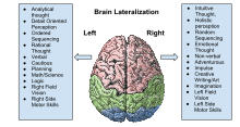
The two hemispheres of the cerebral cortex are linked by the corpus callosum, through which they communicate and coordinate actions and decisions. Communication and coordination between the two hemispheres is essential because each hemisphere has some separate functions. The right hemisphere of the cortex excels at nonverbal and spatial tasks, whereas the left hemisphere is more dominant in verbal tasks, such as speaking and writing. The right hemisphere controls the primary sensory functions of the left side of the body. In a cognitive sense the right hemisphere is responsible for recognizing objects and timing, and in an emotional sense it is responsible for empathy, humour and depression. On the other hand, the left hemisphere controls the primary sensory functions of the right side of the body and is responsible for scientific and maths skills, and logic. The extent of specialised brain function by an area remains under investigation. It is claimed that the difference between the two hemispheres is that the left hemisphere is "analytic" or "logical" while the right hemisphere is "holistic" or "intuitive." Many simple tasks, especially comprehension of inputs, require functions that are specific to both the right and left hemispheres and together form a one direction systematised way of creating an output through the communication and coordination that occurs between hemispheres.

Role of the corpus callosum

The corpus callosum is a structure in the brain along the longitudinal fissure that facilitates much of the communication between the two hemispheres. This structure is composed of white matter: millions of axons that have their dendrites and terminal boutons projecting in both the right and left hemisphere. However, there is evidence that the corpus callosum may also have some inhibitory functions. Post-mortem research on human and monkey brains shows that the corpus callosum is functionally organised. It proves that the right hemisphere is superior for detecting faces. This organisation results in modality-specific regions of the corpus callosum that are responsible for the transfer of different types of information. Research has revealed that the anterior midbody transfers motor information, the posterior midbody transfers somatosensory information, the isthmus transfers auditory information and the splenium transfers visual information. Although much of the interhemispheric transfer occurs at the corpus callosum, there are trace amounts of transfer via subcortical pathways.

Studies of the effects on the visual pathway on split-brained patients has revealed that there is a redundancy gain (the ability of target detection to benefit from multiple copies of the target) in simple reaction time. In a simple response to visual stimuli, split-brained patients experience a faster reaction time to bilateral stimuli than predicted by model. A model proposed by Iacoboni et al. suggests split-brained patients experience asynchronous activity that causes a stronger signal, and thus a decreased reaction time. Iacoboni also suggests there exists dual attention in split-brained patients, which is implying that each cerebral hemisphere has its own attentional system. An alternative approach taken by Reuter-Lorenz et al. suggests that enhanced redundancy gain in the split brain is primarily due to a slowing of responses to unilateral stimuli, rather than a speeding of responses to bilateral ones. It is important to note that the simple reaction time in split-brained patients, even with enhanced redundancy gain, is slower than the reaction time of normal adults.

Functional plasticity

Following a stroke or other injury to the brain, functional deficiencies are common. The deficits are expected to be in areas related to the part of the brain that has been damaged; if a stroke has occurred in the motor cortex, deficits may include paralysis, abnormal posture, or abnormal movement synergies. Significant recovery occurs during the first several weeks after the injury. However, recovery is generally thought not to continue past 6 months. If a specific region of the brain is injured or destroyed, its functions can sometimes be transferred and taken over by a neighbouring region. There is little functional plasticity observed in partial and complete callosotomies; however, much more plasticity can be seen in infant patients receiving a hemispherectomy, which suggests that the opposite hemisphere can adapt some functions typically performed by its opposite pair. In a study done by Anderson, it proved a correlation between the severity of the injury, the age of the individual and their cognitive performance. It was evident that there was more neuroplasticity in older children, even if their injury was extremely severe, than infants who suffered moderate brain injury. In some incidents of any moderate to severe brain injury, it mostly causes developmental impairments and in some of the most severe injuries it can cause a profound impact on their development that can lead to long-term cognitive effects. In the aging brain, it is extremely uncommon for neuroplasticity to occur; "olfactory bulb and hippocampus are two regions of the mammalian brain in which mutations preventing adult neurogenesis were never beneficial, or simply never occurred" (Anderson, 2005).

Corpus callosotomy

Corpus callosotomy is a surgical procedure that sections the corpus callosum, resulting in either the partial or complete disconnection between the two hemispheres. It is typically used as a last resort measure in treatment of intractable epilepsy. The modern procedure typically involves only the anterior third of the corpus callosum; however, if the epileptic seizures continue, the following third is lesioned prior to the remaining third if the seizures persist. This results in a complete callosotomy in which most of the information transfer between hemispheres is lost.

Due to the functional mapping of the corpus callosum, a partial callosotomy has less detrimental effects because it leaves parts of the corpus callosum intact. There is little functional plasticity observed in partial and complete callosotomies on adults, the most neuroplasticity is seen in young children but not in infants.

It is known that when the corpus callosum is severed during an experimental procedure, the experimenter can ask each side of the brain the same question and receive two different answers. When the experimenter asks the right visual field/left hemisphere what they see the participant will respond verbally, whereas if the experimenter asks the left visual field/right hemisphere what they see the participant will not be able to respond verbally but will pick up the appropriate object with their left hand.

Memory

It is known that the right and the left hemisphere have different functions when it comes to memory. The right hemisphere is better at recognizing objects and faces, recalling knowledge that the individual has already learned, or recalling images already seen. The left hemisphere is better at mental manipulation, language production, and semantic priming but was more susceptible to memory confusion than the right hemisphere. The main issue for individuals that have undergone a callosotomy is that because the function of memory is split into two major systems, the individual is more likely to become confused between knowledge they already know and information that they have only inferred.

In tests, memory in either hemisphere of split-brained patients is generally lower than normal, though better than in patients with amnesia, suggesting that the forebrain commissures are important for the formation of some kinds of memory. This suggests that posterior callosal sections that include the hippocampal commissures cause a mild memory deficit (in standardised free-field testing) involving recognition.

Control

In general, split-brained patients behave in a coordinated, purposeful and consistent manner, despite the independent, parallel, usually different and occasionally conflicting processing of the same information from the environment by the two disconnected hemispheres. When two hemispheres receive competing stimuli at the same time, the response mode tends to determine which hemisphere controls behaviour.

Often, split-brained patients are indistinguishable from normal adults. This is due to the compensatory phenomena; split-brained patients progressively acquire a variety of strategies to get around their interhemispheric transfer deficits. One issue that can happen with their body control is that one side of the body is doing the opposite of the other side called the intermanual effect.

Attention

Experiments on covert orienting of spatial attention using the Posner paradigm confirm the existence of two different attentional systems in the two hemispheres. The right hemisphere was found superior to the left hemisphere on modified versions of spatial relations tests and in locations testing, whereas the left hemisphere was more object based. The components of mental imagery are differentially specialised: the right hemisphere was found superior for mental rotation, the left hemisphere superior for image generation. It was also found that the right hemisphere paid more attention to landmarks and scenes whereas the left hemisphere paid more attention to exemplars of categories.

Case studies of split-brain patients

Patient W.J.

Patient W.J. was the first patient to undergo a full corpus callosotomy in 1962, after experiencing fifteen years of convulsions resulting from grand mal seizures. He was a World War II paratrooper who was injured at 30 years old during a bombing raid jump over the Netherlands, and again in a prison camp following his first injury. After returning home, he began to suffer from blackouts in which he would not remember what he was doing or where, and how or when he got there. At age 37, he suffered his first generalised convulsion. One of his worst episodes occurred in 1953, when he suffered a series of convulsions lasting for many days. During these convulsions, his left side would go numb and he would recover quickly, but after the series of convulsions, he never regained complete feeling on his left side.

Before his surgery, both hemispheres functioned and interacted normally, his sensory and motor functions were normal aside from slight hypoesthesia, and he could correctly identify and understand visual stimuli presented to both sides of his visual field. During his surgery in 1962, his surgeons determined that no massa intermedia had developed, and he had undergone atrophy in the part of the right frontal lobe exposed during the procedure. His operation was a success, in that it led to decreases in the frequency and intensity of his seizures.

Patient JW

Funnell et al. (2007) tested patient JW some time before June 2006. They described JW as

a right-handed male who was 47 years old at the time of testing. He successfully completed high school and has no reported learning disabilities. He had his first seizure at the age of 16 and the age of 25, he underwent a two-stage resection of the corpus callosum for relief of intractable epilepsy. Complete sectioning of the corpus callosum has been confirmed by MRI. Post-surgical MRI also revealed no evidence of other neurological damage.

Funnell et al.'s (2007) experiments were to determine each of JW's hemisphere's ability to perform simple addition, subtraction, multiplication and division. For example, in one experiment, on each trial, they presented an arithmetic problem in the center of the screen for 1 second, followed by a central cross hair JW was to look at. After 1 more second, Funnell et al. presented a number to one or the other hemisphere/visual field for 150 ms—too fast for JW to move his eyes. Randomly in half the trials, the number was the correct answer; in the other half of the trials it was the incorrect answer. With the hand on the same side as the number, JW pressed one key if the number was correct and another key if the number was incorrect.

Funnell et al.'s results were that performance of the left hemisphere was highly accurate (around 95%)—much better than performance of the right hemisphere, which was at chance for subtraction, multiplication, and division. Nevertheless the right hemisphere showed better than chance performance for addition (around 58%).

Turk et al. (2002) tested hemispheric differences in JW's recognition of himself and of familiar faces. They used faces that were composites of JW's face and Dr. Michael Gazzaniga's face. Composites ranges from 100% JW, through 50% JW and 50% Gazzaniga, to 100% Gazzaniga. JW pressed keys to say whether a presented face looked like him or Gazzaniga. Turk et al.concluded there are cortical networks in the left hemisphere that play an important role in self-recognition.

Patient VP

Patient VP is a woman who underwent a two-stage callosotomy in 1979 at the age of 27. Although the callosotomy was reported to be complete, follow-up MRI in 1984 revealed spared fibers in the rostrum and splenium. The spared rostral fibers constituted approximately 1.8% of the total cross-sectional area of the corpus callosum and the spared splenial fibers constituted approximately 1% of the area. VP's postsurgery intelligence and memory quotients were within normal limits.

One of the experiments involving VP attempted to investigate systematically the types of visual information that could be transferred via VP's spared splenial fibers. The first experiment was designed to assess VP's ability to make between-field perceptual judgements about simultaneously presented pairs of stimuli. The stimuli were presented in varying positions with respect to the horizontal and vertical midline with VP's vision fixated on a central crosshair. The judgements were based on differences in colour, shape or size. The testing procedure was the same for all three types of stimuli; after presentation of each pair, VP verbally responded "yes" if the two items in the pair were identical and "no" if they were not. The results show that there was no perceptual transfer for colour, size or shape with binomial tests showing that VP's accuracy was not greater than chance.

A second experiment involving VP attempted to investigate what aspects of words transferred between the two hemispheres. The set up was similar to the previous experiment, with VP's vision fixated on a central cross hair. A word pair was presented with one word on each side of the cross-hair for 150 ms. The words presented were in one of four categories: words that looked and sounded like rhymes (e.g. tire and fire), words that looked as if they should rhyme but did not (e.g. cough and dough), words that did not look as if they should rhyme but did (e.g. bake and ache), and words that neither looked nor sounded like rhymes (e.g. keys and fort). After presentation of each word pair, VP responded "yes" if the two words rhymed and "no" if they did not. VP's performance was above chance and she was able to distinguish among the different conditions. When the word pairs did not sound like rhymes, VP was able to say accurately that the words did not rhyme, regardless of whether or not they looked as if they should rhyme. When the words did rhyme, VP was more likely to say they rhymed, particularly if the words also looked as if they should rhyme.

Although VP showed no evidence for transfer of colour, shape or size, there was evidence for transfer of word information. This is consistent with the speculation that the transfer of word information involves fibres in the ventroposterior region of the splenium—the same region in which V.P. had callosal sparing. V.P. is able to integrate words presented to both visual fields, creating a concept that is not suggested by either word. For example, she combines "head" and "stone" to form the integrated concept of a tombstone.

Kim Peek

Kim Peek was arguably the most well-known savant. He was born on November 11, 1951 with an enlarged head, sac-like protrusions of the brain and the membranes that cover it through openings in the skull, a malformed cerebellum, and without a corpus callosum, an anterior commissure, or a posterior commissure. He was able to memorize over 9,000 books, and information from approximately 15 subject areas. These include: world/American history, sports, movies, geography, actors and actresses, the Bible, church history, literature, classical music, area codes/zip codes of the United States, television stations serving these areas, and step by step directions within any major U.S. city. Despite these abilities, he had an IQ of 87, was diagnosed as autistic, was unable to button his shirt, and had difficulties performing everyday tasks. The missing structures of his brain have yet to be linked to his increased abilities, but they can be linked to his ability to read pages of a book in 8–10 seconds. He was able to view the left page of a book with his left visual field and the right page of a book with his right visual fields so he could read both pages simultaneously. He also had developed language areas in both hemispheres, something very uncommon in split-brain patients. Language is processed in areas of the left temporal lobe, and involves a contralateral transfer of information before the brain can process what is being read. In Peek's case, there was no transfer ability—this is what led to his development of language centers in each hemisphere. Many believe this is the reason behind his extremely fast reading capabilities.

Although Peek did not undergo corpus callosotomy, he is considered a natural split-brain patient and is critical to understanding the importance of the corpus callosum. Kim Peek died in 2009.

Alien hand syndrome

    From Wikipedia, the free encyclopedia

    Alien hand syndrome (AHS) or Dr. Strangelove syndrome is a category of conditions in which a person experiences their limbs acting seemingly on their own, without conscious control over the actions. There are a variety of clinical conditions that fall under this category, which most commonly affects the left hand. There are many similar terms for the various forms of the condition, but they are often used inappropriately. The afflicted person may sometimes reach for objects and manipulate them without wanting to do so, even to the point of having to use the controllable hand to restrain the alien hand. While under normal circumstances, thought, as intent, and action can be assumed to be deeply mutually entangled, the occurrence of alien hand syndrome can be usefully conceptualized as a phenomenon reflecting a functional "disentanglement" between thought and action.

    Alien hand syndrome is best documented in cases where a person has had the two hemispheres of their brain surgically separated, a procedure sometimes used to relieve the symptoms of extreme cases of epilepsy and epileptic psychosis, e.g., temporal lobe epilepsy. It also occurs in some cases after brain surgery, stroke, infection, tumor, aneurysm, migraine and specific degenerative brain conditions such as Alzheimer's disease, Corticobasal degeneration and Creutzfeldt–Jakob disease. Other areas of the brain that are associated with alien hand syndrome are the frontal, occipital, and parietal lobes.

    Signs and symptoms

    "Alien behavior" can be distinguished from reflexive behavior in that the former is flexibly purposive while the latter is obligatory. Sometimes the sufferer will not be aware of what the alien hand is doing until it is brought to his or her attention, or until the hand does something that draws their attention to its behavior. There is a clear distinction between the behaviors of the two hands in which the affected hand is viewed as "wayward" and sometimes "disobedient" and generally out of the realm of their own voluntary control, while the unaffected hand is under normal volitional control. At times, particularly in patients who have sustained damage to the corpus callosum that connects the two cerebral hemispheres (see also split-brain), the hands appear to be acting in opposition to each other.

    A related syndrome described by the French neurologist François Lhermitte involves the release through disinhibition of a tendency to compulsively utilize objects that present themselves in the surrounding environment around the patient. The behavior of the patient is, in a sense, obligatorily linked to the "affordances" (using terminology introduced by the American ecological psychologist, James J. Gibson) presented by objects that are located within the immediate peri-personal environment.

    This condition, termed "utilization behavior", is most often associated with extensive bilateral frontal lobe damage and might actually be thought of as "bilateral" alien hand syndrome in which the patient is compulsively directed by external environmental contingencies (e.g. the presence of a hairbrush on the table in front of them elicits the act of brushing the hair) and has no capacity to "hold back" and inhibit pre-potent motor programs that are obligatorily linked to the presence of specific external objects in the peri-personal space of the patient. When the frontal lobe damage is bilateral and generally more extensive, the patient completely loses the ability to act in a self-directed manner and becomes totally dependent upon the surrounding environmental indicators to guide his behavior in a general social context, a condition referred to as "environmental dependency syndrome".

    In order to deal with the alien hand, some patients engage in personification of the affected hand. Usually these names are negative in nature, from mild such as "cheeky" to malicious "monster from the moon". For example, Doody and Jankovic described a patient who named her alien hand "baby Joseph". When the hand engaged in playful, troublesome activities such as pinching her nipples (akin to biting while nursing), she would experience amusement and would instruct baby Joseph to "stop being naughty". Furthermore, Bogen suggested that certain personality characteristics, such as a flamboyant personality, contribute to frequent personification of the affected hand.

    Neuroimaging and pathological research shows that the frontal lobe (in the frontal variant) and corpus callosum (in the callosal variant) are the most common anatomical lesions responsible for the alien hand syndrome. These areas are closely linked in terms of motor planning and its final pathways.

    The callosal variant includes advanced willed motor acts by the non-dominant hand, where patients frequently exhibit "intermanual conflict" in which one hand acts at cross-purposes with the other "good hand". For example, one patient was observed putting a cigarette into her mouth with her intact, "controlled" hand (her right, dominant hand), following which her alien, non-dominant, left hand came up to grasp the cigarette, pull the cigarette out of her mouth, and toss it away before it could be lit by the controlled, dominant, right hand. The patient then surmised that "I guess 'he' doesn't want me to smoke that cigarette." Another patient was observed to be buttoning up her blouse with her controlled dominant hand while the alien non-dominant hand, at the same time, was unbuttoning her blouse. The frontal variant most often affects the dominant hand, but can affect either hand depending on the lateralization of the damage to medial frontal cortex, and includes grasp reflex, impulsive groping toward objects or/and tonic grasping (i.e. difficulty in releasing grip).

    In most cases, classic alien-hand signs derive from damage to the medial frontal cortex, accompanying damage to the corpus callosum. In these patients the main cause of damage is unilateral or bilateral infarction of cortex in the territory supplied by the anterior cerebral artery or associated arteries. Oxygenated blood is supplied by the anterior cerebral artery to most medial portions of the frontal lobes and to the anterior two-thirds of the corpus callosum, and infarction may consequently result in damage to multiple adjacent locations in the brain in the supplied territory. As the medial frontal lobe damage is often linked to lesions of the corpus callosum, frontal variant cases may also present with callosal form signs. Cases of damage restricted to the callosum however, tend not to show frontal alien-hand signs.

    Cause

    The common emerging factor in alien hand syndrome is that the primary motor cortex controlling hand movement is isolated from premotor cortex influences but remains generally intact in its ability to execute movements of the hand.

    A 2009 fMRI study looking at the temporal sequence of activation of components of a cortical network associated with voluntary movement in normal individuals demonstrated "an anterior-to-posterior temporal gradient of activity from supplemental motor area through premotor and motor cortices to the posterior parietal cortex". Therefore, with normal voluntary movement, the emergent sense of agency appears to be associated with an orderly sequence of activation that develops initially in the anteromedial frontal cortex in the vicinity of the supplementary motor complex on the medial surface of the frontal aspect of the hemisphere (including the supplementary motor area) prior to activation of the primary motor cortex in the pre-central gyrus on the lateral aspect of the hemisphere, when the hand movement is being generated. Activation of the primary motor cortex, presumed to be directly involved in the execution of the action via projections into the corticospinal component of the pyramidal tracts, is then followed by activation of the posterior parietal cortex, possibly related to the receipt of recurrent or re-afferent somatosensory feedback generated from the periphery by the movement which would normally interact with the efference copy transmitted from primary motor cortex to permit the movement to be recognized as self-generated rather than imposed by an external force. That is, the efference copy allows the recurrent afferent somatosensory flow from the periphery associated with the self-generated movement to be recognized as re-afference as distinct from ex-afference. Failure of this mechanism may lead to a failure to distinguish between self-generated and externally generated movement of the limb. This anomalous situation in which re-afference from a self-generated movement is mistakenly registered as ex-afference due to a failure to generate and successfully transmit an efference copy to sensory cortex, could readily lead to the interpretation that what is in actuality a self-generated movement has been produced by an external force as a result of the failure to develop a sense of agency in association with emergence of the self-generated movement (see below for a more detailed discussion).

    A 2007 fMRI study examining the difference in functional brain activation patterns associated with alien as compared to non-alien "volitional" movement in a patient with alien hand syndrome found that alien movement involved anomalous isolated activation of the primary motor cortex in the damaged hemisphere contralateral to the alien hand, while non-alien movement involved the normal process of activation described in the preceding paragraph in which primary motor cortex in the intact hemisphere activates in concert with frontal premotor cortex and posterior parietal cortex presumably involved in a normal cortical network generating premotor influences on the primary motor cortex along with immediate post-motor re-afferent activation of the posterior parietal cortex.

    Combining these two fMRI studies, one could hypothesize that the alien behavior that is unaccompanied by a sense of agency emerges due to autonomous activity in the primary motor cortex acting independently of premotor cortex pre-activating influences that would normally be associated with the emergence of a sense of agency linked to the execution of the action.

    As noted above, these ideas can also be linked to the concept of efference copy and re-afference, where efference copy is a signal postulated to be directed from premotor cortex (activated normally in the process associated with emergence of an internally generated movement) over to somatosensory cortex of the parietal region, in advance of the arrival of the "re-afferent" input generated from the moving limb, that is, the afferent return from the moving limb associated with the self-generated movement produced. It is generally thought that a movement is recognized as internally generated when the efference copy signal effectively "cancels out" the re-afference. The afferent return from the limb is effectively correlated with the efference copy signal so that the re-afference can be recognized as such and distinguished from "ex-afference", which would be afferent return from the limb produced by an externally imposed force. When the efference copy is no longer normally generated, then the afferent return from the limb associated with the self-generated movement is mis-perceived as externally produced "ex-afference" since it is no longer correlated with or canceled out by the efference copy. As a result, the development of the sense that a movement is not internally generated even though it actually is (i.e. the failure of the sense of agency to emerge in conjunction with the movement), could indicate a failure of the generation of the efference copy signal associated with the normal premotor process through which the movement is prepared for execution.

    Since there is no disturbance of the sense of ownership of the limb (a concept discussed in the Wikipedia entry on sense of agency) in this situation, and there is no clearly apparent physically ostensible explanation for how the owned limb could be moving in a purposive manner without an associated sense of agency, effectively through its own power, a cognitive dissonance is created which may be resolved through the assumption that the goal-directed limb movement is being directed by an "alien" unidentifiable external force with the capacity for directing goal-directed actions of one's own limb.

    Disconnection

    It is theorized that alien hand syndrome results when disconnection occurs between different parts of the brain that are engaged in different aspects of the control of bodily movement. As a result, different regions of the brain are able to command bodily movements, but cannot generate a conscious feeling of self-control over these movements. As a result, the "sense of agency" that is normally associated with voluntary movement is impaired or lost. There is a dissociation between the process associated with the actual execution of the physical movements of the limb and the process that produces an internal sense of voluntary control over the movements, with this latter process thus normally creating the internal conscious sensation that the movements are being internally initiated, controlled and produced by an active self.

    Recent studies have examined the neural correlates of emergence of the sense of agency under normal circumstances. This appears to involve consistent congruence between what is being produced through efferent outflow to the musculature of the body, and what is being sensed as the presumed product in the periphery of this efferent command signal. In alien hand syndrome, the neural mechanisms involved in establishing that this congruence has occurred may be impaired. This may involve an abnormality in the brain mechanism that differentiates between "re-afference" (i.e., the return of kinesthetic sensation from the self-generated "active" limb movement) and "ex-afference" (i.e., kinesthetic sensation generated from an externally produced 'passive' limb movement in which an active self does not participate). This brain mechanism is proposed to involve the production of a parallel "efference copy" signal that is sent directly to the somatic sensory regions and is transformed into a "corollary discharge", an expected afferent signal from the periphery that would result from the performance driven by the issued efferent signal. The correlation of the corollary discharge signal with the actual afferent signal returned from the periphery can then be used to determine if, in fact, the intended action occurred as expected. When the sensed result of the action is congruent with the predicted result, then the action can be labelled as self-generated and associated with an emergent sense of agency.

    If, however, the neural mechanisms involved in establishing this sensorimotor linkage associated with self-generated action are faulty, it would be expected that the sense of agency with action would not develop as discussed in the previous section.

    Loss of inhibitions

    One theory posed to explain these phenomena proposes that the brain has separable neural "premotor" or "agency" systems for managing the process of transforming intentions into overt action. An anteromedial frontal premotor system is engaged in the process of directing exploratory actions based on "internal" drive by releasing or reducing inhibitory control over such actions.

    A recent paper reporting on neuronal unit recording in the medial frontal cortex in human subjects showed a clear pre-activation of neurons identified in this area up to several hundred milliseconds prior to the onset of an overt self-generated finger movement and the authors were able to develop a computational model whereby volition emerges once a change in internally generated firing rate of neuronal assemblies in this part of the brain crossed a threshold. Damage to this anteromedial premotor system produces disinhibition and release of such exploratory and object acquisition actions which then occur autonomously. A posterolateral temporo-parieto-occipital premotor system has a similar inhibitory control over actions that withdraw from environmental stimuli as well as the ability to excite actions that are contingent upon and driven by external stimulation, as distinct from internal drive. These two intrahemispheric systems, each of which activates an opposing cortical "tropism", interact through mutual inhibition that maintains a dynamic balance between approaching toward (i.e. with "intent-to-capture" in which contact with and grasping onto the attended object is sought) versus withdrawing from (i.e. with "intent-to-escape" in which distancing from the attended object is sought) environmental stimuli in the behavior of the contralateral limbs. Together, these two intrahemispheric agency systems form an integrated trans-hemispheric agency system.

    When the anteromedial frontal "escape" system is damaged, involuntary but purposive movements of an exploratory reach-and-grasp nature—what Denny-Brown referred to as a positive cortical tropism—are released in the contralateral limb. This is referred to as a positive cortical tropism because eliciting sensory stimuli, such as would result from tactile contact on the volar aspect of the fingers and palm of the hand, are linked to the activation of movement that increases or enhances the eliciting stimulation through a positive feedback connection (see discussion above in section entitled "Parietal and Occipital Lobes").

    When the posterolateral parieto-occipital "approach" system is damaged, involuntary purposive movements of a release-and-retract nature, such as levitation and instinctive avoidance – what Denny-Brown referred to as a negative cortical tropism – are released in the contralateral limb. This is referred to as a negative cortical tropism because eliciting sensory stimuli, such as would result from tactile contact on the volar aspect of the fingers and palm of the hand, are linked to the activation of movement that reduces or eliminates the eliciting stimulation through a negative feedback connection (see discussion above in section entitled "Parietal and Occipital Lobes").

    Each intrahemispheric agency system has the potential capability of acting autonomously in its control over the contralateral limb although unitary integrative control of the two hands is maintained through interhemispheric communication between these systems via the projections traversing the corpus callosum at the cortical level and other interhemispheric commissures linking the two hemispheres at the subcortical level.

    Disconnection of hemispheres due to injury

    One major difference between the two hemispheres is the direct connection between the agency system of the dominant hemisphere and the encoding system based primarily in the dominant hemisphere that links action to its production and through to its interpretation with language and language-encoded thought. The overarching unitary conscious agent that emerges in the intact brain is based primarily in the dominant hemisphere and is closely connected to the organization of language capacity. It is proposed that while relational action in the form of embodied inter-subjective behavior precedes linguistic capacity during infant development, a process ensues through the course of development through which linguistic constructs are linked to action elements in order to produce a language-based encoding of action-oriented knowledge.

    When there is a major disconnection between the two hemispheres resulting from callosal injury, the language-linked dominant hemisphere agent which maintains its primary control over the dominant limb loses, to some degree, its direct and linked control over the separate "agent" based in the nondominant hemisphere, and the nondominant limb, which had been previously responsive and "obedient" to the dominant conscious agent. The possibility of purposeful action occurring outside of the realm of influence of the conscious dominant agent can occur and the basic assumption that both hands are controlled through and subject to the dominant agent is proven incorrect. The sense of agency that would normally arise from movement of the nondominant limb now no longer develops, or, at least, is no longer accessible to consciousness. A new explanatory narrative for understanding the situation in which the now inaccessible nondominant hemisphere based agent is capable of activating the nondominant limb is necessitated.

    Under such circumstances, the two separated agents can control simultaneous actions in the two limbs that are directed at opposing purposes although the dominant hand remains linked to the dominant consciously accessible language-linked agent and is viewed as continuing to be under "conscious control" and obedient to conscious will and intent as accessible through thought, while the nondominant hand, directed by an essentially non-verbal agent whose intent can only be inferred by the dominant agent after the fact, is no longer "tied in" and subject to the dominant agent and is thus identified by the conscious language-based dominant agent as having a separate and inaccessible alien agency and associated existence. This theory would explain the emergence of alien behavior in the nondominant limb and intermanual conflict between the two limbs in the presence of damage to the corpus callosum.

    The distinct anteromedial, frontal, and posterolateral temporo-parieto-occipital variants of the alien hand syndrome would be explained by selective injury to either the frontal or the posterior components of the agency systems within a particular hemisphere, with the relevant and specific form of alien behavior developing in the limb contralateral to the damaged hemisphere.

    Diagnosis

    Corpus callosum

    Damage to the corpus callosum can give rise to "purposeful" actions in the sufferer's non-dominant hand (an individual who is left-hemisphere-dominant will experience the left hand becoming alien, and the right hand will turn alien in the person with right-hemisphere dominance).

    In "the callosal variant", the patient's hand counteracts voluntary actions performed by the other, "good" hand. Two phenomena that are often found in patients with callosal alien hand are agonistic dyspraxia and diagonistic dyspraxia.

    Agonistic dyspraxia involves compulsive automatic execution of motor commands by one hand when the patient is asked to perform movements with the other hand. For example, when a patient with callosal damage was instructed to pull a chair forward, the affected hand would decisively and impulsively push the chair backwards. Agonistic dyspraxia can thus be viewed as an involuntary competitive interaction between the two hands directed toward completion of a desired act in which the affected hand competes with the unaffected hand to complete a purposive act originally intended to be performed by the unaffected hand.

    Diagonistic dyspraxia, on the other hand, involves a conflict between the desired act in which the unaffected hand has been engaged and the interfering action of the affected hand which works to oppose the purpose of the desired act intended to be performed by the unaffected hand. For instance, when Akelaitis's patients underwent surgery to the corpus callosum to reduce epileptic seizures, one patient's left alien hand would frequently interfere with the right hand. For instance, while trying to turn over to the next page with the right hand, his left hand would try to close the book.

    In another case of callosal alien hand, the patient did not suffer from intermanual conflict between the hands but rather from a symptom characterized by involuntary mirror movements of the affected hand. When the patient was asked to perform movements with one hand, the other hand would involuntarily perform a mirror image movement which continued even when the involuntary movement was brought to the attention of the patient, and the patient was asked to restrain the mirrored movement. The patient suffered from a ruptured aneurysm near the anterior cerebral artery, which resulted in the right hand being mirrored by the left hand. The patient described the left hand as frequently interfering and taking over anything the patient tried to do with the right hand. For instance, when trying to grasp a glass of water with the right hand with a right side approach, the left hand would involuntary reach out and grasp hold of the glass through a left side approach.

    More recently, Geschwind et al. described the case of a woman who suffered severe coronary heart disease. One week after undergoing coronary artery bypass grafting, she noticed that her left hand started to "live a life of its own". It would unbutton her gown, try to choke her while asleep and would automatically fight with the right hand to answer the phone. She had to physically restrain the affected hand with the right hand to prevent injury, a behavior which has been termed "self-restriction". The left hand also showed signs of severe ideomotor apraxia. It was able to mimic actions but only with the help of mirror movements executed by the right hand (enabling synkinesis). Using magnetic resonance imaging (MRI), Geschwind et al. found damage to the posterior half of the callosal body, sparing the anterior half and the splenium extending slightly into the white matter underlying the right cingulate cortex.

    Park et al. also described two cases of infarction as the origin of alien hand symptoms. Both individuals had suffered an infarction of the anterior cerebral artery (ACA). One individual, a 72 year-old male, had difficulty controlling his hands, as they often moved involuntarily, despite his trying to stabilize them. Furthermore, he often could not let go of objects after grasping them with his palms. The other individual, a 47 year-old female who suffered an ACA in a different location of the artery, complained that her left hand would move on its own and she could not control its movements. Her left hand could also sense when her right hand was holding an object and would involuntarily, forcibly take the object out of her right hand.

    Frontal lobe

    Unilateral injury to the medial aspect of the brain's frontal lobe can trigger reaching, grasping and other purposeful movements in the contralateral hand. With anteromedial frontal lobe injuries, these movements are often exploratory reaching movements in which external objects are frequently grasped and utilized functionally, without the simultaneous perception on the part of the patient that they are "in control" of these movements. Once an object has been acquired and is maintained in the grasp of this "frontal variant" form of alien hand, the patient often has difficulty with voluntarily releasing the object from grasp and can sometimes be seen to be peeling the fingers of the hand back off the grasped object using the opposite controlled hand to enable the release of the grasped object (also referred to as tonic grasping or the "instinctive grasp reaction"). Some (for example, the neurologist Derek Denny-Brown) have referred to this behavior as "magnetic apraxia"

    Goldberg and Bloom described a woman who suffered a large cerebral infarction of the medial surface of the left frontal lobe in the territory of the left anterior cerebral artery which left her with the frontal variant of the alien hand involving the right hand. There were no signs of callosal disconnection nor was there evidence of any callosal damage. The patient displayed frequent grasp reflexes; her right hand would reach out and grab objects without releasing them. In regards to tonic grasping, the more the patient tried to let go of the object, the more the grip of the object tightened. With focused effort the patient was able to let go of the object, but if distracted, the behaviour would re-commence. The patient could also forcibly release the grasped object by peeling her fingers away from contact with the object using the intact left hand. Additionally, the hand would scratch at the patient's leg to the extent that an orthotic device was required to prevent injury. Another patient reported not only tonic grasping towards objects nearby, but the alien hand would take hold of the patient's penis and engage in public masturbation.

    Parietal and occipital lobes

    A distinct "posterior variant" form of alien hand syndrome is associated with damage to the posterolateral parietal lobe and/or occipital lobe of the brain. The movements in this situation tend to be more likely to withdraw the palmar surface of the hand away from sustained environmental contact rather than reaching out to grasp onto objects to produce palmar tactile stimulation, as is most often seen in the frontal form of the condition. In the frontal variant, tactile contact on the ventral surface of the palm and fingers facilitates finger flexion and grasp of the object through a positive feedback loop (i.e. the stimulus generates movement that reinforces, strengthens and sustains the triggering stimulation).

    In contrast, in the posterior variant, tactile contact on the ventral surface of the palm and fingers is actively avoided through facilitation of extension of the fingers and withdrawal of the palm in a negative feedback loop (i.e. the stimulus, and even anticipation of stimulation of the palmar surface of the hand, generates movement of the palm and fingers that reduces and effectively counteracts and eliminates the triggering stimulation, or, in the case of anticipated palmar contact, decreases the likelihood of such contact). Alien movements in the posterior variant of the syndrome also tend to be less coordinated and show a coarse ataxic motion during active movement that is generally not observed in the frontal form of the condition. This is generally thought to be due to an optic form of ataxia since it is facilitated by the visual presence of an object with visual attention directed toward the object. The apparent instability could be due to an unstable interaction between the tactile avoidance tendency biasing toward withdrawal from the object, and the visually based acquisition bias tendency pushing toward an approach to the object.

    The alien limb in the posterior variant of the syndrome may be seen to "levitate" upward into the air withdrawing away from contact surfaces through the activation of anti-gravity musculature. Alien hand movement in the posterior variant may show a typical posture, sometimes referred to as a "parietal hand" or the "instinctive avoidance reaction" (a term introduced by neurologist Derek Denny-Brown as an inverse form of the "magnetic apraxia" seen in the frontal variant, as noted above), in which the digits move into a highly extended position with active extension of the interphalangeal joints of the digits and hyper-extension of the metacarpophalangeal joints, and the palmar surface of the hand is actively pulled back away from approaching objects or up and away from supporting surfaces. The "alien" movements, however, remain purposeful and goal-directed, a point which clearly differentiates these movements from other disorganized non-purposeful forms of involuntary limb movement (e.g. athetosis, chorea, or myoclonus).

    Similarities between frontal and posterior variants

    In both the frontal and the posterior variants of the alien hand syndrome, the patient's reactions to the limb's apparent capability to perform goal-directed actions independent of conscious volition is similar. In both of these variants of alien hand syndrome, the alien hand emerges in the hand contralateral to the damaged hemisphere.

    Treatment

    There is no cure for the alien hand syndrome. However, the symptoms can be reduced and managed to some degree by keeping the alien hand occupied and involved in a task, for example by giving it an object to hold in its grasp. Specific learned tasks can restore voluntary control of the hand to a significant degree. One patient with the "frontal" form of alien hand who would reach out to grasp onto different objects (e.g., door handles) as he was walking was given a cane to hold in the alien hand while walking, even though he really did not need a cane for its usual purpose. With the cane firmly in the grasp of the alien hand, it would generally not release the grasp and drop the cane in order to reach out to grasp onto a different object. Other techniques proven to be effective includes; wedging the hand between the legs or slapping it; warm water application and visual or tactile contact. Additionally, Wu et al. found that an irritating alarm activated by biofeedback reduced the time the alien hand held an object.

    In the presence of unilateral damage to a single cerebral hemisphere, there is generally a gradual reduction in the frequency of alien behaviors observed over time and a gradual restoration of voluntary control over the affected hand. Actually, when AHS originates from focal injury of acute onset, recovery usually occurs within a year. One theory is that neuroplasticity in the bihemispheric and subcortical brain systems involved in voluntary movement production can serve to re-establish the connection between the executive production process and the internal self-generation and registration process. Exactly how this may occur is not well understood, but a process of gradual recovery from alien hand syndrome when the damage is confined to a single cerebral hemisphere has been reported. In some instances, patients may resort to constraining the wayward, undesirable and sometimes embarrassing actions of the impaired hand by voluntarily grasping onto the forearm of the impaired hand using the intact hand. This observed behavior has been termed "self-restriction" or "self-grasping".

    In another approach, the patient is trained to perform a specific task, such as moving the alien hand to contact a specific object or a highly salient environmental target, which is a movement that the patient can learn to generate voluntarily through focused training in order to effectively override the alien behavior. It is possible that some of this training produces a re-organization of premotor systems within the damaged hemisphere, or, alternatively, that ipsilateral control of the limb from the intact hemisphere may be expanded.

    Another method involves simultaneously "muffling" the action of the alien hand and limiting the sensory feedback coming back to the hand from environmental contact by placing it in a restrictive "cloak" such as a specialized soft foam hand orthosis or, alternatively, an everyday oven mitt. Other patients have reported using an orthotic device to restrict perseverative grasping or restraining the alien hand by securing it to the bed pole. Of course, this can limit the degree to which the hand can participate in addressing functional goals for the patient and may be considered to be an unjustifiable restraint.

    Theoretically, this approach could slow down the process through which voluntary control of the hand is restored if the neuroplasticity that underlies recovery involves the recurrent exercise of voluntary will to control the actions of the hand in a functional context and the associated experiential reinforcement through successful willful suppression of the alien behavior.

    History

    The first known case described in the medical literature appeared in a detailed case report published in German in 1908 by the preeminent German neuro-psychiatrist, Kurt Goldstein. In this paper, Goldstein described a right-handed woman who had suffered a stroke affecting her left side from which she had partially recovered by the time she was seen. However, her left arm seemed as though it belonged to another person and performed actions that appeared to occur independent of her will.

    The patient complained of a feeling of "strangeness" in relationship to the goal-directed movements of the left hand and insisted that "someone else" was moving the left hand, and that she was not moving it herself. When the left hand grasped an object, she could not voluntarily release it. The senses of touch and proprioception of the left side were impaired. The left hand would make spontaneous movements, such as wiping the face or rubbing the eyes, but these were relatively infrequent. With significant effort, she was able to move her left arm in response to spoken command, but conscious movements were slower or less precise than similar involuntary motions.

    Goldstein developed a "doctrine of motor apraxia" in which he discussed the generation of voluntary action and proposed a brain structure for temporal and spatial cognition, will and other higher cognitive processes. Goldstein maintained that a structure conceptually organizing both the body and external space was necessary for object perception as well as for voluntary action on external objects.

    In his classic papers reviewing the wide variety of disconnection syndromes associated with focal brain pathology, Norman Geschwind commented that Kurt Goldstein "was perhaps the first to stress the non-unity of the personality in patients with callosal section, and its possible psychiatric effects".

    In popular culture

  • In Stanley Kubrick's 1964 film Dr. Strangelove, the eponymous character played by Peter Sellers apparently suffers from alien hand syndrome, as he can't stop himself from doing the Nazi salute. "Dr. Strangelove syndrome" was suggested as the official name for AHS. This was not approved, though it is sometimes used as an alternative name.
  • In the 1999 American horror comedy film Idle Hands, the teenage boy protagonist finds out that his right hand has become possessed and is responsible for killing his parents and harming others.
  • In the House episode "Both Sides Now", a patient suffers from alien hand syndrome.
  • An episode of Dark Matters: Twisted But True—a documentary show on Discovery Science—described alien hand syndrome and traced its history.
  • The 2017 Indian Tamil dark comedy film Peechankai is about a person who suffers from AHS.
  • In Season 2 of Scream Queens, Dr. Brock Holt appears to suffer from alien hand syndrome.
  • An episode of the NPR show Invisibilia centers on a lady who developed alien hand syndrome after brain surgery.
  • The 2018 Indian Kannada Comedy film Sankashta Kara Ganapathi is about a cartoonist suffering from AHS.

Lateralization of brain function

From Wikipedia, the free encyclopedia
The human brain is divided into two hemispheres–left and right. Scientists continue to explore how some cognitive functions tend to be dominated by one side or the other; that is, how they are lateralized.

The lateralization of brain function is the tendency for some neural functions or cognitive processes to be specialized to one side of the brain or the other. The medial longitudinal fissure separates the human brain into two distinct cerebral hemispheres, connected by the corpus callosum. Although the macrostructure of the two hemispheres appears to be almost identical, different composition of neuronal networks allows for specialized function that is different in each hemisphere.

Lateralization of brain structures is based on general trends expressed in healthy patients; however, there are numerous counterexamples to each generalization. Each human's brain develops differently, leading to unique lateralization in individuals. This is different from specialization, as lateralization refers only to the function of one structure divided between two hemispheres. Specialization is much easier to observe as a trend, since it has a stronger anthropological history.

The best example of an established lateralization is that of Broca's and Wernicke's areas, where both are often found exclusively on the left hemisphere. Function lateralization, such as semantics, intonation, accentuation, and prosody, has since been called into question and largely been found to have a neuronal basis in both hemispheres. Another example is that each hemisphere in the brain tends to represent one side of the body. In the cerebellum, this is the same body side, but in the forebrain this is predominantly the contralateral side.

Lateralized functions

Language

Language functions such as grammar, vocabulary and literal meaning are typically lateralized to the left hemisphere, especially in right-handed individuals. While language production is left-lateralized in up to 90% of right-handers, it is more bilateral, or even right-lateralized, in approximately 50% of left-handers.

Broca's area and Wernicke's area, associated with the production of speech and comprehension of speech, respectively, are located in the left cerebral hemisphere for about 95% of right-handers but about 70% of left-handers. Individuals who speak multiple languages demonstrate separate speech areas for each language.

Sensory processing

The processing of basic sensory information is lateralized by being divided into left and right sides of the body or the space around the body.

In vision, about half the neurons of the optic nerve from each eye cross to project to the opposite hemisphere, and about half do not cross to project to the hemisphere on the same side. This means that the left side of the visual field is processed largely by the visual cortex of the right hemisphere and vice versa for the right side of the visual field.

In hearing, about 90% of the neurons of the auditory nerve from one ear cross to project to the auditory cortex of the opposite hemisphere.

In the sense of touch, most of the neurons from the skin cross to project to the somatosensory cortex of the opposite hemisphere.

Because of this functional division of the left and right sides of the body and of the space that surrounds it, the processing of information in the sensory cortices is essentially identical. That is, the processing of visual and auditory stimuli, spatial manipulation, facial perception, and artistic ability are represented bilaterally. Numerical estimation, comparison and online calculation depend on bilateral parietal regions while exact calculation and fact retrieval are associated with left parietal regions, perhaps due to their ties to linguistic processing.

Value systems

Rather than just being a series of places where different brain modules occur, there are running similarities in the kind of function seen in each side, for instance how right-side impairment of drawing ability making patients draw the parts of the subject matter with wholly incoherent relationships, or where the kind of left-side damage seen in language impairment not damaging the patient's ability to catch the significance of intonation in speech. This has led British psychiatrist Iain McGilchrist to say that the two hemispheres as having different value systems, where the left hemisphere tends to reduce complex matters such as ethics to rules and measures, where the right hemisphere is disposed to the holistic and metaphorical.

Clinical significance

Depression is linked with a hyperactive right hemisphere, with evidence of selective involvement in "processing negative emotions, pessimistic thoughts and unconstructive thinking styles", as well as vigilance, arousal and self-reflection, and a relatively hypoactive left hemisphere, "specifically involved in processing pleasurable experiences" and "relatively more involved in decision-making processes". Additionally, "left hemisphere lesions result in an omissive response bias or error pattern whereas right hemisphere lesions result in a commissive response bias or error pattern." The delusional misidentification syndromes, reduplicative paramnesia and Capgras delusion are also often the result of right hemisphere lesions.

Hemisphere damage

Damage to either the right or left hemisphere, and its resulting deficits provide insight into the function of the damaged area. Left hemisphere damage has many effects on language production and perception. Damage or lesions to the right hemisphere can result in a lack of emotional prosody or intonation when speaking. Right hemisphere damage also has grave effects on understanding discourse. People with damage to the right hemisphere have a reduced ability to generate inferences, comprehend and produce main concepts, and a reduced ability to manage alternative meanings. Furthermore, people with right hemisphere damage often exhibit discourse that is abrupt and perfunctory or verbose and excessive. They can also have pragmatic deficits in situations of turn taking, topic maintenance and shared knowledge.

Lateral brain damage can also affect visual perceptual spatial resolution. People with left hemisphere damage may have impaired perception of high resolution, or detailed, aspects of an image. People with right hemisphere damage may have impaired perception of low resolution, or big picture, aspects of an image.

Plasticity

If a specific region of the brain, or even an entire hemisphere, is injured or destroyed, its functions can sometimes be assumed by a neighboring region in the same hemisphere or the corresponding region in the other hemisphere, depending upon the area damaged and the patient's age. When injury interferes with pathways from one area to another, alternative (indirect) connections may develop to communicate information with detached areas, despite the inefficiencies.

Broca's aphasia

Broca's aphasia is a specific type of expressive aphasia and is so named due to the aphasia that results from damage or lesions to the Broca's area of the brain, that exists most commonly in the left inferior frontal hemisphere. Thus, the aphasia that develops from the lack of functioning of the Broca's area is an expressive and non-fluent aphasia. It is called 'non-fluent' due to the issues that arise because Broca's area is critical for language pronunciation and production. The area controls some motor aspects of speech production and articulation of thoughts to words and as such lesions to the area result in specific non-fluent aphasia.

Wernicke's aphasia

Wernicke's aphasia is the result of damage to the area of the brain that is commonly in the left hemisphere above the Sylvian fissure. Damage to this area causes primarily a deficit in language comprehension. While the ability to speak fluently with normal melodic intonation is spared, the language produced by a person with Wernicke's aphasia is riddled with semantic errors and may sound nonsensical to the listener. Wernicke's aphasia is characterized by phonemic paraphasias, neologism or jargon. Another characteristic of a person with Wernicke's aphasia is that they are unconcerned by the mistakes that they are making.

Society and culture

Misapplication

Terence Hines states that the research on brain lateralization is valid as a research program, though commercial promoters have applied it to promote subjects and products far outside the implications of the research. For example, the implications of the research have no bearing on psychological interventions such as eye movement desensitization and reprocessing (EMDR) and neurolinguistic programming, brain-training equipment, or management training.

Popular psychology

The oversimplification of lateralization in pop psychology. This belief was widely held even in the scientific community for some years.

Some popularizations oversimplify the science about lateralization, by presenting the functional differences between hemispheres as being more absolute than is actually the case.

Sex differences

In the 19th century and to a lesser extent the 20th, it was thought that each side of the brain was associated with a specific gender: the left corresponding with masculinity and the right with femininity and each half could function independently. The right side of the brain was seen as the inferior and thought to be prominent in women, savages, children, criminals, and the insane. A prime example of this in fictional literature can be seen in Robert Louis Stevenson's Strange Case of Dr. Jekyll and Mr. Hyde.

Evolutionary advantage

The widespread lateralization of many vertebrate animals suggests an evolutionary advantage associated with the specialization of each hemisphere.

History

Broca

One of the first indications of brain function lateralization resulted from the research of French physician Pierre Paul Broca, in 1861. His research involved the male patient nicknamed "Tan", who suffered a speech deficit (aphasia); "tan" was one of the few words he could articulate, hence his nickname. In Tan's autopsy, Broca determined he had a syphilitic lesion in the left cerebral hemisphere. This left frontal lobe brain area (Broca's area) is an important speech production region. The motor aspects of speech production deficits caused by damage to Broca's area are known as expressive aphasia. In clinical assessment of this type of aphasia, patients have difficulty producing speech.

Wernicke

German physician Karl Wernicke continued in the vein of Broca's research by studying language deficits unlike expressive aphasia. Wernicke noted that not every deficit was in speech production; some were linguistic. He found that damage to the left posterior, superior temporal gyrus (Wernicke's area) caused language comprehension deficits rather than speech production deficits, a syndrome known as receptive aphasia.

Imaging

These seminal works on hemispheric specialization were done on patients or postmortem brains, raising questions about the potential impact of pathology on the research findings. New methods permit the in vivo comparison of the hemispheres in healthy subjects. Particularly, magnetic resonance imaging (MRI) and positron emission tomography (PET) are important because of their high spatial resolution and ability to image subcortical brain structures.

Movement and sensation

In the 1940s, neurosurgeon Wilder Penfield and his neurologist colleague Herbert Jasper developed a technique of brain mapping to help reduce side effects caused by surgery to treat epilepsy. They stimulated motor and somatosensory cortices of the brain with small electrical currents to activate discrete brain regions. They found that stimulation of one hemisphere's motor cortex produces muscle contraction on the opposite side of the body. Furthermore, the functional map of the motor and sensory cortices is fairly consistent from person to person; Penfield and Jasper's famous pictures of the motor and sensory homunculi were the result.

Split-brain patients

Research by Michael Gazzaniga and Roger Wolcott Sperry in the 1960s on split-brain patients led to an even greater understanding of functional laterality. Split-brain patients are patients who have undergone corpus callosotomy (usually as a treatment for severe epilepsy), a severing of a large part of the corpus callosum. The corpus callosum connects the two hemispheres of the brain and allows them to communicate. When these connections are cut, the two halves of the brain have a reduced capacity to communicate with each other. This led to many interesting behavioral phenomena that allowed Gazzaniga and Sperry to study the contributions of each hemisphere to various cognitive and perceptual processes. One of their main findings was that the right hemisphere was capable of rudimentary language processing, but often has no lexical or grammatical abilities. Eran Zaidel also studied such patients and found some evidence for the right hemisphere having at least some syntactic ability.

Language is primarily localized in the left hemisphere. While the left hemisphere has proven to be more optimized for language, the right hemisphere has the capacity with emotions, such as sarcasm, that can express prosody in sentences when speaking. According to Sheppard and Hillis, "The right hemisphere is critical for perceiving sarcasm (Davis et al., 2016), integrating context required for understanding metaphor, inference, and humour, as well as recognizing and expressing affective or emotional prosody—changes in pitch, rhythm, rate, and loudness that convey emotions". One of the experiments carried out by Gazzaniga involved a split-brain male patient sitting in front of a computer screen while having words and images presented on either side of the screen, and the visual stimuli would go to either the right or left visual field, and thus the left or right brain, respectively. It was observed that if the patient was presented with an image to his left visual field (right brain), he would report not seeing anything. If he was able to feel around for certain objects, he could accurately pick out the correct object, despite not having the ability to verbalize what he saw.

Additional images


Entropy (information theory)

From Wikipedia, the free encyclopedia https://en.wikipedia.org/wiki/Entropy_(information_theory) In info...