Search This Blog

Saturday, December 11, 2021

Opsin

From Wikipedia, the free encyclopedia
 
Three-dimensional structure of bovine rhodopsin. The seven transmembrane domains are shown in varying colors. The chromophore is shown in red.
 
The retinal molecule inside an opsin protein absorbs a photon of light. Absorption of the photon causes retinal to change from its 11-cis-retinal isomer into its all-trans-retinal isomer. This change in shape of retinal pushes against the outer opsin protein to begin a signal cascade, which may eventually result in chemical signaling being sent to the brain as visual perception. The retinal is re-loaded by the body so that signaling can happen again.

Opsins are a group of proteins made light-sensitive via the chromophore retinal (or a variant) found in photoreceptor cells of the retina. Five classical groups of opsins are involved in vision, mediating the conversion of a photon of light into an electrochemical signal, the first step in the visual transduction cascade. Another opsin found in the mammalian retina, melanopsin, is involved in circadian rhythms and pupillary reflex but not in vision.

Opsin classification

Opsins can be classified several ways, including function (vision, phototaxis, photoperiodism, etc.), type of chromophore (retinal, flavine, bilin), molecular structure (tertiary, quaternary), signal output (phosphorylation, reduction, oxidation), etc.

There are two groups of protein termed opsins. Type I opsins are employed by prokaryotes and by some algae (as a component of channelrhodopsins) and fungi, whereas animals use type II opsins. No opsins have been found outside these groups (for instance in plants, or placozoans).

At one time it was thought that type I and type II were related because of structural and functional similarities. With the advent of genetic sequencing it became apparent that sequence identity was no greater than could be accounted for by random chance. However, in recent years new methods have been developed specific to deep phylogeny. As a result, several studies have found evidence of a possible phylogenetic relationship between the two. However, this does not necessarily mean that the last common ancestor of type I and II opsins was itself an opsin, a light sensitive receptor: all animal opsins arose (by gene duplication and divergence) late in the history of the large G-protein coupled receptor (GPCR) gene family, which itself arose after the divergence of plants, fungi, choanflagellates and sponges from the earliest animals. The retinal chromophore is found solely in the opsin branch of this large gene family, meaning its occurrence elsewhere represents convergent evolution, not homology. Microbial rhodopsins are, by sequence, very different from any of the GPCR families. According to one hypothesis, both type-I and type-II opsins belong to the transporter-opsin-G protein-coupled receptor (TOG) superfamily, a proposed clade that includes G protein-coupled receptor (GPCR), Ion-translocating microbial rhodopsin (MR), and seven others.

Type I opsins

Type I opsins (also known as microbial opsins) are seven-transmembrane-domain proteins. Most of them are ion channels or pumps instead of proper receptors and do not bind to a G protein. Type I opsins are found in all three domains of life: Archaea, Bacteria, and Eukaryota. In Eukaryota, type I opsins are found mainly in unicellular organisms such as green algae, and in fungi. In most complex multicellular eukaryotes, type I opsins have been replaced with other light-sensitive molecules such as cryptochrome and phytochrome in plants, and type II opsins in Metazoa (animals).

Microbial opsins are often known by the rhodopsin form of the molecule, i.e., rhodopsin (in the broad sense) = opsin + chromophore. Among the many kinds of microbial opsins are the proton pumps bacteriorhodopsin (BR) and xanthorhodopsin (xR), the chloride pump halorhodopsin (HR) the photosensors sensory rhodopsin I (SRI) and sensory rhodopsin II (SRII), as well as proteorhodopsin (PR), Neurospora opsin I (NOPI), Chlamydomonas sensory rhodopsins A (CSRA), Chlamydomonas sensory rhodopsins B (CSRB), channelrhodopsin (ChR), and archaerhodopsin (Arch).

Several type I opsins, such as proteo- and bacteriorhodopsin, are used by various bacterial groups to harvest energy from light to carry out metabolic processes using a non-chlorophyll-based pathway. Beside that, halorhodopsins of Halobacteria and channelrhodopsins of some algae, e.g. Volvox, serve them as light-gated ion channels, amongst others also for phototactic purposes. Sensory rhodopsins exist in Halobacteria that induce a phototactic response by interacting with transducer membrane-embedded proteins that have no relation to G proteins.

Type I opsins (like channelrhodopsin, halorhodopsin, and archaerhodopsin) are used in optogenetics to switch on or off neuronal activity. Type I opsins are preferred if the neuronal activity should be modulated at higher frequency, because they respond faster than type II opsins. This is because type I opsins are ion channels or proton/ion pumps and thus are activated by light directly, while type II opsins activate G-proteins, which then activate effector enzymes that produce metabolites to open ion channels.

Type II opsins

Type II opsins (or animal opsins) are members of the seven-transmembrane-domain proteins (35–55 kDa) of the G protein-coupled receptor (GPCR) superfamily.

Type II opsins fall phylogenetically into four groups: C-opsins (Ciliary), Cnidops (cnidarian opsins), R-opsins (rhabdomeric), and Go/RGR opsins (also known as RGR/Go or Group 4 opsins). The Go/RGR opsins are divided into four sub-clades: Go-opsins, RGR, Peropsins, and Neuropsins. C-opsins, R-opsins, and the Go/RGR opsins are found only in Bilateria.

Type II visual opsins are traditionally classified as either ciliary or rhabdomeric. Ciliary opsins, found in vertebrates and cnidarians, attach to ciliary structures such as rods and cones. Rhabdomeric opsins are attached to light-gathering organelles called rhabdomeres. This classification cuts across phylogenetic categories (clades) so that both the terms "ciliary" and "rhabdomeric" can be ambiguous. Here, "C-opsins (ciliary)" refers to a clade found exclusively in Bilateria and excludes cnidarian ciliary opsins such as those found in the box jellyfish. Similarly, "R-opsin (rhabdomeric)" includes melanopsin even though it does not occur on rhabdomeres in vertebrates.

C-opsins (ciliary)

Ciliary opsins (or c-opsins) are expressed in ciliary photoreceptor cells, and include the vertebrate visual opsins and encephalopsins. They convert light signals to nerve impulses via cyclic nucleotide gated ion channels, which work by increasing the charge differential across the cell membrane (i.e. hyperpolarization.)

Vertebrate visual opsins

Vertebrate visual opsins are a subset of C-opsins (ciliary). They are expressed in the vertebrate retina and mediate vision. They can be further subdivided into rod opsins and four types of cone opsin. Rod opsins (rhodopsins, usually denoted Rh), are used in dim-light vision, are thermally stable, and are found in the rod photoreceptor cells. Cone opsins, employed in color vision, are less-stable opsins located in the cone photoreceptor cells. Cone opsins are further subdivided according to their absorption maxima (λmax), the wavelength at which the highest light absorption is observed. Evolutionary relationships, deduced using the amino acid sequence of the opsins, are also frequently used to categorize cone opsins into their respective group. Both methods predict four general cone opsin groups in addition to rhodopsin.

Vertebrates typically have four cone opsins (LWS, SWS1, SWS2, and Rh2) inherited from the first vertebrate (and thus predating the first vertebrate), as well as the rod opsin, rhodopsin (Rh1), which emerged after the first vertebrate but before the first Gnathostome (jawed vertebrate). These five opsins emerged through a series of gene duplications beginning with LWS and ending with Rh1. Each one has since evolved into numerous variants and thus constitutes an opsin family or subtype.

Name Abbr. Photo
receptor
λmax Color Human variant
Long-wave sensitive LWS Cone 500–570 nm Green, yellow, red OPN1LW "red" / OPN1MW "green"
Short-wave sensitive 1 SWS1 Cone 355–445 nm Ultraviolet, violet OPN1SW "blue" (extinct in monotremes)
Short-wave sensitive 2 SWS2 Cone 400–470 nm Violet, blue (Extinct in therian mammals)
Rhodopsin-like 2 Rh2 Cone 480–530 nm Green (Extinct in mammals)
Rhodopsin-like 1
(vertebrate rhodopsin)
Rh1 Rod ~500 nm Blue–green OPN2/Rho, human rhodopsin

Humans have the following set of photoreceptor proteins responsible for vision:

  • Rhodopsin (Rh1, OPN2, RHO) – expressed in rod cells, used in night vision
  • Three cone opsins (also known as photopsins) – expressed in cone cells, used in color vision
    • Long-wavelength sensitive (OPN1LW) Opsin – λmax of 560 nm, in the yellow-green region of the electromagnetic spectrum. May be called the "red opsin," "erythrolabe," "L opsin" or "LWS opsin." Note that despite its common name as the "red" opsin, this opsin's peak sensitivity is not in the red region of the spectrum. However, it is more sensitive to red than the other two human opsins. This receptor also has a secondary response in the violet high frequencies.
    • Middle-wavelength sensitive (OPN1MW) Opsin – λmax of 530 nm, in the green region of the electromagnetic spectrum. May be called the "green opsin," "chlorolabe," "M opsin" or "MWS opsin."
    • Short-wavelength sensitive (OPN1SW) Opsin – λmax of 430 nm, in the blue region of the electromagnetic spectrum. May be called the "blue opsin," "cyanolabe," "S opsin" or "SWS opsin."

Pinopsins

The first Pineal Opsin (Pinopsin) was found in the chicken pineal gland. It is a blue sensitive opsin (λmax = 470 nm).

wide range of expression in the brain, most notably in the pineal region

Vertebrate Ancient (VA) opsin

Vertebrate Ancient (VA) opsin has three isoforms VA short (VAS), VA medium (VAM), and VA long (VAL). It is expressed in the inner retina, within the horizontal and amacrine cells, as well as the pineal organ and habenular region of the brain. It is sensitive to approximately 500 nm [14], found in most vertebrate classes, but not in mammals.

Parapinopsins

The first parapinopsin (PP) opsin was found in the parapineal organ of the catfish. The parapinopsin of lamprey is a UV-sensitive opsin (λmax = 370 nm). The teleosts have two groups of parapinopsins, one is sensitive to UV (λmax = 360-370 nm), the other is sensitive to blue (λmax = 460-480 nm) light.[31]

Parietopsins

The first parietopsin was found in the photoreceptor cells of the lizard parietal eye. The lizard parietopsin is green-sensitive (λmax = 522 nm), and despite it is a c-opsin, like the vertebrate visual opsins, it does not induce hyperpolarization via a Gt-protein, but induces depolarization via a Go-protein.

OPN3 (Encephalopsin or Panopsin)

Panopsins are found in many tissues (skin, brain, testes, heart, liver, kidney, skeletal muscle, lung, pancreas and retina). They were originally found in the human and mouse brain and thus called encephalopsin.

The first invertebrate panopsin was found in the ciliary photoreceptor cells of the annelid Platynereis dumerilii and is called c(iliary)-opsin. This c-opsin is UV-sensitive (λmax = 383 nm) and can be tuned by 125 nm at a single amino-acid (range λmax = 377 - 502 nm). Thus, not unsurprisingly, a second but cyan sensitive c-opsin (λmax = 490 nm) exists in Platynereis dumerilii. The first c-opsin mediates in the larva UV induced gravitaxis. The gravitaxis forms with phototaxis a ratio-chromatic depth-gauge. In different depths, the light in water is composed of different wavelengths: First the red (> 600 nm) and the UV and violet (< 420 nm) wavelengths disappear. The higher the depth the narrower the spectrum so that only cyan light (480 nm) is left.[41] Thus, the larvae can determine their depth by color. The color unlike brightness stays almost constant independent of time of day or the weather, for instance if it is cloudy.

Panopsins are also expressed in the brains of some insects. The panopsins of mosquito and pufferfish absorb maximally at 500 nm and 460 nm, respectively. Both activate in vitro Gi and Go proteins.

The panopsins of teleost fish are called: Teleost multiple tissue (TMT) opsins.

Teleost Multiple Tissue (TMT) Opsin

Teleost fish opsins are expressed in many tissues and therefore called Teleost Multiple Tissue (TMT) opsins. TMT opsins form three groups which are most closely related to a fourth groups the panopsins. In fact, TMT opsins in teleost fish are orthologous to the panopsins in the other vertebrates. They also have the same introns and the same place, which confirms that they belong together.

Cnidarian opsins

Cnidaria, which include jellyfish, corals, and sea anemones, are the most basal animals to possess complex eyes. Jellyfish opsins in the rhopalia couple to Gs-proteins raising the intracellular cAMP level. Coral opsins can couple to Gq-proteins and Gc-proteins. Gc-proteins are a subtype of G-proteins specific to cnidarians. The cnidarian opsins have been identified as one group and so called cnidops, however at least some of them belong to the c-opsins, r-opsins, and Go/RGR-opsins found in bilaterians.

r-opsins (rhabdomeric) / Gq-coupled

Rhabdomeric opsins (or r-opsins) are also known as Gq-opsins, because they couple to a Gq-protein. R-opsins are used by molluscs and arthropods. Arthropods appear to attain colour vision in a similar fashion to the vertebrates, by using three (or more) distinct groups of opsins, distinct both in terms of phylogeny and spectral sensitivity. The r-opsin melanopsin is also expressed in vertebrates, where it regulates circadian rhythms and mediates the pupillary reflex.

Unlike c-opsins, r-opsins are associated with canonical transient receptor potential ion channels; these lead to the electric potential difference across a cell membrane being eradicated (i.e. depolarization).

The identification of the crystal structure of squid rhodopsin is likely to further our understanding of its function in this group.

Arthropods use different opsins in their different eye types, but at least in Limulus the opsins expressed in the lateral and the compound eyes are 99% identical and presumably diverged recently.

Melanopsin OPN4

Involved in circadian rhythms, pupillary reflex, and color correction in high-brightness situations. Phylogenetically a member of the R-opsin (rhabdomeric) group, functionally and structurally an r-opsin, but does not occur in rhabdomeres.

Go/RGR (Group 4) opsins

Go/RGR opsins include Go-opsins, RGR-opsins, neuropsins, and peropsins.

Go-opsins

Go-opsins are absent from higher vertebrates and ecdysozoans. They are found in the ciliary photoreceptor cells of the scallop eye and the basal chordate amphioxus. In Platynereis dumerilii however, a Go-opsin is expressed in the rhabdomeric photoreceptor cells of the eyes.

RGR opsins

RGR opsins, also known as Retinal G protein coupled receptors are expressed in the retinal pigment epithelium (RPE) and Müller cells. They preferentially bind all-trans-retinal in the dark instead of 11-cis-retinal. RGR opsins were thought to be photoisomerases but instead, they regulate retinoid traffic and production. In particular, they speed up light-independently the production of 11-cis-retinol (a precursor of 11-cis-retinal) from all-trans-retinyl-esters. However, the all-trans-retinyl-esters are made available light-dependently by RGR-opsins. Whether RGR-opsins regulate this via a G-protein or another signaling mechanism is unknown. The cattle RGR opsin absorbs maximally at different wavelengths depending on the pH-value. At high pH it absorbs maximally blue (469 nm) light and at low pH it absorbs maximally UV (370 nm) light.

Peropsin

Peropsin, a visual pigment-like receptor, is a protein that in humans is encoded by the RRH gene.

Neuropsins

Neuropsins are sensitive to UVA, typically at 380 nm. They are found in the brain, testes, skin, and retina of humans and rodents, as well as in the brain and retina of birds. In birds and rodents they mediate ultraviolet vision. They couple to Gi-proteins. In humans, Neuropsin is encoded by the OPN5 gene. In the human retina, its function is unknown. In the mouse, it photo-entrains the retina and cornea at least ex vivo.

Unclassified

Extraretinal (or extra-ocular) Rhodopsin-Like Opsins (Exo-Rh)

These pineal opsins, found in the Actinopterygii (ray-finned fish) apparently arose as a result of gene duplication from Rh1 (rhodopsin). These opsins appear to serve functions similar to those of pinopsin found in birds and reptiles.

Structure and function

Opsin proteins covalently bind to a vitamin A-based retinaldehyde chromophore through a Schiff base linkage to a lysine residue in the seventh transmembrane alpha helix. In vertebrates, the chromophore is either 11-cis-retinal (A1) or 11-cis-3,4-didehydroretinal (A2) and is found in the retinal binding pocket of the opsin. The absorption of a photon of light results in the photoisomerization of the chromophore from the 11-cis to an all-trans conformation. The photoisomerization induces a conformational change in the opsin protein, causing the activation of the phototransduction cascade. The opsin remains insensitive to light in the trans form. It is regenerated by the replacement of the all-trans retinal by a newly synthesized 11-cis-retinal provided from the retinal epithelial cells. Opsins are functional while bound to either chromophore, with A2-bound opsin λmax being at a longer wavelength than A1-bound opsin.

Opsins contain seven transmembrane α-helical domains connected by three extra-cellular and three cytoplasmic loops. Many amino acid residues, termed functionally conserved residues, are highly conserved between all opsin groups, indicative of important functional roles. All residue positions discussed henceforth are relative to the 348 amino acid bovine rhodopsin crystallized by Palczewski et al. Lys296 is conserved in all known opsins and serves as the site for the Schiff base linkage with the chromophore. Cys138 and Cys110 form a highly conserved disulfide bridge. Glu113 serves as the counterion, stabilizing the protonation of the Schiff linkage between Lys296 and the chromophore. The Glu134-Arg135-Tyr136 is another highly conserved motif, involved in the propagation of the transduction signal once a photon has been absorbed.

Certain amino acid residues, termed spectral tuning sites, have a strong effect on λmax values. Using site-directed mutagenesis, it is possible to selectively mutate these residues and investigate the resulting changes in light absorption properties of the opsin. It is important to differentiate spectral tuning sites, residues that affect the wavelength at which the opsin absorbs light, from functionally conserved sites, residues important for the proper functioning of the opsin. They are not mutually exclusive, but, for practical reasons, it is easier to investigate spectral tuning sites that do not affect opsin functionality. For a comprehensive review of spectral tuning sites see Yokoyama and Deeb. The impact of spectral tuning sites on λmax differs between different opsin groups and between opsin groups of different species.

Evolution of the eye

From Wikipedia, the free encyclopedia
 
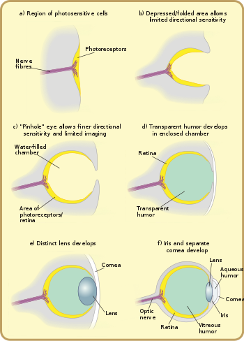
Major stages in the evolution of the eye in vertebrates.

Many researchers have found the evolution of the eye attractive to study because the eye distinctively exemplifies an analogous organ found in many animal forms. Simple light detection is found in bacteria, single-celled organisms, plants and animals. Complex, image-forming eyes have evolved independently several times.

Diverse eyes are known from the Burgess shale of the Middle Cambrian, and from the slightly older Emu Bay Shale. Eyes vary in their visual acuity, the range of wavelengths they can detect, their sensitivity in low light, their ability to detect motion or to resolve objects, and whether they can discriminate colours.

History of research

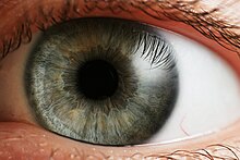
The human eye, showing the iris

In 1802, philosopher William Paley called it a miracle of "design". Charles Darwin himself wrote in his Origin of Species, that the evolution of the eye by natural selection seemed at first glance "absurd in the highest possible degree". However, he went on that despite the difficulty in imagining it, its evolution was perfectly feasible:

... if numerous gradations from a simple and imperfect eye to one complex and perfect can be shown to exist, each grade being useful to its possessor, as is certainly the case; if further, the eye ever varies and the variations be inherited, as is likewise certainly the case and if such variations should be useful to any animal under changing conditions of life, then the difficulty of believing that a perfect and complex eye could be formed by natural selection, though insuperable by our imagination, should not be considered as subversive of the theory.

He suggested a stepwise evolution from "an optic nerve merely coated with pigment, and without any other mechanism" to "a moderately high stage of perfection", and gave examples of existing intermediate steps. Current research is investigating the genetic mechanisms underlying eye development and evolution.

Biologist D.E. Nilsson has independently theorized about four general stages in the evolution of a vertebrate eye from a patch of photoreceptors. Nilsson and S. Pelger estimated in a classic paper that only a few hundred thousand generations are needed to evolve a complex eye in vertebrates. Another researcher, G.C. Young, has used the fossil record to infer evolutionary conclusions, based on the structure of eye orbits and openings in fossilized skulls for blood vessels and nerves to go through. All this adds to the growing amount of evidence that supports Darwin's theory.

Rate of evolution

The first fossils of eyes found to date are from the Ediacaran period (about 555 million years ago). The lower Cambrian had a burst of apparently rapid evolution, called the "Cambrian explosion". One of the many hypotheses for "causes" of the Cambrian explosion is the "Light Switch" theory of Andrew Parker: it holds that the evolution of advanced eyes started an arms race that accelerated evolution. Before the Cambrian explosion, animals may have sensed light, but did not use it for fast locomotion or navigation by vision.

The rate of eye evolution is difficult to estimate because the fossil record, particularly of the lower Cambrian, is poor. How fast a circular patch of photoreceptor cells can evolve into a fully functional vertebrate eye has been estimated based on rates of mutation, relative advantage to the organism, and natural selection. However, the time needed for each state was consistently overestimated and the generation time was set to one year, which is common in small animals. Even with these pessimistic values, the vertebrate eye would still evolve from a patch of photoreceptor cells in less than 364,000 years.

One origin or many?

Whether the eye evolved once or many times depends on the definition of an eye. All eyed animals share much of the genetic machinery for eye development. This suggests that the ancestor of eyed animals had some form of light-sensitive machinery – even if it was not a dedicated optical organ. However, even photoreceptor cells may have evolved more than once from molecularly similar chemoreceptor cells. Probably, photoreceptor cells existed long before the Cambrian explosion. Higher-level similarities – such as the use of the protein crystallin in the independently derived cephalopod and vertebrate lenses – reflect the co-option of a more fundamental protein to a new function within the eye.

A shared trait common to all light-sensitive organs are opsins. Opsins belong to a family of photo-sensitive proteins and fall into nine groups, which already existed in the urbilaterian, the last common ancestor of all bilaterally symmetrical animals. Additionally, the genetic toolkit for positioning eyes is shared by all animals: The PAX6 gene controls where eyes develop in animals ranging from octopuses to mice and fruit flies. Such high-level genes are, by implication, much older than many of the structures that they control today; they must originally have served a different purpose, before they were co-opted for eye development.

Eyes and other sensory organs probably evolved before the brain: There is no need for an information-processing organ (brain) before there is information to process. A living example are cubozoan jellyfish that possess eyes comparable to vertebrate and cephalopod camera eyes despite lacking a brain.

Stages of eye evolution

The stigma (2) of the euglena hides a light-sensitive spot.

The earliest predecessors of the eye were photoreceptor proteins that sense light, found even in unicellular organisms, called "eyespots". Eyespots can sense only ambient brightness: they can distinguish light from dark, sufficient for photoperiodism and daily synchronization of circadian rhythms. They are insufficient for vision, as they cannot distinguish shapes or determine the direction light is coming from. Eyespots are found in nearly all major animal groups, and are common among unicellular organisms, including euglena. The euglena's eyespot, called a stigma, is located at its anterior end. It is a small splotch of red pigment which shades a collection of light sensitive crystals. Together with the leading flagellum, the eyespot allows the organism to move in response to light, often toward the light to assist in photosynthesis, and to predict day and night, the primary function of circadian rhythms. Visual pigments are located in the brains of more complex organisms, and are thought to have a role in synchronising spawning with lunar cycles. By detecting the subtle changes in night-time illumination, organisms could synchronise the release of sperm and eggs to maximise the probability of fertilisation.

Vision itself relies on a basic biochemistry which is common to all eyes. However, how this biochemical toolkit is used to interpret an organism's environment varies widely: eyes have a wide range of structures and forms, all of which have evolved quite late relative to the underlying proteins and molecules.

At a cellular level, there appear to be two main "designs" of eyes, one possessed by the protostomes (molluscs, annelid worms and arthropods), the other by the deuterostomes (chordates and echinoderms).

The functional unit of the eye is the photoreceptor cell, which contains the opsin proteins and responds to light by initiating a nerve impulse. The light sensitive opsins are borne on a hairy layer, to maximise the surface area. The nature of these "hairs" differs, with two basic forms underlying photoreceptor structure: microvilli and cilia. In the eyes of protostomes, they are microvilli: extensions or protrusions of the cellular membrane. But in the eyes of deuterostomes, they are derived from cilia, which are separate structures. However, outside the eyes an organism may use the other type of photoreceptor cells, for instance the clamworm Platynereis dumerilii uses microvilliar cells in the eyes but has additionally deep brain ciliary photoreceptor cells. The actual derivation may be more complicated, as some microvilli contain traces of cilia – but other observations appear to support a fundamental difference between protostomes and deuterostomes. These considerations centre on the response of the cells to light – some use sodium to cause the electric signal that will form a nerve impulse, and others use potassium; further, protostomes on the whole construct a signal by allowing more sodium to pass through their cell walls, whereas deuterostomes allow less through.

This suggests that when the two lineages diverged in the Precambrian, they had only very primitive light receptors, which developed into more complex eyes independently.

Early eyes

The basic light-processing unit of eyes is the photoreceptor cell, a specialized cell containing two types of molecules bound to each other and located in a membrane: the opsin, a light-sensitive protein; and a chromophore, the pigment that absorbs light. Groups of such cells are termed "eyespots", and have evolved independently somewhere between 40 and 65 times. These eyespots permit animals to gain only a basic sense of the direction and intensity of light, but not enough to discriminate an object from its surroundings.

Developing an optical system that can discriminate the direction of light to within a few degrees is apparently much more difficult, and only six of the thirty-some phyla possess such a system. However, these phyla account for 96% of living species.

The planarian has "cup" eyespots that can slightly distinguish light direction.

These complex optical systems started out as the multicellular eyepatch gradually depressed into a cup, which first granted the ability to discriminate brightness in directions, then in finer and finer directions as the pit deepened. While flat eyepatches were ineffective at determining the direction of light, as a beam of light would activate exactly the same patch of photo-sensitive cells regardless of its direction, the "cup" shape of the pit eyes allowed limited directional differentiation by changing which cells the lights would hit depending upon the light's angle. Pit eyes, which had arisen by the Cambrian period, were seen in ancient snails, and are found in some snails and other invertebrates living today, such as planaria. Planaria can slightly differentiate the direction and intensity of light because of their cup-shaped, heavily pigmented retina cells, which shield the light-sensitive cells from exposure in all directions except for the single opening for the light. However, this proto-eye is still much more useful for detecting the absence or presence of light than its direction; this gradually changes as the eye's pit deepens and the number of photoreceptive cells grows, allowing for increasingly precise visual information.

When a photon is absorbed by the chromophore, a chemical reaction causes the photon's energy to be transduced into electrical energy and relayed, in higher animals, to the nervous system. These photoreceptor cells form part of the retina, a thin layer of cells that relays visual information, including the light and day-length information needed by the circadian rhythm system, to the brain. However, some jellyfish, such as Cladonema (Cladonematidae), have elaborate eyes but no brain. Their eyes transmit a message directly to the muscles without the intermediate processing provided by a brain.

During the Cambrian explosion, the development of the eye accelerated rapidly, with radical improvements in image-processing and detection of light direction.

The nautilus eye functions similarly to a pinhole camera.

After the photosensitive cell region invaginated, there came a point when reducing the width of the light opening became more efficient at increasing visual resolution than continued deepening of the cup. By reducing the size of the opening, organisms achieved true imaging, allowing for fine directional sensing and even some shape-sensing. Eyes of this nature are currently found in the nautilus. Lacking a cornea or lens, they provide poor resolution and dim imaging, but are still, for the purpose of vision, a major improvement over the early eyepatches.

Overgrowths of transparent cells prevented contamination and parasitic infestation. The chamber contents, now segregated, could slowly specialize into a transparent humour, for optimizations such as colour filtering, higher refractive index, blocking of ultraviolet radiation, or the ability to operate in and out of water. The layer may, in certain classes, be related to the moulting of the organism's shell or skin. An example of this can be observed in Onychophorans where the cuticula of the shell continues to the cornea. The cornea is composed of either one or two cuticular layers depending on how recently the animal has moulted. Along with the lens and two humors, the cornea is responsible for converging light and aiding the focusing of it on the back of the retina. The cornea protects the eyeball while at the same time accounting for approximately 2/3 of the eye's total refractive power.

It is likely that a key reason eyes specialize in detecting a specific, narrow range of wavelengths on the electromagnetic spectrum—the visible spectrum—is that the earliest species to develop photosensitivity were aquatic, and water filters out electromagnetic radiation except for a range of wavelengths, the shorter of which we refer to as blue, through to longer wavelengths we identify as red. This same light-filtering property of water also influenced the photosensitivity of plants.

Lens formation and diversification

Light from a distant object and a near object being focused by changing the curvature of the lens

In a lensless eye, the light emanating from a distant point hits the back of the eye with about the same size as the eye's aperture. With the addition of a lens this incoming light is concentrated on a smaller surface area, without reducing the overall intensity of the stimulus. The focal length of an early lobopod with lens-containing simple eyes focused the image behind the retina, so while no part of the image could be brought into focus, the intensity of light allowed the organism to see in deeper (and therefore darker) waters. A subsequent increase of the lens's refractive index probably resulted in an in-focus image being formed.

The development of the lens in camera-type eyes probably followed a different trajectory. The transparent cells over a pinhole eye's aperture split into two layers, with liquid in between. The liquid originally served as a circulatory fluid for oxygen, nutrients, wastes, and immune functions, allowing greater total thickness and higher mechanical protection. In addition, multiple interfaces between solids and liquids increase optical power, allowing wider viewing angles and greater imaging resolution. Again, the division of layers may have originated with the shedding of skin; intracellular fluid may infill naturally depending on layer depth.

Note that this optical layout has not been found, nor is it expected to be found. Fossilization rarely preserves soft tissues, and even if it did, the new humour would almost certainly close as the remains desiccated, or as sediment overburden forced the layers together, making the fossilized eye resemble the previous layout.

Compound eye of Antarctic krill

Vertebrate lenses are composed of adapted epithelial cells which have high concentrations of the protein crystallin. These crystallins belong to two major families, the α-crystallins and the βγ-crystallins. Both categories of proteins were originally used for other functions in organisms, but eventually adapted for vision in animal eyes. In the embryo, the lens is living tissue, but the cellular machinery is not transparent so must be removed before the organism can see. Removing the machinery means the lens is composed of dead cells, packed with crystallins. These crystallins are special because they have the unique characteristics required for transparency and function in the lens such as tight packing, resistance to crystallization, and extreme longevity, as they must survive for the entirety of the organism's life. The refractive index gradient which makes the lens useful is caused by the radial shift in crystallin concentration in different parts of the lens, rather than by the specific type of protein: it is not the presence of crystallin, but the relative distribution of it, that renders the lens useful.

It is biologically difficult to maintain a transparent layer of cells. Deposition of transparent, nonliving, material eased the need for nutrient supply and waste removal. Trilobites used calcite, a mineral which today is known to be used for vision only in a single species of brittle star. In other compound eyes and camera eyes, the material is crystallin. A gap between tissue layers naturally forms a biconvex shape, which is optically and mechanically ideal for substances of normal refractive index. A biconvex lens confers not only optical resolution, but aperture and low-light ability, as resolution is now decoupled from hole size – which slowly increases again, free from the circulatory constraints.

Independently, a transparent layer and a nontransparent layer may split forward from the lens: a separate cornea and iris. (These may happen before or after crystal deposition, or not at all.) Separation of the forward layer again forms a humour, the aqueous humour. This increases refractive power and again eases circulatory problems. Formation of a nontransparent ring allows more blood vessels, more circulation, and larger eye sizes. This flap around the perimeter of the lens also masks optical imperfections, which are more common at lens edges. The need to mask lens imperfections gradually increases with lens curvature and power, overall lens and eye size, and the resolution and aperture needs of the organism, driven by hunting or survival requirements. This type is now functionally identical to the eye of most vertebrates, including humans. Indeed, "the basic pattern of all vertebrate eyes is similar.

Other developments

Color vision

Five classes of visual opsins are found in vertebrates. All but one of these developed prior to the divergence of Cyclostomata and fish. The five opsin classes are variously adapted depending on the light spectrum encountered. As light travels through water, longer wavelengths, such as reds and yellows, are absorbed more quickly than the shorter wavelengths of the greens and blues. This creates a gradient in the spectral power density, with the average wavelength becoming shorter as water depth increases. The visual opsins in fish are more sensitive to the range of light in their habitat and depth. However, land environments do not vary in wavelength composition, so that the opsin sensitivities among land vertebrates does not vary much. This directly contributes to the significant presence of communication colors. Color vision gives distinct selective advantages, such as better recognition of predators, food, and mates. Indeed, it is possible that simple sensory-neural mechanisms may selectively control general behavior patterns, such as escape, foraging, and hiding. Many examples of wavelength-specific behaviors have been identified, in two primary groups: Below 450 nm, associated with direct light, and above 450 nm, associated with reflected light. As opsin molecules were tuned to detect different wavelengths of light, at some point color vision developed when the photoreceptor cells used differently tuned opsins. This may have happened at any of the early stages of the eye's evolution, and may have disappeared and reevolved as relative selective pressures on the lineage varied.

Polarization vision

Polarization is the organization of disordered light into linear arrangements, which occurs when light passes through slit like filters, as well as when passing into a new medium. Sensitivity to polarized light is especially useful for organisms whose habitats are located more than a few meters under water. In this environment, color vision is less dependable, and therefore a weaker selective factor. While most photoreceptors have the ability to distinguish partially polarized light, terrestrial vertebrates' membranes are orientated perpendicularly, such that they are insensitive to polarized light. However, some fish can discern polarized light, demonstrating that they possess some linear photoreceptors. Additionally, cuttlefish are capable of perceiving the polarization of light with high visual fidelity, although they appear to lack any significant capacity for color differentiation. Like color vision, sensitivity to polarization can aid in an organism's ability to differentiate surrounding objects and individuals. Because of the marginal reflective interference of polarized light, it is often used for orientation and navigation, as well as distinguishing concealed objects, such as disguised prey.

Focusing mechanism

By utilizing the iris sphincter muscle, some species move the lens back and forth, some stretch the lens flatter. Another mechanism regulates focusing chemically and independently of these two, by controlling growth of the eye and maintaining focal length. In addition, the pupil shape can be used to predict the focal system being utilized. A slit pupil can indicate the common multifocal system, while a circular pupil usually specifies a monofocal system. When using a circular form, the pupil will constrict under bright light, increasing the focal length, and will dilate when dark in order to decrease the depth of focus. Note that a focusing method is not a requirement. As photographers know, focal errors increase as aperture increases. Thus, countless organisms with small eyes are active in direct sunlight and survive with no focus mechanism at all. As a species grows larger, or transitions to dimmer environments, a means of focusing need only appear gradually.

Placement

Predators generally have eyes on the front of their heads for better depth perception to focus on prey. Prey animals' eyes tend to be on the side of the head giving a wide field of view to detect predators from any direction. Flatfish are predators which lie on their side on the bottom, and have eyes placed asymmetrically on the same side of the head. A transitional fossil from the common symmetric position is Amphistium.

Lens (anatomy)

From Wikipedia, the free encyclopedia
 
Lens
Focus in an eye.svg
Light from a single point of a distant object and light from a single point of a near object being brought to a focus by changing the curvature of the lens.
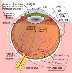
Schematic diagram of the human eye en.svg
Schematic diagram of the human eye.
Details
Part ofEyeball
SystemVisual system
FunctionRefract light
Identifiers
Latinlens crystallin
MeSHD007908
TA98A15.2.05.001
TA26798
FMA58241

The lens is a transparent biconvex structure in the eye that, along with the cornea, helps to refract light to be focused on the retina. By changing shape, it functions to change the focal length of the eye so that it can focus on objects at various distances, thus allowing a sharp real image of the object of interest to be formed on the retina. This adjustment of the lens is known as accommodation (see also below). Accommodation is similar to the focusing of a photographic camera via movement of its lenses. The lens is flatter on its anterior side than on its posterior side.

The lens is also known as the aquula (Latin, a little stream, dim. of aqua, water) or crystalline lens. In humans, the refractive power of the lens in its natural environment is approximately 18 dioptres, roughly one-third of the eye's total power.

Structure

The lens is part of the anterior segment of the human eye. In front of the lens is the iris, which regulates the amount of light entering into the eye. The lens is suspended in place by the suspensory ligament of the lens, a ring of fibrous tissue that attaches to the lens at its equator and connects it to the ciliary body. Posterior to the lens is the vitreous body, which, along with the aqueous humor on the anterior surface, bathes the lens. The lens has an ellipsoid, biconvex shape. The anterior surface is less curved than the posterior. In the adult, the lens is typically circa 10 mm in diameter and has an axial length of about 4 mm, though it is important to note that the size and shape can change due to accommodation and because the lens continues to grow throughout a person's lifetime.

Microanatomy

The lens has three main parts: the lens capsule, the lens epithelium, and the lens fibers. The lens capsule forms the outermost layer of the lens and the lens fibers form the bulk of the interior of the lens. The cells of the lens epithelium, located between the lens capsule and the outermost layer of lens fibers, are found only on the anterior side of the lens. The lens itself lacks nerves, blood vessels, or connective tissue.

Lens capsule

The lens capsule is a smooth, transparent basement membrane that completely surrounds the lens. The capsule is elastic and is composed of collagen. It is synthesized by the lens epithelium and its main components are type IV collagen and sulfated glycosaminoglycans (GAGs). The capsule is very elastic and so allows the lens to assume a more spherical shape when not under the tension of the zonular fibers (also called suspensory ligaments), which connect the lens capsule to the ciliary body. The capsule varies from 2 to 28 micrometres in thickness, being thickest near the equator and thinnest near the posterior pole.

Lens epithelium

The lens epithelium, located in the anterior portion of the lens between the lens capsule and the lens fibers, is a simple cuboidal epithelium. The cells of the lens epithelium regulate most of the homeostatic functions of the lens. As ions, nutrients, and liquid enter the lens from the aqueous humor, Na+/K+-ATPase pumps in the lens epithelial cells pump ions out of the lens to maintain appropriate lens osmotic concentration and volume, with equatorially positioned lens epithelium cells contributing most to this current. The activity of the Na+/K+-ATPases keeps water and current flowing through the lens from the poles and exiting through the equatorial regions.

The cells of the lens epithelium also serve as the progenitors for new lens fibers. It constantly lays down fibers in the embryo, fetus, infant, and adult, and continues to lay down fibers for lifelong growth.[6]

Lens fibers

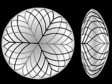
Pattern of lens fibers (anterior and lateral aspect)

The lens fibers form the bulk of the lens. They are long, thin, transparent cells, firmly packed, with diameters typically 4–7 micrometres and lengths of up to 12 mm long. The lens fibers stretch lengthwise from the posterior to the anterior poles and, when cut horizontally, are arranged in concentric layers rather like the layers of an onion. If cut along the equator, it appears as a honeycomb. The middle of each fiber lies on the equator. These tightly packed layers of lens fibers are referred to as laminae. The lens fibers are linked together via gap junctions and interdigitations of the cells that resemble "ball and socket" forms.

The lens is split into regions depending on the age of the lens fibers of a particular layer. Moving outwards from the central, oldest layer, the lens is split into an embryonic nucleus, the fetal nucleus, the adult nucleus, and the outer cortex. New lens fibers, generated from the lens epithelium, are added to the outer cortex. Mature lens fibers have no organelles or nuclei.

Development

Development of the human lens begins at the 4 mm embryonic stage. Unlike the rest of the eye, which is derived mostly from the neural ectoderm, the lens is derived from the surface ectoderm. The first stage of lens differentiation takes place when the optic vesicle, which is formed from outpocketings in the neural ectoderm, comes in proximity to the surface ectoderm. The optic vesicle induces nearby surface ectoderm to form the lens placode. At the 4 mm stage, the lens placode is a single monolayer of columnar cells.

As development progresses, the lens placode begins to deepen and invaginate. As the placode continues to deepen, the opening to the surface ectoderm constricts and the lens cells forms a structure known as the lens vesicle. By the 10 mm stage, the lens vesicle has completely separated from the surface ectoderm.

After the 10 mm stage, signals from the developing neural retina induces the cells closest to the posterior end of the lens vesicle begin to elongate toward the anterior end of the vesicle. These signals also induce the synthesis of crystallins. These elongating cells eventually fill in the lumen of the vesicle to form the primary fibers, which become the embryonic nucleus in the mature lens. The cells of the anterior portion of the lens vesicle give rise to the lens epithelium.

Additional secondary fibers are derived from lens epithelial cells located toward the equatorial region of the lens. These cells lengthen anteriorly and posteriorly to encircle the primary fibers. The new fibers grow longer than those of the primary layer, but as the lens gets larger, the ends of the newer fibers cannot reach the posterior or anterior poles of the lens. The lens fibers that do not reach the poles form tight, interdigitating seams with neighboring fibers. These seams are readily visible and are termed sutures. The suture patterns become more complex as more layers of lens fibers are added to the outer portion of the lens.

The lens continues to grow after birth, with the new secondary fibers being added as outer layers. New lens fibers are generated from the equatorial cells of the lens epithelium, in a region referred to as the germinative zone. The lens epithelial cells elongate, lose contact with the capsule and epithelium, synthesize crystallin, and then finally lose their nuclei (enucleate) as they become mature lens fibers. From development through early adulthood, the addition of secondary lens fibers results in the lens growing more ellipsoid in shape; after about age 20, however, the lens grows rounder with time and the iris is very important for this development.

Several proteins control the embryonic development of the lens: among these, primarily, PAX6, considered the master regulator gene of this organ. Other effectors of proper lens development include the Wnt signaling components BCL9 and Pygo2.

Variation

In many aquatic vertebrates, the lens is considerably thicker, almost spherical, to increase the refraction. This difference compensates for the smaller angle of refraction between the eye's cornea and the watery medium, as they have similar refractive indices. Even among terrestrial animals, however, the lens of primates such as humans is unusually flat.

In reptiles and birds, the ciliary body touches the lens with a number of pads on its inner surface, in addition to the zonular fibres. These pads compress and release the lens to modify its shape while focusing on objects at different distances; the zonular fibres perform this function in mammals. In fish and amphibians, the lens is fixed in shape, and focusing is instead achieved by moving the lens forwards or backwards within the eye.

In cartilaginous fish, the zonular fibres are replaced by a membrane, including a small muscle at the underside of the lens. This muscle pulls the lens forward from its relaxed position when focusing on nearby objects. In teleosts, by contrast, a muscle projects from a vascular structure in the floor of the eye, called the falciform process, and serves to pull the lens backwards from the relaxed position to focus on distant objects. While amphibians move the lens forward, as do cartilaginous fish, the muscles involved are not homologous with those of either type of fish. In frogs, there are two muscles, one above and one below the lens, while other amphibians have only the lower muscle.

In the most primitive vertebrates, the lampreys and hagfish, the lens is not attached to the outer surface of the eyeball at all. There is no aqueous humor in these fish, and the vitreous body simply presses the lens against the surface of the cornea. To focus its eyes, a lamprey flattens the cornea using muscles outside of the eye and pushes the lens backwards.

Function

Accommodation

An image that is partially in focus, but mostly out of focus in varying degrees.

The lens is flexible and its curvature is controlled by ciliary muscles through the zonules. By changing the curvature of the lens, one can focus the eye on objects at different distances from it. This process is called accommodation. At short focal distance the ciliary muscle contracts, zonule fibers loosen, and the lens thickens, resulting in a rounder shape and thus higher refractive power. Changing focus to an object at a greater distance requires the relaxation of the lens and thus increasing the focal distance.

The refractive index of human lens varies from approximately 1.406 in the central layers down to 1.386 in less dense layers of the lens. This index gradient enhances the optical power of the lens.

Aquatic animals must rely entirely on their lens for both focusing and to provide almost the entire refractive power of the eye as the water-cornea interface does not have a large enough difference in indices of refraction to provide significant refractive power. As such, lenses in aquatic eyes tend to be much rounder and harder.

Crystallins and transparency

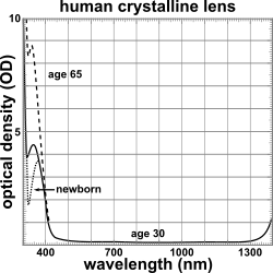
Graph showing optical density (OD) of the human crystalline lens for newborn, 30-year-old, and 65-year-old from wavelengths 300-1400 nm.

Crystallins are water-soluble proteins that compose over 90% of the protein within the lens. The three main crystallin types found in the human eye are α-, β-, and γ-crystallins. Crystallins tend to form soluble, high-molecular weight aggregates that pack tightly in lens fibers, thus increasing the index of refraction of the lens while maintaining its transparency. β and γ crystallins are found primarily in the lens, while subunits of α -crystallin have been isolated from other parts of the eye and the body. α-crystallin proteins belong to a larger superfamily of molecular chaperone proteins, and so it is believed that the crystallin proteins were evolutionarily recruited from chaperone proteins for optical purposes. The chaperone functions of α-crystallin may also help maintain the lens proteins, which must last a human for their entire lifetime.

Another important factor in maintaining the transparency of the lens is the absence of light-scattering organelles such as the nucleus, endoplasmic reticulum, and mitochondria within the mature lens fibers. Lens fibers also have a very extensive cytoskeleton that maintains the precise shape and packing of the lens fibers; disruptions/mutations in certain cytoskeletal elements can lead to the loss of transparency.

The lens blocks most ultraviolet light in the wavelength range of 300–400 nm; shorter wavelengths are blocked by the cornea. The pigment responsible for blocking the light is 3-hydroxykynurenine glucoside, a product of tryptophan catabolism in the lens epithelium. High intensity ultraviolet light can harm the retina, and artificial intraocular lenses are therefore manufactured to also block ultraviolet light. People lacking a lens (a condition known as aphakia) perceive ultraviolet light as whitish blue or whitish-violet.

Nourishment

The lens is metabolically active and requires nourishment in order to maintain its growth and transparency. Compared to other tissues in the eye, however, the lens has considerably lower energy demands.

By nine weeks into human development, the lens is surrounded and nourished by a net of vessels, the tunica vasculosa lentis, which is derived from the hyaloid artery. Beginning in the fourth month of development, the hyaloid artery and its related vasculature begin to atrophy and completely disappear by birth. In the postnatal eye, Cloquet's canal marks the former location of the hyaloid artery.

After regression of the hyaloid artery, the lens receives all its nourishment from the aqueous humor. Nutrients diffuse in and waste diffuses out through a constant flow of fluid from the anterior/posterior poles of the lens and out of the equatorial regions, a dynamic that is maintained by the Na+/K+-ATPase pumps located in the equatorially positioned cells of the lens epithelium.

Glucose is the primary energy source for the lens. As mature lens fibers do not have mitochondria, approximately 80% of the glucose is metabolized via anaerobic metabolism. The remaining fraction of glucose is shunted primarily down the pentose phosphate pathway. The lack of aerobic respiration means that the lens consumes very little oxygen as well.

Clinical significance

  • Cataracts are opacities of the lens. While some are small and do not require any treatment, others may be large enough to block light and obstruct vision. Cataracts usually develop as the aging lens becomes more and more opaque, but cataracts can also form congenitally or after injury to the lens. Nuclear sclerosis is a type of age-related cataract. Diabetes is another risk factor for cataract. Cataract surgery involves the removal of the lens and insertion of an artificial intraocular lens.
  • Presbyopia is the age-related loss of accommodation, which is marked by the inability of the eye to focus on nearby objects. The exact mechanism is still unknown, but age-related changes in the hardness, shape, and size of the lens have all been linked to the condition.
  • Ectopia lentis is the displacement of the lens from its normal position.
  • Aphakia is the absence of the lens from the eye. Aphakia can be the result of surgery or injury, or it can be congenital.

Entropy (information theory)

From Wikipedia, the free encyclopedia https://en.wikipedia.org/wiki/Entropy_(information_theory) In info...