Search This Blog

Wednesday, November 2, 2022

Social model of disability

From Wikipedia, the free encyclopedia

The social model of disability identifies systemic barriers, derogatory attitudes, and social exclusion (intentional or inadvertent), which make it difficult or impossible for disabled people to attain their valued functionings. The social model of disability diverges from the dominant medical model of disability, which is a functional analysis of the body as a machine to be fixed in order to conform with normative values. While physical, sensory, intellectual, or psychological variations may result in individual functional differences, these do not necessarily have to lead to disability unless society fails to take account of and include people intentionally with respect to their individual needs. The origin of the approach can be traced to the 1960s, and the specific term emerged from the United Kingdom in the 1980s.

The social model of disability seeks to redefine disability to refer to the restrictions caused by society when it does not give equitable social and structural support according to disabled peoples' structural needs. As a simple example, if a person is unable to climb stairs, the medical model focuses on making the individual physically able to climb stairs. The social model tries to make stair-climbing unnecessary, such as by making society adapt to their needs, and assist them by replacing the stairs with a wheelchair-accessible ramp. According to the social model, the person remains disabled with respect to climbing stairs, but the disability is negligible and no longer disabling in that scenario, because the person can get to the same locations without climbing any stairs.

History

Disability rights movement

There is a hint from before the 1970s that the interaction between disability and society was beginning to be considered. British politician and disability rights campaigner Alf Morris wrote in 1969 (emphasis added):

When the title of my Bill was announced, I was frequently asked what kind of improvements for the chronically sick and disabled I had in mind. It always seemed best to begin with the problems of access. I explained that I wanted to remove the severe and gratuitous social handicaps inflicted on disabled people, and often on their families and friends, not just by their exclusion from town and county halls, art galleries, libraries and many of the universities, but even from pubs, restaurants, theatres, cinemas and other places of entertainment ... I explained that I and my friends were concerned to stop society from treating disabled people as if they were a separate species.

The history of the social model of disability begins with the history of the disability rights movement. Around 1970, various groups in North America, including sociologists, disabled people, and disability-focused political groups, began to pull away from the accepted medical lens of viewing disability. Instead, they began to discuss things like oppression, civil rights, and accessibility. This change in discourse resulted in conceptualizations of disability that was rooted in social constructs.

In 1975, the UK organization Union of the Physically Impaired Against Segregation (UPIAS) claimed: "In our view it is society which disables physically impaired people. Disability is something imposed on top of our impairments by the way we are unnecessarily isolated and excluded from full participation in society." This became known as the social interpretation, or social definition, of disability.

Mike Oliver

In 1983, the disabled academic Mike Oliver coined the phrase social model of disability in reference to these ideological developments. Oliver focused on the idea of an individual model versus a social model. Oliver's seminal 1990 book The Politics of Disablement is widely cited as a major moment in the adoption of this model. The book included just three pages about the social model of disability.

Developments

The "social model" was extended and developed by academics and activists in Australia, the UK, the US, and other countries to include all disabled people, including those who have learning disabilities, intellectual disabilities, or emotional, mental health or behavioural problems.

Tool for cultural analysis

The social model has become a key tool in the analysis of the cultural representation of disability; from literature, to radio, to charity-imagery to cinema. The social model has become the key conceptual analysis in challenging, for examples, stereotypes and archetypes of disabled people by revealing how conventional imagery reinforces the oppression of disabled people. Key theorists include Paul Darke (cinema), Lois Keith (literature), Leonard Davis (Deaf culture), Jenny Sealey (theatre) and Mary-Pat O'Malley (radio).

Components and usage

A fundamental aspect of the social model concerns equality. The struggle for equality is often compared to the struggles of other socially marginalized groups. Equal rights are said to empower people with the "ability" to make decisions and the opportunity to live life to the fullest. A related phrase often used by disability rights activists, as with other social activism, is "Nothing About Us Without Us".

The social model of disability focuses on changes required in society. These might be in terms of:

  • Attitudes, for example a more positive attitude towards certain mental traits or behaviors, or not underestimating the potential quality of life of disabled people,
  • Social support, for example help dealing with barriers; resources, aids, or positive discrimination to provide equal access, for example providing someone to explain work culture for an autistic employee,
  • Information, for example using suitable formats (e.g. braille) or levels (e.g. simplicity of language) or coverage (e.g. explaining issues others may take for granted),
  • Physical structures, for example buildings with sloped access and elevators, or
  • Flexible work hours for people with circadian rhythm sleep disorders.

Limitations

Oliver did not intend the social model of disability to be an all-encompassing theory of disability, but rather a starting point in reframing how society views disability. This model was conceived of as a tool that could be used to improve the lives of disabled people, rather than a complete explanation for every experience and circumstance.

It has been criticized for underplaying the role of disabilities. It has also been criticized for not promoting the normal differences between disabled people, who can be any age, gender, race, and sexual orientation, and instead presenting them as a monolithic, insufficiently individuated group of people.

As an identity

In the late 20th century and early 21st century, the social model of disability became a dominant identity for disabled people in the UK.

The social model of disability implies that attempts to change, "fix", or "cure" individuals, especially when used against the wishes of the individual, can be discriminatory and prejudiced. This attitude, which may be seen as stemming from a medical model and a subjective value system, can harm the self-esteem and social inclusion of those constantly subjected to it (e.g. being told they are not as good or valuable, in an overall and core sense, as others). Some communities have actively resisted "treatments", while, for example, defending a unique culture or set of abilities. In the Deaf community, sign language is valued even if most people do not know it, and some parents argue against cochlear implants for deaf infants who cannot consent to them. Autistic people may say that their "unusual" behavior, which they say can serve an important purpose to them, should not have to be suppressed to please others. They argue instead for acceptance of neurodiversity and accommodation to different needs and goals. Some people diagnosed with a mental disorder argue that they are just different and don't necessarily conform. The biopsychosocial model of disease/disability is an attempt by practitioners to address this.

The Neurodiversity label has been used by various mental-disability rights advocates within the context of the social model of disability. The label has been applied to other neurodevelopmental conditions apart from autism, such as attention deficit hyperactivity disorder (ADHD), developmental speech disorders, dyslexia, dysgraphia, dyspraxia, dyscalculia, dysnomia, intellectual disability, and Tourette syndrome, as well as schizophrenia, bipolar disorder, and some mental health conditions such as schizoaffective disorder, antisocial personality disorder, dissociative disorders, and obsessive–compulsive disorder.

The social model implies that practices such as eugenics are founded on social values and a prejudiced understanding of the potential and value of those labeled disabled. "Over 200,000 disabled people were some of the earlier victims of the Holocaust, after Communists, other political enemies, and homosexuals."

A 1986 article stated:

It is important that we do not allow ourselves to be dismissed as if we all come under this one great metaphysical category 'the disabled'. The effect of this is a depersonalization, a sweeping dismissal of our individuality, and a denial of our right to be seen as people with our own uniqueness, rather than as the anonymous constituents of a category or group. These words that lump us all together – 'the disabled', 'spina bifida', 'tetraplegic', 'muscular dystrophy', – are nothing more than terminological rubbish bins into which all the important things about us as people get thrown away.

Economic aspects

The social model also relates to economic empowerment, proposing that people can be disabled by a lack of resources to meet their needs. For example, a disabled person may need support services to be able to participate fully in society, and can become disabled if society cuts access to those support services, perhaps in the name of government austerity measures.

The social model addresses other issues, such as the underestimation of the potential of disabled people to contribute to society and add economic value to society if they are given equal rights and equally suitable facilities and opportunities as others. Economic research on companies that attempt to accommodate disability in their workforce suggest they outperform competitors.

In Autumn 2001, the UK Office for National Statistics identified that approximately one-fifth of the working-age population was disabled, equating to an estimated 7.1 million disabled people, compared to an estimated 29.8 million nondisabled people. This analysis also provided insight into some of the reasons why disabled people weren't in the labor market, such as that the reduction in disability benefits in entering the labor market would not make it worthwhile to enter into employment. A three-pronged approach was suggested: "incentives to work via the tax and benefit system, for example through the Disabled Person's Tax Credit; helping people back into work, for example via the New Deal for Disabled People; and tackling discrimination in the workplace via anti-discrimination policy. Underpinning this are the Disability Discrimination Act (DDA) 1995 and the Disability Rights Commission."

Canada and the United States have operated under the premise that social assistance benefits should not exceed the amount of money earned through labour in order to give citizens an incentive to search for and maintain employment. This has led to widespread poverty amongst disabled citizens. In the 1950s, disability pensions were established and included various forms of direct economic assistance; however, compensation was low. Since the 1970s, both governments have viewed unemployed, disabled citizens as excess labor due to continuous high unemployment rates and have made minimal attempts to increase employment, keeping disabled people at poverty-level incomes due to the 'incentive' principle. Poverty is the most debilitating circumstance disabled people face, resulting in the inability to afford proper medical, technological and other assistance necessary to participate in society.

Law and public policy

In the United Kingdom, the Disability Discrimination Act defines disability using the medical model - disabled people are defined as people with certain conditions or limitations on their ability to carry out "normal day-to-day activities." But the requirement of employers and service providers to make "reasonable adjustments" to their policies or practices, or physical aspects of their premises, follows the social model. By making adjustments, employers and service providers are removing the barriers that disable, according to the social model. In 2006, amendments to the act called for local authorities and others to actively promote disability equality; this was enforced via the formation of the Disability Equality Duty in December 2006. In 2010, The Disability Discrimination Act (1995) was amalgamated into the Equality Act 2010, along with other pertinent discrimination legislation. The Equality Act of 2010 extends the law on discrimination to indirect discrimination. For example, if a carer of a disabled person is discriminated against, this is now also unlawful. Since October 2010, when it came into effect, employers may not legally ask questions about illness or disability at interviews for a job or for a referee to comment on such in a reference, except where there is a need to make reasonable adjustments for an interview to proceed. Following an offer of a job, an employer can lawfully ask such questions.

In the United States, the Americans with Disabilities Act of 1990 (ADA), is a wide-ranging civil rights law that prohibits discrimination based on disability in a wide range of settings. The ADA was the first civil rights law of its kind in the world and affords protections against discrimination to disabled Americans. The law was modeled after the Civil Rights Act of 1964, which made discrimination based on race, religion, sex, national origin, and other characteristics illegal. It requires that mass transportation, commercial buildings, and public accommodations be accessible to disabled people.

In 2007, the European Court of Justice in the Chacón Navas v Eurest Colectividades SA court case, defined disability narrowly according to a medical definition that excluded temporary illness, when considering the Directive establishing a general framework for equal treatment in employment and occupation (Council Directive 2000/78/EC). The directive did not provide for any definition of disability, despite discourse in policy documents previously in the EU about endorsing the social model of disability. This allowed the Court of Justice to take a narrow medical definition.

Technology

Over the last several decades, technology has transformed networks, services, and communication by promoting the rise of telecommunications, computer use, etc. This Digital Revolution has changed how people work, learn, and interact, moving these basic human activities to technological platforms. However, many people who use such technology experience a form of disability. Even if it is not physically visible, those with, for example cognitive impairments, hand tremors, or vision impairments, have some form of disability that prohibit them from fully accessing technology in the way that those without a "technological disability" do.

In "Disability and New Media," Katie Ellis and Mike Kent state that "technology is often presented as a source of liberation; however, developments associated with Web 2.0 show that this is not always the case." They go on to state that the technological advancement of Web 2.0 is tethered to social ideology and stigma which "routinely disables people with disability."

In "Digital Disability: The Social Construction of Disability in New Media," Gregg Goggin and Christopher Newell call for an innovative understanding of new media and disability issues. They trace developments ranging from telecommunications to assistive technologies to offer a technoscience of disability ,which offers a global perspective on how disabled people are represented as users, consumers, viewers, or listeners of new media, by policymakers, corporations, programmers, and disabled people themselves.

Social construction of disability

The social construction of disability comes from a paradigm that suggests that society's beliefs about a particular community, group, or population are grounded in the power structures inherent in that society at any given time. These are often steeped in historical representations of the issue and social expectations surrounding concepts, such as disability, thereby enabling a social construct around what society deems disabled and healthy.

Ideas surrounding disability stem from societal attitudes, often connected to who is deserving or undeserving, and deemed productive to society at any given time. For example, in the medieval period, a person's moral behavior established disability. Disability was a divine punishment or side effect of a moral failing; being physically or biologically different was not enough to be considered disabled. Only during the European Enlightenment did society change its definition of disability to be more related to biology. However, what most Western Europeans considered to be healthy determined the new biological definition of health.

2000 Paralympics

Since the invention of the television in the early 1900s, this medium has held a pervasive influence on public outlook on many aspects of society, disability being one of them. One example is how the 2000 Paralympics were televised, in contrast to the Olympics. The 2000 Sydney Paralympic Games, one of the biggest in history, was barely acknowledged by mainstream media prior to the event. The Sydney Paralympic organizers worked extensively to try to solicit coverage of the Games. For more than two years, they negotiated with Channel 7 to broadcast the competitions. Channel 7 proposed that if the Paralympics paid them $3 million in case of lack of advertising revenue, they would agree to broadcast the event. Eventually, the Australian Broadcasting Company (ABC) and Channel 7 announced they would be broadcasting the Games and Channel 7 would "complement" the coverage with a highlights package that ran daily on its pay-TV Channel. ABC also promised to broadcast at least 60 minutes of daily highlights. Later, ABC finally agreed to air a live broadcast of the opening and closing ceremonies. The opening and closing ceremonies were quite popular amongst viewers, watched by 2.5 million; however the rest of the games were not popular. While the Olympics were covered live throughout the entire event, the Paralympics were not seen as important enough for the same live coverage before the initial showing. By separating the Olympics and Paralympics, and thus indicating that one is less valuable than the other, disability is socially constructed.

Applications

Applying the social model of disability can change goals and care plans. For example, with the medical model of disability, the goal may be to help a child acquire typical abilities and to reduce impairment. With the social model, the goal may be to have a child be included in the normal life of the community, such as attending birthday parties and other social events, regardless of the level of function.

Education

It has been suggested that disability education tries to restore the idea of a moral community, one in which the members question what constitutes a good life, reimagine education, see physical and mental conditions as part of a range of abilities, consider that different talents are distributed in different ways, and understand that all talents should be recognized. In this system, all students would be included in the educational network instead of being set apart as special cases, and it would be acknowledged that all humans have individual needs.

X-ray microtomography

From Wikipedia, the free encyclopedia
 
0:46
3D rendering of a µCT scan of a leaf piece, resolution circa 40 µm/voxel.
 
Two phase µCT analysis of Ti2AlC/Al MAX phase composite

X-ray microtomography, like tomography and X-ray computed tomography, uses X-rays to create cross-sections of a physical object that can be used to recreate a virtual model (3D model) without destroying the original object. The prefix micro- (symbol: µ) is used to indicate that the pixel sizes of the cross-sections are in the micrometre range. These pixel sizes have also resulted in the terms high-resolution X-ray tomography, micro–computed tomography (micro-CT or µCT), and similar terms. Sometimes the terms high-resolution CT (HRCT) and micro-CT are differentiated, but in other cases the term high-resolution micro-CT is used. Virtually all tomography today is computed tomography.

Micro-CT has applications both in medical imaging and in industrial computed tomography. In general, there are two types of scanner setups. In one setup, the X-ray source and detector are typically stationary during the scan while the sample/animal rotates. The second setup, much more like a clinical CT scanner, is gantry based where the animal/specimen is stationary in space while the X-ray tube and detector rotate around. These scanners are typically used for small animals (in vivo scanners), biomedical samples, foods, microfossils, and other studies for which minute detail is desired.

The first X-ray microtomography system was conceived and built by Jim Elliott in the early 1980s. The first published X-ray microtomographic images were reconstructed slices of a small tropical snail, with pixel size about 50 micrometers.

Working principle

Imaging system

Fan beam reconstruction

The fan-beam system is based on a one-dimensional (1D) X-ray detector and an electronic X-ray source, creating 2D cross-sections of the object. Typically used in human computed tomography systems.

Cone beam reconstruction

The cone-beam system is based on a 2D X-ray detector (camera) and an electronic X-ray source, creating projection images that later will be used to reconstruct the image cross-sections.

Open/Closed systems

Open X-ray system

In an open system, X-rays may escape or leak out, thus the operator must stay behind a shield, have special protective clothing, or operate the scanner from a distance or a different room. Typical examples of these scanners are the human versions, or designed for big objects.

Closed X-ray system

In a closed system, X-ray shielding is put around the scanner so the operator can put the scanner on a desk or special table. Although the scanner is shielded, care must be taken and the operator usually carries a dosimeter, since X-rays have a tendency to be absorbed by metal and then re-emitted like an antenna. Although a typical scanner will produce a relatively harmless volume of X-rays, repeated scannings in a short timeframe could pose a danger. Digital detectors with small pixel pitches and micro-focus x-ray tubes are usually employed to yield in high resolution images.

Closed systems tend to become very heavy because lead is used to shield the X-rays. Therefore, the smaller scanners only have a small space for samples.

3D image reconstruction

The principle

Because microtomography scanners offer isotropic, or near isotropic, resolution, display of images does not need to be restricted to the conventional axial images. Instead, it is possible for a software program to build a volume by 'stacking' the individual slices one on top of the other. The program may then display the volume in an alternative manner.

Image reconstruction software

For X-ray microtomography, powerful open source software is available, such as the ASTRA toolbox. The ASTRA Toolbox is a MATLAB and python toolbox of high-performance GPU primitives for 2D and 3D tomography, from 2009–2014 developed by iMinds-Vision Lab, University of Antwerp and since 2014 jointly developed by iMinds-VisionLab, UAntwerpen and CWI, Amsterdam. The toolbox supports parallel, fan, and cone beam, with highly flexible source/detector positioning. A large number of reconstruction algorithms are available, including FBP, ART, SIRT, SART, CGLS.

For 3D visualization, tomviz is a popular open-source tool for tomography.

Volume rendering

Volume rendering is a technique used to display a 2D projection of a 3D discretely sampled data set, as produced by a microtomography scanner. Usually these are acquired in a regular pattern (e.g., one slice every millimeter) and usually have a regular number of image pixels in a regular pattern. This is an example of a regular volumetric grid, with each volume element, or voxel represented by a single value that is obtained by sampling the immediate area surrounding the voxel.

Image segmentation

Where different structures have similar threshold density, it can become impossible to separate them simply by adjusting volume rendering parameters. The solution is called segmentation, a manual or automatic procedure that can remove the unwanted structures from the image.

Typical use

Archaeology

Biomedical

  • Both in vitro and in vivo small animal imaging
  • Neurons
  • Human skin samples
  • Bone samples, including teeth, ranging in size from rodents to human biopsies
  • Lung imaging using respiratory gating
  • Cardiovascular imaging using cardiac gating
  • Imaging of the human eye, ocular microstructures and tumors
  • Tumor imaging (may require contrast agents)
  • Soft tissue imaging
  • Insects – Insect development
  • Parasitology – migration of parasites, parasite morphology

Developmental biology

  • Tracing the development of the extinct Tasmanian tiger during growth in the pouch
  • Model and non-model organisms (elephants, zebrafish, and whales)

Electronics

  • Small electronic components. E.g. DRAM IC in plastic case.

Microdevices

Composite materials and metallic foams

  • Ceramics and Ceramic–Metal composites. Microstructural analysis and failure investigation
  • Composite material with glass fibers 10 to 12 micrometres in diameter

Polymers, plastics

Diamonds

  • Detecting defects in a diamond and finding the best way to cut it.

Food and seeds

  • 3-D imaging of foods using X-ray microtomography
  • Analysing heat and drought stress on food crops

Wood and paper

Building materials

Geology

In geology it is used to analyze micro pores in the reservoir rocks, it can used in microfacies analysis for sequence stratigraphy. In petroleum exploration it is used to model the petroleum flow under micro pores and nano particles.

It can give a resolution up to 1 nm.

Fossils

Microfossils

X-ray microtomography of a radiolarian, Triplococcus acanthicus
This is a microfossil from the Middle Ordovician with four nested spheres. The innermost sphere is highlighted red. Each segment is shown at the same scale.
  • Benthonic foraminifers

Palaeography

  • Digitally unfolding letters of correspondence which employed letterlocking.

Space

Stereo images

  • Visualizing with blue and green or blue filters to see depth

Others

Neuroregeneration

From Wikipedia, the free encyclopedia
https://en.wikipedia.org/wiki/Neuroregeneration

Neuroregeneration refers to the regrowth or repair of nervous tissues, cells or cell products. Such mechanisms may include generation of new neurons, glia, axons, myelin, or synapses. Neuroregeneration differs between the peripheral nervous system (PNS) and the central nervous system (CNS) by the functional mechanisms involved, especially in the extent and speed of repair. When an axon is damaged, the distal segment undergoes Wallerian degeneration, losing its myelin sheath. The proximal segment can either die by apoptosis or undergo the chromatolytic reaction, which is an attempt at repair. In the CNS, synaptic stripping occurs as glial foot processes invade the dead synapse.

Nervous system injuries affect over 90,000 people every year. It is estimated that spinal cord injuries alone affect 10,000 each year. As a result of this high incidence of neurological injuries, nerve regeneration and repair, a subfield of neural tissue engineering, is becoming a rapidly growing field dedicated to the discovery of new ways to recover nerve functionality after injury. The nervous system is divided into two parts: the central nervous system, which consists of the brain and spinal cord, and the peripheral nervous system, which consists of cranial and spinal nerves along with their associated ganglia. While the peripheral nervous system has an intrinsic ability for repair and regeneration, the central nervous system is, for the most part, incapable of self-repair and regeneration. There is currently no treatment for recovering human nerve function after injury to the central nervous system. In addition, multiple attempts at nerve re-growth across the PNS-CNS transition have not been successful. There is simply not enough knowledge about regeneration in the central nervous system. In addition, although the peripheral nervous system has the capability for regeneration, much research still needs to be done to optimize the environment for maximum regrowth potential. Neuroregeneration is important clinically, as it is part of the pathogenesis of many diseases, including multiple sclerosis.

Peripheral nervous system regeneration

Guillain–Barré syndrome – nerve damage

Neuroregeneration in the peripheral nervous system (PNS) occurs to a significant degree. After an injury to the axon, peripheral neurons activate a variety of signaling pathways which turn on pro-growth genes, leading to reformation of a functional growth cone and regeneration. The growth of these axons is also governed by chemotactic factors secreted from Schwann cells. Injury to the peripheral nervous system immediately elicits the migration of phagocytes, Schwann cells, and macrophages to the lesion site in order to clear away debris such as damaged tissue which is inhibitory to regeneration. When a nerve axon is severed, the end still attached to the cell body is labeled the proximal segment, while the other end is called the distal segment. After injury, the proximal end swells and experiences some retrograde degeneration, but once the debris is cleared, it begins to sprout axons and the presence of growth cones can be detected. The proximal axons are able to regrow as long as the cell body is intact, and they have made contact with the Schwann cells in the endoneurium (also known as the endoneurial tube or channel). Human axon growth rates can reach 2 mm/day in small nerves and 5 mm/day in large nerves. The distal segment, however, experiences Wallerian degeneration within hours of the injury; the axons and myelin degenerate, but the endoneurium remains. In the later stages of regeneration the remaining endoneurial tube directs axon growth back to the correct targets. During Wallerian degeneration, Schwann cells grow in ordered columns along the endoneurial tube, creating a band of Büngner cells that protects and preserves the endoneurial channel. Also, macrophages and Schwann cells release neurotrophic factors that enhance re-growth.

Central nervous system regeneration

Unlike peripheral nervous system injury, injury to the central nervous system is not followed by extensive regeneration. It is limited by the inhibitory influences of the glial and extracellular environment. The hostile, non-permissive growth environment is, in part, created by the migration of myelin-associated inhibitors, astrocytes, oligodendrocytes, oligodendrocyte precursors, and microglia. The environment within the CNS, especially following trauma, counteracts the repair of myelin and neurons. Growth factors are not expressed or re-expressed; for instance, the extracellular matrix is lacking laminins. Glial scars rapidly form, and the glia actually produce factors that inhibit remyelination and axon repair; for instance, NOGO and NI-35. The axons themselves also lose the potential for growth with age, due to a decrease in GAP43 expression, among others.

Slower degeneration of the distal segment than that which occurs in the peripheral nervous system also contributes to the inhibitory environment because inhibitory myelin and axonal debris are not cleared away as quickly. All these factors contribute to the formation of what is known as a glial scar, which axons cannot grow across. The proximal segment attempts to regenerate after injury, but its growth is hindered by the environment. It is important to note that central nervous system axons have been proven to regrow in permissive environments; therefore, the primary problem to central nervous system axonal regeneration is crossing or eliminating the inhibitory lesion site. Another problem is that the morphology and functional properties of central nervous system neurons are highly complex, for this reason a neuron functionally identical cannot be replaced by one of another type (Llinás' law).

Inhibition of axonal regrowth

Glial cell scar formation is induced following damage to the nervous system. In the central nervous system, this glial scar formation significantly inhibits nerve regeneration, which leads to a loss of function. Several families of molecules are released that promote and drive glial scar formation. For instance, transforming growth factors B-1 and -2, interleukins, and cytokines play a role in the initiation of scar formation. The accumulation of reactive astrocytes at the site of injury and the up regulation of molecules that are inhibitory for neurite outgrowth contribute to the failure of neuroregeneration. The up-regulated molecules alter the composition of the extracellular matrix in a way that has been shown to inhibit neurite outgrowth extension. This scar formation involves several cell types and families of molecules.

Chondroitin sulfate proteoglycan

In response to scar-inducing factors, astrocytes up regulate the production of chondroitin sulfate proteoglycans. Astrocytes are a predominant type of glial cell in the central nervous system that provide many functions including damage mitigation, repair, and glial scar formation. The RhoA pathway is involved. Chondroitin sulfate proteoglycans (CSPGs) have been shown to be up regulated in the central nervous system (CNS) following injury. Repeating disaccharides of glucuronic acid and galactosamine, glycosaminoglycans (CS-GAGs), are covalently coupled to the protein core CSPGs. CSPGs have been shown to inhibit regeneration in vitro and in vivo, but the role that the CSPG core protein vs. CS-GAGs had not been studied until recently.

Keratan sulfate proteoglycans

Like the chondroitin sulfate proteoglycans, keratan sulfate proteoglycan (KSPG) production is up regulated in reactive astrocytes as part of glial scar formation. KSPGs have also been shown to inhibit neurite outgrowth extension, limiting nerve regeneration. Keratan sulfate, also called keratosulfate, is formed from repeating disaccharide galactose units and N-acetylglucosamines. It is also 6-sulfated. This sulfation is crucial to the elongation of the keratan sulfate chain. A study was done using N-acetylglucosamine 6-O-sulfotransferase-1 deficient mice. The wild type mouse showed a significant up regulation of mRNA expressing N-acetylglucosamine 6-O-sulfotransferase-1 at the site of cortical injury. However, in the N-acetylglucosamine 6-O-sulfotransferase-1 deficient mice, the expression of keratan sulfate was significantly decreased when compared to the wild type mice. Similarly, glial scar formation was significantly reduced in the N-acetylglucosamine 6-O-sulfotransferase-1 mice, and as a result, nerve regeneration was less inhibited.

Other inhibitory factors

Proteins of oligodendritic or glial debris origin that influence neuroregeneration:

  • NOGO –The protein family Nogo, particularly Nogo-A, has been identified as an inhibitor of remyelination in the CNS, especially in autoimmune mediated demyelination, such as found in experimental autoimmune encephalomyelitis (EAE), and multiple sclerosis (MS). Nogo A functions via either its amino-Nogo terminus through an unknown receptor, or by its Nogo-66 terminus through NgR1, p75, TROY or LINGO1. Antagonising this inhibitor results in improved remyelination, as it is involved in the RhoA pathway.
  • NI-35 a non-permissive growth factor from myelin.
  • MAGMyelin-associated glycoprotein acts via the receptors NgR2, GT1b, NgR1, p75, TROY and LINGO1.
  • OMgpOligodendrocyte myelin glycoprotein
  • Ephrin B3 functions through the EphA4 receptor and inhibits remyelination.
  • Sema 4D(Semaphorin 4D) functions through the PlexinB1 receptor and inhibits remyelination.
  • Sema 3A (Semaphorin 3A) is present in the scar that forms in both central nervous system and peripheral nerve injuries and contributes to the outgrowth-inhibitory properties of these scars

Clinical treatments

Surgery

Surgery can be done in case a peripheral nerve has become cut or otherwise divided. This is called peripheral nerve reconstruction. The injured nerve is identified and exposed so that normal nerve tissue can be examined above and below the level of injury, usually with magnification, using either loupes or an operating microscope. If a large segment of nerve is harmed, as can happen in a crush or stretch injury, the nerve will need to be exposed over a larger area. Injured portions of the nerve are removed. The cut nerve endings are then carefully reapproximated using very small sutures. The nerve repair must be covered by healthy tissue, which can be as simple as closing the skin or it can require moving skin or muscle to provide healthy padded coverage over the nerve. The type of anesthesia used depends on the complexity of the injury. A surgical tourniquet is almost always used.

Prognosis

The expectations after surgical repair of a divided peripheral nerve depends on several factors:

  • Age: Recovery of a nerve after surgical repair depends mainly on the age of the patient. Young children can recover close-to-normal nerve function. In contrast, a patient over 60 years old with a cut nerve in the hand would expect to recover only protective sensation; that is, the ability to distinguish hot/cold or sharp/dull.
  • The mechanism of injury: Sharp injuries, such as a knife wound, damage only a very short segment of the nerve, availing for direct suture. In contrast, nerves that are divided by stretch or crush may be damaged over long segments. These nerve injuries are more difficult to treat and generally have a poorer outcome. In addition, associated injuries, like injury to bone, muscle and skin, can make nerve recovery more difficult.
  • The level of injury: After a nerve is repaired, the regenerating nerve endings must grow all the way to their target. For example, a nerve injured at the wrist that normally provides sensation to the thumb must grow to the end of the thumb in order to provide sensation. The return of function decreases with increased distance over which a nerve must grow.

Autologous nerve grafting

Currently, autologous nerve grafting, or a nerve autograft, is known as the gold standard for clinical treatments used to repair large lesion gaps in the peripheral nervous system. It is important that nerves are not repaired under tension, which could otherwise happen if cut ends are reapproximated across a gap. Nerve segments are taken from another part of the body (the donor site) and inserted into the lesion to provide endoneurial tubes for axonal regeneration across the gap. However, this is not a perfect treatment; often the outcome is only limited function recovery. Also, partial de-innervation is frequently experienced at the donor site, and multiple surgeries are required to harvest the tissue and implant it.

When appropriate, a nearby donor may be used to supply innervation to lesioned nerves. Trauma to the donor can be minimized by utilizing a technique known as end-to-side repair. In this procedure, an epineurial window is created in the donor nerve and the proximal stump of the lesioned nerve is sutured over the window. Regenerating axons are redirected into the stump. Efficacy of this technique is partially dependent upon the degree of partial neurectomy performed on the donor, with increasing degrees of neurectomy giving rise to increasing axon regeneration within the lesioned nerve, but with the consequence of increasing deficit to the donor.

Some evidence suggests that local delivery of soluble neurotrophic factors at the site of autologous nerve grafting may enhance axon regeneration within the graft and help expedite functional recovery of a paralyzed target. Other evidence suggests that gene-therapy induced expression of neurotrophic factors within the target muscle itself can also help enhance axon regeneration. Accelerating neuroregeneration and the reinnervation of a denervated target is critically important in order to reduce the possibility of permanent paralysis due to muscular atrophy.

Allografts and xenografts

Variations on the nerve autograft include the allograft and the xenograft. In allografts, the tissue for the graft is taken from another person, the donor, and implanted in the recipient. Xenografts involve taking donor tissue from another species. Allografts and xenografts have the same disadvantages as autografts, but in addition, tissue rejection from immune responses must also be taken into account. Often immunosuppression is required with these grafts. Disease transmission also becomes a factor when introducing tissue from another person or animal. Overall, allografts and xenografts do not match the quality of outcomes seen with autografts, but they are necessary when there is a lack of autologous nerve tissue.

Nerve guidance conduit

Because of the limited functionality received from autografts, the current gold standard for nerve regeneration and repair, recent neural tissue engineering research has focused on the development of bioartificial nerve guidance conduits in order to guide axonal regrowth. The creation of artificial nerve conduits is also known as entubulation because the nerve ends and intervening gap are enclosed within a tube composed of biological or synthetic materials.

Immunisation

A direction of research is towards the use of drugs that target remyelinating inhibitor proteins, or other inhibitors. Possible strategies include vaccination against these proteins (active immunisation), or treatment with previously created antibodies (passive immunisation). These strategies appear promising on animal models with experimental autoimmune encephalomyelitis (EAE), a model of MS. Monoclonal antibodies have also been used against inhibitory factors such as NI-35 and NOGO.

Regeneration (biology)

From Wikipedia, the free encyclopedia
 
Sunflower sea star regenerates its arms.
 
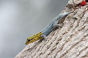
Dwarf yellow-headed gecko with regenerating tail

In biology, regeneration is the process of renewal, restoration, and tissue growth that makes genomes, cells, organisms, and ecosystems resilient to natural fluctuations or events that cause disturbance or damage. Every species is capable of regeneration, from bacteria to humans. Regeneration can either be complete where the new tissue is the same as the lost tissue, or incomplete where after the necrotic tissue comes fibrosis.

At its most elementary level, regeneration is mediated by the molecular processes of gene regulation and involves the cellular processes of cell proliferation, morphogenesis and cell differentiation. Regeneration in biology, however, mainly refers to the morphogenic processes that characterize the phenotypic plasticity of traits allowing multi-cellular organisms to repair and maintain the integrity of their physiological and morphological states. Above the genetic level, regeneration is fundamentally regulated by asexual cellular processes. Regeneration is different from reproduction. For example, hydra perform regeneration but reproduce by the method of budding.

The hydra and the planarian flatworm have long served as model organisms for their highly adaptive regenerative capabilities. Once wounded, their cells become activated and restore the organs back to their pre-existing state. The Caudata ("urodeles"; salamanders and newts), an order of tailed amphibians, is possibly the most adept vertebrate group at regeneration, given their capability of regenerating limbs, tails, jaws, eyes and a variety of internal structures. The regeneration of organs is a common and widespread adaptive capability among metazoan creatures. In a related context, some animals are able to reproduce asexually through fragmentation, budding, or fission. A planarian parent, for example, will constrict, split in the middle, and each half generates a new end to form two clones of the original.

Echinoderms (such as the sea star), crayfish, many reptiles, and amphibians exhibit remarkable examples of tissue regeneration. The case of autotomy, for example, serves as a defensive function as the animal detaches a limb or tail to avoid capture. After the limb or tail has been autotomized, cells move into action and the tissues will regenerate. In some cases a shed limb can itself regenerate a new individual. Limited regeneration of limbs occurs in most fishes and salamanders, and tail regeneration takes place in larval frogs and toads (but not adults). The whole limb of a salamander or a triton will grow again and again after amputation. In reptiles, chelonians, crocodilians and snakes are unable to regenerate lost parts, but many (not all) kinds of lizards, geckos and iguanas possess regeneration capacity in a high degree. Usually, it involves dropping a section of their tail and regenerating it as part of a defense mechanism. While escaping a predator, if the predator catches the tail, it will disconnect.

Ecosystems

Ecosystems can be regenerative. Following a disturbance, such as a fire or pest outbreak in a forest, pioneering species will occupy, compete for space, and establish themselves in the newly opened habitat. The new growth of seedlings and community assembly process is known as regeneration in ecology.

Cellular molecular fundamentals

Pattern formation in the morphogenesis of an animal is regulated by genetic induction factors that put cells to work after damage has occurred. Neural cells, for example, express growth-associated proteins, such as GAP-43, tubulin, actin, an array of novel neuropeptides, and cytokines that induce a cellular physiological response to regenerate from the damage. Many of the genes that are involved in the original development of tissues are reinitialized during the regenerative process. Cells in the primordia of zebrafish fins, for example, express four genes from the homeobox msx family during development and regeneration.

Tissues

"Strategies include the rearrangement of pre-existing tissue, the use of adult somatic stem cells and the dedifferentiation and/or transdifferentiation of cells, and more than one mode can operate in different tissues of the same animal. All these strategies result in the re-establishment of appropriate tissue polarity, structure and form." During the developmental process, genes are activated that serve to modify the properties of cell as they differentiate into different tissues. Development and regeneration involves the coordination and organization of populations cells into a blastema, which is "a mound of stem cells from which regeneration begins". Dedifferentiation of cells means that they lose their tissue-specific characteristics as tissues remodel during the regeneration process. This should not be confused with the transdifferentiation of cells which is when they lose their tissue-specific characteristics during the regeneration process, and then re-differentiate to a different kind of cell.

In animals

Arthropods

Limb regeneration

Many arthropods can regenerate limbs and other appendages following either injury or autotomy. Regeneration capacity is constrained by the developmental stage and ability to molt.

Crustaceans, which continually molt, can regenerate throughout their lifetimes. While molting cycles are generally hormonally regulated, limb amputation induces premature molting.

Hemimetabolous insects such as crickets can regenerate limbs as nymphs, before their final molt.

Holometabolous insects can regenerate appendages as larvae prior to the final molt and metamorphosis. Beetle larvae, for example, can regenerate amputated limbs. Fruit fly larvae do not have limbs but can regenerate their appendage primordia, imaginal discs. In both systems, the regrowth of the new tissue delays pupation.

Mechanisms underlying appendage limb regeneration in insects and crustaceans are highly conserved. During limb regeneration species in both taxa form a blastema that proliferates and grows to repattern the missing tissue.

Venom regeneration

Arachnids, including scorpions, are known to regenerate their venom, although the content of the regenerated venom is different from the original venom during its regeneration, as the venom volume is replaced before the active proteins are all replenished.

Fruit fly model

The fruit fly Drosophila melanogaster is a useful model organism to understand the molecular mechanisms that control regeneration, especially gut and germline regeneration. In these tissues, resident stem cells continually renew lost cells. The Hippo signaling pathway was discovered in flies and was found to be required for midgut regeneration. Later, this conserved signaling pathway was also found to be essential for regeneration of many mammalian tissues, including heart, liver, skin, and lung, and intestine.

Annelids

Many annelids (segmented worms) are capable of regeneration. For example, Chaetopterus variopedatus and Branchiomma nigromaculata can regenerate both anterior and posterior body parts after latitudinal bisection. The relationship between somatic and germline stem cell regeneration has been studied at the molecular level in the annelid Capitella teleta. Leeches, however, appear incapable of segmental regeneration. Furthermore, their close relatives, the branchiobdellids, are also incapable of segmental regeneration. However, certain individuals, like the lumbriculids, can regenerate from only a few segments. Segmental regeneration in these animals is epimorphic and occurs through blastema formation. Segmental regeneration has been gained and lost during annelid evolution, as seen in oligochaetes, where head regeneration has been lost three separate times.

Along with epimorphosis, some polychaetes like Sabella pavonina experience morphallactic regeneration. Morphallaxis involves the de-differentiation, transformation, and re-differentation of cells to regenerate tissues. How prominent morphallactic regeneration is in oligochaetes is currently not well understood. Although relatively under-reported, it is possible that morphallaxis is a common mode of inter-segment regeneration in annelids. Following regeneration in L. variegatus, past posterior segments sometimes become anterior in the new body orientation, consistent with morphallaxis.

Following amputation, most annelids are capable of sealing their body via rapid muscular contraction. Constriction of body muscle can lead to infection prevention. In certain species, such as Limnodrilus, autolysis can be seen within hours after amputation in the ectoderm and mesoderm. Amputation is also thought to cause a large migration of cells to the injury site, and these form a wound plug.

Echinoderms

Tissue regeneration is widespread among echinoderms and has been well documented in starfish (Asteroidea), sea cucumbers (Holothuroidea), and sea urchins (Echinoidea). Appendage regeneration in echinoderms has been studied since at least the 19th century. In addition to appendages, some species can regenerate internal organs and parts of their central nervous system. In response to injury starfish can autotomize damaged appendages. Autotomy is the self-amputation of a body part, usually an appendage.  Depending on severity, starfish will then go through a four-week process where the appendage will be regenerated. Some species must retain mouth cells to regenerate an appendage, due to the need for energy. The first organs to regenerate, in all species documented to date, are associated with the digestive tract. Thus, most knowledge about visceral regeneration in holothurians concerns this system.

Planaria (Platyhelminthes)

Regeneration research using Planarians began in the late 1800s and was popularized by T.H. Morgan at the beginning of the 20th century. Alejandro Sanchez-Alvarado and Philip Newmark transformed planarians into a model genetic organism in the beginning of the 20th century to study the molecular mechanisms underlying regeneration in these animals. Planarians exhibit an extraordinary ability to regenerate lost body parts. For example, a planarian split lengthwise or crosswise will regenerate into two separate individuals. In one experiment, T.H. Morgan found that a piece corresponding to 1/279th of a planarian or a fragment with as few as 10,000 cells can successfully regenerate into a new worm within one to two weeks. After amputation, stump cells form a blastema formed from neoblasts, pluripotent cells found throughout the planarian body. New tissue grows from neoblasts with neoblasts comprising between 20 and 30% of all planarian cells. Recent work has confirmed that neoblasts are totipotent since one single neoblast can regenerate an entire irradiated animal that has been rendered incapable of regeneration. In order to prevent starvation a planarian will use their own cells for energy, this phenomenon is known as de-growth.

Amphibians

Limb regeneration in the axolotl and newt has been extensively studied and researched. The nineteenth century studies of this subject are reviewed in Holland (2021). Urodele amphibians, such as salamanders and newts, display the highest regenerative ability among tetrapods. As such, they can fully regenerate their limbs, tail, jaws, and retina via epimorphic regeneration leading to functional replacement with new tissue. Salamander limb regeneration occurs in two main steps. First, the local cells dedifferentiate at the wound site into progenitor to form a blastema. Second, the blastemal cells will undergo cell proliferation, patterning, cell differentiation and tissue growth using similar genetic mechanisms that deployed during embryonic development. Ultimately, blastemal cells will generate all the cells for the new structure.

Axolotls can regenerate a variety of structures, including their limbs.

After amputation, the epidermis migrates to cover the stump in 1–2 hours, forming a structure called the wound epithelium (WE). Epidermal cells continue to migrate over the WE, resulting in a thickened, specialized signaling center called the apical epithelial cap (AEC). Over the next several days there are changes in the underlying stump tissues that result in the formation of a blastema (a mass of dedifferentiated proliferating cells). As the blastema forms, pattern formation genes – such as HoxA and HoxD – are activated as they were when the limb was formed in the embryo. The positional identity of the distal tip of the limb (i.e. the autopod, which is the hand or foot) is formed first in the blastema. Intermediate positional identities between the stump and the distal tip are then filled in through a process called intercalation. Motor neurons, muscle, and blood vessels grow with the regenerated limb, and reestablish the connections that were present prior to amputation. The time that this entire process takes varies according to the age of the animal, ranging from about a month to around three months in the adult and then the limb becomes fully functional. Researchers at Australian Regenerative Medicine Institute at Monash University have published that when macrophages, which eat up material debris, were removed, salamanders lost their ability to regenerate and formed scarred tissue instead.

In spite of the historically few researchers studying limb regeneration, remarkable progress has been made recently in establishing the neotenous amphibian the axolotl (Ambystoma mexicanum) as a model genetic organism. This progress has been facilitated by advances in genomics, bioinformatics, and somatic cell transgenesis in other fields, that have created the opportunity to investigate the mechanisms of important biological properties, such as limb regeneration, in the axolotl. The Ambystoma Genetic Stock Center (AGSC) is a self-sustaining, breeding colony of the axolotl supported by the National Science Foundation as a Living Stock Collection. Located at the University of Kentucky, the AGSC is dedicated to supplying genetically well-characterized axolotl embryos, larvae, and adults to laboratories throughout the United States and abroad. An NIH-funded NCRR grant has led to the establishment of the Ambystoma EST database, the Salamander Genome Project (SGP) that has led to the creation of the first amphibian gene map and several annotated molecular data bases, and the creation of the research community web portal.

Frog model

Anurans (frogs) can only regenerate their limbs during embryonic development. Reactive oxygen species (ROS) appear to be required for a regeneration response in the anuran larvae. ROS production is essential to activate the Wnt signaling pathway, which has been associated with regeneration in other systems.

Once the limb skeleton has developed in frogs, regeneration does not occur (Xenopus can grow a cartilaginous spike after amputation). The adult Xenopus laevis is used as a model organism for regenerative medicine. In 2022, a cocktail of drugs and hormones (1,4-DPCA, BDNF, growth hormone, resolvin D5, and retinoic acid), in a single dose lasting 24 hours, was shown to trigger long-term leg regenration in adult X. laevis. Instead of a single spike, a paddle-shaped growth is obtained at the end of the limb by 18 months.

Hydra

Hydra is a genus of freshwater polyp in the phylum Cnidaria with highly proliferative stem cells that gives them the ability to regenerate their entire body. Any fragment larger than a few hundred epithelial cells that is isolated from the body has the ability to regenerate into a smaller version of itself. The high proportion of stem cells in the hydra supports its efficient regenerative ability.

Regeneration among hydra occurs as foot regeneration arising from the basal part of the body, and head regeneration, arising from the apical region. Regeneration tissues that are cut from the gastric region contain polarity, which allows them to distinguish between regenerating a head in the apical end and a foot in the basal end so that both regions are present in the newly regenerated organism. Head regeneration requires complex reconstruction of the area, while foot regeneration is much simpler, similar to tissue repair. In both foot and head regeneration, however, there are two distinct molecular cascades that occur once the tissue is wounded: early injury response and a subsequent, signal-driven pathway of the regenerating tissue that leads to cellular differentiation. This early-injury response includes epithelial cell stretching for wound closure, the migration of interstitial progenitors towards the wound, cell death, phagocytosis of cell debris, and reconstruction of the extracellular matrix.

Regeneration in hydra has been defined as morphallaxis, the process where regeneration results from remodeling of existing material without cellular proliferation. If a hydra is cut into two pieces, the remaining severed sections form two fully functional and independent hydra, approximately the same size as the two smaller severed sections. This occurs through the exchange and rearrangement of soft tissues without the formation of new material.

Aves (birds)

Owing to a limited literature on the subject, birds are believed to have very limited regenerative abilities as adults. Some studies on roosters have suggested that birds can adequately regenerate some parts of the limbs and depending on the conditions in which regeneration takes place, such as age of the animal, the inter-relationship of the injured tissue with other muscles, and the type of operation, can involve complete regeneration of some musculoskeletal structure. Werber and Goldschmidt (1909) found that the goose and duck were capable of regenerating their beaks after partial amputation and Sidorova (1962) observed liver regeneration via hypertrophy in roosters. Birds are also capable of regenerating the hair cells in their cochlea following noise damage or ototoxic drug damage. Despite this evidence, contemporary studies suggest reparative regeneration in avian species is limited to periods during embryonic development. An array of molecular biology techniques have been successful in manipulating cellular pathways known to contribute to spontaneous regeneration in chick embryos. For instance, removing a portion of the elbow joint in a chick embryo via window excision or slice excision and comparing joint tissue specific markers and cartilage markers showed that window excision allowed 10 out of 20 limbs to regenerate and expressed joint genes similarly to a developing embryo. In contrast, slice excision did not allow the joint to regenerate due to the fusion of the skeletal elements seen by an expression of cartilage markers.

Similar to the physiological regeneration of hair in mammals, birds can regenerate their feathers in order to repair damaged feathers or to attract mates with their plumage. Typically, seasonal changes that are associated with breeding seasons will prompt a hormonal signal for birds to begin regenerating feathers. This has been experimentally induced using thyroid hormones in the Rhode Island Red Fowls.

Mammals

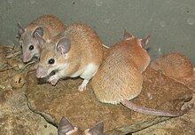
Spiny mice (Acomys cahirinus pictured here) can regenerate skin, cartilage, nerves and muscle.

Mammals are capable of cellular and physiological regeneration, but have generally poor reparative regenerative ability across the group. Examples of physiological regeneration in mammals include epithelial renewal (e.g., skin and intestinal tract), red blood cell replacement, antler regeneration and hair cycling. Male deer lose their antlers annually during the months of January to April then through regeneration are able to regrow them as an example of physiological regeneration. A deer antler is the only appendage of a mammal that can be regrown every year. While reparative regeneration is a rare phenomenon in mammals, it does occur. A well-documented example is regeneration of the digit tip distal to the nail bed. Reparative regeneration has also been observed in rabbits, pikas and African spiny mice. In 2012, researchers discovered that two species of African Spiny Mice, Acomys kempi and Acomys percivali, were capable of completely regenerating the autotomically released or otherwise damaged tissue. These species can regrow hair follicles, skin, sweat glands, fur and cartilage. In addition to these two species, subsequent studies demonstrated that Acomys cahirinus could regenerate skin and excised tissue in the ear pinna.

Despite these examples, it is generally accepted that adult mammals have limited regenerative capacity compared to most vertebrate embryos/larvae, adult salamanders and fish. But the regeneration therapy approach of Robert O. Becker, using electrical stimulation, has shown promising results for rats and mammals in general.

Some researchers have also claimed that the MRL mouse strain exhibits enhanced regenerative abilities. Work comparing the differential gene expression of scarless healing MRL mice and a poorly-healing C57BL/6 mouse strain, identified 36 genes differentiating the healing process between MRL mice and other mice. Study of the regenerative process in these animals is aimed at discovering how to duplicate them in humans, such as deactivation of the p21 gene. However, recent work has shown that MRL mice actually close small ear holes with scar tissue, rather than regeneration as originally claimed.

MRL mice are not protected against myocardial infarction; heart regeneration in adult mammals (neocardiogenesis) is limited, because heart muscle cells are nearly all terminally differentiated. MRL mice show the same amount of cardiac injury and scar formation as normal mice after a heart attack. However, recent studies provide evidence that this may not always be the case, and that MRL mice can regenerate after heart damage.

Humans

The regrowth of lost tissues or organs in the human body is being researched. Some tissues such as skin regrow quite readily; others have been thought to have little or no capacity for regeneration, but ongoing research suggests that there is some hope for a variety of tissues and organs. Human organs that have been regenerated include the bladder, vagina and the penis.

As are all metazoans, humans are capable of physiological regeneration (i.e. the replacement of cells during homeostatic maintenance that does not necessitate injury). For example, the regeneration of red blood cells via erythropoiesis occurs through the maturation of erythrocytes from hematopoietic stem cells in the bone marrow, their subsequent circulation for around 90 days in the blood stream, and their eventual cell-death in the spleen. Another example of physiological regeneration is the sloughing and rebuilding of a functional endometrium during each menstrual cycle in females in response to varying levels of circulating estrogen and progesterone.

However, humans are limited in their capacity for reparative regeneration, which occurs in response to injury. One of the most studied regenerative responses in humans is the hypertrophy of the liver following liver injury. For example, the original mass of the liver is re-established in direct proportion to the amount of liver removed following partial hepatectomy, which indicates that signals from the body regulate liver mass precisely, both positively and negatively, until the desired mass is reached. This response is considered cellular regeneration (a form of compensatory hypertrophy) where the function and mass of the liver is regenerated through the proliferation of existing mature hepatic cells (mainly hepatocytes), but the exact morphology of the liver is not regained. This process is driven by growth factor and cytokine regulated pathways. The normal sequence of inflammation and regeneration does not function accurately in cancer. Specifically, cytokine stimulation of cells leads to expression of genes that change cellular functions and suppress the immune response.

Adult neurogenesis is also a form of cellular regeneration. For example, hippocampal neuron renewal occurs in normal adult humans at an annual turnover rate of 1.75% of neurons. Cardiac myocyte renewal has been found to occur in normal adult humans, and at a higher rate in adults following acute heart injury such as infarction. Even in adult myocardium following infarction, proliferation is only found in around 1% of myocytes around the area of injury, which is not enough to restore function of cardiac muscle. However, this may be an important target for regenerative medicine as it implies that regeneration of cardiomyocytes, and consequently of myocardium, can be induced.

Another example of reparative regeneration in humans is fingertip regeneration, which occurs after phalange amputation distal to the nail bed (especially in children) and rib regeneration, which occurs following osteotomy for scoliosis treatment (though usually regeneration is only partial and may take up to one year).

Yet another example of regeneration in humans is vas deferens regeneration, which occurs after a vasectomy and which results in vasectomy failure.

Reptiles

The ability and degree of regeneration in reptiles differs among the various species, but the most notable and well-studied occurrence is tail-regeneration in lizards. In addition to lizards, regeneration has been observed in the tails and maxillary bone of crocodiles and adult neurogenesis has also been noted. Tail regeneration has never been observed in snakes. Lizards possess the highest regenerative capacity as a group. Following autotomous tail loss, epimorphic regeneration of a new tail proceeds through a blastema-mediated process that results in a functionally and morphologically similar structure.

Chondrichthyes

It has been estimated that the average shark loses about 30,000 to 40,000 teeth in a lifetime. Leopard sharks routinely replace their teeth every 9–12 days and this is an example of physiological regeneration. This can occur because shark teeth are not attached to a bone, but instead are developed within a bony cavity.

Rhodopsin regeneration has been studied in skates and rays. After complete photo-bleaching, rhodopsin can completely regenerate within 2 hours in the retina.

White bamboo sharks can regenerate at least two-thirds of their liver and this has been linked to three micro RNAs, xtr-miR-125b, fru-miR-204, and has-miR-142-3p_R-. In one study, two-thirds of the liver was removed and within 24 hours more than half of the liver had undergone hypertrophy.

Some sharks can regenerate scales and even skin following damage. Within two weeks of skin wounding, mucus is secreted into the wound and this initiates the healing process. One study showed that the majority of the wounded area was regenerated within 4 months, but the regenerated area also showed a high degree of variability.

Inequality (mathematics)

From Wikipedia, the free encyclopedia https://en.wikipedia.org/wiki/Inequality...