Search This Blog

Tuesday, December 3, 2019

Metalloprotein

From Wikipedia, the free encyclopedia
 
The structure of hemoglobin. The heme cofactor, containing the metal iron, shown in green.
 
Metalloprotein is a generic term for a protein that contains a metal ion cofactor. A large proportion of all proteins are part of this category. For instance, at least 1000 human proteins (out of ~20,000) contain zinc-binding protein domains although there may be up to 3000 human zinc metalloproteins.

Abundance

It is estimated that approximately half of all proteins contain a metal. In another estimate, about one quarter to one third of all proteins are proposed to require metals to carry out their functions. Thus, metalloproteins have many different functions in cells, such as storage and transport of proteins, enzymes and signal transduction proteins, or infectious diseases. The abundance of metal binding proteins may be inherent to the amino acids that proteins use, as even artificial proteins without evolutionary history will readily bind metals.

Most metals in the human body are bound to proteins. For instance, the relatively high concentration of iron in the human body is mostly due to the iron in hemoglobin.

Metal concentrations in humans organs (ppm = ug/g ash)

Liver Kidney Lung Heart Brain Muscle
Mn (manganese) 138 79 29 27 22 <4-40 font="">
Fe (iron) 16,769 7,168 24,967 5530 4100 3,500
Co (cobalt) <2-13 font=""> <2 font=""> <2-8 font=""> --- <2 font=""> 150 (?)
Ni (nickel) <5 font=""> <5-12 font=""> <5 font=""> <5 font=""> <5 font=""> <15 font="">
Cu (copper) 882 379 220 350 401 85-305
Zn (zinc) 5,543 5,018 1,470 2,772 915 4,688

Coordination chemistry principles

In metalloproteins, metal ions are usually coordinated by nitrogen, oxygen or sulfur centers belonging to amino acid residues of the protein. These donor groups are often provided by side-chains on the amino acid residues. Especially important are the imidazole substituent in histidine residues, thiolate substituents in cysteine residues, and carboxylate groups provided by aspartate. Given the diversity of the metalloproteome, virtually all amino acid residues have been shown to bind metal centers. The peptide backbone also provides donor groups; these include deprotonated amides and the amide carbonyl oxygen centers. Lead(II) binding in natural and artificial proteins has been reviewed.

In addition to donor groups that are provided by amino acid residues, many organic cofactors function as ligands. Perhaps most famous are the tetradentate N4 macrocyclic ligands incorporated into the heme protein. Inorganic ligands such as sulfide and oxide are also common.

Storage and transport metalloproteins

These are the second stage product of protein hydrolysis obtained by treatment with slightly stronger acids and alkalies.

Oxygen carriers

Hemoglobin, which is the principal oxygen-carrier in humans, has four subunits in which the iron(II) ion is coordinated by the planar macrocyclic ligand protoporphyrin IX (PIX) and the imidazole nitrogen atom of a histidine residue. The sixth coordination site contains a water molecule or a dioxygen molecule. By contrast the protein myoglobin, found in muscle cells, has only one such unit. The active site is located in a hydrophobic pocket. This is important as without it the iron(II) would be irreversibly oxidized to iron(III). The equilibrium constant for the formation of HbO2 is such that oxygen is taken up or released depending on the partial pressure of oxygen in the lungs or in muscle. In hemoglobin the four subunits show a cooperativity effect that allows for easy oxygen transfer from hemoglobin to myoglobin.

In both hemoglobin and myoglobin it is sometimes incorrectly stated that the oxygenated species contains iron(III). It is now known that the diamagnetic nature of these species is because the iron(II) atom is in the low-spin state. In oxyhemoglobin the iron atom is located in the plane of the porphyrin ring, but in the paramagnetic deoxyhemoglobin the iron atom lies above the plane of the ring. This change in spin state is a cooperative effect due to the higher crystal field splitting and smaller ionic radius of Fe2+ in the oxyhemoglobin moiety.

Hemerythrin is another iron-containing oxygen carrier. The oxygen binding site is a binuclear iron center. The iron atoms are coordinated to the protein through the carboxylate side chains of a glutamate and aspartate and five histidine residues. The uptake of O2 by hemerythrin is accompanied by two-electron oxidation of the reduced binuclear center to produce bound peroxide (OOH). The mechanism of oxygen uptake and release have been worked out in detail.

Hemocyanins carry oxygen in the blood of most mollusks, and some arthropods such as the horseshoe crab. They are second only to hemoglobin in biological popularity of use in oxygen transport. On oxygenation the two copper(I) atoms at the active site are oxidized to copper(II) and the dioxygen molecules are reduced to peroxide, O2−
2
.

Chlorocruorin (as the larger carrier erythrocruorin) is an oxygen-binding hemeprotein present in the blood plasma of many annelids, particularly certain marine polychaetes.

Cytochromes

Oxidation and reduction reactions are not common in organic chemistry as few organic molecules can act as oxidizing or reducing agents. Iron(II), on the other hand, can easily be oxidized to iron(III). This functionality is used in cytochromes, which function as electron-transfer vectors. The presence of the metal ion allows metalloenzymes to perform functions such as redox reactions that cannot easily be performed by the limited set of functional groups found in amino acids. The iron atom in most cytochromes is contained in a heme group. The differences between those cytochromes lies in the different side-chains. For instance cytochrome a has a heme a prosthetic group and cytochrome b has a heme b prosthetic group. These differences result in different Fe2+/Fe3+ redox potentials such that various cytochromes are involved in the mitochondrial electron transport chain.

Cytochrome P450 enzymes perform the function of inserting an oxygen atom into a C−H bond, an oxidation reaction.

Rubredoxin active site.

Rubredoxin

Rubredoxin is an electron-carrier found in sulfur-metabolizing bacteria and archaea. The active site contains an iron ion coordinated by the sulfur atoms of four cysteine residues forming an almost regular tetrahedron. Rubredoxins perform one-electron transfer processes. The oxidation state of the iron atom changes between the +2 and +3 states. In both oxidation states the metal is high spin, which helps to minimize structural changes.

Plastocyanin

The copper site in plastocyanin
 
Plastocyanin is one of the family of blue copper proteins that are involved in electron transfer reactions. The copper-binding site is described as distorted trigonal pyramidal. The trigonal plane of the pyramidal base is composed of two nitrogen atoms (N1 and N2) from separate histidines and a sulfur (S1) from a cysteine. Sulfur (S2) from an axial methionine forms the apex. The distortion occurs in the bond lengths between the copper and sulfur ligands. The Cu−S1 contact is shorter (207 pm) than Cu−S2 (282 pm). The elongated Cu−S2 bonding destabilizes the Cu(II) form and increases the redox potential of the protein. The blue color (597 nm peak absorption) is due to the Cu−S1 bond where S(pπ) to Cu(dx2y2) charge transfer occurs.

In the reduced form of plastocyanin, His-87 will become protonated with a pKa of 4.4. Protonation prevents it acting as a ligand and the copper site geometry becomes trigonal planar.

Metal-ion storage and transfer

Iron

Iron is stored as iron(III) in ferritin. The exact nature of the binding site has not yet been determined. The iron appears to be present as a hydrolysis product such as FeO(OH). Iron is transported by transferrin whose binding site consists of two tyrosines, one aspartic acid and one histidine. The human body has no mechanism for iron excretion. This can lead to iron overload problems in patients treated with blood transfusions, as, for instance, with β-thalassemia. Iron is actually excreted in urine and is also concentrated in bile which is excreted in feces.

Copper

Ceruloplasmin is the major copper-carrying protein in the blood. Ceruloplasmin exhibits oxidase activity, which is associated with possible oxidation of Fe(II) into Fe(III), therefore assisting in its transport in the blood plasma in association with transferrin, which can carry iron only in the Fe(III) state.

Calcium

Osteopontin is involved in mineralization in the extracellular matrices of bones and teeth.

Metalloenzymes

Metalloenzymes all have one feature in common, namely that the metal ion is bound to the protein with one labile coordination site. As with all enzymes, the shape of the active site is crucial. The metal ion is usually located in a pocket whose shape fits the substrate. The metal ion catalyzes reactions that are difficult to achieve in organic chemistry.

Carbonic anhydrase

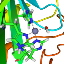
Active site of carbonic anhydrase. The three coordinating histidine residues are shown in green, hydroxide in red and white, and the zinc in gray.
 
CO2 + H2O ⇌ H2CO3
This reaction is very slow in the absence of a catalyst, but quite fast in the presence of the hydroxide ion
CO2 + OHHCO
3
A reaction similar to this is almost instantaneous with carbonic anhydrase. The structure of the active site in carbonic anhydrases is well known from a number of crystal structures. It consists of a zinc ion coordinated by three imidazole nitrogen atoms from three histidine units. The fourth coordination site is occupied by a water molecule. The coordination sphere of the zinc ion is approximately tetrahedral. The positively-charged zinc ion polarizes the coordinated water molecule, and nucleophilic attack by the negatively-charged hydroxide portion on carbon dioxide (carbonic anhydride) proceeds rapidly. The catalytic cycle produces the bicarbonate ion and the hydrogen ion as the equilibrium
H2CO3HCO
3
+ H+
favours dissociation of carbonic acid at biological pH values.

Vitamin B12-dependent enzymes

The cobalt-containing Vitamin B12 (also known as cobalamin) catalyzes the transfer of methyl (−CH3) groups between two molecules, which involves the breaking of C−C bonds, a process that is energetically expensive in organic reactions. The metal ion lowers the activation energy for the process by forming a transient Co−CH3 bond. The structure of the coenzyme was famously determined by Dorothy Hodgkin and co-workers, for which she received a Nobel Prize in Chemistry. It consists of a cobalt(II) ion coordinated to four nitrogen atoms of a corrin ring and a fifth nitrogen atom from an imidazole group. In the resting state there is a Co−C sigma bond with the 5′ carbon atom of adenosine. This is a naturally occurring organometallic compound, which explains its function in trans-methylation reactions, such as the reaction carried out by methionine synthase.

Nitrogenase (nitrogen fixation)

The fixation of atmospheric nitrogen is a very energy-intensive process, as it involves breaking the very stable triple bond between the nitrogen atoms. The enzyme nitrogenase is one of the few enzymes that can catalyze the process. The enzyme occurs in Rhizobium bacteria. There are three components to its action: a molybdenum atom at the active site, iron–sulfur clusters that are involved in transporting the electrons needed to reduce the nitrogen, and an abundant energy source in the form of magnesium ATP. This last is provided by a symbiotic relationship between the bacteria and a host plant, often a legume. The relationship is symbiotic because the plant supplies the energy by photosynthesis and benefits by obtaining the fixed nitrogen. The reaction may be written symbolically as
N2 + 16 MgATP + 8 e → 2 NH3 + 16 MgADP +16 Pi + H2
where Pi stands for inorganic phosphate. The precise structure of the active site has been difficult to determine. It appears to contain a MoFe7S8 cluster that is able to bind the dinitrogen molecule and, presumably, enable the reduction process to begin. The electrons are transported by the associated "P" cluster, which contains two cubical Fe4S4 clusters joined by sulfur bridges.

Superoxide dismutase

Structure of a human superoxide dismutase 2 tetramer
 
The superoxide ion, O
2
is generated in biological systems by reduction of molecular oxygen. It has an unpaired electron, so it behaves as a free radical. It is a powerful oxidizing agent. These properties render the superoxide ion very toxic and are deployed to advantage by phagocytes to kill invading microorganisms. Otherwise, the superoxide ion must be destroyed before it does unwanted damage in a cell. The superoxide dismutase enzymes perform this function very efficiently.

The formal oxidation state of the oxygen atoms is −​12. In solutions at neutral pH, the superoxide ion disproportionates to molecular oxygen and hydrogen peroxide.
O
2
+ 2 H+ → O2 + H2O2
In biology this type of reaction is called a dismutation reaction. It involves both oxidation and reduction of superoxide ions. The superoxide dismutase (SOD) group of enzymes increase the rate of reaction to near the diffusion-limited rate. The key to the action of these enzymes is a metal ion with variable oxidation state that can act either as an oxidizing agent or as a reducing agent.
Oxidation: M(n+1)+ + O
2
→ Mn+ + O2
Reduction: Mn+ + O
2
+ 2 H+ → M(n+1)+ + H2O2.
In human SOD the active metal is copper, as Cu(II) or Cu(I), coordinated tetrahedrally by four histidine residues. This enzyme also contains zinc ions for stabilization and is activated by copper chaperone for superoxide dismutase (CCS). Other isozymes may contain iron, manganese or nickel. Ni-SOD is particularly interesting as it involves nickel(III), an unusual oxidation state for this element. The active site nickel geometry cycles from square planar Ni(II), with thiolate (Cys2 and Cys6) and backbone nitrogen (His1 and Cys2) ligands, to square pyramidal Ni(III) with an added axial His1 side chain ligand.

Chlorophyll-containing proteins

Hemoglobin (left) and chlorophyll (right), two extremely different molecules when it comes to function, are quite similar when it comes to its atomic shape. There are only three major structural differences; a magnesium atom (Mg) in chlorophyll, as opposed to iron (Fe) in hemoglobin. Additionally, chlorophyll has an extended isoprenoid tail and an additional aliphatic cyclic structure off the macrocycle.
 
Chlorophyll plays a crucial role in photosynthesis. It contains a magnesium enclosed in a chlorin ring. However, the magnesium ion is not directly involved in the photosynthetic function and can be replaced by other divalent ions with little loss of activity. Rather, the photon is absorbed by the chlorin ring, whose electronic structure is well-adapted for this purpose. 

Initially, the absorption of a photon causes an electron to be excited into a singlet state of the Q band. The excited state undergoes an intersystem crossing from the singlet state to a triplet state in which there are two electrons with parallel spin. This species is, in effect, a free radical, and is very reactive and allows an electron to be transferred to acceptors that are adjacent to the chlorophyll in the chloroplast. In the process chlorophyll is oxidized. Later in the photosynthetic cycle, chlorophyll is reduced back again. This reduction ultimately draws electrons from water, yielding molecular oxygen as a final oxidation product.

Hydrogenase

Hydrogenases are subclassified into three different types based on the active site metal content: iron–iron hydrogenase, nickel–iron hydrogenase, and iron hydrogenase. All hydrogenases catalyze reversible H2 uptake, but while the [FeFe] and [NiFe] hydrogenases are true redox catalysts, driving H2 oxidation and H+ reduction
H2 ⇌ 2 H+ + 2 e
the [Fe] hydrogenases catalyze the reversible heterolytic cleavage of H2.
H2 ⇌ H+ + H
The active site structures of the three types of hydrogenase enzymes.

Ribozyme and deoxyribozyme

Since discovery of ribozymes by Thomas Cech and Sidney Altman in the early 1980s, ribozymes have been shown to be a distinct class of metalloenzymes. Many ribozymes require metal ions in their active sites for chemical catalysis; hence they are called metalloenzymes. Additionally, metal ions are essential for structural stabilization of ribozymes. Group I intron is the most studied ribozyme which has three metals participating in catalysis. Other known ribozymes include group II intron, RNase P, and several small viral ribozymes (such as hammerhead, hairpin, HDV, and VS) and the large subunit of ribosomes. Recently, four new classes of ribozymes have been discovered (named twister, twister sister, pistol and hatchet) which are all self-cleaving ribozymes.

Deoxyribozymes, also called DNAzymes or catalytic DNA, are artificial catalytic DNA molecules that were first produced in 1994 and gained a rapid increase of interest since then. Almost all DNAzymes require metal ions in order to function; thus they are classified as metalloenzymes. Although ribozymes mostly catalyze cleavage of RNA substrates, a variety of reactions can be catalyzed by DNAzymes including RNA/DNA cleavage, RNA/DNA ligation, amino acid phosphorylation and dephosphorylation, and carbon–carbon bond formation. Yet, DNAzymes that catalyze RNA cleavage reaction are the most extensively explored ones. 10-23 DNAzyme, discovered in 1997, is one of the most studied catalytic DNAs with clinical applications as a therapeutic agent. Several metal-specific DNAzymes have been reported including the GR-5 DNAzyme (lead-specific), the CA1-3 DNAzymes (copper-specific), the 39E DNAzyme (uranyl-specific) and the NaA43 DNAzyme (sodium-specific).

Signal-transduction metalloproteins

Calmodulin

EF-hand motif
 
Calmodulin is an example of a signal-transduction protein. It is a small protein that contains four EF-hand motifs, each of which is able to bind a Ca2+ ion. 

In an EF-hand loop the calcium ion is coordinated in a pentagonal bipyramidal configuration. Six glutamic acid and aspartic acid residues involved in the binding are in positions 1, 3, 5, 7 and 9 of the polypeptide chain. At position 12, there is a glutamate or aspartate ligand that behaves as a (bidentate ligand), providing two oxygen atoms. The ninth residue in the loop is necessarily glycine due to the conformational requirements of the backbone. The coordination sphere of the calcium ion contains only carboxylate oxygen atoms and no nitrogen atoms. This is consistent with the hard nature of the calcium ion.

The protein has two approximately symmetrical domains, separated by a flexible "hinge" region. Binding of calcium causes a conformational change to occur in the protein. Calmodulin participates in an intracellular signaling system by acting as a diffusible second messenger to the initial stimuli.

Troponin

In both cardiac and skeletal muscles, muscular force production is controlled primarily by changes in the intracellular calcium concentration. In general, when calcium rises, the muscles contract and, when calcium falls, the muscles relax. Troponin, along with actin and tropomyosin, is the protein complex to which calcium binds to trigger the production of muscular force.

Transcription factors

Zinc finger. The zinc ion (green) is coordinated by two histidine residues and two cysteine residues.
 
Many transcription factors contain a structure known as a zinc finger, this is a structural module where a region of protein folds around a zinc ion. The zinc does not directly contact the DNA that these proteins bind to. Instead, the cofactor is essential for the stability of the tightly-folded protein chain. In these proteins, the zinc ion is usually coordinated by pairs of cysteine and histidine side-chains.

Other metalloenzymes

There are two types of carbon monoxide dehydrogenase: one contains iron and molybdenum, the other contains iron and nickel. Parallels and differences in catalytic strategies have been reviewed.

Pb2+ (lead) can replace Ca2+ (calcium) as, for example, with calmodulin or Zn2+ (zinc) as with metallocarboxypeptidases.
 
Some other metalloenzymes are given in the following table, according to the metal involved.
 
Ion Examples of enzymes containing this ion
Magnesium Glucose 6-phosphatase
Hexokinase
DNA polymerase
Vanadium vanabins
Manganese Arginase
Oxygen-evolving complex
Iron Catalase
Hydrogenase
IRE-BP
Aconitase
Cobalt Nitrile hydratase
Methionyl aminopeptidase
Methylmalonyl-CoA mutase
Isobutyryl-CoA mutase
Nickel Urease
Hydrogenase
Methyl-coenzyme M reductase (MCR)
Copper Cytochrome oxidase
Laccase
Nitrous-oxide reductase
Nitrite reductase
Zinc Alcohol dehydrogenase
Carboxypeptidase
Aminopeptidase
Beta amyloid
Cadmium Metallothionein
Thiolate proteins
Molybdenum Nitrate reductase
Sulfite oxidase
Xanthine oxidase
DMSO reductase
Tungsten Acetylene hydratase
various Metallothionein
Phosphatase

Gel electrophoresis of proteins

From Wikipedia, the free encyclopedia
 
Proteins separated by SDS-PAGE, Coomassie Brilliant Blue staining
 
Protein electrophoresis is a method for analysing the proteins in a fluid or an extract. The electrophoresis may be performed with a small volume of sample in a number of alternative ways with or without a supporting medium: SDS polyacrylamide gel electrophoresis (in short: gel electrophoresis, PAGE, or SDS-electrophoresis), free-flow electrophoresis, electrofocusing, isotachophoresis, affinity electrophoresis, immunoelectrophoresis, counterelectrophoresis, and capillary electrophoresis. Each method has many variations with individual advantages and limitations. Gel electrophoresis is often performed in combination with electroblotting immunoblotting to give additional information about a specific protein. Because of practical limitations, protein electrophoresis is generally not suited as a preparative method.

Denaturing gel methods

SDS-PAGE

SDS-PAGE, sodium dodecyl sulfate polyacrylamide gel electrophoresis, describes a collection of related techniques to separate proteins according to their electrophoretic mobility (a function of the molecular weight of a polypeptide chain) while in the denatured (unfolded) state. In most proteins, the binding of SDS to the polypeptide chain imparts an even distribution of charge per unit mass, thereby resulting in a fractionation by approximate size during electrophoresis. 

SDS is a strong detergent agent used to denature native proteins to unfolded, individual polypeptides. When a protein mixture is heated to 100 °C in presence of SDS, the detergent wraps around the polypeptide backbone. In this process, the intrinsic charges of polypeptides becomes negligible when compared to the negative charges contributed by SDS. Thus polypeptides after treatment become rod-like structures possessing a uniform charge density, that is same net negative charge per unit length. The electrophoretic mobilities of these proteins will be a linear function of the logarithms of their molecular weights.

Native gel methods

Native gels, also known as non-denaturing gels, analyze proteins that are still in their folded state. Thus, the electrophoretic mobility depends not only on the charge-to-mass ratio, but also on the physical shape and size of the protein.

Blue native PAGE

BN-PAGE is a native PAGE technique, where the Coomassie Brilliant Blue dye provides the necessary charges to the protein complexes for the electrophoretic separation. The disadvantage of Coomassie is that in binding to proteins it can act like a detergent causing complexes to dissociate. Another drawback is the potential quenching of chemoluminescence (e.g. in subsequent western blot detection or activity assays) or fluorescence of proteins with prosthetic groups (e.g. heme or chlorophyll) or labelled with fluorescent dyes.

Clear native PAGE

CN-PAGE (commonly referred to as Native PAGE) separates acidic water-soluble and membrane proteins in a polyacrylamide gradient gel. It uses no charged dye so the electrophoretic mobility of proteins in CN-PAGE (in contrast to the charge shift technique BN-PAGE) is related to the intrinsic charge of the proteins. The migration distance depends on the protein charge, its size and the pore size of the gel. In many cases this method has lower resolution than BN-PAGE, but CN-PAGE offers advantages whenever Coomassie dye would interfere with further analytical techniques, for example it has been described as a very efficient microscale separation technique for FRET analyses. Also CN-PAGE is milder than BN-PAGE so it can retain labile supramolecular assemblies of membrane protein complexes that are dissociated under the conditions of BN-PAGE.

Quantitative native PAGE

The folded protein complexes of interest separate cleanly and predictably due to the specific properties of the polyacrylamide gel. The separated proteins are continuously eluted into a physiological eluent and transported to a fraction collector. In four to five PAGE fractions each the metal cofactors can be identified and absolutely quantified by high-resolution ICP-MS. The respective structures of the isolated metalloproteins can be determined by solution NMR spectroscopy.

Buffer systems

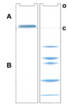
Postulated migration of proteins in a Laemmli gel system A: Stacking gel, B: Resolving gel, o: sample application c: discontinuities in the buffer and electrophoretic matrix
 
Most protein separations are performed using a "discontinuous" (or DISC) buffer system that significantly enhances the sharpness of the bands within the gel. During electrophoresis in a discontinuous gel system, an ion gradient is formed in the early stage of electrophoresis that causes all of the proteins to focus into a single sharp band. The formation of the ion gradient is achieved by choosing a pH value at which the ions of the buffer are only moderately charged compared to the SDS-coated proteins. These conditions provide an environment in which Kohlrausch's reactions determine the molar conductivity. As a result, SDS-coated proteins are concentrated to several fold in a thin zone of the order of 19 μm within a few minutes. At this stage all proteins migrate at the same migration speed by isotachophoresis. This occurs in a region of the gel that has larger pores so that the gel matrix does not retard the migration during the focusing or "stacking" event. Separation of the proteins by size is achieved in the lower, "resolving" region of the gel. The resolving gel typically has a much smaller pore size, which leads to a sieving effect that now determines the electrophoretic mobility of the proteins. At the same time, the separating part of the gel also has a pH value in which the buffer ions on average carry a greater charge, causing them to "outrun" the SDS-covered proteins and eliminate the ion gradient and thereby the stacking effect.

A very widespread discontinuous buffer system is the tris-glycine or "Laemmli" system that stacks at a pH of 6.8 and resolves at a pH of ~8.3-9.0. A drawback of this system is that these pH values may promote disulfide bond formation between cysteine residues in the proteins because the pKa of cysteine ranges from 8-9 and because reducing agent present in the loading buffer doesn't co-migrate with the proteins. Recent advances in buffering technology alleviate this problem by resolving the proteins at a pH well below the pKa of cysteine (e.g., bis-tris, pH 6.5) and include reducing agents (e.g. sodium bisulfite) that move into the gel ahead of the proteins to maintain a reducing environment. An additional benefit of using buffers with lower pH values is that the acrylamide gel is more stable at lower pH values, so the gels can be stored for long periods of time before use.

SDS gradient gel electrophoresis of proteins

As voltage is applied, the anions (and negatively charged sample molecules) migrate toward the positive electrode (anode) in the lower chamber, the leading ion is Cl ( high mobility and high concentration); glycinate is the trailing ion (low mobility and low concentration). SDS-protein particles do not migrate freely at the border between the Cl of the gel buffer and the Gly of the cathode buffer. Friedrich Kohlrausch found that Ohm's law also applies to dissolved electrolytes. Because of the voltage drop between the Cl and Glycine-buffers, proteins are compressed (stacked) into micrometer thin layers. The boundary moves through a pore gradient and the protein stack gradually disperses due to a frictional resistance increase of the gel matrix. Stacking and unstacking occurs continuously in the gradient gel, for every protein at a different position. For a complete protein unstacking the polyacrylamide-gel concentration must exceed 16% T. The two-gel system of "Laemmli" is a simple gradient gel. The pH discontinuity of the buffers is of no significance for the separation quality, and a "stacking-gel" with a different pH is not needed.

Visualization

The most popular protein stain is Coomassie Brilliant Blue. It is an anionic dye, which non-specifically binds to proteins. Proteins in the gel are fixed by acetic acid and simultaneously stained. The excess dye incorporated into the gel can be removed by destaining with the same solution without the dye. The proteins are detected as blue bands on a clear background.

When more sensitive method than staining by Coomassie is needed silver staining is usually used. Silver staining is a sensitive procedure to detect trace amounts of proteins in gels, but can also visualize nucleic acid or polysaccharides.

Visualization methods without using a dye such as Coomassie and silver are available on the market. For example Bio-Rad Laboratories markets ”stain-free” gels for SDS-PAGE gel electrophoresis.

Similarly as in nucleic acid gel electrophoresis, tracking dye is often used. Anionic dyes of a known electrophoretic mobility are usually included in the sample buffer. A very common tracking dye is Bromophenol blue. This dye is coloured at alkali and neutral pH and is a small negatively charged molecule that moves towards the anode. Being a highly mobile molecule it moves ahead of most proteins.

Medical applications

Schematic representation of a protein electrophoresis gel.
 
Serum protein electrophoresis showing a paraprotein (peak in the gamma zone) in a patient with multiple myeloma.
 
In medicine, protein electrophoresis is a method of analysing the proteins mainly in blood serum. Before the widespread use of gel electrophoresis, protein electrophoresis was performed as free-flow electrophoresis (on paper) or as immunoelectrophoresis. 

Traditionally, two classes of blood proteins are considered: serum albumin and globulin. They are generally equal in proportion, but albumin as a molecule is much smaller and lightly, negatively-charged, leading to an accumulation of albumin on the electrophoretic gel. A small band before albumin represents transthyretin (also named prealbumin). Some forms of medication or body chemicals can cause their own band, but it usually is small. Abnormal bands (spikes) are seen in monoclonal gammopathy of undetermined significance and multiple myeloma, and are useful in the diagnosis of these conditions. 

The globulins are classified by their banding pattern (with their main representatives):
Normal present medical procedure involves determination of numerous proteins in plasma including hormones and enzymes, some of them also determined by electrophoresis. However, gel electrophoresis is mainly a research tool, also when the subject is blood proteins.

What are common treatments for phenylketonuria (PKU)?

 https://www.nichd.nih.gov/health/topics/pku/conditioninfo/treatments

There is no cure for PKU, but treatment can prevent intellectual disabilities and other health problems.1 A person with PKU should receive treatment at a medical center that specializes in the disorder. (Visit the Resources and Publications section for ways to locate a center.)

The PKU Diet

People with PKU need to follow a diet that limits foods with phenylalanine. The diet should be followed carefully and be started as soon after birth as possible. In the past, experts believed that it was safe for people to stop following the diet as they got older. However, they now recommend that people with PKU stay on the diet throughout their lives for better physical and mental health.

It is especially important for a pregnant woman with PKU to strictly follow the low-phenylalanine diet throughout her pregnancy to ensure the healthy development of her infant.

People with PKU need to avoid various high-protein foods, including:
  • Milk and cheese
  • Eggs
  • Nuts
  • Soybeans
  • Beans
  • Chicken, beef, or pork
  • Fish
  • Peas
  • Beer
People with PKU also need to avoid the sweetener aspartame, which is in some foods, drinks, medications, and vitamins. Aspartame releases phenylalanine when it is digested, so it raises the level of phenylalanine in a person's blood.

Often, people with PKU also have to limit their intake of lower-protein foods, such as certain fruits and vegetables. However, a PKU diet can include low-protein noodles and other special products.

The amount of phenylalanine that is safe to consume differs for each person. Therefore, a person with PKU needs to work with a health care professional to develop an individualized diet. The goal is to eat only the amount of phenylalanine necessary for healthy growth and body processes but not any extra. Frequent blood tests and doctor visits are necessary to help determine how well the diet is working. Some relaxation of the diet may be possible as a child gets older, but the recommendation today is lifelong adherence to the diet. Following the diet is especially important during pregnancy.
However, the PKU diet can be very challenging. Getting support from friends and family or a support group can help. Sticking with the diet ensures better functioning and improved overall health.

A PKU Formula

People who follow the PKU diet will not get enough essential nutrients from food. Therefore, they must drink a special formula.

A newborn who is diagnosed with PKU should receive special infant formula. The formula may be mixed with a small amount of breast milk or regular infant formula to make sure the child gets enough phenylalanine for normal development but not enough to cause harm.

Older children and adults receive a different formula to meet their nutritional needs. This formula should be consumed every day throughout a person's life.

In addition to the formula, health care professionals may recommend other supplements. For example, fish oil may be recommended to help with fine motor coordination and other aspects of development.1

Medication for PKU

The U.S. Food and Drug Administration (FDA) has approved the drug sapropterin dihydrochloride (Kuvan®) for the treatment of PKU. Kuvan® is a form of BH4, which is a substance in the body that helps break down phenylalanine. However, having too little BH4 is only one reason a person may not break down phenylalanine. Therefore, Kuvan® only helps some people reduce the phenylalanine in their blood. Even if the medication helps, it will not decrease the phenylalanine to the desired amount and must be used together with the PKU diet.

When the FDA approved Kuvan®, the agency suggested that research on the medication continue to determine its long-term safety and effectiveness.

Other Treatments for PKU

NICHD-supported researchers and other scientists are exploring additional treatments for PKU. These treatments include large neutral amino acid supplementation, which may help prevent phenylalanine from entering the brain, and enzyme replacement therapy, which uses a substance similar to the enzyme that usually breaks down phenylalanine. Researchers are also investigating the possibility of using gene therapy, which involves injecting new genes to break down phenylalanine. That would result in the breakdown of phenylalanine and decreased blood phenylalanine levels.

Phenylketonuria

From Wikipedia, the free encyclopedia
https://en.wikipedia.org/wiki/Phenylketonuria
 
Phenylketonuria
Other namesPhenylalanine hydroxylase deficiency, PAH deficiency, Følling disease
L-Phenylalanin - L-Phenylalanine.svg
Phenylalanine
SpecialtyMedical genetics, pediatrics
SymptomsWithout treatment intellectual disability, seizures, behavioral problems, mental disorders, musty smell
Usual onsetAt birth
TypesClassic, variant
CausesGenetic (autosomal recessive)
Diagnostic methodNewborn screening programs in many countries
TreatmentDiet low in foods that contain phenylalanine, special supplements
MedicationSapropterin dihydrochloride, pegvaliase
PrognosisNormal health with treatment
Frequency~1 in 12,000 newborns

Phenylketonuria (PKU) is an inborn error of metabolism that results in decreased metabolism of the amino acid phenylalanine. Untreated, PKU can lead to intellectual disability, seizures, behavioral problems, and mental disorders. It may also result in a musty smell and lighter skin. A baby born to a mother who has poorly treated PKU may have heart problems, a small head, and low birth weight.

Phenylketonuria is a genetic disorder inherited from a person's parents. It is due to mutations in the PAH gene, which results in low levels of the enzyme phenylalanine hydroxylase. This results in the buildup of dietary phenylalanine to potentially toxic levels. It is autosomal recessive, meaning that both copies of the gene must be mutated for the condition to develop. There are two main types, classic PKU and variant PKU, depending on whether any enzyme function remains. Those with one copy of a mutated gene typically do not have symptoms. Many countries have newborn screening programs for the disease.

Treatment is with a diet low in foods that contain phenylalanine and special supplements. Babies should use a special formula with a small amount of breast milk. The diet should begin as soon as possible after birth and be continued for at least 10 years, if not lifelong. People who are diagnosed early and maintain a strict diet can have normal health and a normal life span. Effectiveness is monitored through periodic blood tests. The medication sapropterin dihydrochloride may be useful in some.

Phenylketonuria affects about 1 in 12,000 babies. Males and females are affected equally. The disease was discovered in 1934 by Ivar Asbjørn Følling, with the importance of diet determined in 1953. Gene therapy, while promising, requires a great deal more study as of 2014.

Signs and symptoms

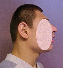
Abnormally small head (microcephaly)
 
Untreated PKU can lead to intellectual disability, seizures, behavioral problems, and mental disorders. It may also result in a musty smell and lighter skin. A baby born to a mother who has poorly treated PKU may have heart problems, a small head, and low birth weight.

Because the mother's body is able to break down phenylalanine during pregnancy, infants with PKU are normal at birth. The disease is not detectable by physical examination at that time, because no damage has yet been done. Newborn screening is performed to detect the disease and initiate treatment before any damage is done. The blood sample is usually taken by a heel prick, typically performed 2–7 days after birth. This test can reveal elevated phenylalanine levels after one or two days of normal infant feeding.

If a child is not diagnosed during the routine newborn screening test and a phenylalanine restricted diet is not introduced, then phenylalanine levels in the blood will increase over time. Toxic levels of phenylalanine (and insufficient levels of tyrosine) can interfere with infant development in ways which have permanent effects. The disease may present clinically with seizures, hypopigmentation (excessively fair hair and skin), and a "musty odor" to the baby's sweat and urine (due to phenylacetate, a carboxylic acid produced by the oxidation of phenylketone). In most cases, a repeat test should be done at approximately two weeks of age to verify the initial test and uncover any phenylketonuria that was initially missed.

Untreated children often fail to attain early developmental milestones, develop microcephaly, and demonstrate progressive impairment of cerebral function. Hyperactivity, EEG abnormalities, and seizures, and severe learning disabilities are major clinical problems later in life. A characteristic "musty or mousy" odor on the skin, as well as a predisposition for eczema, persist throughout life in the absence of treatment.

The damage done to the brain if PKU is untreated during the first months of life is not reversible. It is critical to control the diet of infants with PKU very carefully so that the brain has an opportunity to develop normally. Affected children who are detected at birth and treated are much less likely to develop neurological problems or have seizures and intellectual disability (though such clinical disorders are still possible.)

In general, however, outcomes for people treated for PKU are good. Treated people may have no detectable physical, neurological, or developmental problems at all.

Genetics

Phenylketonuria is inherited in an autosomal recessive fashion
 
PKU is an autosomal recessive metabolic genetic disorder. As an autosomal recessive disorder, two PKU alleles are required for an individual to experience symptoms of the disease. If both parents are carriers for PKU, there is a 25% chance any child they have will be born with the disorder, a 50% chance the child will be a carrier, and a 25% chance the child will neither develop nor be a carrier for the disease.

PKU is characterized by homozygous or compound heterozygous mutations in the gene for the hepatic enzyme phenylalanine hydroxylase (PAH), rendering it nonfunctional. This enzyme is necessary to metabolize the amino acid phenylalanine (Phe) to the amino acid tyrosine (Tyr). When PAH activity is reduced, phenylalanine accumulates and is converted into phenylpyruvate (also known as phenylketone), which can be detected in the urine.

Carriers of a single PKU allele do not exhibit symptoms of the disease but appear to be protected to some extent against the fungal toxin ochratoxin A. This accounts for the persistence of the allele in certain populations in that it confers a selective advantage—in other words, being a heterozygote is advantageous.

The PAH gene is located on chromosome 12 in the bands 12q22-q24.2. As of 2000, around 400 disease-causing mutations had been found in the PAH gene. This is an example of allelic genetic heterogeneity.

Pathophysiology

When Phe cannot be metabolized by the body, a typical diet that would be healthy for people without PKU causes abnormally high levels of Phe to accumulate in the blood, which is toxic to the brain. If left untreated, complications of PKU include severe intellectual disability, brain function abnormalities, microcephaly, mood disorders, irregular motor functioning, and behavioral problems such as attention deficit hyperactivity disorder, as well as physical symptoms such as a "musty" odor, eczema, and unusually light skin and hair coloration.

Classical PKU

Classical PKU, and its less severe forms "mild PKU" and "mild hyperphenylalaninemia" are caused by a mutated gene for the enzyme phenylalanine hydroxylase (PAH), which converts the amino acid phenylalanine ("Phe") to other essential compounds in the body, in particular tyrosine. Tyrosine is a conditionally essential amino acid for PKU patients because without PAH it cannot be produced in the body through the breakdown of phenylalanine. Tyrosine is necessary for the production of neurotransmitters like epinephrine, norepinephrine, and dopamine.

PAH deficiency causes a spectrum of disorders, including classic phenylketonuria (PKU) and mild hyperphenylalaninemia (also known as "hyperphe" or "mild HPA"), a less severe accumulation of phenylalanine. Patients with "hyperphe" may have more functional PAH enzyme and be able to tolerate larger amounts of phenylalanine in their diets than those with classic PKU, but unless dietary intake is at least somewhat restricted, their blood Phe levels are still higher than the levels in people with normal PAH activity.

Phenylalanine is a large, neutral amino acid (LNAA). LNAAs compete for transport across the blood–brain barrier (BBB) via the large neutral amino acid transporter (LNAAT). If phenylalanine is in excess in the blood, it will saturate the transporter. Excessive levels of phenylalanine tend to decrease the levels of other LNAAs in the brain. As these amino acids are necessary for protein and neurotransmitter synthesis, Phe buildup hinders the development of the brain, causing intellectual disability.

Recent research suggests that neurocognitive, psychosocial, quality of life, growth, nutrition, bone pathology are slightly suboptimal even for patients who are treated and maintain their Phe levels in the target range, if their diet is not supplemented with other amino acids.

Classic PKU dramatically affects myelination and white matter tracts in untreated infants; this may be one major cause of neurological disorders associated with phenylketonuria. Differences in white matter development are observable with magnetic resonance imaging. Abnormalities in gray matter can also be detected, particularly in the motor and pre-motor cortex, thalamus and the hippocampus.

It was recently suggested that PKU may resemble amyloid diseases, such as Alzheimer's disease and Parkinson's disease, due to the formation of toxic amyloid-like assemblies of phenylalanine.

Other non-PAH mutations can also cause PKU.

Tetrahydrobiopterin-deficient hyperphenylalaninemia

A rarer form of hyperphenylalaninemia is tetrahydrobiopterin deficiency, which occurs when the PAH enzyme is normal, and a defect is found in the biosynthesis or recycling of the cofactor tetrahydrobiopterin (BH4). BH4 is necessary for proper activity of the enzyme PAH, and this coenzyme can be supplemented as treatment. Those who suffer from this form of hyperphenylalaninemia may have a deficiency of tyrosine (which is created from phenylalanine by PAH), in which case treatment is supplementation of tyrosine to account for this deficiency. 

Levels of dopamine can be used to distinguish between these two types. Tetrahydrobiopterin is required to convert Phe to Tyr and is required to convert Tyr to L-DOPA via the enzyme tyrosine hydroxylase. L-DOPA, in turn, is converted to dopamine. Low levels of dopamine lead to high levels of prolactin. By contrast, in classical PKU (without dihydrobiopterin involvement), prolactin levels would be relatively normal.

Tetrahydrobiopterin deficiency can be caused by defects in four genes. They are known as HPABH4A, HPABH4B, HPABH4C, and HPABH4D.

Metabolic pathways

 
Pathophysiology of phenylketonuria, which is due to the absence of functional phenylalanine hydroxylase (classical subtype) or functional enzymes for the recycling of tetrahydrobiopterin (new variant subtype) utilized in the first step of the metabolic pathway.

The enzyme phenylalanine hydroxylase normally converts the amino acid phenylalanine into the amino acid tyrosine. If this reaction does not take place, phenylalanine accumulates and tyrosine is deficient. Excessive phenylalanine can be metabolized into phenylketones through the minor route, a transaminase pathway with glutamate. Metabolites include phenylacetate, phenylpyruvate and phenethylamine. Elevated levels of phenylalanine in the blood and detection of phenylketones in the urine is diagnostic, however most patients are diagnosed via newborn screening.

Screening

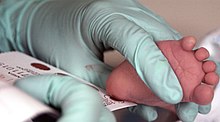
Blood is taken from a two-week-old baby to test for phenylketonuria
 
PKU is commonly included in the newborn screening panel of many countries, with varied detection techniques. Most babies in developed countries are screened for PKU soon after birth. Screening for PKU is done with bacterial inhibition assay (Guthrie test), immunoassays using fluorometric or photometric detection, or amino acid measurement using tandem mass spectrometry (MS/MS). Measurements done using MS/MS determine the concentration of Phe and the ratio of Phe to tyrosine, the ratio will be elevated in PKU.

Treatment

PKU is not curable. However, if PKU is diagnosed early enough, an affected newborn can grow up with normal brain development by managing and controlling phenylalanine ("Phe") levels through diet, or a combination of diet and medication.

Diet

People who follow the prescribed dietary treatment from birth may have no symptoms. Their PKU would be detectable only by a blood test. People must adhere to a special diet low in Phe for optimal brain development. Since Phe is necessary for the synthesis of many proteins, it is required for appropriate growth, but levels must be strictly controlled. 

For people who do not have phenylketonuria, the U.S. Institute of Medicine set recommended at least 33 mg/kg body weight/day phenylalanine plus tyrosine for adults 19 years and older. For people with PKU, a recommendation for children up to age 10 years is 200 to 500 mg/d; for older children and adults 220 to 1200 mg/day. Where in the range depends on body weight and age, and on monitoring blood concentration.

Optimal health ranges (or "target ranges") are between 120 and 360 µmol/L or equivalently 2 to 6 mg/dL, and aimed to be achieved during at least the first 10 years, to allow the brain to develop normally. 

The age at which people with PKU may safely go off diet is subject to some debate. The diet should be maintained at least until the age of eight or ten. Some evidence supports discontinued after 10 years as a normal diet after that does not appear to have negative effects. One study however has shown temporarily detrimental effects when off the diet. There is no evidence for permanent brain damage in people who have gone off diet in adulthood. In case of mild neurocognitive impairment, the re-introduction of diet is indicated. 

The diet requires restricting or eliminating foods high in Phe, such as soybeans, egg whites, shrimp, chicken breast, spirulina, watercress, fish, nuts, crayfish, lobster, tuna, turkey, legumes, and lowfat cottage cheese. Starchy foods, such as potatoes and corn are generally acceptable in controlled amounts, but the quantity of Phe consumed from these foods must be monitored. A food diary is usually kept to record the amount of Phe consumed with each meal, snack, or drink. An "exchange" system can be used to calculate the amount of Phe in a food from the protein content identified on a nutritional information label. Lower-protein "medical food" substitutes are often used in place of normal bread, pasta, and other grain-based foods, which contain a significant amount of Phe. Many fruits and vegetables are lower in Phe and can be eaten in larger quantities. Infants may still be breastfed to provide all of the benefits of breastmilk, but the quantity must also be monitored and supplementation for missing nutrients will be required. The sweetener aspartame, present in many diet foods and soft drinks, must also be avoided, as aspartame is metabolised into phenylalanine.

Different people can tolerate different amounts of Phe in their diet. Regular blood tests are used to determine the effects of dietary Phe intake on blood Phe level.

Nutritional supplements

Supplementary "protein substitute" formulas are typically prescribed for people PKU (starting in infancy) to provide the amino acids and other necessary nutrients that would otherwise be lacking in a low-phenylalanine diet. Tyrosine, which is normally derived from phenylalanine and which is necessary for normal brain function, is usually supplemented. Consumption of the protein substitute formulas can actually reduce phenylalanine levels, probably because it stops the process of protein catabolism from releasing Phe stored in the muscles and other tissues into the blood. Many PKU patients have their highest Phe levels after a period of fasting (such as overnight), because fasting triggers catabolism. A diet that is low in phenylalanine but does not include protein substitutes may also fail to lower blood Phe levels, since a nutritionally insufficient diet may also trigger catabolism. For all these reasons, the prescription formula is an important part of the treatment for patients with classic PKU.

Tentative evidence supports dietary supplementation with large neutral amino acids (LNAAs). The LNAAs (e.g. leu, tyr, trp, met, his, ile, val, thr) may compete with phe for specific carrier proteins that transport LNAAs across the intestinal mucosa into the blood and across the blood–brain barrier into the brain. Its use is really only indicated in adults who will not follow an appropriate diet.

Another interesting treatment strategy is casein glycomacropeptide (CGMP), which is a milk peptide naturally free of Phe in its pure form CGMP can substitute for the main part of the free amino acids in the PKU diet and provides several beneficial nutritional effects compared to free amino acids. The fact that CGMP is a peptide ensures that the absorption rate of its amino acids is prolonged compared to free amino acids and thereby results in improved protein retention and increased satiety compared to free amino acids. Another important benefit of CGMP is that the taste is significantly improved[37] when CGMP substitutes part of the free amino acids and this may help ensure improved compliance to the PKU diet. 

Furthermore, CGMP contains a high amount of the Phe-lowering LNAAs, which constitutes about 41 g per 100 g protein and will therefore help maintain plasma phe levels in the target range.

Enzyme substitutes

In 2018, the FDA approved an enzyme substitute called pegvaliase which metabolizes phenylalanine. It is for adults who are poorly managed on other treatments.

Tetrahydrobiopterin (BH4) (a cofactor for the oxidation of phenylalanine) when taken by mouth can reduce blood levels of this amino acid in some people. Most people, however, with the "classical" sequence of mutations, will have little or no benefit.

Mothers

For women with PKU, it is important for the health of their children to maintain low Phe levels before and during pregnancy. Though the developing fetus may only be a carrier of the PKU gene, the intrauterine environment can have very high levels of phenylalanine, which can cross the placenta. The child may develop congenital heart disease, growth retardation, microcephaly and intellectual disability as a result. PKU-affected women themselves are not at risk of additional complications during pregnancy. 

In most countries, women with PKU who wish to have children are advised to lower their blood Phe levels (typically to between 2 and 6 mg/dL) before they become pregnant, and carefully control their levels throughout the pregnancy. This is achieved by performing regular blood tests and adhering very strictly to a diet, in general monitored on a day-to-day basis by a specialist metabolic dietitian. In many cases, as the fetus' liver begins to develop and produce PAH normally, the mother's blood Phe levels will drop, requiring an increased intake to remain within the safe range of 2–6 mg/dL. The mother's daily Phe intake may double or even triple by the end of the pregnancy, as a result. When maternal blood Phe levels fall below 2 mg/dL, anecdotal reports indicate that the mothers may suffer adverse effects, including headaches, nausea, hair loss, and general malaise. When low phenylalanine levels are maintained for the duration of pregnancy, there are no elevated levels of risk of birth defects compared with a baby born to a non-PKU mother.

Epidemiology

Country Incidence
Australia 1 in 10,000
Brazil 1 in 8,690
Canada 1 in 22,000
China 1 in 17,000
Czechoslovakia 1 in 7,000
Denmark 1 in 12,000
Finland 1 in 200,000
France 1 in 13,500
India 1 in 18,300
Ireland 1 in 4,500
Italy 1 in 17,000
Japan 1 in 125,000
Korea 1 in 41,000
Netherlands 1 in 18,000
Norway 1 in 14,500
Philippines 1 in 102,000
Poland 1 in 8,000
Scotland 1 in 5,300
Spain 1 in 20,000
Sweden 1 in 20,000
Turkey 1 in 2,600
United Kingdom 1 in 10,000
United States 1 in 25,000
The average number of new cases of PKU varies in different human populations. United States Caucasians are affected at a rate of 1 in 10,000.[50] Turkey has the highest documented rate in the world, with 1 in 2,600 births, while countries such as Finland and Japan have extremely low rates with fewer than one case of PKU in 100,000 births. A 1987 study from Slovakia reports a Roma population with an extremely high incidence of PKU (one case in 40 births) due to extensive inbreeding.[51] It is the most common amino acid metabolic problem in the United Kingdom.[citation needed]

History

Before the causes of PKU were understood, PKU caused severe disability in most people who inherited the relevant mutations. Nobel and Pulitzer Prize winning author Pearl S. Buck had a daughter named Carol who lived with PKU before treatment was available, and wrote a moving account of its effects in a book called The Child Who Never Grew. Many untreated PKU patients born before widespread newborn screening are still alive, largely in dependent living homes/institutions.

Phenylketonuria was discovered by the Norwegian physician Ivar Asbjørn Følling in 1934 when he noticed hyperphenylalaninemia (HPA) was associated with intellectual disability. In Norway, this disorder is known as Følling's disease, named after its discoverer. Følling was one of the first physicians to apply detailed chemical analysis to the study of disease.

In 1934 at Rikshospitalet, Følling saw a young woman named Borgny Egeland. She had two children, Liv and Dag, who had been normal at birth but subsequently developed intellectual disability. When Dag was about a year old, the mother noticed a strong smell to his urine. Følling obtained urine samples from the children and, after many tests, he found that the substance causing the odor in the urine was phenylpyruvic acid. The children, he concluded, had excess phenylpyruvic acid in the urine, the condition which came to be called phenylketonuria (PKU).

His careful analysis of the urine of the two affected siblings led him to request many physicians near Oslo to test the urine of other affected patients. This led to the discovery of the same substance he had found in eight other patients. He conducted tests and found reactions that gave rise to benzaldehyde and benzoic acid, which led him to conclude that the compound contained a benzene ring. Further testing showed the melting point to be the same as phenylpyruvic acid, which indicated that the substance was in the urine.

In 1954, Horst Bickel, Evelyn Hickmans and John Gerrard published a paper that described how they created a diet that was low in phenylalanine and the patient recovered. Bickel, Gerrard and Hickmans were awarded the John Scott Medal in 1962 for their discovery.

PKU was the first disorder to be routinely diagnosed through widespread newborn screening. Robert Guthrie introduced the newborn screening test for PKU in the early 1960s. With the knowledge that PKU could be detected before symptoms were evident, and treatment initiated, screening was quickly adopted around the world. Ireland was the first country to introduce a national screening programme in February 1966, Austria also started screening in 1966 and England in 1968.

In 2017 the European Guidelines were published. They were called for by the patient organizations such as the European Society for Phenylketonuria and Allied Disorders Treated as Phenylketonuria. They have received some critical reception.

Etymology and pronunciation

Research

Other therapies are currently under investigation, including gene therapy

Biomarin is currently conducting clinical trials to investigate PEG-PAL (PEGylated recombinant phenylalanine ammonia lyase or ‘PAL’) is an enzyme substitution therapy in which the missing PAH enzyme is replaced with an analogous enzyme that also breaks down Phe. PEG-PAL is now in Phase 2 clinical development.

Operator (computer programming)

From Wikipedia, the free encyclopedia https://en.wikipedia.org/wiki/Operator_(computer_programmin...