Search This Blog

Thursday, January 24, 2019

Nervous system network models

From Wikipedia, the free encyclopedia

Network of human nervous system comprises nodes (for example, neurons) that are connected by links (for example, synapses). The connectivity may be viewed anatomically, functionally, or electrophysiologically. These are presented in several Wikipedia articles that include Connectionism (a.k.a. Parallel Distributed Processing (PDP)), Biological neural network, Artificial neural network (a.k.a. Neural network), Computational neuroscience, as well as in several books by Ascoli, G. A. (2002), Sterratt, D., Graham, B., Gillies, A., & Willshaw, D. (2011), Gerstner, W., & Kistler, W. (2002), and Rumelhart, J. L., McClelland, J. L., and PDP Research Group (1986) among others. The focus of this article is a comprehensive view of modeling a neural network (technically neuronal network based on neuron model). Once an approach based on the perspective and connectivity is chosen, the models are developed at microscopic (ion and neuron), mesoscopic (functional or population), or macroscopic (system) levels. Computational modeling refers to models that are developed using computing tools.

Introduction

The nervous system consists networks made up of neurons and synapses connected to and controlling tissues as well as impacting human thoughts and behavior. In modeling neural networks of the nervous system one has to consider many factors. The brain and the neural network should be considered as an integrated and self-contained firmware system that includes hardware (organs), software (programs), memory (short term and long term), database (centralized and distributed), and a complex network of active elements (such as neurons, synapses, and tissues) and passive elements (such as parts of visual and auditory system) that carry information within and in-and-out of the body.
Why does one want to model the brain and neural network? Although highly sophisticated computer systems have been developed and used in all walks of life, they are nowhere close to the human system in hardware and software capabilities. So, scientists have been at work to understand the human operation system and try to simulate its functionalities. In order to accomplish this, one needs to model its components and functions and validate its performance with real life. Computational models of a well simulated nervous system enable learning the nervous system and apply it to real life problem solutions.

What is brain and what is neural network? "Network connectivity and models" below addresses the former question from an evolutionary perspective. The answer to the second question is based on the neural doctrine proposed by Ramon y Cajal (1894). He hypothesized that the elementary biological unit is an active cell, called neuron, and the human machine is run by a vast network that connects these neurons, called neural (or neuronal) network. The neural network is integrated with the human organs to form the human machine comprising the nervous system. 

Innumerable number of models of various aspects of the nervous system has been developed and there are several Wikipedia articles identified above that have been generated on the subject. The purpose of this article is to present a comprehensive view of all the models and provide the reader, especially a novice, to the neuroscience, with reference to the various sources.

Network characteristics

The basic structural unit of the neural network is connectivity of one neuron to another via an active junction, called synapse. Neurons of widely divergent characteristics are connected to each other via synapses, whose characteristics are also of diverse chemical and electrical properties. In presenting a comprehensive view of all possible modeling of the brain and neural network, an approach is to organize the material based on the characteristics of the networks and the goals that need to be accomplished. The latter could be either for understanding the brain and the nervous system better or to apply the knowledge gained from the total or partial nervous system to real world applications such as artificial intelligence, Neuroethics or improvements in medical science for society.

Network connectivity and models

On a high level representation, the neurons can be viewed as connected to other neurons to form a neural network in one of three ways. A specific network can be represented as a physiologically (or anatomically) connected network and modeled that way. There are several approaches to this (see Ascoli, G.A. (2002) Sporns, O. (2007), Connectionism, Rumelhart, J. L., McClelland, J. L., and PDP Research Group (1986), Arbib, M. A. (2007)). Or, it can form a functional network that serves a certain function and modeled accordingly (Honey, C. J., Kotter, R., Breakspear, R., & Sporns, O. (2007), Arbib, M. A. (2007)). A third way is to hypothesize a theory of the functioning of the biological components of the neural system by a mathematical model, in the form of a set of mathematical equations. The variables of the equation are some or all of the neurobiological properties of the entity being modeled, such as the dimensions of the dendrite or the stimulation rate of action potential along the axon in a neuron. The mathematical equations are solved using computational techniques and the results are validated with either simulation or experimental processes. This approach to modeling is called computational neuroscience. This methodology is used to model components from the ionic level to system level of the brain. This method is applicable for modeling integrated system of biological components that carry information signal from one neuron to another via intermediate active neurons that can pass the signal through or create new or additional signals. The computational neuroscience approach is extensively used and is based on two generic models, one of cell membrane potential Goldman (1943) and Hodgkin and Katz (1949), and the other based on Hodgkin-Huxley model of action potential (information signal).

Modeling levels

Sterratt, D., Graham, B., Gillies, A., & Willshaw, D. (2011) classify the biological model of neuroscience into nine levels from ion channels to nervous system level based on size and function. Table 1 is based on this for neuronal networks. 

Level Size Description and Functions
Nervous system > 1 m Total system controlling thought, behavior, and sensory & motor functions
Subsystem 10 cm Subsystem associated with one or more functions
Neural network 1 cm Neural networks for system, subsystem, and functions
Microcircuit 1 mm Networks of multilevel neurons, e.g., visual subsystem
Neuron 100 µm Elementary biological unit of neuronal network
Dendric subunit 10 µm Arbor of receptors in neuron
Synapse 1 µm Junction or connectivity between neurons
Signalling pathway 1 nm Link between connecting neurons
Ion channels 1 pm Channels that act as gateway causing voltage change

Sporns, O. (2007) presents in his article on brain connectivity, modeling based on structural and functional types. A network that connects at neuron and synaptic level falls into the microscale. If the neurons are grouped into population of columns and minicolumns, the level is defined as mesoscale. The macroscale representation considers the network as regions of the brain connected by inter-regional pathways.

Arbib, M. A. (2007) considers in the modular model, a hierarchical formulation of the system into modules and sub-modules.

Signaling modes

The neuronal signal comprises a stream of short electrical pulses of about 100 millivolt amplitude and about 1 to 2 millisecond duration (Gerstner, W., & Kistler, W. (2002) Chapter 1). The individual pulses are action potentials or spikes and the chain of pulses is called spike train. The action potential does not contain any information. A combination of the timing of the start of the spike train, the rate or frequency of the spikes, and the number and pattern of spikes in the spike train determine the coding of the information content or the signal message.

The neuron cell has three components – dendrites, soma, and axon as shown in Figure 1. Dendrites, which have the shape of a tree with branches, called arbor, receive the message from other neurons with which the neuron is connected via synapses. The action potential received by each dendrite from the synapse is called the postsynaptic potential. The cumulative sum of the postsynaptic potentials is fed to the soma. The ionic components of the fluid inside and outside maintain the cell membrane at a resting potential of about 65 millivolts. When the cumulative postsynaptic potential exceeds the resting potential, an action potential is generated by the cell body or soma and propagated along the axon. The axon may have one or more terminals and these terminals transmit neurotransmitters to the synapses with which the neuron is connected. Depending on the stimulus received by the dendrites, soma may generate one or more well-separated action potentials or spike train. If the stimulus drives the membrane to a positive potential, it is an excitatory neuron; and if it drives the resting potential further in the negative direction, it is an inhibitory neuron.

Figure 1. Neuron anatomy for network model
 
The generation of the action potential is called the “firing.” The firing neuron described above is called a spiking neuron. We will model the electrical circuit of the neuron in Section 3.6. There are two types of spiking neurons. If the stimulus remains above the threshold level and the output is a spike train, it is called the Integrate-and-Fire (IF) neuron model. If output is modeled as dependent on the impulse response of the circuit, then it is called the Spike Response Model (SRM) (Gestner, W. (1995)). 

The spiking neuron model assumes that frequency (inverse of the rate at which spikes are generated) of spiking train starts at 0 and increases with the stimulus current. There is another hypothetical model that formulates the firing to happen at the threshold, but there is a quantum jump in frequency in contrast to smooth rise in frequency as in the spiking neuron model. This model is called the rate model. Gerstner, W., & Kistler, W. (2002), and Sterratt, D., Graham, B., Gillies, A., & Willshaw, D. (2011) are good sources for a detailed treatment of spiking neuron models and rate neuron models.

Biological vs. artificial neural network

The concept of artificial neural network (ANN) was introduced by McColloch, W. S. & Pitts, W. (1943) for models based on behavior of biological neurons. Norbert Wiener (1961)  gave this new field the popular name of cybernetics, whose principle is the interdisciplinary relationship among engineering, biology, control systems, brain functions, and computer science. With the computer science field advancing, the von Neumann-type computer was introduced early in the neuroscience study. But it was not suitable for symbolic processing, nondeterministic computations, dynamic executions, parallel distributed processing, and management of extensive knowledge bases, which are needed for biological neural network applications; and the direction of mind-like machine development changed to a learning machine. Computing technology has since advanced extensively and computational neuroscience is now able to handle mathematical models developed for biological neural network. Research and development are progressing in both artificial and biological neural networks including efforts to merge the two.

Nervous system models

Evolutionary brain model

The “triune theory of the brain” McLean, P. (2003) is one of several models used to theorize the organizational structure of the brain. The most ancient neural structure of the brain is the brain stem or “lizard brain.” The second phase is limbic or paleo-mammalian brain and performs the four functions needed for animal survival – fighting, feeding, fleeing, and fornicating. The third phase is the neocortex or the neo-mammalian brain. The higher cognitive functions which distinguish humans from other animals are primarily in the cortex. The reptilian brain controls muscles, balance, and autonomic functions, such as breathing and heartbeat. This part of the brain is active, even in deep sleep. The limbic system includes the hypothalamus, hippocampus, and amygdala. The neocortex includes the cortex and the cerebrum. It corresponds to the brain of primates and, specifically, the human species. Each of the three brains is connected by nerves to the other two, but each seems to operate as its own brain system with distinct capacities.

PDP / connectionist model

The connectionist model evolved out of Parallel Distributed Processing framework that formulates a metatheory from which specific models can be generated for specific applications. PDP approach (Rumelhart, J. L., McClelland, J. L., and PDP Research Group (1986)) is a distributed parallel processing of many inter-related operations, somewhat similar to what’s happening in the human nervous system. The individual entities are defined as units and the units are connected to form a network. Thus, in the application to nervous system, one representation could be such that the units are the neurons and the links are the synapses.

Brain connectivity model

There are three types of brain connectivity models of a network (Sporns, O. (2007)). “Anatomical (or structural) connectivity” describes a network with anatomical links having specified relationship between connected “units.” If the dependent properties are stochastic, it is defined as “functional connectivity.” “Effective connectivity” has causal interactions between distinct units in the system. As stated earlier, brain connectivity can be described at three levels. At microlevel, it connects neurons through electrical or chemical synapses. A column of neurons can be considered as a unit in the mesolevel and regions of the brain comprising a large number of neurons and neuron populations as units in the macrolevel. The links in the latter case are the inter-regional pathways, forming large-scale connectivity. 

Figure 2 Types of brain connectivity
 
Figure 2 shows the three types of connectivity. The analysis is done using the directed graphs (see Sporns, O. (2007) and Hilgetag, C. C. (2002)). In the structural brain connectivity type, the connectivity is a sparse and directed graph. The functional brain connectivity has bidirectional graphs. The effective brain connectivity is bidirectional with interactive cause and effect relationships. Another representation of the connectivity is by matrix representation (See Sporns, O. (2007)). Hilgetag, C. C. (2002) describes the computational analysis of brain connectivity.

Modular models of brain function

Arbib, M. A. (2007) describes the modular models as follows. “Modular models of the brain aid the understanding of a complex system by decomposing it into structural modules (e.g., brain regions, layers, columns) or functional modules (schemas) and exploring the patterns of competition and cooperation that yield the overall function.” This definition is not the same as that defined in functional connectivity. The modular approach is intended to build cognitive models and is, in complexity, between the anatomically defined brain regions (defined as macrolevel in brain connectivity) and the computational model at the neuron level.

There are three views of modules for modeling. They are (1) modules for brain structures, (2) modules as schemas, and (3) modules as interfaces. Figure 3 presents the modular design of a model for reflex control of saccades (Arbib, M. A. (2007)). It involves two main modules, one for superior colliculus (SC), and one for brain stem. Each of these is decomposed into sub-modules, with each sub-module defining an array of physiologically defined neurons. In Figure 3(b) the model of Figure 3(a) is embedded into a far larger model which embraces various regions of cerebral cortex (represented by the modules Pre-LIP Vis, Ctx., LIP, PFC, and FEF), thalamus, and basal ganglia. While the model may indeed be analyzed at this top level of modular decomposition, we need to further decompose basal ganglia, BG, as shown in Figure 3(c) if we are to tease apart the role of dopamine in differentially modulating (the 2 arrows shown arising from SNc) the direct and indirect pathways within the basal ganglia (Crowley, M. (1997)). Neural Simulation Language (NSL) has been developed to provide a simulation system for large-scale general neural networks. It provides an environment to develop an object-oriented approach to brain modeling. NSL supports neural models having as basic data structure neural layers with similar properties and similar connection patterns. Models developed using NSL are documented in Brain Operation Database (BODB) as hierarchically organized modules that can be decomposed into lower levels.

Artificial neural networks

As mentioned in Section 2.4, development of artificial neural network (ANN), or neural network as it is now called, started as simulation of biological neuron network and ended up using artificial neurons. Major development work has gone into industrial applications with learning process. Complex problems were addressed by simplifying the assumptions. Algorithms were developed to achieve a neurological related performance, such as learning from experience. Since the background and overview have been covered in the other internal references, the discussion here is limited to the types of models. The models are at the system or network level.

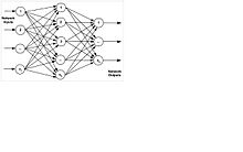
The four main features of an ANN are topology, data flow, types of input values, and forms of activation (Meireles, M. R. G. (2003), Munakata, T. (1998)). Topology can be multilayered, single-layered, or recurrent. Data flow can be recurrent with feedback or non-recurrent with feedforward model. The inputs are binary, bipolar, or continuous. The activation is linear, step, or sigmoid. Multilayer Perceptron (MLP) is the most popular of all the types, which is generally trained with back-propagation of error algorithm. Each neuron output is connected to every neuron in subsequent layers connected in cascade and with no connections between neurons in the same layer. Figure 4 shows a basic MLP topology (Meireles, M. R. G. (2003)), and a basic telecommunication network (Subramanian, M. (2010)) that most are familiar with. We can equate the routers at the nodes in telecommunication network to neurons in MLP technology and the links to synapses.

Figure 4(a) Basic telecommunication network
 
Figure 4(b) Basic MLP technology model Figure 4. Telecommunication network and neural network topologies

Computational neuron models

Computational science is an interdisciplinary field that combines engineering, biology, control systems, brain functions, physical sciences, and computer science. It has fundamental development models done at the lower levels of ions, neurons, and synapses, as well as information propagation between neurons. These models have established the enabling technology for higher-level models to be developed. They are based on chemical and electrical activities in the neurons for which electrical equivalent circuits are generated. A simple model for the neuron with predominantly potassium ions inside the cell and sodium ions outside establishes an electric potential on the membrane under equilibrium, i.e., no external activity, condition. This is called the resting membrane potential, which can be determined by Nernst Equation (Nernst, W. (1888)). An equivalent electrical circuit for a patch of membrane, for example an axon or dendrite, is shown in Figure 5. EK and ENa are the potentials associated with the potassium and sodium channels respectively and RK and RNa are the resistances associated with them. C is the capacitance of the membrane and I is the source current, which could be the test source or the signal source (action potential). The resting potential for potassium-sodium channels in a neuron is about -65 millivolts.

Figure 5 Membrane model

The membrane model is for a small section of the cell membrane; for larger sections it can be extended by adding similar sections, called compartments, with the parameter values being the same or different. The compartments are cascaded by a resistance, called axial resistance. Figure 6 shows a compartmental model of a neuron that is developed over the membrane model. Dendrites are the postsynaptic receptors receiving inputs from other neurons; and the axon with one or more axon terminals transmits neurotransmitters to other neurons. 

Figure 6 Neuron model

The second building block is the Hodgkin-Huxley (HH) model of the action potential. When the membrane potential from the dendrites exceeds the resting membrane potential, a pulse is generated by the neuron cell and propagated along the axon. This pulse is called the action potential and HH model is a set of equations that is made to fit the experimental data by the design of the model and the choice of the parameter values.

Models for more complex neurons containing other types of ions can be derived by adding to the equivalent circuit additional battery and resistance pairs for each ionic channel. The ionic channel could be passive or active as they could be gated by voltage or be ligands. The extended HH model has been developed to handle the active channel situation.

Although there are neurons that are physiologically connected to each other, information is transmitted at most of the synapses by chemical process across a cleft. Synapses are also computationally modeled. The next level of complexity is that of stream of action potentials, which are generated, whose pattern contains the coding information of the signal being transmitted. There are basically two types of action potentials, or spikes as they are called, that are generated. One is “integrate-and-fire” (the one we have so far addressed) and the other which is rate based. The latter is a stream whose rate varies. The signal going across the synapses could be modeled either as a deterministic or a stochastic process based on the application (See Section 3.7). Another anatomical complication is when a population of neurons, such as a column of neurons in visionary system, needs to be handled. This is done by considering the collective behavior of the group (Kotter, R., Nielson, P., Dyhrfjeld-Johnson, J., Sommer, F. T., & Northoff, G. (2002)).

Spiking neuron models

The action potential or the spike does not itself carry any information. It is the stream of spikes, called spike train, that carry the information in its number and pattern of spikes and timing of spikes. The postsynaptic potential can be either positive, the excitatory synapse or negative, inhibitory synapse. In modeling, the postsynaptic potentials received by the dendrites in the postsynaptic neuron are integrated and when the integrated potential exceeds the resting potential, the neuron fires an action potential along its axon. This model is the Integrate-and-Fire (IF) model that was mentioned in Section 2.3. Closely related to IF model is a model called Spike Response Model (SRM) (Gerstner, W. (1995) Pages 738-758) that is dependent on impulse function response convoluted with the input stimulus signal. This forms a base for a large number of models developed for spiking neural networks. 

The IF and SR model of spike train occurs in Type I neurons, in which the spike rate or spike frequency of the occurrence increases smoothly with the increase in stimulus current starting from zero. Another phenomenon of spike train generation happens in Type II neurons, where firing occurs at the resting potential threshold, but with a quantum jump to a non-zero frequency. Models have been developed using the rate (frequency) of the spike train and are called rate-based models. 

What is important for understanding the functions of the nervous system is how the message is coded and transported by the action potential in the neuron. There are two theories on how the signal that is being propagated is coded in the spikes as to whether it is pulse code or rate code. In the former, it is the time delay of the first spike from the time of stimulus as seen by the postsynaptic receiver that determines the coding. In the rate code, it is average rate of the spike that influences the coding. It is not certain as to which is really the actual physiological phenomenon in each case. However, both cases can be modeled computationally and the parameters varied to match the experimental result. The pulse mode is more complex to model and numerous detailed neuron models and population models are described by Gerstner and Kistler in Parts I and II of Gerstner, W., & Kistler, W. (2002) and Chapter 8 of Sterratt, D., Graham, B., Gillies, A., & Willshaw, D. (2011). Another important characteristic associated with SR model is the spike-time-dependent-plasticity. It is based on Hebb’s postulate on plasticity of synapse, which states that “the neurons that fire together wire together.” This causes the synapse to be a long-term potentiation (LTP) or long-term depression (LTD). The former is the strengthening of the synapse between two neurons if the postsynaptic spike temporally follows immediately after the presynaptic spike. Latter is the case if it is reverse, i.e., the presynaptic spike occurs after the postsynaptic spike. Gerstner, W. & Kistler, W. (2002) in Chapter 10 and Sterratt, D., Graham, B., Gillies, A., & Willshaw, D. (2011) in Chapter 7 discuss the various models related to Hebbian models on plasticity and coding.

Nervous system network models

The challenge involved in developing models for small, medium, and large networks is one of reducing the complexity by making valid simplifying assumptions in and extending the Hodgkin-Huxley neuronal model appropriately to design those models ( see Chapter 9 of Sterratt, D., Graham, B., Gillies, A., & Willshaw, D. (2011), Kotter, R., Nielson, P., Dyhrfjeld-Johnson, J., Sommer, F. T., & Northoff, G. (2002), and Chapter 9 of Gerstner, W., & Kistler, W. (2002)). Network models can be classified as either network of neurons propagating through different levels of cortex or neuron populations interconnected as multilevel neurons. The spatial positioning of neuron could be 1-, 2- or 3-dimensional; the latter ones are called small-world networks as they are related to local region. The neuron could be either excitatory or inhibitory, but not both. Modeling design depends on whether it is artificial neuron or biological neuron of neuronal model. Type I or Type II choice needs to be made for the firing mode. Signaling in neurons could be rate-based neurons, spiking response neurons, or deep-brain stimulated. The network can be designed as feedforward or recurrent type. The network needs to be scaled for the computational resource capabilities. Large-scale thalamocortical systems are handled in the manner of the Blue Brain project (Markam, H. (2006)).

Nervous system development models

No generalized modeling concepts exist for modeling the development of anatomical physiology and morphology similar to the one of behavior of neuronal network, which is based on HH model. Shankle, W. R., Hara, J., Fallon, J. H., and Landing, B. H. (2002) describe the application of neuroanatomical data of the developing human cerebral cortex to computational models. Sterratt, D., Graham, B., Gillies, A., & Willshaw, D. (2011) discuss aspects of the nervous system of computational modeling in the development of nerve cell morphology, cell physiology, cell patterning, patterns of ocular dominance, and connection between nerve cell and muscle, and retinotopic maps. Carreira-Perpinan, M. A. & Goodhill, G. J. (2002) deal with the optimization of the computerized models of the visual cortex.

Modeling tools

With the enormous number of models that have been created, tools have been developed for dissemination of the information, as well as platforms to develop models. Several generalized tools, such as GENESIS, NEURON, XPP, and NEOSIM are available and are discussed by Hucka, M. (2002).

Lateralization of brain function

From Wikipedia, the free encyclopedia

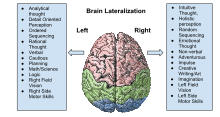
Diagram of the human brain.
The human brain is divided into two hemispheres–left and right. Scientists continue to explore how some cognitive functions tend to be dominated by one side or the other; that is, how they are lateralized.
 
The lateralization of brain function is the tendency for some neural functions or cognitive processes to be specialized to one side of the brain or the other. The medial longitudinal fissure separates the human brain into two distinct cerebral hemispheres, connected by the corpus callosum. Although the macrostructure of the two hemispheres appears to be almost identical, different composition of neuronal networks allows for specialized function that is different in each hemisphere. Lateralization of brain structures is based on general trends expressed in healthy patients; however, there are numerous counterexamples to each generalization. Each human's brain develops differently leading to unique lateralization in individuals. This is different from specialization as lateralization refers only to the function of one structure divided between two hemispheres. Specialization is much easier to observe as a trend since it has a stronger anthropological history. The best example of an established lateralization is that of Broca's and Wernicke's areas where both are often found exclusively on the left hemisphere. These areas frequently correspond to handedness, however, meaning that the localization of these areas is regularly found on the hemisphere corresponding to the dominant hand (anatomically on the opposite side). Function lateralization such as semantics, intonation, accentuation, prosody, etc. has since been called into question and largely been found to have a neuronal basis in both hemispheres. Another example is that each hemisphere in the brain tends to represent one side of the body. In the cerebellum this is the same bodyside, but in the forebrain this is predominantly the contralateral side.

Lateralized functions

Language

Language functions such as grammar, vocabulary and literal meaning are typically lateralized to the left hemisphere, especially in right handed individuals. While language production is left-lateralized in up to 90% of right-handers, it is more bilateral, or even right-lateralized, in approximately 50% of left-handers.

Broca's area and Wernicke's area, two areas associated with the production of speech, are located in the left cerebral hemisphere for about 95% of right-handers, but about 70% of left-handers.

Auditory and visual processing

The processing of visual and auditory stimuli, spatial manipulation, facial perception, and artistic ability are represented bilaterally. Numerical estimation, comparison and online calculation depend on bilateral parietal regions while exact calculation and fact retrieval are associated with left parietal regions, perhaps due to their ties to linguistic processing.

Clinical significance

Depression is linked with a hyperactive right hemisphere, with evidence of selective involvement in "processing negative emotions, pessimistic thoughts and unconstructive thinking styles", as well as vigilance, arousal and self-reflection, and a relatively hypoactive left hemisphere, "specifically involved in processing pleasurable experiences" and "relatively more involved in decision-making processes". Additionally, "left hemisphere lesions result in an omissive response bias or error pattern whereas right hemisphere lesions result in a commissive response bias or error pattern." The delusional misidentification syndromes, reduplicative paramnesia and Capgras delusion are also often the result of right hemisphere lesions.

Hemisphere damage

Damage to either the right or left hemisphere, and its resulting deficits provide insight into the function of the damaged area. Left hemisphere damage has many effects on language production and perception. Damage or lesions to the right hemisphere can result in a lack of emotional prosody or intonation when speaking. Right hemisphere damage also has grave effects on understanding discourse. People with damage to the right hemisphere have a reduced ability to generate inferences, comprehend and produce main concepts, and a reduced ability to manage alternative meanings. Furthermore, people with right hemisphere damage often exhibit discourse that is abrupt and perfunctory or verbose and excessive. They can also have pragmatic deficits in situations of turn taking, topic maintenance and shared knowledge. 

Lateral brain damage can also affect visual perceptual spatial resolution. People with left hemisphere damage may have impaired perception of high resolution, or detailed, aspects of an image. People with right hemisphere damage may have impaired perception of low resolution, or big picture, aspects of an image.

Plasticity

If a specific region of the brain, or even an entire hemisphere, is injured or destroyed, its functions can sometimes be assumed by a neighboring region in the same hemisphere or the corresponding region in the other hemisphere, depending upon the area damaged and the patient's age. When injury interferes with pathways from one area to another, alternative (indirect) connections may develop to communicate information with detached areas, despite the inefficiencies.

Broca's aphasia

Broca's aphasia is a specific type of expressive aphasia and is so named due to the aphasia that results from damage or lesions to the Broca's area of the brain, that exists most commonly in the left inferior frontal hemisphere. Thus, the aphasia that develops from the lack of functioning of the Broca's area is an expressive and non-fluent aphasia. It is called 'non-fluent' due the issues that arise because Broca's area is critical for language pronunciation and production. The area controls some motor aspects of speech production and articulation of thoughts to words and as such lesions to the area result in the specific non-fluent aphasia.

Wernicke's aphasia

Wernicke's aphasia is the result of damage to the area of the brain that is commonly in the left hemisphere above the sylvian fissure. Damage to this area causes primarily a deficit in language comprehension. While the ability to speak fluently with normal melodic intonation is spared, the language produced by a person with Wernicke's aphasia is riddled with semantic errors, and may sound nonsensical to the listener. Wernicke's aphasia is characterized by phonemic paraphasias, neologism or jargon. Another characteristic of a person with Wernicke's aphasia is that they are unconcerned by the mistakes that they are making.

Society and culture

Misapplication

Terence Hines states that the research on brain lateralization is valid as a research program, though commercial promoters have applied it to promote subjects and products far outside the implications of the research. For example, the implications of the research have no bearing on psychological interventions such as EMDR and neurolinguistic programming, brain-training equipment, or management training.

Pop psychology

The oversimplification of lateralization in pop psychology. This belief was widely held even in the scientific community for some years.
 
Some popularizations oversimplify the science about lateralization, by presenting the functional differences between hemispheres as being more absolute than is actually the case.

Sex differences

In the 19th century and to a lesser extent the 20th, it was thought that each side of the brain was associated with a specific gender: the left corresponding with masculinity and the right with femininity and each half could function independently. The right side of the brain was seen as the inferior and thought to be prominent in women, savages, children, criminals, and the insane. A prime example of this in fictional literature can be seen in Robert Louis Stevenson's Strange Case of Dr. Jekyll and Mr. Hyde.

Evolutionary advantage

The widespread lateralization of many vertebrate animals indicates an evolutionary advantage associated with the specialization of each hemisphere.

History

Broca

One of the first indications of brain function lateralization resulted from the research of French physician Pierre Paul Broca, in 1861. His research involved the male patient nicknamed "Tan", who suffered a speech deficit (aphasia); "tan" was one of the few words he could articulate, hence his nickname. In Tan's autopsy, Broca determined he had a syphilitic lesion in the left cerebral hemisphere. This left frontal lobe brain area (Broca's area) is an important speech production region. The motor aspects of speech production deficits caused by damage to Broca's area are known as expressive aphasia. In clinical assessment of this aphasia, it is noted that the patient cannot clearly articulate the language being employed.

Wernicke

German physician Karl Wernicke continued in the vein of Broca's research by studying language deficits unlike expressive aphasia. Wernicke noted that not every deficit was in speech production; some were linguistic. He found that damage to the left posterior, superior temporal gyrus (Wernicke's area) caused language comprehension deficits rather than speech production deficits, a syndrome known as receptive aphasia.

Imaging

These seminal works on hemispheric specialization were done on patients or postmortem brains, raising questions about the potential impact of pathology on the research findings. New methods permit the in vivo comparison of the hemispheres in healthy subjects. Particularly, magnetic resonance imaging (MRI) and positron emission tomography (PET) are important because of their high spatial resolution and ability to image subcortical brain structures.

Movement and sensation

In the 1940s, neurosurgeon Wilder Penfield and his neurologist colleague Herbert Jasper developed a technique of brain mapping to help reduce side effects caused by surgery to treat epilepsy. They stimulated motor and somatosensory cortices of the brain with small electrical currents to activate discrete brain regions. They found that stimulation of one hemisphere's motor cortex produces muscle contraction on the opposite side of the body. Furthermore, the functional map of the motor and sensory cortices is fairly consistent from person to person; Penfield and Jasper's famous pictures of the motor and sensory homunculi were the result.

Split-brain patients

Research by Michael Gazzaniga and Roger Wolcott Sperry in the 1960s on split-brain patients led to an even greater understanding of functional laterality. Split-brain patients are patients who have undergone corpus callosotomy (usually as a treatment for severe epilepsy), a severing of a large part of the corpus callosum. The corpus callosum connects the two hemispheres of the brain and allows them to communicate. When these connections are cut, the two halves of the brain have a reduced capacity to communicate with each other. This led to many interesting behavioral phenomena that allowed Gazzaniga and Sperry to study the contributions of each hemisphere to various cognitive and perceptual processes. One of their main findings was that the right hemisphere was capable of rudimentary language processing, but often has no lexical or grammatical abilities. Eran Zaidel also studied such patients and found some evidence for the right hemisphere having at least some syntactic ability. 

Language is primarily localized in the left hemisphere. One of the experiments carried out by Gazzaniga involved a split-brain male patient sitting in front of a computer screen while having words and images presented on either side of the screen and the visual stimuli would go to either the right or left visual field, and thus the left or right brain, respectively. It was observed that if the patient was presented with an image to his left visual field (right brain), he would report not seeing anything. If he was able to feel around for certain objects, he could accurately pick out the correct object, despite not having the ability to verbalize what he saw. This led to confirmation that the left brain is localized for language whereas the right brain does not have this capability, and when the corpus callosum is cut, the two hemispheres cannot communicate in order for situation-pertinent speech to be produced.

Additional images

Eureka effect

From Wikipedia, the free encyclopedia

A 16th century woodcut of Archimedes' eureka moment
 
The eureka effect (also known as the Aha! moment or eureka moment) refers to the common human experience of suddenly understanding a previously incomprehensible problem or concept. Some research describes the Aha! effect (also known as insight or epiphany) as a memory advantage, but conflicting results exist as to where exactly it occurs in the brain, and it is difficult to predict under what circumstances one can predict an Aha! moment

Insight is a psychological term that attempts to describe the process in problem solving when a previously unsolvable puzzle becomes suddenly clear and obvious. Often this transition from not understanding to spontaneous comprehension is accompanied by an exclamation of joy or satisfaction, an Aha! moment. A person utilizing insight to solve a problem is able to give accurate, discrete, all-or-nothing type responses, whereas individuals not using the insight process are more likely to produce partial, incomplete responses.

A recent theoretical account of the Aha! moment started with four defining attributes of this experience. First, the Aha! moment appears suddenly; second, the solution to a problem can be processed smoothly, or fluently; third, the Aha! moment elicits positive affect; fourth, a person experiencing the Aha! moment is convinced that a solution is true. These four attributes are not separate but can be combined because the experience of processing fluency, especially when it occurs surprisingly (for example, because it is sudden), elicits both positive affect and judged truth.

Insight can be conceptualized as a two phase process. The first phase of an Aha! experience requires the problem solver to come upon an impasse, where they become stuck and even though they may seemingly have explored all the possibilities, are still unable to retrieve or generate a solution. The second phase occurs suddenly and unexpectedly. After a break in mental fixation or re-evaluating the problem, the answer is retrieved. Some research suggest that insight problems are difficult to solve because of our mental fixation on the inappropriate aspects of the problem content. In order to solve insight problems, one must "think outside the box". It is this elaborate rehearsal that may cause people to have better memory for Aha! moments. Insight is believed to occur with a break in mental fixation, allowing the solution to appear transparent and obvious.

History and etymology

The effect is named from a story about the ancient Greek polymath Archimedes. In the story, Archimedes was asked (c. 250 BC) by the local king to determine whether a crown was pure gold. During a subsequent trip to a public bath, Archimedes noted that water was displaced when his body sank into the bath, and particularly that the volume of water displaced equaled the volume of his body immersed in the water. Having discovered how to measure the volume of an irregular object, and conceiving of a method to solve the king's problem, Archimedes allegedly leaped out and ran home naked, shouting "eureka" (I have found it). This story is now thought to be fictional, because it was first mentioned by the Roman writer Vitruvius nearly 200 years after the date of the alleged event, and because the method described by Vitruvius would not have worked. However, Archimedes certainly did important, original work in hydrostatics, notably in his On Floating Bodies.

Research

Initial research

Research on the Aha! moment dates back more than 100 years, to the Gestalt psychologists' first experiments on chimpanzee cognition. In his 1921 book, Wolfgang Köhler described the first instance of insightful thinking in animals: One of his chimpanzees, Sultan, was presented with the task of reaching a banana that had been strung up high on the ceiling so that it was impossible to reach by jumping. After several failed attempts to reach the banana, Sultan sulked in the corner for a while, then suddenly jumped up and stacked a few boxes upon each other, climbed them and thus was able to grab the banana. This observation was interpreted as insightful thinking. Köhler's work was continued by Karl Duncker and Max Wertheimer

The Eureka effect was later also described by Pamela Auble, Jeffrey Franks and Salvatore Soraci in 1979. The subject would be presented with an initially confusing sentence such as "The haystack was important because the cloth ripped". After a certain period of time of non-comprehension by the reader, the cue word (parachute) would be presented, the reader could comprehend the sentence, and this resulted in better recall on memory tests. Subjects spend a considerable amount of time attempting to solve the problem, and initially it was hypothesized that elaboration towards comprehension may play a role in increased recall. There was no evidence that elaboration had any effect for recall. It was found that both "easy" and "hard" sentences that resulted in an Aha! effect had significantly better recall rates than sentences that subjects were able to comprehend immediately. In fact equal recall rates were obtained for both "easy" and "hard" sentences which were initially non-comprehensible. It seems to be this non-comprehension to comprehension which results in better recall. The essence of the aha feeling underling insight problem solving was systemically empirically investigated by Danek et al. and Shen and his colleagues.

How people solve insight problems

Currently there are two theories for how people arrive at the solution for insight problems. The first is the progress monitoring theory. The person will analyze the distance from their current state to the goal state. Once a person realizes that they cannot solve the problem while on their current path, they will seek alternative solutions. In insight problems this usually occurs late in the puzzle. The second way that people attempt to solve these puzzles is the representational change theory. The problem solver initially has a low probability for success because they use inappropriate knowledge as they set unnecessary constraints on the problem. Once the person relaxes his or her constraints, they can bring previously unavailable knowledge into working memory to solve the problem. The person also utilizes chunk decomposition, where he or she will separate meaningful chunks into their component pieces. Both constraint relaxation and chunk decomposition allow for a change in representation, that is, a change in the distribution of activation across working memory, at which point they may exclaim, "Aha!" Currently both theories have support, with the progress monitoring theory being more suited to multiple step problems, and the representational change theory more suited to single step problems.

The Eureka effect on memory occurs only when there is an initial confusion. When subjects were presented with a clue word before the confusing sentence was presented, there was no effect on recall. If the clue was provided after the sentence was presented, an increase in recall occurred.

Memory

It had been determined that recall is greater for items that were generated by the subject versus if the subject was presented with the stimuli. There seems to be a memory advantage for instances where people are able to produce an answer themselves, recall was higher when Aha! reactions occurred. They tested sentences that were initially hard to understand, but when presented with a cued word, the comprehension became more apparent. Other evidence was found indicating that effort in processing visual stimuli was recalled more frequently than the stimuli that were simply presented. This study was done using connect-the-dots or verbal instruction to produce either a nonsense or real image. It is believed that effort made to comprehend something when encoding induces activation of alternative cues that later participate in recall.

Cerebral lateralization

Functional magnetic resonance imaging and electroencephalogram studies have found that problem solving requiring insight involves increased activity in the right cerebral hemisphere as compared with problem solving not requiring insight. In particular, increased activity was found in the right hemisphere anterior superior temporal gyrus.

Sleep

Some unconscious processing may take place while a person is asleep, and there are several cases of scientific discoveries coming to people in their dreams. Friedrich August Kekulé von Stradonitz said that the ring structure of benzene came to him in a dream where a snake was eating its own tail. Studies have shown increased performance at insight problems if the subjects slept during a break between receiving the problem and solving it. Sleep may function to restructure problems, and allow new insights to be reached. Henri Poincaré stated that he valued sleep as a time for "unconscious thought" that helped him break through problems.

Other theories

Professor Stellan Ohlsson believes that at the beginning of the problem-solving process, some salient features of the problem are incorporated into a mental representation of the problem. In the first step of solving the problem, it is considered in the light of previous experience. Eventually, an impasse is reached, where all approaches to the problem have failed, and the person becomes frustrated. Ohlsson believes that this impasse drives unconscious processes which change the mental representation of a problem, and cause novel solutions to occur.

General procedure for conducting ERP and EEG studies

When studying insight, or the Aha! effect, ERP or EEG general methods are used. Initially a baseline measurement is taken, which generally asks the subject to simply remember an answer to a question. Following this, subjects are asked to focus on the screen while a logogriph is shown, and then they are given time with a blank screen to get the answer, once they do they are required to press a key. After which the answer appears on the screen. The subjects are then asked to press one key to indicate that they thought of the correct answer and another to indicate if they got the answer wrong, finally, not to press a key at all if they were unsure or did not know the answer.

Evidence in EEG studies

Resting-state neural activity has a standing influence on cognitive strategies used when solving problems, particularly in the case of deriving solutions by methodical search or by sudden insight.[3] The two cognitive strategies used involve both search and analysis of current state of a problem, to the goal state of that problem, while insight problems are a sudden awareness of the solution to a problem.

Subjects studied were first recorded on the base-line resting state of thinking. After being tested using the method described in the General Procedure for Conducting ERP and EEG Studies, the ratio of insight versus non-insight solution were made to determine whether an individual is classified as a high insight (HI) or a low insight (LI) individual. Discriminating between HI and LI individuals were important as both groups use different cognitive strategies to solve anagram problems used in this study. Right hemisphere activation is believed to be involved in Aha! effects, so it comes as no surprise that HI individuals would show greater activation in the right hemisphere than the left hemisphere when compared to the LI individuals. Evidence was found to support this idea, there was greater activation in HI subjects at the right dorsal-frontal (low-alpha band), right inferior-frontal (beta and gamma bands) and the right parietal (gamma band) areas. As for LI subjects, left inferior-frontal and left anterior-temporal areas were active (low-alpha band).

There were also differences in attention between individuals of HI and LI. It has been suggested that individuals who are highly creative exhibit diffuse attention, thus allowing them a greater range of environmental stimuli. It was found that individuals who displayed HI would have less resting state occipital alpha-band activity, meaning there would be less inhibition of the visual system. Individuals that were less creative were found to focus their attention, thus causing them to sample less of their environment. Although, LI individuals were shown to have more occipital beta activity, consistent with heightened focused attention.

Evidence in ERP studies

These results are more reflective of models, rather than empirical evidence, as source localization is hard to determine precisely. Due to the nature of these studies that use Chinese logographs, there is a difficulty in an exact translation; a language barrier certainly exists.

There are some difficulties that exist in brain imaging when it comes to insight, thus making it hard to discuss neural mechanisms. Issues include: that insight occurs when an unwarranted mental fixation is broken and when novel task related associations are formed on top of old cognitive skills.

One theory discussed found that "Aha" answers produced more negative ERP results, N380 in the ACC, than the "No-Aha" answers, 250–500 ms, after an answer was produced. The authors suspected that this N380 in the ACC, which plays the role of a warning sign of breaking the mental set, was a reflection of the Aha! effect. Another study was done showed that an Aha! effect was elicited at N320 which has a strong activation in the central-posterior region. These previous studies reflective the premise of the study, that the Aha! effect occurs in the anterior cingulate cortex, while this study finds results indicating the posterior cingulate cortex is responsible. It was found that there was a N350 in the posterior cingulate cortex for successful guessing of logographs, not in the anterior cingulate cortex. The posterior cingulate cortex seems to play a more non-executive function in monitoring and inhibiting the mind set and cognitive function.

Another significant finding of this study done by Qiu and Zhang (2008), was a late positive component (LPC) in successful guessing of the logograph and then recognition of the answer at 600 and 700 ms, post-stimulus, in the parahippocampal gyrus (BA34). The data suggests that the parahippocampus is involved in searching of a correct answer by manipulating it in working memory, and integrating relationships between the base of the target logograph. The parahippocampal gyrus may reflect the formation of novel associations while solving insight problem.

Another ERP study is fairly similar to the Qiu and Zhang, 2008 study, however, this study claims to have anterior cingulate cortex activation at N380, which may be responsible for the mediation of breaking the mental set. Other areas of interest were prefrontal cortex (PFC), the posterior parietal cortex, and the medial temporal lobe. If subjects failed to solve the riddle, and then were shown the correct answer, they displayed the feeling of insight, which reflected the electroencephalogram recordings. 

Overall, it is quite apparent that there are many aspects that can explain the Aha! effect. No particular area has been determined but from the information gathered, it seems that insight occurs in many parts of the brain, within a given time period.

Evidence in fMRI studies

A study with the goal of recording the activity that occurs in the brain during an Aha! moment using fMRIs was conducted in 2003 by Jing Luo and Kazuhisa Niki. Participants in this study were presented with a series of Japanese riddles, and asked to rate their impressions toward each question using the following scale: (1) I can understand this question very well and know the answer; (2) I can understand this question very well and feel it is interesting, but I do not know the answer; or (3) I cannot understand this question and do not know the answer. This scale allowed the researchers to only look at participants who would experience an Aha! moment upon viewing the answer to the riddle. In previous studies on insight, researchers have found that participants reported feelings of insight when they viewed the answer to an unsolved riddle or problem. Luo and Niki had the goal of recording these feelings of insight in their participants using fMRIs. This method allowed the researchers to directly observe the activity that was occurring in the participant's brains during an Aha! moment.
An example of a Japanese riddle used in the study: The thing that can move heavy logs, but cannot move a small nailA river.
Participants were given 3 minutes to respond to each riddle, before the answer to the riddle was revealed. If the participant experienced an Aha! moment upon viewing the correct answer, any brain activity would be recorded on the fMRI. The fMRI results for this study showed that when participants were given the answer to an unsolved riddle, the activity in their right hippocampus increased significantly during these Aha! moments. This increased activity in the right hippocampus may be attributed to the formation of new associations between old nodes. These new associations will in turn strengthen memory for the riddles and their solutions. 

Although various studies using EEGs, ERPs, and fMRI's report activation in a variety of areas in the brain during Aha! moments, this activity occurs predominantly in the right hemisphere. More details on the neural basis of insight see a recent review named "New advances in the neural correlates of insight: A decade in review of the insightful brain"

Insight problems and problems with insight

Insight problems

The Nine Dot Problem

The Nine Dot Problem with solution. Most individuals fail to draw lines beyond the dots that compose the square, and are unable to solve this puzzle.
 
The Nine Dot Problem is a classic spatial problem used by psychologists to study insight. The problem consists of a 3 × 3 square created by 9 black dots. The task is to connect all 9 dots using exactly 4 straight lines, without retracing or removing one's pen from the paper. Kershaw & Ohlsson report that in a laboratory setting with a time limit of 2 or 3 minutes, the expected solution rate is 0%.

The difficulty with the Nine Dot Problem is that it requires respondents to look beyond the conventional figure-ground relationships that create subtle, illusory spatial constraints and (literally) "think outside of the box". Breaking the spatial constraints shows a shift in attention in working memory and utilizing new knowledge factors to solve the puzzle.

Verbal riddles

Verbal riddles are becoming popular problems in insight research. 

Example: "A man was washing windows on a high-rise building when he fell from the 40-foot ladder to the concrete path below. Amazingly, he was unhurt. Why? [Answer] He slipped from the bottom rung!"

Matchstick arithmetic

Matchstick arithmetic, which was developed and used by G. Knoblich, involves matchsticks that are arranged to show a simple but incorrect math equation in Roman numerals. The task is to correct the equation by moving only one matchstick. 

Two examples of Matchstick Arithmetic Problems.

Anagrams

Anagrams involve manipulating the order of a given set of letters in order to create one or many words. The original set of letters may be a word itself, or simply a jumble.

Example: Santa can be transformed to spell Satan.

Rebus puzzles

Rebus puzzles, also called "wordies", involve verbal and visual cues that force the respondent to restructure and "read between the lines" (almost literally) to solve the puzzle.

Some examples:
  1. Puzzle: you just me [Answer: just between you and me]
  2. Puzzle: PUNISHMENT [Answer: capital punishment]
  3. Puzzle:
   i i i 

   OOOOO
[Answer: circles under the eyes]

Remote Associates Test (RAT)

The Remote Associates Test (known as the RAT) was developed by Martha Mednick in 1962 to test creativity. However, it has recently been utilized in insight research. 

The test consists of presenting participants with a set of words, such as lick, mine, and shaker. The task is to identify the word that connects these three seemingly unrelated ones. In this example, the answer is salt. The link between words is associative, and does not follow rules of logic, concept formation or problem solving, and thus requires the respondent to work outside of these common heuristical constraints. 

Performance on the RAT is known to correlate with performance on other standard insight problems.

The Eight Coin Problem

In this problem a set of 8 coins is arranged on a table in a certain configuration, and the subject is told to move 2 coins so that all coins touch exactly three others. The difficulty in this problem comes from thinking of the problem in a purely 2-dimensional way, when a 3-dimensional approach is the only way to solve the problem.

Problems with insight

Insight research is problematic because of the ambiguity and lack of agreement among psychologists of its definition. This could largely be explained by the phenomenological nature of insight, and the difficulty in catalyzing its occurrence, as well as the ways in which it is experimentally "triggered". 

An example where another solution than the evident solution (number 6) must be found.
 
The pool of insight problems currently employed by psychologists is small and tepid, and due to its heterogeneity and often high difficulty level, is not conducive of validity or reliability. 

One of the biggest issues surrounding insight problems is that for most participants, they're simply too difficult. For many problems, this difficulty revolves around the requisite restructuring or re-conceptualization of the problem or possible solutions, for example, drawing lines beyond the square composed of dots in the Nine-Dot Problem. 

Furthermore, there are issues related to the taxonomy of insight problems. Puzzles and problems that are utilized in experiments to elicit insight may be classified in two ways. "Pure" insight problems are those that necessitate the use of insight, whereas "hybrid" insight problems are those that can be solved by other methods, such as the trial and error. As Weisberg (1996) points out, the existence of hybrid problems in insight research poses a significant threat to any evidence gleaned from studies that employ them. While the phenomenological experience of insight can help to differentiate insight-solving from non-insight solving (by asking the respondent to describe how they solved the problem, for example), the risk that non-insight solving has been mistaken for insight solving still exists. Likewise, issues surrounding the validity of insight evidence is also threatened by the characteristically small sample sizes. Experimenters may recruit an initially adequate sample size, but because of the level of difficulty inherent to insight problems, only a small fraction of any sample will successfully solve the puzzle or task given to them; placing serious limits on usable data. In the case of studies using hybrid problems, the final sample is at even greater risk of being very small by way of having to exclude whatever percentage of respondents solved their given puzzle without utilizing insight.

The Aha! effect and scientific discovery

There are several examples of scientific discoveries being made after a sudden flash of insight. One of the key insights in developing his special theory of relativity came to Albert Einstein while talking to his friend Michele Besso:
I started the conversation with him in the following way: "Recently I have been working on a difficult problem. Today I come here to battle against that problem with you." We discussed every aspect of this problem. Then suddenly I understood where the key to this problem lay. Next day I came back to him again and said to him, without even saying hello, "Thank you. I've completely solved the problem."
However, Einstein has said that the whole idea of special relativity did not come to him as a sudden, single eureka moment, and that he was "led to it by steps arising from the individual laws derived from experience". Similarly, Carl Friedrich Gauss said after a eureka moment: "I have the result, only I do not yet know how to get to it."

Sir Alec Jeffreys had a eureka moment in his lab in Leicester after looking at the X-ray film image of a DNA experiment at 9:05 am on Monday 10 September 1984, which unexpectedly showed both similarities and differences between the DNA of different members of his technician's family. Within about half an hour, he realized the scope of DNA profiling, which uses variations in the genetic code to identify individuals. The method has become important in forensic science to assist detective work, and in resolving paternity and immigration disputes. It can also be applied to non-human species, such as in wildlife population genetics studies. Before his methods were commercialized in 1987, Jeffreys' laboratory was the only center carrying out DNA fingerprinting in the world.

Lie group

From Wikipedia, the free encyclopedia https://en.wikipedia.org/wiki/Lie_group In mathematics , a Lie gro...