Search This Blog

Friday, May 11, 2018

Sex-determination system

From Wikipedia, the free encyclopedia

Some chromosomal sex determination systems

A sex-determination system is a biological system that determines the development of sexual characteristics in an organism. Most organisms that create their offspring using sexual reproduction have two sexes. Occasionally, there are hermaphrodites in place of one or both sexes. There are also some species that are only one sex due to parthenogenesis, the act of a female reproducing without fertilization.

In many species, sex determination is genetic: males and females have different alleles or even different genes that specify their sexual morphology. In animals this is often accompanied by chromosomal differences, generally through combinations of XY, ZW, XO, ZO chromosomes, or haplodiploidy. The sexual differentiation is generally triggered by a main gene (a "sex locus"), with a multitude of other genes following in a domino effect.

In other cases, sex of a fetus is determined by environmental variables (such as temperature). The details of some sex-determination systems are not yet fully understood. Although, they do provide concrete analysis of complete biological sex-determinism. Hypothesis for future fetal biological system analysis include complete-reproduction-system initialized signals that can be measured during pregnancies to more accurately determine whether a determined sex of a fetus is male, or female. Such analysis of biological systems could also signal whether the fetus is hermaphrodite, which includes total or partial of both male and female reproduction organs.

Some species such as various flowers and fish do not have a fixed sex, and instead go through life cycles and change sex based on genetic cues during corresponding life stages of their type. This could be due to environmental factors such as seasons and temperature. Human fetus genitals can sometimes develop abnormalities during maternal pregnancies due to mutations in the fetuses sex-determinism system, resulting in the fetus becoming a hermaphrodite.

Chromosomal systems

XX/XY sex chromosomes


Drosophila sex-chromosomes

Human male XY chromosomes after G-banding

The XX/XY sex-determination system is the most familiar, as it is found in humans. The XX/XY system is found in most other mammals, as well as some insects. In this system, most females have two of the same kind of sex chromosome (XX), while most males have two distinct sex chromosomes (XY). The X and Y sex chromosomes are different in shape and size from each other, unlike the rest of the chromosomes (autosomes), and are sometimes called allosomes. In some species, such as humans, organisms remain sex indifferent for a time after they're created; in others, however, such as fruit flies, sexual differentiation occurs as soon as the egg is fertilized.[1]

Y-centered sex determination

Some species (including humans) have a gene SRY on the Y chromosome that determines maleness. Members of SRY-reliant species can have uncommon XY chromosomal combinations such as XXY and still live.[1] Human sex is determined by the presence or absence of a Y chromosome with a functional SRY gene. Once the SRY gene is activated, cells create testosterone and anti-müllerian hormone which typically ensures the development of a single, male reproductive system.[1] In typical XX embryos, cells secrete estrogen, which drives the body toward the female pathway.

In Y-centered sex determination, the SRY gene is the main gene in determining male characteristics, but multiple genes are required to develop testes. In XY mice, lack of the gene DAX1 on the X chromosome results in sterility, but in humans it causes adrenal hypoplasia congenita.[2] However, when an extra DAX1 gene is placed on the X chromosome, the result is a female, despite the existence of SRY.[3] Even when there are normal sex chromosomes in XX females, duplication or expression of SOX9 causes testes to develop.[4][5] Gradual sex reversal in developed mice can also occur when the gene FOXL2 is removed from females.[6] Even though the gene DMRT1 is used by birds as their sex locus, species who have XY chromosomes also rely upon DMRT1, contained on chromosome 9, for sexual differentiation at some point in their formation.[1]

X-centered sex determination

Some species, such as fruit flies, use the presence of two X chromosomes to determine femaleness.[7] Species that use the number of Xs to determine sex are nonviable with an extra X chromosome.

Other variants of XX/XY sex determination

Some fish have variants of the XY sex-determination system, as well as the regular system. For example, while having an XY format, Xiphophorus nezahualcoyotl and X. milleri also have a second Y chromosome, known as Y', that creates XY' females and YY' males.[8]

At least one monotreme, the platypus, presents a particular sex determination scheme that in some ways resembles that of the ZW sex chromosomes of birds and lacks the SRY gene. The platypus has ten sex chromosomes; males have an XYXYXYXYXY pattern while females have ten X chromosomes. Although it is an XY system, the platypus' sex chromosomes share no homologues with eutherian sex chromosomes.[9] Instead, homologues with eutherian sex chromosomes lie on the platypus chromosome 6, which means that the eutherian sex chromosomes were autosomes at the time that the monotremes diverged from the therian mammals (marsupials and eutherian mammals). However, homologues to the avian DMRT1 gene on platypus sex chromosomes X3 and X5 suggest that it is possible the sex-determining gene for the platypus is the same one that is involved in bird sex-determination. More research must be conducted in order to determine the exact sex determining gene of the platypus.[10]


Heredity of sex chromosomes in XO sex determination

XX/X0 sex chromosomes

In this variant of the XY system, females have two copies of the sex chromosome (XX) but males have only one (X0). The 0 denotes the absence of a second sex chromosome. Generally in this method, the sex is determined by amount of genes expressed across the two chromosomes. This system is observed in a number of insects, including the grasshoppers and crickets of order Orthoptera and in cockroaches (order Blattodea). A small number of mammals also lack a Y chromosome. These include the Amami spiny rat (Tokudaia osimensis) and the Tokunoshima spiny rat (Tokudaia tokunoshimensis) and Sorex araneus, a shrew species. Transcaucasian mole voles (Ellobius lutescens) also have a form of XO determination, in which both sexes lack a second sex chromosome.[3] The mechanism of sex determination is not yet understood.[11]

The nematode C. elegans is male with one sex chromosome (X0); with a pair of chromosomes (XX) it is a hermaphrodite.[12] Its main sex gene is XOL, which encodes XOL-1 and also controls the expression of the genes TRA-2 and HER-1. These genes reduce male gene activation and increase it, respectively.[13]

ZW sex chromosomes

The ZW sex-determination system is found in birds, some reptiles, and some insects and other organisms. The ZW sex-determination system is reversed compared to the XY system: females have two different kinds of chromosomes (ZW), and males have two of the same kind of chromosomes (ZZ). In the chicken, this was found to be dependent on the expression of DMRT1.[14] In birds, the genes FET1 and ASW are found on the W chromosome for females, similar to how the Y chromosome contains SRY.[1] However, not all species depend upon the W for their sex. For example, there are moths and butterflies that are ZW, but some have been found female with ZO, as well as female with ZZW.[12] Also, while mammals deactivate one of their extra X chromosomes when female, it appears that in the case of Lepidoptera, the males produce double the normal amount of enzymes, due to having two Z's.[12] Because the use of ZW sex determination is varied, it is still unknown how exactly most species determine their sex.[12] However, reportedly, the silkworm Bombyx mori uses a single female-specific piRNA as the primary determiner of sex.[15] Despite the similarities between the ZW and XY systems, these sex chromosomes evolved separately. In the case of the chicken, their Z chromosome is more similar to humans' autosome 9.[16] The chicken's Z chromosome also seems to be related to the X chromosome of the platypus.[17] When a ZW species, such as the Komodo dragon, reproduces parthenogenetically, usually only males are produced. This is due to the fact that the haploid eggs double their chromosomes, resulting in ZZ or WW. The ZZ become males, but the WW are not viable and are not brought to term.[18]

UV sex chromosomes

In some Bryophyte and some algae species, the gametophyte stage of the life cycle, rather than being hermaphrodite, occurs as separate male or female individuals that produce male and female gametes respectively. When meiosis occurs in the sporophyte generation of the life cycle, the sex chromosomes known as U and V assort in spores that carry either the U chromosome and give rise to female gametophytes, or the V chromosome and give rise to male gametophytes.[19] [20]


Haplodiploid sex chromosomes

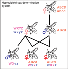
Haplodiploidy

Haplodiploidy is found in insects belonging to Hymenoptera, such as ants and bees. Unfertilized eggs develop into haploid individuals, which are the males. Diploid individuals are generally female but may be sterile males. Males cannot have sons or fathers. If a queen bee mates with one drone, her daughters share ¾ of their genes with each other, not ½ as in the XY and ZW systems. This may be significant for the development of eusociality, as it increases the significance of kin selection, but it is debated.[21] Most females in the Hymenoptera order can decide the sex of their offspring by holding received sperm in their spermatheca and either releasing it into their oviduct or not. This allows them to create more workers, depending on the status of the colony.[22]

Environmental systems


All alligators determine the sex of their offspring by the temperature of the nest.

Temperature-dependent

Many other sex-determination systems exist. In some species of reptiles, including alligators, some turtles, and the tuatara, sex is determined by the temperature at which the egg is incubated during a temperature-sensitive period. There are no examples of temperature-dependent sex determination (TSD) in birds. Megapodes had formerly been thought to exhibit this phenomenon, but were found to actually have different temperature-dependent embryo mortality rates for each sex.[23] For some species with TSD, sex determination is achieved by exposure to hotter temperatures resulting in the offspring being one sex and cooler temperatures resulting in the other. This type of TSD is called Pattern I. For others species using TSD, it is exposure to temperatures on both extremes that results in offspring of one sex, and exposure to moderate temperatures that results in offspring of the opposite sex, called Pattern II TSD. The specific temperatures required to produce each sex are known as the female-promoting temperature and the male-promoting temperature.[24] When the temperature stays near the threshold during the temperature sensitive period, the sex ratio is varied between the two sexes.[25] Some species' temperature standards are based on when a particular enzyme is created. These species that rely upon temperature for their sex determination do not have the SRY gene, but have other genes such as DAX1, DMRT1, and SOX9 that are expressed or not expressed depending on the temperature.[24] The sex of some species, such as the Nile tilapia, Australian skink lizard, and Australian dragon lizard, is initially determined by chromosomes, but can later be changed by the temperature of incubation.[8]

It is unknown how exactly temperature-dependent sex determination evolved.[26] It could have evolved through certain sexes being more suited to certain areas that fit the temperature requirements. For example, a warmer area could be more suitable for nesting, so more females are produced to increase the amount that nest next season.[26] Environmental sex determination preceded the genetically determined systems of birds and mammals; it is thought that a temperature-dependent amniote was the common ancestor of amniotes with sex chromosomes.[citation needed]

Other systems

There are other environmental sex determination systems including location-dependent determination systems as seen in the marine worm Bonellia viridis – larvae become males if they make physical contact with a female, and females if they end up on the bare sea floor. This is triggered by the presence of a chemical produced by the females, bonellin.[27] Some species, such as some snails, practice sex change: adults start out male, then become female. In tropical clown fish, the dominant individual in a group becomes female while the other ones are male, and bluehead wrasses (Thalassoma bifasciatum) are the reverse. Some species, however, have no sex-determination system. Hermaphrodite species include the common earthworm and certain species of snails. A few species of fish, reptiles, and insects reproduce by parthenogenesis and are female altogether. There are some reptiles, such as the boa constrictor and Komodo dragon that can reproduce both sexually and asexually, depending on whether a mate is available.[28]

Other unusual systems include those of the swordtail fish[clarification needed];[8] the Chironomus midges[clarification needed][citation needed]; the platypus, which has 10 sex chromosomes[9] but lacks the mammalian sex-determining gene SRY, meaning that the process of sex determination in the platypus remains unknown;[10] the juvenile hermaphroditism of zebrafish, with an unknown trigger;[8] and the platyfish, which has W, X, and Y chromosomes. This allows WY, WX, or XX females and YY or XY males.[8]

Evolution

Origin of sex chromosomes


The ends of the XY chromosomes, highlighted here in green, are all that is left of the original autosomes that can still cross-over with each other.

The accepted hypothesis of XY and ZW sex chromosome evolution is that they evolved at the same time, in two different branches.[29][30] However, there is some evidence to suggest that there could have been transitions between ZW and XY, such as in Xiphophorus maculatus, which have both ZW and XY systems in the same population, despite the fact that ZW and XY have different gene locations.[31][32] A recent theoretical model raises the possibility of both transitions between the XY/XX and ZZ/ZW system and environmental sex determination[33] The platypus' genes also back up the possible evolutionary link between XY and ZW, because they have the DMRT1 gene possessed by birds on their X chromosomes.[34] Regardless, XY and ZW follow a similar route. All sex chromosomes started out as an original autosome of an original amniote that relied upon temperature to determine the sex of offspring. After the mammals separated, the branch further split into Lepidosauria and Archosauromorpha. These two groups both evolved the ZW system separately, as evidenced by the existence of different sex chromosomal locations.[30] In mammals, one of the autosome pair, now Y, mutated its SOX3 gene into the SRY gene, causing that chromosome to designate sex.[30][34][35] After this mutation, the SRY-containing chromosome inverted and was no longer completely homologous with its partner. The regions of the X and Y chromosomes that are still homologous to one another are known as the pseudoautosomal region.[36] Once it inverted, the Y chromosome became unable to remedy deleterious mutations, and thus degenerated.[30] There is some concern that the Y chromosome will shrink further and stop functioning in ten million years: but the Y chromosome has been strictly conserved after its initial rapid gene loss.[37][38]

There are some species, such as the medaka fish, that evolved sex chromosomes separately; their Y chromosome never inverted and can still swap genes with the X. These species are still in an early phase of evolution with regard to their sex chromosomes. Because the Y does not have male-specific genes and can interact with the X, XY and YY females can be formed as well as XX males.[8]

Thursday, May 10, 2018

Testis-determining factor

From Wikipedia, the free encyclopedia
SRY
PBB Protein SRY image.jpg
Available structures
PDB Human UniProt search: PDBe RCSB
Identifiers
Aliases SRY, SRXX1, SRXY1, TDF, TDY, Testis determining factor, sex determining region Y, Sex-determining region of Y-chromosome, Sex-determining region Y
External IDs OMIM: 480000 HomoloGene: 48168 GeneCards: SRY

Y chromosome (human)
Chr. Y chromosome (human)[1]
Y chromosome (human)
Genomic location for SRY
Band Yp11.2 Start 2,786,855 bp[1]
End 2,787,699 bp[1]

PBB GE SRY 207893 at fs.png
More reference expression data
Orthologs
Species Human Mouse
Entrez


Ensembl


UniProt


RefSeq (mRNA)

NM_003140

n/a
RefSeq (protein)

NP_003131

n/a
Location (UCSC) Chr Y: 2.79 – 2.79 Mb n/a
PubMed search [2] n/a


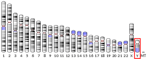
In human, the SRY gene is located on short (p) arm of the Y chromosome at position 11.2

Testis-determining factor (TDF), also known as sex-determining region Y (SRY) protein, is a DNA-binding protein (also known as gene-regulatory protein/transcription factor) encoded by the SRY gene that is responsible for the initiation of male sex determination in humans.[3] SRY is an intronless sex-determining gene on the Y chromosome in therians (placental mammals and marsupials);[4] mutations in this gene lead to a range of sex-related disorders with varying effects on an individual's phenotype and genotype.

TDF is a member of the SOX (SRY-like box) gene family of DNA-binding proteins. When complexed with the SF1 protein, TDF acts as a transcription factor that can upregulate other transcription factors, most importantly SOX9.[5] Its expression causes the development of primary sex cords, which later develop into seminiferous tubules. These cords form in the central part of the yet-undifferentiated gonad, turning it into a testis. The now-induced Leydig cells of the testis then start secreting testosterone, while the Sertoli cells produce anti-Müllerian hormone.[6] SRY gene effects normally take place 6–8 weeks after foetus formation and inhibits the female anatomical structural growth in males. It also works towards developing the dominant male characteristics.

Gene evolution and regulation

Evolution

SRY may have arisen from a gene duplication of the X chromosome bound gene SOX3, a member of the Sox family.[7] This duplication occurred after the split between monotremes and therians. Monotremes lack SRY and some of their sex chromosomes share homology with bird sex chromosomes.[8] SRY is a quickly evolving gene and its regulation has been difficult to study because sex determination is not a highly conserved phenomenon within the animal kingdom. [9]

Regulation

SRY gene has little in common with sex determination genes of other model organisms, and mice are the main model research organisms that can be utilized for its study. Understanding its regulation is further complicated because even between mammalian species, there is little protein sequence conservation. The only conserved group between mice and other mammals is the High-mobility group (HMG) box region that is responsible for DNA binding. Mutations in this region result in sex reversal, where the opposite sex is produced.[10] Because there is little conservation, the SRY promoter, regulatory elements and regulation are not well understood. Within related mammalian groups there are homologies within the first 400-600 base pairs upstream from the translational start site. In vitro studies of human SRY promoter have shown that a region of at least 310 bp upstream to translational start site are required for SRY promoter function. It's been shown that binding of three transcription factors, Steroidogenic factor 1 (SF1), Specificity Protein 1 (Sp1 transcription factor) and Wilms tumor protein 1 (WT1), to the human promoter sequence, influence expression of SRY.[10]

The promoter region has two Sp1 binding sites, at -150 and -13 that function as regulatory sites. Sp1 is a transcription factor that binds GC-rich consensus sequences, and mutation of the SRY binding sites leads to a 90% reduction in gene transcription. Studies of SF1 have resulted in less definite results. Mutations of SF1 can lead to sex reversal and deletion lead to incomplete gonad development. However, it's not clear how SF1 interacts with the SR1 promoter directly.[11] The promoter region also has two WT1 binding sites at -78 and -87 bp from the ATG codon. WT1 is transcription factor that has four C-terminal Zinc fingers and an N-terminal Pro/Glu-rich region and primarily functions as an activator. Mutation of the Zinc fingers or inactivation of WT1 results in reduced male gonad size. Deletion of the gene resulted in complete sex reversal. It is not clear how WT1 functions to up-regulate SRY, but some research suggests that it helps stabilize message processing.[11] However, there are complications to this hypothesis, because WT1 also is responsible for expression of an antagonist of male development, DAX1, which stands for Dosage-sensitive sex reversal, Adrenal hypoplasia critical region, on chromosome X, gene 1 . An additional copy of DAX1 in mice leads to sex reversal. It is not clear how DAX1 functions, and many different pathways have been suggested, including SRY transcriptional destabilization and RNA binding. There is evidence from work on suppression of male development that DAX1 can interfere with function of SF1, and in turn transcription of SRY by recruiting corepressors.[10]

There is also evidence that GATA binding protein 4 (GATA4) and FOG2 contribute to activation of SRY by associating with its promoter. How these proteins regulate SRY transcription is not clear, but FOG2 and GATA4 mutants have significantly lower levels of SRY transcription.[12] FOGs have zinc finger motifs that can bind DNA, but there is no evidence of FOG2 interaction with SRY. Studies suggest that FOG2 and GATA4 associate with nucleosome remodeling proteins that could lead to its activation.[13]

Function

During gestation, the cells of the primordial gonad that lie along the urogenital ridge are in a bipotential state, meaning they possess the ability to become either male cells (Sertoli and Leydig cells) or female cells (follicle cells and Theca cells). TDF initiates testis differentiation by activating male-specific transcription factors that allow these bipotential cells to differentiate and proliferate. TDF accomplishes this by upregulating SOX9, a transcription factor with a DNA-binding site very similar to TDF's. SOX9 leads to the upregulation of fibroblast growth factor 9 (Fgf9), which in turn leads to further upregulation of SOX9 . Once proper SOX9 levels are reached, the bipotential cells of the gonad begin to differentiate into Sertoli cells. Additionally, cells expressing TDF will continue to proliferate to form the primordial testis. While this constitutes the basic series of events, this brief review should be taken with caution since there are many more factors that influence sex differentiation.

Action in the nucleus

The TDF protein consists of three main regions. The central region encompasses the HMG (high-mobility group) domain, which contains nuclear localization sequences and acts as the DNA-binding domain. The C-terminal domain has no conserved structure, and the N-terminal domain can be phosphorylated to enhance DNA-binding.[11] The process begins with nuclear localization of TDF by acetylation of the nuclear localization signal regions, which allows for the binding of importin β and calmodulin to TDF, facilitating its import into the nucleus. Once in the nucleus, TDF and SF1 (steroidogenic factor 1, another transcriptional regulator) complex and bind to TESCO (testis-specific enhancer of Sox9 core), the testes-specific enhancer element of the Sox9 gene in Sertoli cell precursors, located upstream of the Sox9 gene transcription start site.[5] Specifically, it is the HMG region of TDF that binds to the minor groove of the DNA target sequence, causing the DNA to bend and unwind. The establishment of this particular DNA “architecture” facilitates the transcription of the Sox9 gene.[11] SOX9 protein then initiates a positive feedback loop, involving SOX9 acting as its own transcription factor and resulting in the synthesis of large amounts of SOX9.[11]

SOX9 and testes differentiation

The SF1 protein, on its own, leads to minimal transcription of the SOX9 gene in both the XX and XY bipotential gonadal cells along the urogenital ridge. However, binding of the TDF-SF1 complex to the testis-specific enhancer (TESCO) on SOX9 leads to significant up-regulation of the gene in only the XY gonad, while transcription in the XX gonad remains negligible. Part of this up-regulation is accomplished by SOX9 itself through a positive feedback loop; like TDF, SOX9 complexes with SF1 and binds to the TESCO enhancer, leading to further expression of SOX9 in the XY gonad. Two other proteins, FGF9 (fibroblast growth factor 9) and PDG2 (prostaglandin D2), also maintain this up-regulation. Although their exact pathways are not fully understood, they have been proven to be essential for the continued expression of SOX9 at the levels necessary for testes development.[5]

SOX9 and TDF are believed to be responsible for the cell-autonomous differentiation of supporting cell precursors in the gonads into Sertoli cells, the beginning of testes development. These initial Sertoli cells, in the center of the gonad, are hypothesized to be the starting point for a wave of FGF9 that spreads throughout the developing XY gonad, leading to further differentiation of Sertoli cells via the up-regulation of SOX9.[14] SOX9 and TDF are also believed to be responsible for many of the later processes of testis development (such as Leydig cell differentiation, sex cord formation, and formation of testis-specific vasculature), although exact mechanisms remain unclear.[15] It has been shown, however, that SOX9, in the presence of PDG2, acts directly on Amh (encoding anti-Müllerian hormone) and is capable of inducing testis formation in XX mice gonads, indicating its vital to testes development.[14]

Influence on sex

Embryos are gonadally identical, regardless of genetic sex, until a certain point in development when the testis-determining factor causes male sex organs to develop. Therefore, SRY plays an important role in sex determination. A typical male karyotype is XY. Individuals who inherit a normal Y chromosome and multiple X chromosomes are generally male (such as in Klinefelter Syndrome, which has an XXY karyotype). Atypical genetic recombination during crossover when a sperm cell is developing can result in karyotypes that do not match their phenotypic expression.

Most of the time, when a developing sperm cell undergoes crossover during its meiosis, the SRY gene stays on the Y chromosome. If it is transferred to the X chromosome, however, the resulting Y chromosome will not have an SRY gene and can no longer initiate testis development. Offspring which inherit this Y chromosome will have Swyer syndrome, characterized by an XY karyotype and a female phenotype. The X chromosome that results from this crossover event now has a SRY gene, and therefore the ability to initiate testis development. Offspring who inherit this X chromosome will have a condition called XX male syndrome, characterized by an XX karyotype, and a male phenotype. While most XX males develop testis, it is possible for them to experience incomplete differentiation resulting in the formation of both testicular and ovarian tissues in the same individual. XX male syndrome results in infertility, most likely caused by the inactivation (either random or non-random) of the X chromosome containing the SRY in some cells.[16]

While the presence or absence of SRY has generally determined whether or not testis development occurs, it has been suggested that there are other factors that affect the functionality of SRY.[17] Therefore, there are individuals who have the SRY gene, but still develop as females, either because the gene itself is defective or mutated, or because one of the contributing factors is defective.[18] This can happen in individuals exhibiting a XY, XXY, or XX SRY-positive karyotype.

Role in other diseases

SRY has been shown to interact with the androgen receptor and individuals with XY karyotype and a functional SRY gene can have an outwardly female phenotype due to an underlying androgen insensitivity syndrome (AIS).[19] Individuals with AIS are unable to respond to androgens properly due to a defect in their androgen receptor gene, and affected individuals can have complete or partial AIS.[20] SRY has also been linked to the fact that males are more likely than females to develop dopamine-related diseases such as schizophrenia and Parkinson's disease. SRY encodes a protein that controls the concentration of dopamine, the neurotransmitter that carries signals from the brain that control movement and coordination.[21]

Use in Olympic screening

One of the most controversial uses of this discovery was as a means for gender verification at the Olympic Games, under a system implemented by the International Olympic Committee in 1992. Athletes with an SRY gene were not permitted to participate as females, although all athletes in whom this was "detected" at the 1996 Summer Olympics were ruled false positives and were not disqualified. Specifically, eight female participants (out of a total of 3387) at these games were found to have the SRY gene. However, after further investigation of their genetic conditions, all these athletes were verified as female and allowed to compete. These athletes were found to have either partial or full androgen insensitivity, despite having an SRY gene, making them phenotypically female and giving them no advantage over other female competitors.[22] In the late 1990s, a number of relevant professional societies in United States called for elimination of gender verification, including the American Medical Association, stating that the method used was uncertain and ineffective.[23] Chromosomal screening was eliminated as of the 2000 Summer Olympics,[23][24][25] but this was later followed by other forms of testing based on hormone levels.

Ongoing research

Despite the progress made during the past several decades in the study of sex determination, the SRY gene, and the TDF protein, work is still being done to further our understanding in these areas. There remain factors that need to be identified in the sex-determining molecular network, and the chromosomal changes involved in many other human sex-reversal cases are still unknown. Scientists continue to search for additional sex-determining genes, using techniques such as microarray screening of the genital ridge genes at varying developmental stages, mutagenesis screens in mice for sex-reversal phenotypes, and identifying the genes that transcription factors act on using chromatin immunoprecipitation.[11]

FOXP2

From Wikipedia, the free encyclopedia

FOXP2
Protein FOXP2 PDB 2a07.png
Available structures
PDB Human UniProt search: PDBe RCSB
Identifiers
Aliases FOXP2, CAGH44, SPCH1, TNRC10, forkhead box P2
External IDs OMIM: 605317 HomoloGene: 33482 GeneCards: FOXP2

Chromosome 7 (human)
Chr. Chromosome 7 (human)[1]
Chromosome 7 (human)
Genomic location for FOXP2
Band 7q31.1 Start 114,086,327 bp[1]
End 114,693,772 bp[1]

PBB GE FOXP2 gnf1h09377 at fs.png
More reference expression data
Orthologs
Species Human Mouse
Entrez


Ensembl


UniProt


RefSeq (mRNA)

n/a
RefSeq (protein)

n/a
Location (UCSC) Chr 7: 114.09 – 114.69 Mb n/a
PubMed search [2] n/a


FOXP2 gene is located on the long (q) arm of chromosome 7 at position 31.

Forkhead box protein P2 (FOXP2) is a protein that, in humans, is encoded by the FOXP2 gene, also known as CAGH44, SPCH1 or TNRC10, and is required for proper development of speech and language.[3] The gene is shared with many vertebrates, where it generally plays a role in communication (for instance, the development of bird song).

Initially identified as the genetic factor of speech disorder in KE family, FOXP2 is the first gene discovered associated with speech and language.[4] The gene is located on chromosome 7 (7q31, at the SPCH1 locus), and is expressed in fetal and adult brain, heart, lung and gut.[5][6] FOXP2 orthologs[7] have also been identified in other mammals for which complete genome data are available. The FOXP2 protein contains a forkhead-box DNA-binding domain, making it a member of the FOX group of transcription factors, involved in regulation of gene expression. In addition to this characteristic forkhead-box domain, the protein contains a polyglutamine tract, a zinc finger and a leucine zipper. The gene is more active in females than in males, to which could be attributed better language learning in females.[8]

In humans, mutations of FOXP2 cause a severe speech and language disorder.[3][9] Versions of FOXP2 exist in similar forms in distantly related vertebrates; functional studies of the gene in mice[10] and in songbirds[11] indicate that it is important for modulating plasticity of neural circuits.[12] Outside the brain FOXP2 has also been implicated in development of other tissues such as the lung and gut.[13]

FOXP2 is popularly dubbed the "language gene", but this is only partly correct since there are other genes involved in language development.[14] It directly regulates a number of other genes, including CNTNAP2, CTBP1, and SRPX2.[15][16]

Two amino acid substitutions distinguish the human FOXP2 protein from that found in chimpanzees,[17] but only one of these two changes is unique to humans.[13] Evidence from genetically manipulated mice[18] and human neuronal cell models[19] suggests that these changes affect the neural functions of FOXP2.

Discovery

FOXP2 and its gene were discovered as a result of investigations on an English family known as the KE family, half of whom (fifteen individuals across three generations) suffered from a speech and language disorder called developmental verbal dyspraxia. Their case was studied at the Institute of Child Health of University College London.[20] In 1990 Myrna Gopnik, Professor of Linguistics at McGill University, reported that the disorder-affected KE family had severe speech impediment with incomprehensible talk, largely characterized by grammatical deficits.[21] She hypothesized that the basis was not of learning or cognitive disability, but due to genetic factors affecting mainly grammatical ability.[22] (Her hypothesis led to a popularised existence of "grammar gene" and a controversial notion of grammar-specific disorder.[23][24]) In 1995, the University of Oxford and the Institute of Child Health researchers found that the disorder was purely genetic.[25] Remarkably, the inheritance of the disorder from one generation to the next was consistent with autosomal dominant inheritance, i.e., mutation of only a single gene on an autosome (non-sex chromosome) acting in a dominant fashion. This is one of the few known examples of Mendelian (monogenic) inheritance for a disorder affecting speech and language skills, which typically have a complex basis involving multiple genetic risk factors.[26]

In 1998, Oxford University geneticists Simon Fisher, Anthony Monaco, Cecilia S. L. Lai, Jane A. Hurst, and Faraneh Vargha-Khadem identified an autosomal dominant monogenic inheritance that is localized on a small region of chromosome 7 from DNA samples taken from the affected and unaffected members.[5] The chromosomal region (locus) contained 70 genes.[27] The locus was given the official name "SPCH1" (for speech-and-language-disorder-1) by the Human Genome Nomenclature committee. Mapping and sequencing of the chromosomal region was performed with the aid of bacterial artificial chromosome clones.[6] Around this time, the researchers identified an individual who was unrelated to the KE family, but had a similar type of speech and language disorder. In this case the child, known as CS, carried a chromosomal rearrangement (a translocation) in which part of chromosome 7 had become exchanged with part of chromosome 5. The site of breakage of chromosome 7 was located within the SPCH1 region.[6]

In 2001, the team identified in CS that the mutation is in the middle of a protein-coding gene.[3] Using a combination of bioinformatics and RNA analyses, they discovered that the gene codes for a novel protein belonging to the forkhead-box (FOX) group of transcription factors. As such, it was assigned with the official name of FOXP2. When the researchers sequenced the FOXP2 gene in the KE family, they found a heterozygous point mutation shared by all the affected individuals, but not in unaffected members of the family and other people.[3] This mutation is due to an amino-acid substitution that inhibits the DNA-binding domain of the FOXP2 protein.[28] Further screening of the gene identified multiple additional cases of FOXP2 disruption, including different point mutations[9] and chromosomal rearrangements,[29] providing evidence that damage to one copy of this gene is sufficient to derail speech and language development.

Function

Foxp2 is expressed in the developing cerebellum and the hindbrain of the embryonic day 13.5 mouse. Allen Brain Atlases

FOXP2 is required for proper brain and lung development. Knockout mice with only one functional copy of the FOXP2 gene have significantly reduced vocalizations as pups.[30] Knockout mice with no functional copies of FOXP2 are runted, display abnormalities in brain regions such as the Purkinje layer, and die an average of 21 days after birth from inadequate lung development.[13]

FOXP2 is expressed in many areas of the brain[17] including the basal ganglia and inferior frontal cortex where it is essential for brain maturation and speech and language development.[15]

A knockout mouse model has been used to examine FOXP2's role in brain development and how mutations in the two copies of FOXP2 affect vocalization. Mutations in one copy result in reduced speech while abnormalities in both copies cause major brain and lung developmental issues.[13]

The expression of FOXP2 is subject to post-transcriptional regulation, particularly micro RNA, which binds to multiple miRNA binding-sites in the neocortex, causing the repression of FOXP2 3’UTR.[31]

Clinical significance

There are several abnormalities linked to FOXP2. The most common mutation results in severe speech impairment known as developmental verbal dyspraxia (DVD) which is caused by a translocation in the 7q31.2 region [t(5;7)(q22;q31.2)].[3][6] A missense mutation causing an arginine-to-histidine substitution (R553H) in the DNA-binding domain is thought to be the abnormality in KE.[32] A heterozygous nonsense mutation, R328X variant, produces a truncated protein involved in speech and language difficulties in one KE individual and two of their close family members.[9] R553H and R328X mutations also affected nuclear localization, DNA-binding, and the transactivation (increased gene expression) properties of FOXP2.[33][34] Although DVD associated with FOXP2 disruptions are thought to be rare (~2% by one estimate),[9] a faulty copy of FOXP2 in individuals always causes speech and language problems.

Several cases of developmental verbal dyspraxia in humans have been linked to mutations in the FOXP2 gene.[29][35][36][37] Such individuals have little or no cognitive handicaps but are unable to correctly perform the coordinated movements required for speech. fMRI analysis of these individuals performing silent verb generation and spoken word repetition tasks showed underactivation of Broca's area and the putamen, brain centers thought to be involved in language tasks. Because of this, FOXP2 has been dubbed the "language gene". People with this mutation also experience symptoms not related to language (not surprisingly, as FOXP2 is known to affect development in other parts of the body as well).[38] Scientists have also looked for associations between FOXP2 and autism, and both positive and negative findings have been reported.[39][40]

There is some evidence that the linguistic impairments associated with a mutation of the FOXP2 gene are not simply the result of a fundamental deficit in motor control. For examples, the impairments include difficulties in comprehension. Brain imaging of affected individuals indicates functional abnormalities in language-related cortical and basal/ganglia regions, demonstrating that the problems extend beyond the motor system.

Evolution

Human FOXP2 gene and evolutionary conservation is shown in a multiple alignment (at bottom of figure) in this image from the UCSC Genome Browser. Note that conservation tends to cluster around coding regions (exons).

The FOXP2 gene is highly conserved in mammals.[41] Human gene differs from non-human primates by the substitution of two amino acids, threonine to asparagine substitution at position 303 (T303N) and asparagine to serine substitution at position 325 (N325S).[32] In mice it differs from that of humans by three substitutions, and in zebra finch by seven amino acids.[17][42][43] One of the two amino acid difference between human and chimps also arose independently in carnivores and bats.[13][44] Similar FOXP2 proteins can be found in songbirds, fish, and reptiles such as alligators.[45][46]

DNA sampling from Homo neanderthalensis bones indicates that their FOXP2 gene is a little different, though largely similar to those of Homo sapiens (i.e. humans).[47][48]

The FOXP2 gene showed indications of recent positive selection.[41][49] Some researchers have speculated that positive selection is crucial for the evolution of language in humans.[17] Others, however, have been unable to find a clear association between species with learned vocalizations and similar mutations in FOXP2.[45][46] Insertion of both human mutations into mice, whose version of FOXP2 otherwise differs from the human and chimpanzee versions in only one additional base pair, causes changes in vocalizations as well as other behavioral changes, such as a reduction in exploratory tendencies. A reduction in dopamine levels and changes in the morphology of certain nerve cells are also observed.[18]

However, FOXP2 is extremely diverse in echolocating bats.[50] Twenty-two sequences of non-bat eutherian mammals revealed a total number of 20 nonsynonymous mutations in contrast to half that number of bat sequences, which showed 44 nonsynonymous mutations.[44] Interestingly, all cetaceans share three amino acid substitutions, but no differences were found between echolocating toothed whales and non-echolocating baleen cetaceans.[44] Within bats, however, amino acid variation correlated with different echolocating types.[44]

Interactions

FOXP2 interacts with a regulatory gene CTBP1.[51] It also downregulates CNTNAP2 gene, a member of the neurexin family found in neurons. The target gene is associated with common forms of language impairment.[52] It regulates the repeat-containing protein X-linked 2 (SRPX2), which is an epilepsy and language-associated gene in humans, and sound-controlling gene in mice.[53]

Mice

In a mouse FOXP2 knockout study, loss of both copies of the gene caused severe motor impairment related to cerebellar abnormalities and lack of ultrasonic vocalisations normally elicited when pups are removed from their mothers.[30] These vocalizations have important communicative roles in mother-offspring interactions. Loss of one copy was associated with impairment of ultrasonic vocalisations and a modest developmental delay. Male mice on encountering female mice produce complex ultrasonic vocalisations that have characteristics of song.[54] Mice that have the R552H point mutation carried by the KE family show cerebellar reduction and abnormal synaptic plasticity in striatal and cerebellar circuits.[10]

Birds

In songbirds, FOXP2 most likely regulates genes involved in neuroplasticity.[11][55] Gene knockdown of FOXP2 in Area X of the basal ganglia in songbirds results in incomplete and inaccurate song imitation.[11] Overexpression of FoxP2 was accomplished through injection of adeno-associated virus serotype 1 (AAV1) into Area X of the brain. This overexpression produced similar effects to that of knockdown; juvenile zebra finch birds were unable to accurately imitate their tutors.[56] Similarly, in adult canaries higher FOXP2 levels also correlate with song changes.[43]

Levels of FOXP2 in adult zebra finches are significantly higher when males direct their song to females than when they sing song in other contexts.[55] “Directed” singing refers to when a male is singing to a female usually for a courtship display. “Undirected” singing occurs when for example, a male sings when other males are present or is alone.[57] Studies have found that FoxP2 levels vary depending on the social context. When the birds were singing undirected song, there was a decrease of FoxP2 expression in Area X. This downregulation was not observed and FoxP2 levels remained stable in birds singing directed song.[58]

Differences between song-learning and non-song-learning birds have been shown to be caused by differences in FOXP2 gene expression, rather than differences in the amino acid sequence of the FOXP2 protein.

FOXP2 also has possible implications in the development of bat echolocation.[32][44][59]

Operator (computer programming)

From Wikipedia, the free encyclopedia https://en.wikipedia.org/wiki/Operator_(computer_programmin...