Search This Blog

Thursday, April 24, 2025

Telophase

From Wikipedia, the free encyclopedia
https://en.wikipedia.org/wiki/Telophase
This image describes the final stage in mitosis, telophase.
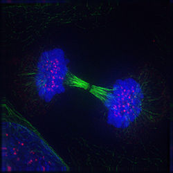
Fluorescence micrograph of a human cell in telophase showing chromosomes (DNA) in blue, microtubules in green and kinetochores in pink

Telophase (from Ancient Greek τέλος (télos) 'end, result, completion' and φάσις (phásis) 'appearance') is the final stage in both meiosis and mitosis in a eukaryotic cell. During telophase, the effects of prophase and prometaphase (the nucleolus and nuclear membrane disintegrating) are reversed. As chromosomes reach the cell poles, a nuclear envelope is re-assembled around each set of chromatids, the nucleoli reappear, and chromosomes begin to decondense back into the expanded chromatin that is present during interphase. The mitotic spindle is disassembled and remaining spindle microtubules are depolymerized. Telophase accounts for approximately 2% of the cell cycle's duration.

Cytokinesis typically begins before late telophase and, when complete, segregates the two daughter nuclei between a pair of separate daughter cells.

Telophase is primarily driven by the dephosphorylation of mitotic cyclin-dependent kinase (Cdk) substrates.

Dephosphorylation of Cdk substrates

The phosphorylation of the protein targets of M-Cdks (Mitotic Cyclin-dependent Kinases) drives spindle assembly, chromosome condensation and nuclear envelope breakdown in early mitosis. The dephosphorylation of these same substrates drives spindle disassembly, chromosome decondensation and the reformation of daughter nuclei in telophase. Establishing a degree of dephosphorylation permissive to telophase events requires both the inactivation of Cdks and the activation of phosphatases.

Cdk inactivation is primarily the result of the destruction of its associated cyclin. Cyclins are targeted for proteolytic degradation by the anaphase promoting complex (APC), also known as the cyclosome, a ubiquitin-ligase. The active, CDC20-bound APC (APC/CCDC20) targets mitotic cyclins for degradation starting in anaphase. Experimental addition of non-degradable M-cyclin to cells induces cell cycle arrest in a post-anaphase/pre-telophase-like state with condensed chromosomes segregated to cell poles, an intact mitotic spindle, and no reformation of the nuclear envelope. This has been shown in frog (Xenopus) eggs, fruit flies (Drosophilla melanogaster), budding (Saccharomyces cerevisiae) and fission (Schizosaccharomyces pombe) yeast, and in multiple human cell lines.

The requirement for phosphatase activation can be seen in budding yeast, which do not have redundant phosphatases for mitotic exit and rely on the phosphatase cdc14. Blocking cdc14 activation in these cells results in the same phenotypic arrest as does blocking M-cyclin degradation.

Historically, it has been thought that anaphase and telophase are events that occur passively after satisfaction of the spindle-assembly checkpoint (SAC) that defines the metaphase-anaphase transition. However, the existence of differential phases to cdc14 activity between anaphase and telophase is suggestive of additional, unexplored late-mitotic checkpoints. Cdc14 is activated by its release into the nucleus, from sequestration in the nucleolus, and subsequent export into the cytoplasm. The Cdc-14 Early Anaphase Release pathway, which stabilizes the spindle, also releases cdc14 from the nucleolus but restricts it to the nucleus. Complete release and maintained activation of cdc14 is achieved by the separate Mitotic Exit Network (MEN) pathway to a sufficient degree (to trigger the spindle disassembly and nuclear envelope assembly) only after late anaphase.

Cdc14-mediated dephosphorylation activates downstream regulatory processes unique to telophase. For example, the dephosphorylation of CDH1 allows the APC/C to bind CDH1. APC/CCDH1 targets CDC20 for proteolysis, resulting in a cellular switch from APC/CCDC20 to APC/CCDH1 activity. The ubiquitination of mitotic cyclins continues along with that of APC/CCDH1-specific targets such as the yeast mitotic spindle component, Ase1, and cdc5, the degradation of which is required for the return of cells to the G1 phase.

Additional mechanisms driving telophase

A shift in the whole-cell phosphoprotein profile is only the broadest of many regulatory mechanisms contributing to the onset of individual telophase events.

  • The anaphase-mediated distancing of chromosomes from the metaphase plate may trigger spatial cues for the onset of telophase.
  • An important regulator and effector of telophase is cdc48 (homologous to yeast cdc48 is human p97, both structurally and functionally), a protein that mechanically employs its ATPase activity to alter target protein conformation. Cdc48 is necessary for spindle disassembly, nuclear envelope assembly, and chromosome decondensation. Cdc48 modifies proteins structurally involved in these processes and also some ubiquitinated proteins which are thus targeted to the proteasome.

Mitotic spindle disassembly

Stages of late M phase in a vertebrate cell

The breaking of the mitotic spindle, common to the completion of mitosis in all eukaryotes, is the event most often used to define the anaphase-B to telophase transition, although the initiation of nuclear reassembly tends to precede that of spindle disassembly.

Spindle disassembly is an irreversible process which must effect not the ultimate degradation, but the reorganization of constituent microtubules; microtubules are detached from kinetochores and spindle pole bodies and return to their interphase states.

Spindle depolymerization during telophase occurs from the plus end and is, in this way, a reversal of spindle assembly. Subsequent microtubule array assembly is, unlike that of the polarized spindle, interpolar. This is especially apparent in animal cells which must immediately, following mitotic spindle disassembly, establish the antiparallel bundle of microtubules known as the central spindle in order to regulate cytokinesis. The ATPase p97 is required for the establishment of the relatively stable and long interphase microtubule arrays following disassembly of the highly dynamic and relatively short mitotic ones.

While spindle assembly has been well studied and characterized as a process where tentative structures are edified by the SAC, the molecular basis of spindle disassembly is not understood in comparable detail. The late-mitotic dephosphorylation cascade of M-Cdk substrates by the MEN is broadly held to be responsible for spindle disassembly. The phosphorylation states of microtubule stabilizing and destabilizing factors, as well as microtubule nucleators are key regulators of their activities. For example, NuMA is a minus-end crosslinking protein and Cdk substrate whose dissociation from the microtubule is effected by its dephosphorylation during telophase.

A general model for spindle disassembly in yeast is that the three functionally overlapping subprocesses of spindle disengagement, destabilization, and depolymerization are primarily effected by APC/CCDH1, microtubule-stabilizer-specific kinases, and plus-end directed microtubule depolymerases, respectively. These effectors are known to be highly conserved between yeast and higher eukaryotes. The APC/CCDH1 targets crosslinking microtubule-associated proteins (NuMA, Ase1, Cin1 and more). AuroraB (yeast IpI1) phosphorylates the spindle-associated stabilizing protein EB1 (yeast Bim1), which then dissociates from microtubules, and the destabilizer She1, which then associates with microtubules. Kinesin8 (yeast Kip3), an ATP-dependent depolymerase, accelerate microtubule depolymerization at the plus end. It was shown the concurrent disruption of these mechanisms, but not of any one, results in dramatic spindle hyperstability during telophase, suggesting functional overlap despite the diversity of the mechanisms.

Nuclear envelope reassembly

The main components of the nuclear envelope are a double membrane, nuclear pore complexes, and a nuclear lamina internal to the inner nuclear membrane. These components are dismantled during prophase and prometaphase and reconstructed during telophase, when the nuclear envelope reforms on the surface of separated sister chromatids. The nuclear membrane is fragmented and partly absorbed by the endoplasmic reticulum during prometaphase and the targeting of inner nuclear membrane protein-containing ER vesicles to the chromatin occurs during telophase in a reversal of this process. Membrane-forming vesicles aggregate directly to the surface of chromatin, where they fuse laterally into a continuous membrane.

Ran-GTP is required for early nuclear envelope assembly at the surface of the chromosomes: it releases envelope components sequestered by importin β during early mitosis. Ran-GTP localizes near chromosomes throughout mitosis, but does not trigger the dissociation of nuclear envelope proteins from importin β until M-Cdk targets are dephosphorylated in telophase. These envelope components include several nuclear pore components, the most studied of which is the nuclear pore scaffold protein ELYS, which can recognize DNA regions rich in A:T base pairs (in vitro), and may therefore bind directly to the DNA. However, experiments in Xenopus egg extracts have concluded that ELYS fails to associate with bare DNA and will only directly bind histone dimers and nucleosomes. After binding to chromatin, ELYS recruits other components of the nuclear pore scaffold and nuclear pore trans-membrane proteins. The nuclear pore complex is assembled and integrated in the nuclear envelope in an organized manner, consecutively adding Nup107-160, POM121, and FG Nups.

It is debated whether the mechanism of nuclear membrane reassembly involves initial nuclear pore assembly and subsequent recruitment of membrane vesicles around the pores or if the nuclear envelope forms primarily from extended ER cisternae, preceding nuclear pore assembly:

  • In cells where the nuclear membrane fragments into non-ER vesicles during mitosis, a Ran-GTP–dependent pathway can direct these discrete vesicle populations to chromatin where they fuse to reform the nuclear envelope.
  • In cells where the nuclear membrane is absorbed into the endoplasmic reticulum during mitosis, reassembly involves the lateral expansion around the chromatin with stabilization of the expanding membrane over the surface of the chromatin. Studies claiming this mechanism is a prerequisite to nuclear pore formation have found that bare-chromatin-associated Nup107–160 complexes are present in single units instead of as assembled pre-pores.

The envelope smoothens and expands following its enclosure of the whole chromatid set. This probably occurs due to the nuclear pores' import of lamin, which can be retained within a continuous membrane. The nuclear envelopes of Xenopus egg extracts failed to smoothen when nuclear import of lamin was inhibited, remaining wrinkled and closely bound to condensed chromosomes. However, in the case of ER lateral expansion, nuclear import is initiated before completion of the nuclear envelope reassembly, leading to a temporary intra-nuclear protein gradient between the distal and medial aspects of the forming nucleus.

Lamin subunits disassembled in prophase are inactivated and sequestered during mitosis. Lamina reassembly is triggered by lamin dephosphorylation (and additionally by methyl-esterification of COOH residues on lamin-B). Lamin-B can target chromatin as early as mid-anaphase. During telophase, when nuclear import is reestablished, lamin-A enters the reforming nucleus but continues to slowly assemble into the peripheral lamina over several hours in throughout the G1 phase.

Xenopus egg extracts and human cancer cell lines have been the primary models used for studying nuclear envelope reassembly.

Yeast lack lamins; their nuclear envelope remains intact throughout mitosis and nuclear division happens during cytokinesis.

Chromosome decondensation

Chromosome decondensation (also known as relaxation or decompaction) into expanded chromatin is necessary for the cell's resumption of interphase processes, and occurs in parallel to nuclear envelope assembly during telophase in many eukaryotes. MEN-mediated Cdk dephosphorylation is necessary for chromosome decondensation.

In vertebrates, chromosome decondensation is initiated only after nuclear import is reestablished. If lamin transport through nuclear pores is prevented, chromosomes remain condensed following cytokinesis, and cells fail to reenter the next S phase. In mammals, DNA licensing for S phase (the association of chromatin to the multiple protein factors necessary for its replication) also occurs coincidentally with the maturation of the nuclear envelope during late telophase. This can be attributed to and provides evidence for the nuclear import machinery's reestablishment of interphase nuclear and cytoplasmic protein localizations during telophase.

Prophase

From Wikipedia, the free encyclopedia
https://en.wikipedia.org/wiki/Prophase

Prophase is the first step of cell division in mitosis. As it occurs after G2 of interphase, DNA has been already replicated when prophase begins.
Fluorescence microscope image of two mouse cell nuclei in prophase (scale bar is 5 μm).

Prophase (from Ancient Greek προ- (pro-) 'before' and φάσις (phásis) 'appearance') is the first stage of cell division in both mitosis and meiosis. Beginning after interphase, DNA has already been replicated when the cell enters prophase. The main occurrences in prophase are the condensation of the chromatin reticulum and the disappearance of the nucleolus.

Staining and microscopy

Microscopy can be used to visualize condensed chromosomes as they move through meiosis and mitosis.

Various DNA stains are used to treat cells such that condensing chromosomes can be visualized as the move through prophase.

The giemsa G-banding technique is commonly used to identify mammalian chromosomes, but utilizing the technology on plant cells was originally difficult due to the high degree of chromosome compaction in plant cells. G-banding was fully realized for plant chromosomes in 1990. During both meiotic and mitotic prophase, giemsa staining can be applied to cells to elicit G-banding in chromosomes. Silver staining, a more modern technology, in conjunction with giemsa staining can be used to image the synaptonemal complex throughout the various stages of meiotic prophase. To perform G-banding, chromosomes must be fixed, and thus it is not possible to perform on living cells.

Fluorescent stains such as DAPI can be used in both live plant and animal cells. These stains do not band chromosomes, but instead allow for DNA probing of specific regions and genes. Use of fluorescent microscopy has vastly improved spatial resolution.

Mitotic prophase

Prophase is the first stage of mitosis in animal cells, and the second stage of mitosis in plant cells. At the start of prophase there are two identical copies of each chromosome in the cell due to replication in interphase. These copies are referred to as sister chromatids and are attached by DNA element called the centromere. The main events of prophase are: the condensation of chromosomes, the movement of the centrosomes, the formation of the mitotic spindle, and the beginning of nucleoli break down.

Condensation of chromosomes

DNA that was replicated in interphase is condensed from DNA strands with lengths reaching 0.7 μm down to 0.2-0.3 μm. This process employs the condensin complex. Condensed chromosomes consist of two sister chromatids joined at the centromere.

Movement of centrosomes

During prophase in animal cells, centrosomes move far enough apart to be resolved using a light microscope. Microtubule activity in each centrosome is increased due to recruitment of γ-tubulin. Replicated centrosomes from interphase move apart towards opposite poles of the cell, powered by centrosome associated motor proteins. Interdigitated interpolar microtubules from each centrosome interact with each other, helping to move the centrosomes to opposite poles.

Formation of the mitotic spindle

Microtubules involved in the interphase scaffolding break down as the replicated centrosomes separate. The movement of centrosomes to opposite poles is accompanied in animal cells by the organization of individual radial microtubule arrays (asters) by each centriole. Interpolar microtubules from both centrosomes interact, joining the sets of microtubules and forming the basic structure of the mitotic spindle. Plant cells do not have centrosomes and the chromosomes can nucleate microtubule assembly into the mitotic apparatus. In plant cells, microtubules gather at opposite poles and begin to form the spindle apparatus at locations called foci. The mitotic spindle is of great importance in the process of mitosis and will eventually segregate the sister chromatids in metaphase.

Beginning of nucleoli breakdown

The nucleoli begin to break down in prophase, resulting in the discontinuation of ribosome production. This indicates a redirection of cellular energy from general cellular metabolism to cellular division. The nuclear envelope stays intact during this process.

Meiotic prophase

Meiosis involves two rounds of chromosome segregation and thus undergoes prophase twice, resulting in prophase I and prophase II. Prophase I is the most complex phase in all of meiosis because homologous chromosomes must pair and exchange genetic information. Prophase II is very similar to mitotic prophase.

Prophase I

Prophase I is divided into five phases: leptotene, zygotene, pachytene, diplotene, and diakinesis. In addition to the events that occur in mitotic prophase, several crucial events occur within these phases such as pairing of homologous chromosomes and the reciprocal exchange of genetic material between these homologous chromosomes. Prophase I occurs at different speeds dependent on species and sex. Many species arrest meiosis in diplotene of prophase I until ovulation. In humans, decades can pass as oocytes remain arrested in prophase I only to quickly complete meiosis I prior to ovulation.

Leptotene

In the first stage of prophase I, leptotene (from the Greek for "delicate"), chromosomes begin to condense. Each chromosome is in a diploid state and consists of two sister chromatids; however, the chromatin of the sister chromatids is not yet condensed enough to be resolvable in microscopy. Homologous regions within homologous chromosome pairs begin to associate with each other.

Zygotene

In the second phase of prophase I, zygotene (from the Greek for "conjugation"), all maternally and paternally derived chromosomes have found their homologous partner. The homologous pairs then undergo synapsis,a process by which the synaptonemal complex (a proteinaceous structure) aligns corresponding regions of genetic information on maternally and paternally derived non-sister chromatids of homologous chromosome pairs. The paired homologous chromosome bound by the synaptonemal complex are referred to as bivalents or tetrads. Sex (X and Y) chromosomes do not fully synapse because only a small region of the chromosomes are homologous.

The nucleolus moves from a central to a peripheral position in the nucleus.

Pachytene

The third phase of prophase I, pachytene (from the Greek for "thick"), begins at the completion of synapsis. Chromatin has condensed enough that chromosomes can now be resolved in microscopy. Structures called recombination nodules form on the synaptonemal complex of bivalents. These recombination nodules facilitate genetic exchange between the non-sister chromatids of the synaptonemal complex in an event known as crossing-over or genetic recombination. Multiple recombination events can occur on each bivalent. In humans, an average of 2-3 events occur on each chromosome.[

Diplotene

In the fourth phase of prophase I, diplotene (from the Greek for "twofold"), crossing-over is completed. Homologous chromosomes retain a full set of genetic information; however, the homologous chromosomes are now of mixed maternal and paternal descent. Visible junctions called chiasmata hold the homologous chromosomes together at locations where recombination occurred as the synaptonemal complex dissolves. It is at this stage where meiotic arrest occurs in many species.

Diakinesis

In the fifth and final phase of prophase I, diakinesis (from the Greek for "double movement"), full chromatin condensation has occurred and all four sister chromatids can be seen in bivalents with microscopy. The rest of the phase resemble the early stages of mitotic prometaphase, as the meiotic prophase ends with the spindle apparatus beginning to form, and the nuclear membrane beginning to break down.

Prophase II

Prophase II of meiosis is very similar to prophase of mitosis. The most noticeable difference is that prophase II occurs with a haploid number of chromosomes as opposed to the diploid number in mitotic prophase. In both animal and plant cells chromosomes may de-condense during telophase I requiring them to re-condense in prophase II. If chromosomes do not need to re-condense, prophase II often proceeds very quickly as is seen in the model organism Arabidopsis.

Prophase I arrest

Female mammals and birds are born possessing all the oocytes needed for future ovulations, and these oocytes are arrested at the prophase I stage of meiosis. In humans, as an example, oocytes are formed between three and four months of gestation within the fetus and are therefore present at birth. During this prophase I arrested stage (dictyate), which may last for decades, four copies of the genome are present in the oocytes. The adaptive significance of prophase I arrest is still not fully understood. However, it has been proposed that the arrest of oocytes at the four genome copy stage may provide the informational redundancy needed to repair damage in the DNA of the germline. The repair process used appears to be homologous recombinational repair Prophase arrested oocytes have a high capability for efficient repair of DNA damages. DNA repair capability appears to be a key quality control mechanism in the female germ line and a critical determinant of fertility.

Differences in plant and animal cell prophase

Arabidopsis thaliana cell in preprophase, prophase and prometaphase. Preprophase band is present along the cell wall from images 1–3, is fading in image 4, and disappears by image 5.

The most notable difference between prophase in plant cells and animal cells occurs because plant cells lack centrioles. The organization of the spindle apparatus is associated instead with foci at opposite poles of the cell or is mediated by chromosomes. Another notable difference is preprophase, an additional step in plant mitosis that results in formation of the preprophase band, a structure composed of microtubules. In mitotic prophase I of plants, this band disappears.

Cell checkpoints

Prophase I in meiosis is the most complex iteration of prophase that occurs in both plant cells and animal cells. To ensure pairing of homologous chromosomes and recombination of genetic material occurs properly, there are cellular checkpoints in place. The meiotic checkpoint network is a DNA damage response system that controls double strand break repair, chromatin structure, and the movement and pairing of chromosomes. The system consists of multiple pathways (including the meiotic recombination checkpoint) that prevent the cell from entering metaphase I with errors due to recombination.

Conserved sequence

From Wikipedia, the free encyclopedia
https://en.wikipedia.org/wiki/Conserved_sequence
A multiple sequence alignment of five mammalian histone H1 proteins
Sequences are the amino acids for residues 120-180 of the proteins. Residues that are conserved across all sequences are highlighted in grey. Below each site (i.e., position) of the protein sequence alignment is a key denoting conserved sites (*), sites with conservative replacements (:), sites with semi-conservative replacements (.), and sites with non-conservative replacements ( ).

In evolutionary biology, conserved sequences are identical or similar sequences in nucleic acids (DNA and RNA) or proteins across species (orthologous sequences), or within a genome (paralogous sequences), or between donor and receptor taxa (xenologous sequences). Conservation indicates that a sequence has been maintained by natural selection.

A highly conserved sequence is one that has remained relatively unchanged far back up the phylogenetic tree, and hence far back in geological time. Examples of highly conserved sequences include the RNA components of ribosomes present in all domains of life, the homeobox sequences widespread amongst eukaryotes, and the tmRNA in bacteria. The study of sequence conservation overlaps with the fields of genomics, proteomics, evolutionary biology, phylogenetics, bioinformatics and mathematics.

History

The discovery of the role of DNA in heredity, and observations by Frederick Sanger of variation between animal insulins in 1949, prompted early molecular biologists to study taxonomy from a molecular perspective. Studies in the 1960s used DNA hybridization and protein cross-reactivity techniques to measure similarity between known orthologous proteins, such as hemoglobin and cytochrome c. In 1965, Émile Zuckerkandl and Linus Pauling introduced the concept of the molecular clock, proposing that steady rates of amino acid replacement could be used to estimate the time since two organisms diverged. While initial phylogenies closely matched the fossil record, observations that some genes appeared to evolve at different rates led to the development of theories of molecular evolution. Margaret Dayhoff's 1966 comparison of ferredoxin sequences showed that natural selection would act to conserve and optimise protein sequences essential to life.

Mechanisms

Over many generations, nucleic acid sequences in the genome of an evolutionary lineage can gradually change over time due to random mutations and deletions. Sequences may also recombine or be deleted due to chromosomal rearrangements. Conserved sequences are sequences which persist in the genome despite such forces, and have slower rates of mutation than the background mutation rate.

Conservation can occur in coding and non-coding nucleic acid sequences. Highly conserved DNA sequences are thought to have functional value, although the role for many highly conserved non-coding DNA sequences is poorly understood. The extent to which a sequence is conserved can be affected by varying selection pressures, its robustness to mutation, population size and genetic drift. Many functional sequences are also modular, containing regions which may be subject to independent selection pressures, such as protein domains.

Coding sequence

In coding sequences, the nucleic acid and amino acid sequence may be conserved to different extents, as the degeneracy of the genetic code means that synonymous mutations in a coding sequence do not affect the amino acid sequence of its protein product.

Amino acid sequences can be conserved to maintain the structure or function of a protein or domain. Conserved proteins undergo fewer amino acid replacements, or are more likely to substitute amino acids with similar biochemical properties. Within a sequence, amino acids that are important for folding, structural stability, or that form a binding site may be more highly conserved.

The nucleic acid sequence of a protein coding gene may also be conserved by other selective pressures. The codon usage bias in some organisms may restrict the types of synonymous mutations in a sequence. Nucleic acid sequences that cause secondary structure in the mRNA of a coding gene may be selected against, as some structures may negatively affect translation, or conserved where the mRNA also acts as a functional non-coding RNA.

Non-coding

Non-coding sequences important for gene regulation, such as the binding or recognition sites of ribosomes and transcription factors, may be conserved within a genome. For example, the promoter of a conserved gene or operon may also be conserved. As with proteins, nucleic acids that are important for the structure and function of non-coding RNA (ncRNA) can also be conserved. However, sequence conservation in ncRNAs is generally poor compared to protein-coding sequences, and base pairs that contribute to structure or function are often conserved instead.

Identification

Conserved sequences are typically identified by bioinformatics approaches based on sequence alignment. Advances in high-throughput DNA sequencing and protein mass spectrometry has substantially increased the availability of protein sequences and whole genomes for comparison since the early 2000s.

Conserved sequences may be identified by homology search, using tools such as BLAST, HMMER, OrthologR, and Infernal. Homology search tools may take an individual nucleic acid or protein sequence as input, or use statistical models generated from multiple sequence alignments of known related sequences. Statistical models such as profile-HMMs, and RNA covariance models which also incorporate structural information, can be helpful when searching for more distantly related sequences. Input sequences are then aligned against a database of sequences from related individuals or other species. The resulting alignments are then scored based on the number of matching amino acids or bases, and the number of gaps or deletions generated by the alignment. Acceptable conservative substitutions may be identified using substitution matrices such as PAM and BLOSUM. Highly scoring alignments are assumed to be from homologous sequences. The conservation of a sequence may then be inferred by detection of highly similar homologs over a broad phylogenetic range.

Multiple sequence alignment

A sequence logo for the LexA-binding motif of gram-positive bacteria. As the adenosine at position 5 is highly conserved, it appears larger than other characters.

Multiple sequence alignments can be used to visualise conserved sequences. The CLUSTAL format includes a plain-text key to annotate conserved columns of the alignment, denoting conserved sequence (*), conservative mutations (:), semi-conservative mutations (.), and non-conservative mutations ( ) Sequence logos can also show conserved sequence by representing the proportions of characters at each point in the alignment by height.

Genome alignment

This image from the ECR browser shows the result of aligning different vertebrate genomes to the human genome at the conserved OTX2 gene. Top: Gene annotations of exons and introns of the OTX2 gene. For each genome, sequence similarity (%) compared to the human genome is plotted. Tracks show the zebrafish, dog, chicken, western clawed frog, opossum, mouse, rhesus macaque and chimpanzee genomes. The peaks show regions of high sequence similarity across all genomes, showing that this sequence is highly conserved.

Whole genome alignments (WGAs) may also be used to identify highly conserved regions across species. Currently the accuracy and scalability of WGA tools remains limited due to the computational complexity of dealing with rearrangements, repeat regions and the large size of many eukaryotic genomes. However, WGAs of 30 or more closely related bacteria (prokaryotes) are now increasingly feasible.

Scoring systems

Other approaches use measurements of conservation based on statistical tests that attempt to identify sequences which mutate differently to an expected background (neutral) mutation rate.

The GERP (Genomic Evolutionary Rate Profiling) framework scores conservation of genetic sequences across species. This approach estimates the rate of neutral mutation in a set of species from a multiple sequence alignment, and then identifies regions of the sequence that exhibit fewer mutations than expected. These regions are then assigned scores based on the difference between the observed mutation rate and expected background mutation rate. A high GERP score then indicates a highly conserved sequence.

LIST (Local Identity and Shared Taxa) is based on the assumption that variations observed in species closely related to human are more significant when assessing conservation compared to those in distantly related species. Thus, LIST utilizes the local alignment identity around each position to identify relevant sequences in the multiple sequence alignment (MSA) and then it estimates conservation based on the taxonomy distances of these sequences to human. Unlike other tools, LIST ignores the count/frequency of variations in the MSA.

Aminode combines multiple alignments with phylogenetic analysis to analyze changes in homologous proteins and produce a plot that indicates the local rates of evolutionary changes. This approach identifies the Evolutionarily Constrained Regions in a protein, which are segments that are subject to purifying selection and are typically critical for normal protein function.

Other approaches such as PhyloP and PhyloHMM incorporate statistical phylogenetics methods to compare probability distributions of substitution rates, which allows the detection of both conservation and accelerated mutation. First, a background probability distribution is generated of the number of substitutions expected to occur for a column in a multiple sequence alignment, based on a phylogenetic tree. The estimated evolutionary relationships between the species of interest are used to calculate the significance of any substitutions (i.e. a substitution between two closely related species may be less likely to occur than distantly related ones, and therefore more significant). To detect conservation, a probability distribution is calculated for a subset of the multiple sequence alignment, and compared to the background distribution using a statistical test such as a likelihood-ratio test or score test. P-values generated from comparing the two distributions are then used to identify conserved regions. PhyloHMM uses hidden Markov models to generate probability distributions. The PhyloP software package compares probability distributions using a likelihood-ratio test or score test, as well as using a GERP-like scoring system.

Extreme conservation

Ultra-conserved elements

Ultra-conserved elements or UCEs are sequences that are highly similar or identical across multiple taxonomic groupings. These were first discovered in vertebrates, and have subsequently been identified within widely-differing taxa.[44] While the origin and function of UCEs are poorly understood, they have been used to investigate deep-time divergences in amniotes, insects, and between animals and plants.

Universally conserved genes

The most highly conserved genes are those that can be found in all organisms. These consist mainly of the ncRNAs and proteins required for transcription and translation, which are assumed to have been conserved from the last universal common ancestor of all life.

Genes or gene families that have been found to be universally conserved include GTP-binding elongation factors, Methionine aminopeptidase 2, Serine hydroxymethyltransferase, and ATP transporters. Components of the transcription machinery, such as RNA polymerase and helicases, and of the translation machinery, such as ribosomal RNAs, tRNAs and ribosomal proteins are also universally conserved.

Applications

Phylogenetics and taxonomy

Sets of conserved sequences are often used for generating phylogenetic trees, as it can be assumed that organisms with similar sequences are closely related. The choice of sequences may vary depending on the taxonomic scope of the study. For example, the most highly conserved genes such as the 16S RNA and other ribosomal sequences are useful for reconstructing deep phylogenetic relationships and identifying bacterial phyla in metagenomics studies. Sequences that are conserved within a clade but undergo some mutations, such as housekeeping genes, can be used to study species relationships. The internal transcribed spacer (ITS) region, which is required for spacing conserved rRNA genes but undergoes rapid evolution, is commonly used to classify fungi and strains of rapidly evolving bacteria.

Medical research

As highly conserved sequences often have important biological functions, they can be useful a starting point for identifying the cause of genetic diseases. Many congenital metabolic disorders and Lysosomal storage diseases are the result of changes to individual conserved genes, resulting in missing or faulty enzymes that are the underlying cause of the symptoms of the disease. Genetic diseases may be predicted by identifying sequences that are conserved between humans and lab organisms such as mice or fruit flies, and studying the effects of knock-outs of these genes. Genome-wide association studies can also be used to identify variation in conserved sequences associated with disease or health outcomes. More than two dozen novel potential susceptibility loci have been discovered for Alzehimer's disease.

Functional annotation

Identifying conserved sequences can be used to discover and predict functional sequences such as genes. Conserved sequences with a known function, such as protein domains, can also be used to predict the function of a sequence. Databases of conserved protein domains such as Pfam and the Conserved Domain Database can be used to annotate functional domains in predicted protein coding genes.

Chromomere

From Wikipedia, the free encyclopedia

A chromomere, also known as an idiomere, is one of the serially aligned beads or granules of a eukaryotic chromosome, resulting from local coiling of a continuous DNA thread.  Chromomeres are regions of chromatin that have been compacted through localized contraction. In areas of chromatin with the absence of transcription, condensing of DNA and protein complexes will result in the formation of chromomeres. It is visible on a chromosome during the prophase of meiosis and mitosis. Giant banded (Polytene) chromosomes resulting from the replication of the chromosomes and the synapsis of homologs without cell division is a process called endomitosis. These chromosomes consist of more than 1000 copies of the same chromatid that are aligned and produce alternating dark and light bands when stained. The dark bands are the chromomere.

It is unknown when chromomeres first appear on the chromosome. Chromomeres can be observed best when chromosomes are highly condensed. The chromomeres are present during leptotene phase of prophase I during meiosis. During zygotene phase of prophase I, the chromomeres of homologs align with each other to form homologous rough pairing (homology searching). These chromomeres helps provide a unique identity for each homologous pairs. They appear as dense granules during leptotene stage

There are more than 2000 chromomeres on 20 chromosomes of maize.

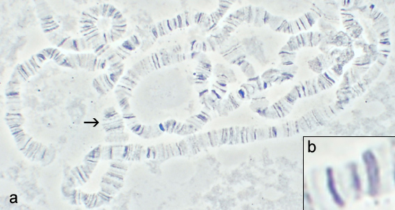
Polytene chromosome of Drosophila. (b) Displays the chromomeric and interchromomeric bands of the chromosome.

Physical properties

Chromomeres are organized in a discontinuous linear pattern along the condensed chromosomes (pachytene chromosomes) during the prophase stage of meiosis. The linear pattern of chromomeres is linked to the arrangement of genes along the chromosome. Chromomeres contain genes and sometimes clusters of genes within their structure. Aggregates of chromomeres are known as chromonemata.

Cohesive proteins SMC3 and hRAD21(plays a role in sister chromatid cohesion) are found within chromomeres at high concentrations, and maintain the proper structure of chromomeres. The protein XCAP-D2 is also present at high concentrations within the chromomere, and acts as a condensin component. High concentrations of tandem repeats of the heterochromatin protein HP1β builds up within the chromomere. In regions where loops attach to chromomeres, there is hyperacetylation of histone 4. The extra acetylation loosens chromatin from a condensed form, making it more accessible to proteins involved in transcription.

Functions

Chromomeres are known as the structural subunit of a chromosome. The arrangement of chromomere structure can aid in control of gene expression.

Maps of chromomeres can be made for use in genetic and evolutionary studies. Chromomeric maps can be used to locate the exact position of genes on a chromosome. Chromomeric maps can be used to analyze chromosome aberrations, and find correlations between the aberrations and their effects on genes near breakpoints.Lampbrush chromosomes and chromomeres

Chromomeres display different properties and behaviours when associated with lampbrush chromosomes. Although found all across the lampbrush chromosome, they are not organized in a clear pattern along as they are in normal pachytene chromosomes of meiosis.  The two sister chromatids of a lampbrush chromosome separate fully, forming lateral loops that extend from chromomeres, and act as transcription complexes. The lateral loops are areas where the chromosomes are transcriptionally active. The loops define where the chromomere will be positioned. The chromomere is an organization of regions of non-transcribed chromatin. These regions of chromatin that have not been transcribed are located at the ends of the loops that were formed by the sister chromatids of a lampbrush chromosome. Each chromomere can have up to several pairs of loops from lampbrush chromosomes originating from it, as well as micro-loops that cannot be detected with a light microscope.

Chromomeres of lampbrush chromosomes act as structural units, and are not considered to be genetic units participating in transcription. The arrangement of a chromosome into chromomeric units happens almost simultaneously with the start of transcription. The loop structures extending from chromomeres are maintained by a high level of transcriptional activity and structural proteins of the chromosome. Chromatin within the chromomere are held in position by a variety of histone modifications and epigenetic markers.

Methyl-CpG-binding protein 2 (MeCP2) is a protein that binds to methylated DNA. MeCP2 has been found to associate most strongly with transcriptionally inactive chromomere domains. MeCP2 binding patterns to chromomere domains are proportional to the density of chromomeric DNA found on a chromosome. The binding of MeCP2 to chromomeric regions represents regions of chromatin that are highly dynamic on the lampbrush chromosome.

Nucleosome

From Wikipedia, the free encyclopedia
https://en.wikipedia.org/wiki/Nucleosome
Basic units of chromatin structure

A nucleosome is the basic structural unit of DNA packaging in eukaryotes. The structure of a nucleosome consists of a segment of DNA wound around eight histone proteins and resembles thread wrapped around a spool. The nucleosome is the fundamental subunit of chromatin. Each nucleosome is composed of a little less than two turns of DNA wrapped around a set of eight proteins called histones, which are known as a histone octamer. Each histone octamer is composed of two copies each of the histone proteins H2A, H2B, H3, and H4.

DNA must be compacted into nucleosomes to fit within the cell nucleus. In addition to nucleosome wrapping, eukaryotic chromatin is further compacted by being folded into a series of more complex structures, eventually forming a chromosome. Each human cell contains about 30 million nucleosomes.

Nucleosomes are thought to carry epigenetically inherited information in the form of covalent modifications of their core histones. Nucleosome positions in the genome are not random, and it is important to know where each nucleosome is located because this determines the accessibility of the DNA to regulatory proteins.

Nucleosomes were first observed as particles in the electron microscope by Don and Ada Olins in 1974, and their existence and structure (as histone octamers surrounded by approximately 200 base pairs of DNA) were proposed by Roger Kornberg. The role of the nucleosome as a regulator of transcription was demonstrated by Lorch et al. in vitro in 1987 and by Han and Grunstein and Clark-Adams et al. in vivo in 1988.

The nucleosome core particle consists of approximately 146 base pairs (bp) of DNA wrapped in 1.67 left-handed superhelical turns around a histone octamer, consisting of 2 copies each of the core histones H2A, H2B, H3, and H4. Core particles are connected by stretches of linker DNA, which can be up to about 80 bp long. Technically, a nucleosome is defined as the core particle plus one of these linker regions; however the word is often synonymous with the core particle. Genome-wide nucleosome positioning maps are now available for many model organisms and human cells.

Linker histones such as H1 and its isoforms are involved in chromatin compaction and sit at the base of the nucleosome near the DNA entry and exit binding to the linker region of the DNA. Non-condensed nucleosomes without the linker histone resemble "beads on a string of DNA" under an electron microscope.

In contrast to most eukaryotic cells, mature sperm cells largely use protamines to package their genomic DNA, most likely to achieve an even higher packaging ratio. Histone equivalents and a simplified chromatin structure have also been found in Archaea, suggesting that eukaryotes are not the only organisms that use nucleosomes.

Structure

Structure of the core particle

The crystal structure of the nucleosome core particle consisting of H2A , H2B , H3 and H4 core histones, and DNA. The view is from the top through the superhelical axis.

Overview

Pioneering structural studies in the 1980s by Aaron Klug's group provided the first evidence that an octamer of histone proteins wraps DNA around itself in about 1.7 turns of a left-handed superhelix. In 1997 the first near atomic resolution crystal structure of the nucleosome was solved by the Richmond group at the ETH Zurich, showing the most important details of the particle. The human alpha satellite palindromic DNA critical to achieving the 1997 nucleosome crystal structure was developed by the Bunick group at Oak Ridge National Laboratory in Tennessee. The structures of over 20 different nucleosome core particles have been solved to date, including those containing histone variants and histones from different species. The structure of the nucleosome core particle is remarkably conserved, and even a change of over 100 residues between frog and yeast histones results in electron density maps with an overall root mean square deviation of only 1.6Å.

The nucleosome core particle (NCP)

The nucleosome core particle (shown in the figure) consists of about 146 base pair of DNA wrapped in 1.67 left-handed superhelical turns around the histone octamer, consisting of 2 copies each of the core histones H2A, H2B, H3, and H4. Adjacent nucleosomes are joined by a stretch of free DNA termed linker DNA (which varies from 10 - 80 bp in length depending on species and tissue type). The whole structure generates a cylinder of diameter 11 nm and a height of 5.5 nm.

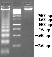
Apoptotic DNA laddering. Digested chromatin is in the first lane; the second contains DNA standard to compare lengths.
Scheme of nucleosome organization
The crystal structure of the nucleosome core particle (PDB: 1EQZ​)

Nucleosome core particles are observed when chromatin in interphase is treated to cause the chromatin to unfold partially. The resulting image, via an electron microscope, is "beads on a string". The string is the DNA, while each bead in the nucleosome is a core particle. The nucleosome core particle is composed of DNA and histone proteins.

Partial DNAse digestion of chromatin reveals its nucleosome structure. Because DNA portions of nucleosome core particles are less accessible for DNAse than linking sections, DNA gets digested into fragments of lengths equal to multiplicity of distance between nucleosomes (180, 360, 540 base pairs etc.). Hence a very characteristic pattern similar to a ladder is visible during gel electrophoresis of that DNA. Such digestion can occur also under natural conditions during apoptosis ("cell suicide" or programmed cell death), because autodestruction of DNA typically is its role.

Protein interactions within the nucleosome

The core histone proteins contains a characteristic structural motif termed the "histone fold", which consists of three alpha-helices (α1-3) separated by two loops (L1-2). In solution, the histones form H2A-H2B heterodimers and H3-H4 heterotetramers. Histones dimerise about their long α2 helices in an anti-parallel orientation, and, in the case of H3 and H4, two such dimers form a 4-helix bundle stabilised by extensive H3-H3' interaction. The H2A/H2B dimer binds onto the H3/H4 tetramer due to interactions between H4 and H2B, which include the formation of a hydrophobic cluster. The histone octamer is formed by a central H3/H4 tetramer sandwiched between two H2A/H2B dimers. Due to the highly basic charge of all four core histones, the histone octamer is stable only in the presence of DNA or very high salt concentrations.

Histone - DNA interactions

The nucleosome contains over 120 direct protein-DNA interactions and several hundred water-mediated ones. Direct protein - DNA interactions are not spread evenly about the octamer surface but rather located at discrete sites. These are due to the formation of two types of DNA binding sites within the octamer; the α1α1 site, which uses the α1 helix from two adjacent histones, and the L1L2 site formed by the L1 and L2 loops. Salt links and hydrogen bonding between both side-chain basic and hydroxyl groups and main-chain amides with the DNA backbone phosphates form the bulk of interactions with the DNA. This is important, given that the ubiquitous distribution of nucleosomes along genomes requires it to be a non-sequence-specific DNA-binding factor. Although nucleosomes tend to prefer some DNA sequences over others, they are capable of binding practically to any sequence, which is thought to be due to the flexibility in the formation of these water-mediated interactions. In addition, non-polar interactions are made between protein side-chains and the deoxyribose groups, and an arginine side-chain intercalates into the DNA minor groove at all 14 sites where it faces the octamer surface. The distribution and strength of DNA-binding sites about the octamer surface distorts the DNA within the nucleosome core. The DNA is non-uniformly bent and also contains twist defects. The twist of free B-form DNA in solution is 10.5 bp per turn. However, the overall twist of nucleosomal DNA is only 10.2 bp per turn, varying from a value of 9.4 to 10.9 bp per turn.

Histone tail domains

The histone tail extensions constitute up to 30% by mass of histones, but are not visible in the crystal structures of nucleosomes due to their high intrinsic flexibility, and have been thought to be largely unstructured. The N-terminal tails of histones H3 and H2B pass through a channel formed by the minor grooves of the two DNA strands, protruding from the DNA every 20 bp. The N-terminal tail of histone H4, on the other hand, has a region of highly basic amino acids (16–25), which, in the crystal structure, forms an interaction with the highly acidic surface region of a H2A-H2B dimer of another nucleosome, being potentially relevant for the higher-order structure of nucleosomes. This interaction is thought to occur under physiological conditions also, and suggests that acetylation of the H4 tail distorts the higher-order structure of chromatin.

Higher order structure

The current chromatin compaction model

The organization of the DNA that is achieved by the nucleosome cannot fully explain the packaging of DNA observed in the cell nucleus. Further compaction of chromatin into the cell nucleus is necessary, but it is not yet well understood. The current understanding is that repeating nucleosomes with intervening "linker" DNA form a 10-nm-fiber, described as "beads on a string", and have a packing ratio of about five to ten. A chain of nucleosomes can be arranged in a 30 nm fiber, a compacted structure with a packing ratio of ~50 and whose formation is dependent on the presence of the H1 histone.

A crystal structure of a tetranucleosome has been presented and used to build up a proposed structure of the 30 nm fiber as a two-start helix. There is still a certain amount of contention regarding this model, as it is incompatible with recent electron microscopy data. Beyond this, the structure of chromatin is poorly understood, but it is classically suggested that the 30 nm fiber is arranged into loops along a central protein scaffold to form transcriptionally active euchromatin. Further compaction leads to transcriptionally inactive heterochromatin.

Dynamics

Although the nucleosome is a very stable protein-DNA complex, it is not static and has been shown to undergo a number of different structural re-arrangements including nucleosome sliding and DNA site exposure. Depending on the context, nucleosomes can inhibit or facilitate transcription factor binding. Nucleosome positions are controlled by three major contributions: First, the intrinsic binding affinity of the histone octamer depends on the DNA sequence. Second, the nucleosome can be displaced or recruited by the competitive or cooperative binding of other protein factors. Third, the nucleosome may be actively translocated by ATP-dependent remodeling complexes.

Nucleosome sliding

When incubated thermally, nucleosomes reconstituted onto the 5S DNA positioning sequence were able to reposition themselves translationally onto adjacent sequences. This repositioning does not require disruption of the histone octamer but is consistent with nucleosomes being able to "slide" along the DNA in cis. CTCF binding sites act as nucleosome positioning anchors so that, when used to align various genomic signals, multiple flanking nucleosomes can be readily identified. Although nucleosomes are intrinsically mobile, eukaryotes have evolved a large family of ATP-dependent chromatin remodelling enzymes to alter chromatin structure, many of which do so via nucleosome sliding. Nucleosome sliding is one of the possible mechanism for large scale tissue specific expression of genes. The transcription start site for genes expressed in a particular tissue, are nucleosome depleted while, the same set of genes in other tissue where they are not expressed, are nucleosome bound.

DNA site exposure

Nucleosomal DNA is in equilibrium between a wrapped and unwrapped state. DNA within the nucleosome remains fully wrapped for only 250 ms before it is unwrapped for 10-50 ms and then rapidly rewrapped, as measured using time-resolved FRET. This implies that DNA does not need to be actively dissociated from the nucleosome but that there is a significant fraction of time during which it is fully accessible. Introducing a DNA-binding sequence within the nucleosome increases the accessibility of adjacent regions of DNA when bound.

This propensity for DNA within the nucleosome to "breathe" has important functional consequences for all DNA-binding proteins that operate in a chromatin environment. In particular, the dynamic breathing of nucleosomes plays an important role in restricting the advancement of RNA polymerase II during transcription elongation.

Nucleosome free region

Promoters of active genes have nucleosome free regions (NFR). This allows for promoter DNA accessibility to various proteins, such as transcription factors. Nucleosome free region typically spans for 200 nucleotides in S. cerevisiae Well-positioned nucleosomes form boundaries of NFR. These nucleosomes are called +1-nucleosome and −1-nucleosome and are located at canonical distances downstream and upstream, respectively, from transcription start site. +1-nucleosome and several downstream nucleosomes also tend to incorporate H2A.Z histone variant.

Modulating nucleosome structure

Eukaryotic genomes are ubiquitously associated into chromatin; however, cells must spatially and temporally regulate specific loci independently of bulk chromatin. In order to achieve the high level of control required to co-ordinate nuclear processes such as DNA replication, repair, and transcription, cells have developed a variety of means to locally and specifically modulate chromatin structure and function. This can involve covalent modification of histones, the incorporation of histone variants, and non-covalent remodelling by ATP-dependent remodeling enzymes.

Histone post-translational modifications

Histone tails and their function in chromatin formation

Since they were discovered in the mid-1960s, histone modifications have been predicted to affect transcription. The fact that most of the early post-translational modifications found were concentrated within the tail extensions that protrude from the nucleosome core lead to two main theories regarding the mechanism of histone modification. The first of the theories suggested that they may affect electrostatic interactions between the histone tails and DNA to "loosen" chromatin structure. Later it was proposed that combinations of these modifications may create binding epitopes with which to recruit other proteins. Recently, given that more modifications have been found in the structured regions of histones, it has been put forward that these modifications may affect histone-DNA and histone-histone  interactions within the nucleosome core. Modifications (such as acetylation or phosphorylation) that lower the charge of the globular histone core are predicted to "loosen" core-DNA association; the strength of the effect depends on location of the modification within the core. Some modifications have been shown to be correlated with gene silencing; others seem to be correlated with gene activation. Common modifications include acetylation, methylation, or ubiquitination of lysine; methylation of arginine; and phosphorylation of serine. The information stored in this way is considered epigenetic, since it is not encoded in the DNA but is still inherited to daughter cells. The maintenance of a repressed or activated status of a gene is often necessary for cellular differentiation.

Histone variants

Although histones are remarkably conserved throughout evolution, several variant forms have been identified. This diversification of histone function is restricted to H2A and H3, with H2B and H4 being mostly invariant. H2A can be replaced by H2AZ (which leads to reduced nucleosome stability) or H2AX (which is associated with DNA repair and T cell differentiation), whereas the inactive X chromosomes in mammals are enriched in macroH2A. H3 can be replaced by H3.3 (which correlates with activate genes and regulatory elements) and in centromeres H3 is replaced by CENPA.

ATP-dependent nucleosome remodeling

A number of distinct reactions are associated with the term ATP-dependent chromatin remodeling. Remodeling enzymes have been shown to slide nucleosomes along DNA, disrupt histone-DNA contacts to the extent of destabilizing the H2A/H2B dimer and to generate negative superhelical torsion in DNA and chromatin. Recently, the Swr1 remodeling enzyme has been shown to introduce the variant histone H2A.Z into nucleosomes. At present, it is not clear if all of these represent distinct reactions or merely alternative outcomes of a common mechanism. What is shared between all, and indeed the hallmark of ATP-dependent chromatin remodeling, is that they all result in altered DNA accessibility.

Studies looking at gene activation in vivo and, more astonishingly, remodeling in vitro have revealed that chromatin remodeling events and transcription-factor binding are cyclical and periodic in nature. While the consequences of this for the reaction mechanism of chromatin remodeling are not known, the dynamic nature of the system may allow it to respond faster to external stimuli. A recent study indicates that nucleosome positions change significantly during mouse embryonic stem cell development, and these changes are related to binding of developmental transcription factors.

Dynamic nucleosome remodelling across the Yeast genome

Studies in 2007 have catalogued nucleosome positions in yeast and shown that nucleosomes are depleted in promoter regions and origins of replication. About 80% of the yeast genome appears to be covered by nucleosomes and the pattern of nucleosome positioning clearly relates to DNA regions that regulate transcription, regions that are transcribed and regions that initiate DNA replication. Most recently, a new study examined dynamic changes in nucleosome repositioning during a global transcriptional reprogramming event to elucidate the effects on nucleosome displacement during genome-wide transcriptional changes in yeast (Saccharomyces cerevisiae). The results suggested that nucleosomes that were localized to promoter regions are displaced in response to stress (like heat shock). In addition, the removal of nucleosomes usually corresponded to transcriptional activation and the replacement of nucleosomes usually corresponded to transcriptional repression, presumably because transcription factor binding sites became more or less accessible, respectively. In general, only one or two nucleosomes were repositioned at the promoter to effect these transcriptional changes. However, even in chromosomal regions that were not associated with transcriptional changes, nucleosome repositioning was observed, suggesting that the covering and uncovering of transcriptional DNA does not necessarily produce a transcriptional event. After transcription, the rDNA region has to protected from any damage, it suggested HMGB proteins play a major role in protecting the nucleosome free region.

DNA Twist Defects

DNA twist defects are when the addition of one or a few base pairs from one DNA segment are transferred to the next segment resulting in a change of the DNA twist. This will not only change the twist of the DNA but it will also change the length. This twist defect eventually moves around the nucleosome through the transferring of the base pair, this means DNA twists can cause nucleosome sliding. Nucleosome crystal structures have shown that superhelix location 2 and 5 on the nucleosome are commonly found to be where DNA twist defects occur as these are common remodeler binding sites. There are a variety of chromatin remodelers but all share the existence of an ATPase motor which facilitates chromatin sliding on DNA through the binding and hydrolysis of ATP. ATPase has an open and closed state. When the ATPase motor is changing from open and closed states, the DNA duplex changes geometry and exhibits base pair tilting. The initiation of the twist defects via the ATPase motor causes tension to accumulate around the remodeler site. The tension is released when the sliding of DNA has been completed throughout the nucleosome via the spread of two twist defects (one on each strand) in opposite directions.

Nucleosome assembly in vitro

Diagram of nucleosome assembly

Nucleosomes can be assembled in vitro by either using purified native or recombinant histones. One standard technique of loading the DNA around the histones involves the use of salt dialysis. A reaction consisting of the histone octamers and a naked DNA template can be incubated together at a salt concentration of 2 M. By steadily decreasing the salt concentration, the DNA will equilibrate to a position where it is wrapped around the histone octamers, forming nucleosomes. In appropriate conditions, this reconstitution process allows for the nucleosome positioning affinity of a given sequence to be mapped experimentally.

Disulfide crosslinked nucleosome core particles

A recent advance in the production of nucleosome core particles with enhanced stability involves site-specific disulfide crosslinks. Two different crosslinks can be introduced into the nucleosome core particle. A first one crosslinks the two copies of H2A via an introduced cysteine (N38C) resulting in histone octamer which is stable against H2A/H2B dimer loss during nucleosome reconstitution. A second crosslink can be introduced between the H3 N-terminal histone tail and the nucleosome DNA ends via an incorporated convertible nucleotide. The DNA-histone octamer crosslink stabilizes the nucleosome core particle against DNA dissociation at very low particle concentrations and at elevated salt concentrations.

Nucleosome assembly in vivo

Steps in nucleosome assembly

Nucleosomes are the basic packing unit of genomic DNA built from histone proteins around which DNA is coiled. They serve as a scaffold for formation of higher order chromatin structure as well as for a layer of regulatory control of gene expression. Nucleosomes are quickly assembled onto newly synthesized DNA behind the replication fork.

H3 and H4

Histones H3 and H4 from disassembled old nucleosomes are kept in the vicinity and randomly distributed on the newly synthesized DNA. They are assembled by the chromatin assembly factor 1 (CAF-1) complex, which consists of three subunits (p150, p60, and p48). Newly synthesized H3 and H4 are assembled by the replication coupling assembly factor (RCAF). RCAF contains the subunit Asf1, which binds to newly synthesized H3 and H4 proteins. The old H3 and H4 proteins retain their chemical modifications which contributes to the passing down of the epigenetic signature. The newly synthesized H3 and H4 proteins are gradually acetylated at different lysine residues as part of the chromatin maturation process. It is also thought that the old H3 and H4 proteins in the new nucleosomes recruit histone modifying enzymes that mark the new histones, contributing to epigenetic memory.

H2A and H2B

In contrast to old H3 and H4, the old H2A and H2B histone proteins are released and degraded; therefore, newly assembled H2A and H2B proteins are incorporated into new nucleosomes. H2A and H2B are assembled into dimers which are then loaded onto nucleosomes by the nucleosome assembly protein-1 (NAP-1) which also assists with nucleosome sliding. The nucleosomes are also spaced by ATP-dependent nucleosome-remodeling complexes containing enzymes such as Isw1 Ino80, and Chd1, and subsequently assembled into higher order structure.

Pain in animals

From Wikipedia, the free encyclopedia https://en.wikipedia.org/wiki/Pain_in_animals ...