Search This Blog

Wednesday, August 28, 2024

Cell (biology)

From Wikipedia, the free encyclopedia
Cell
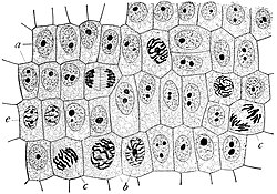
Onion (Allium cepa) root cells in different phases of the cell cycle (drawn by E. B. Wilson, 1900)
 
A eukaryotic cell (left) and prokaryotic cell (right)

The cell is the basic structural and functional unit of all forms of life. Every cell consists of cytoplasm enclosed within a membrane; many cells contain organelles, each with a specific function. The term comes from the Latin word cellula meaning 'small room'. Most cells are only visible under a microscope. Cells emerged on Earth about 4 billion years ago. All cells are capable of replication, protein synthesis, and motility.

Cells are broadly categorized into two types: eukaryotic cells, which possess a nucleus, and prokaryotic cells, which lack a nucleus but have a nucleoid region. Prokaryotes are single-celled organisms such as bacteria, whereas eukaryotes can be either single-celled, such as amoebae, or multicellular, such as some algae, plants, animals, and fungi. Eukaryotic cells contain organelles including mitochondria, which provide energy for cell functions; chloroplasts, which create sugars by photosynthesis, in plants; and ribosomes, which synthesise proteins.

Cells were discovered by Robert Hooke in 1665, who named them after their resemblance to cells inhabited by Christian monks in a monastery. Cell theory, developed in 1839 by Matthias Jakob Schleiden and Theodor Schwann, states that all organisms are composed of one or more cells, that cells are the fundamental unit of structure and function in all living organisms, and that all cells come from pre-existing cells.

Cell types

Cells are broadly categorized into two types: eukaryotic cells, which possess a nucleus, and prokaryotic cells, which lack a nucleus but have a nucleoid region. Prokaryotes are single-celled organisms, whereas eukaryotes can be either single-celled or multicellular.

Prokaryotic cells

Structure of a typical prokaryotic cell

Prokaryotes include bacteria and archaea, two of the three domains of life. Prokaryotic cells were the first form of life on Earth, characterized by having vital biological processes including cell signaling. They are simpler and smaller than eukaryotic cells, and lack a nucleus, and other membrane-bound organelles. The DNA of a prokaryotic cell consists of a single circular chromosome that is in direct contact with the cytoplasm. The nuclear region in the cytoplasm is called the nucleoid. Most prokaryotes are the smallest of all organisms, ranging from 0.5 to 2.0 μm in diameter.

A prokaryotic cell has three regions:

  • Enclosing the cell is the cell envelope, generally consisting of a plasma membrane covered by a cell wall which, for some bacteria, may be further covered by a third layer called a capsule. Though most prokaryotes have both a cell membrane and a cell wall, there are exceptions such as Mycoplasma (bacteria) and Thermoplasma (archaea) which only possess the cell membrane layer. The envelope gives rigidity to the cell and separates the interior of the cell from its environment, serving as a protective filter. The cell wall consists of peptidoglycan in bacteria and acts as an additional barrier against exterior forces. It also prevents the cell from expanding and bursting (cytolysis) from osmotic pressure due to a hypotonic environment. Some eukaryotic cells (plant cells and fungal cells) also have a cell wall.
  • Inside the cell is the cytoplasmic region that contains the genome (DNA), ribosomes and various sorts of inclusions. The genetic material is freely found in the cytoplasm. Prokaryotes can carry extrachromosomal DNA elements called plasmids, which are usually circular. Linear bacterial plasmids have been identified in several species of spirochete bacteria, including members of the genus Borrelia notably Borrelia burgdorferi, which causes Lyme disease. Though not forming a nucleus, the DNA is condensed in a nucleoid. Plasmids encode additional genes, such as antibiotic resistance genes.
  • On the outside, some prokaryotes have flagella and pili that project from the cell's surface. These are structures made of proteins that facilitate movement and communication between cells.

Eukaryotic cells

Structure of a typical animal cell
Structure of a typical plant cell

Plants, animals, fungi, slime moulds, protozoa, and algae are all eukaryotic. These cells are about fifteen times wider than a typical prokaryote and can be as much as a thousand times greater in volume. The main distinguishing feature of eukaryotes as compared to prokaryotes is compartmentalization: the presence of membrane-bound organelles (compartments) in which specific activities take place. Most important among these is a cell nucleus, an organelle that houses the cell's DNA. This nucleus gives the eukaryote its name, which means "true kernel (nucleus)". Some of the other differences are:

  • The plasma membrane resembles that of prokaryotes in function, with minor differences in the setup. Cell walls may or may not be present.
  • The eukaryotic DNA is organized in one or more linear molecules, called chromosomes, which are associated with histone proteins. All chromosomal DNA is stored in the cell nucleus, separated from the cytoplasm by a membrane. Some eukaryotic organelles such as mitochondria also contain some DNA.
  • Many eukaryotic cells are ciliated with primary cilia. Primary cilia play important roles in chemosensation, mechanosensation, and thermosensation. Each cilium may thus be "viewed as a sensory cellular antennae that coordinates a large number of cellular signaling pathways, sometimes coupling the signaling to ciliary motility or alternatively to cell division and differentiation."
  • Motile eukaryotes can move using motile cilia or flagella. Motile cells are absent in conifers and flowering plants. Eukaryotic flagella are more complex than those of prokaryotes.
Comparison of features of prokaryotic and eukaryotic cells

Prokaryotes Eukaryotes
Typical organisms bacteria, archaea protists, algae, fungi, plants, animals
Typical size ~ 1–5 μm ~ 10–100 μm
Type of nucleus nucleoid region; no true nucleus true nucleus with double membrane
DNA circular (usually) linear molecules (chromosomes) with histone proteins
RNA/protein synthesis coupled in the cytoplasm RNA synthesis in the nucleus
protein synthesis in the cytoplasm
Ribosomes 50S and 30S 60S and 40S
Cytoplasmic structure very few structures highly structured by endomembranes and a cytoskeleton
Cell movement flagella made of flagellin flagella and cilia containing microtubules; lamellipodia and filopodia containing actin
Mitochondria none one to several thousand
Chloroplasts none in algae and plants
Organization usually single cells single cells, colonies, higher multicellular organisms with specialized cells
Cell division binary fission (simple division) mitosis (fission or budding)
meiosis
Chromosomes single chromosome more than one chromosome
Membranes cell membrane Cell membrane and membrane-bound organelles

Many groups of eukaryotes are single-celled. Among the many-celled groups are animals and plants. The number of cells in these groups vary with species; it has been estimated that the human body contains around 37 trillion (3.72×10¹³) cells, and more recent studies put this number at around 30 trillion (~36 trillion cells in the male, ~28 trillion in the female).

Subcellular components

All cells, whether prokaryotic or eukaryotic, have a membrane that envelops the cell, regulates what moves in and out (selectively permeable), and maintains the electric potential of the cell. Inside the membrane, the cytoplasm takes up most of the cell's volume. Except red blood cells, which lack a cell nucleus and most organelles to accommodate maximum space for hemoglobin, all cells possess DNA, the hereditary material of genes, and RNA, containing the information necessary to build various proteins such as enzymes, the cell's primary machinery. There are also other kinds of biomolecules in cells. This article lists these primary cellular components, then briefly describes their function.

Cell membrane

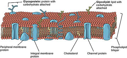
Detailed diagram of lipid bilayer of cell membrane

The cell membrane, or plasma membrane, is a selectively permeable biological membrane that surrounds the cytoplasm of a cell. In animals, the plasma membrane is the outer boundary of the cell, while in plants and prokaryotes it is usually covered by a cell wall. This membrane serves to separate and protect a cell from its surrounding environment and is made mostly from a double layer of phospholipids, which are amphiphilic (partly hydrophobic and partly hydrophilic). Hence, the layer is called a phospholipid bilayer, or sometimes a fluid mosaic membrane. Embedded within this membrane is a macromolecular structure called the porosome the universal secretory portal in cells and a variety of protein molecules that act as channels and pumps that move different molecules into and out of the cell. The membrane is semi-permeable, and selectively permeable, in that it can either let a substance (molecule or ion) pass through freely, to a limited extent or not at all. Cell surface membranes also contain receptor proteins that allow cells to detect external signaling molecules such as hormones.

Cytoskeleton

A fluorescent image of an endothelial cell. Nuclei are stained blue, mitochondria are stained red, and microfilaments are stained green.

The cytoskeleton acts to organize and maintain the cell's shape; anchors organelles in place; helps during endocytosis, the uptake of external materials by a cell, and cytokinesis, the separation of daughter cells after cell division; and moves parts of the cell in processes of growth and mobility. The eukaryotic cytoskeleton is composed of microtubules, intermediate filaments and microfilaments. In the cytoskeleton of a neuron the intermediate filaments are known as neurofilaments. There are a great number of proteins associated with them, each controlling a cell's structure by directing, bundling, and aligning filaments. The prokaryotic cytoskeleton is less well-studied but is involved in the maintenance of cell shape, polarity and cytokinesis. The subunit protein of microfilaments is a small, monomeric protein called actin. The subunit of microtubules is a dimeric molecule called tubulin. Intermediate filaments are heteropolymers whose subunits vary among the cell types in different tissues. Some of the subunit proteins of intermediate filaments include vimentin, desmin, lamin (lamins A, B and C), keratin (multiple acidic and basic keratins), and neurofilament proteins (NF–L, NF–M).

Genetic material

Deoxyribonucleic acid (DNA)

Two different kinds of genetic material exist: deoxyribonucleic acid (DNA) and ribonucleic acid (RNA). Cells use DNA for their long-term information storage. The biological information contained in an organism is encoded in its DNA sequence. RNA is used for information transport (e.g., mRNA) and enzymatic functions (e.g., ribosomal RNA). Transfer RNA (tRNA) molecules are used to add amino acids during protein translation.

Prokaryotic genetic material is organized in a simple circular bacterial chromosome in the nucleoid region of the cytoplasm. Eukaryotic genetic material is divided into different, linear molecules called chromosomes inside a discrete nucleus, usually with additional genetic material in some organelles like mitochondria and chloroplasts (see endosymbiotic theory).

A human cell has genetic material contained in the cell nucleus (the nuclear genome) and in the mitochondria (the mitochondrial genome). In humans, the nuclear genome is divided into 46 linear DNA molecules called chromosomes, including 22 homologous chromosome pairs and a pair of sex chromosomes. The mitochondrial genome is a circular DNA molecule distinct from nuclear DNA. Although the mitochondrial DNA is very small compared to nuclear chromosomes, it codes for 13 proteins involved in mitochondrial energy production and specific tRNAs.

Foreign genetic material (most commonly DNA) can also be artificially introduced into the cell by a process called transfection. This can be transient, if the DNA is not inserted into the cell's genome, or stable, if it is. Certain viruses also insert their genetic material into the genome.

Organelles

Organelles are parts of the cell that are adapted and/or specialized for carrying out one or more vital functions, analogous to the organs of the human body (such as the heart, lung, and kidney, with each organ performing a different function). Both eukaryotic and prokaryotic cells have organelles, but prokaryotic organelles are generally simpler and are not membrane-bound.

There are several types of organelles in a cell. Some (such as the nucleus and Golgi apparatus) are typically solitary, while others (such as mitochondria, chloroplasts, peroxisomes and lysosomes) can be numerous (hundreds to thousands). The cytosol is the gelatinous fluid that fills the cell and surrounds the organelles.

Eukaryotic
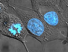
Human cancer cells, specifically HeLa cells, with DNA stained blue. The central and rightmost cell are in interphase, so their DNA is diffuse and the entire nuclei are labelled. The cell on the left is going through mitosis and its chromosomes have condensed.
  • Cell nucleus: A cell's information center, the cell nucleus is the most conspicuous organelle found in a eukaryotic cell. It houses the cell's chromosomes, and is the place where almost all DNA replication and RNA synthesis (transcription) occur. The nucleus is spherical and separated from the cytoplasm by a double membrane called the nuclear envelope, space between these two membrane is called perinuclear space. The nuclear envelope isolates and protects a cell's DNA from various molecules that could accidentally damage its structure or interfere with its processing. During processing, DNA is transcribed, or copied into a special RNA, called messenger RNA (mRNA). This mRNA is then transported out of the nucleus, where it is translated into a specific protein molecule. The nucleolus is a specialized region within the nucleus where ribosome subunits are assembled. In prokaryotes, DNA processing takes place in the cytoplasm.
  • Mitochondria and chloroplasts: generate energy for the cell. Mitochondria are self-replicating double membrane-bound organelles that occur in various numbers, shapes, and sizes in the cytoplasm of all eukaryotic cells. Respiration occurs in the cell mitochondria, which generate the cell's energy by oxidative phosphorylation, using oxygen to release energy stored in cellular nutrients (typically pertaining to glucose) to generate ATP (aerobic respiration). Mitochondria multiply by binary fission, like prokaryotes. Chloroplasts can only be found in plants and algae, and they capture the sun's energy to make carbohydrates through photosynthesis.
Diagram of the endomembrane system
  • Endoplasmic reticulum: The endoplasmic reticulum (ER) is a transport network for molecules targeted for certain modifications and specific destinations, as compared to molecules that float freely in the cytoplasm. The ER has two forms: the rough ER, which has ribosomes on its surface that secrete proteins into the ER, and the smooth ER, which lacks ribosomes. The smooth ER plays a role in calcium sequestration and release and also helps in synthesis of lipid.
  • Golgi apparatus: The primary function of the Golgi apparatus is to process and package the macromolecules such as proteins and lipids that are synthesized by the cell.
  • Lysosomes and peroxisomes: Lysosomes contain digestive enzymes (acid hydrolases). They digest excess or worn-out organelles, food particles, and engulfed viruses or bacteria. Peroxisomes have enzymes that rid the cell of toxic peroxides, Lysosomes are optimally active in an acidic environment. The cell could not house these destructive enzymes if they were not contained in a membrane-bound system.
  • Centrosome: the cytoskeleton organizer: The centrosome produces the microtubules of a cell—a key component of the cytoskeleton. It directs the transport through the ER and the Golgi apparatus. Centrosomes are composed of two centrioles which lie perpendicular to each other in which each has an organization like a cartwheel, which separate during cell division and help in the formation of the mitotic spindle. A single centrosome is present in the animal cells. They are also found in some fungi and algae cells.
  • Vacuoles: Vacuoles sequester waste products and in plant cells store water. They are often described as liquid filled spaces and are surrounded by a membrane. Some cells, most notably Amoeba, have contractile vacuoles, which can pump water out of the cell if there is too much water. The vacuoles of plant cells and fungal cells are usually larger than those of animal cells. Vacuoles of plant cells are surrounded by a membrane which transports ions against concentration gradients.

Eukaryotic and prokaryotic

  • Ribosomes: The ribosome is a large complex of RNA and protein molecules. They each consist of two subunits, and act as an assembly line where RNA from the nucleus is used to synthesise proteins from amino acids. Ribosomes can be found either floating freely or bound to a membrane (the rough endoplasmatic reticulum in eukaryotes, or the cell membrane in prokaryotes).
  • Plastids: Plastid are membrane-bound organelle generally found in plant cells and euglenoids and contain specific pigments, thus affecting the colour of the plant and organism. And these pigments also helps in food storage and tapping of light energy. There are three types of plastids based upon the specific pigments. Chloroplasts contain chlorophyll and some carotenoid pigments which helps in the tapping of light energy during photosynthesis. Chromoplasts contain fat-soluble carotenoid pigments like orange carotene and yellow xanthophylls which helps in synthesis and storage. Leucoplasts are non-pigmented plastids and helps in storage of nutrients.

Structures outside the cell membrane

Many cells also have structures which exist wholly or partially outside the cell membrane. These structures are notable because they are not protected from the external environment by the cell membrane. In order to assemble these structures, their components must be carried across the cell membrane by export processes.

Cell wall

Many types of prokaryotic and eukaryotic cells have a cell wall. The cell wall acts to protect the cell mechanically and chemically from its environment, and is an additional layer of protection to the cell membrane. Different types of cell have cell walls made up of different materials; plant cell walls are primarily made up of cellulose, fungi cell walls are made up of chitin and bacteria cell walls are made up of peptidoglycan.

Prokaryotic

Capsule

A gelatinous capsule is present in some bacteria outside the cell membrane and cell wall. The capsule may be polysaccharide as in pneumococci, meningococci or polypeptide as Bacillus anthracis or hyaluronic acid as in streptococci. Capsules are not marked by normal staining protocols and can be detected by India ink or methyl blue, which allows for higher contrast between the cells for observation.

Flagella

Flagella are organelles for cellular mobility. The bacterial flagellum stretches from cytoplasm through the cell membrane(s) and extrudes through the cell wall. They are long and thick thread-like appendages, protein in nature. A different type of flagellum is found in archaea and a different type is found in eukaryotes.

Fimbriae

A fimbria (plural fimbriae also known as a pilus, plural pili) is a short, thin, hair-like filament found on the surface of bacteria. Fimbriae are formed of a protein called pilin (antigenic) and are responsible for the attachment of bacteria to specific receptors on human cells (cell adhesion). There are special types of pili involved in bacterial conjugation.

Cellular processes
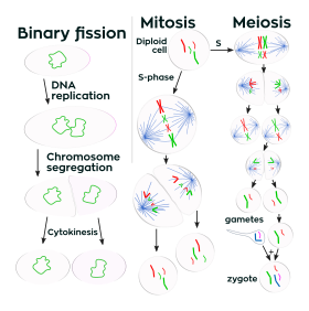
Prokaryotes divide by binary fission, while eukaryotes divide by mitosis or meiosis.

Replication

Cell division involves a single cell (called a mother cell) dividing into two daughter cells. This leads to growth in multicellular organisms (the growth of tissue) and to procreation (vegetative reproduction) in unicellular organisms. Prokaryotic cells divide by binary fission, while eukaryotic cells usually undergo a process of nuclear division, called mitosis, followed by division of the cell, called cytokinesis. A diploid cell may also undergo meiosis to produce haploid cells, usually four. Haploid cells serve as gametes in multicellular organisms, fusing to form new diploid cells.

DNA replication, or the process of duplicating a cell's genome, always happens when a cell divides through mitosis or binary fission. This occurs during the S phase of the cell cycle.

In meiosis, the DNA is replicated only once, while the cell divides twice. DNA replication only occurs before meiosis I. DNA replication does not occur when the cells divide the second time, in meiosis II. Replication, like all cellular activities, requires specialized proteins for carrying out the job.

DNA repair

Cells of all organisms contain enzyme systems that scan their DNA for damage and carry out repair processes when it is detected. Diverse repair processes have evolved in organisms ranging from bacteria to humans. The widespread prevalence of these repair processes indicates the importance of maintaining cellular DNA in an undamaged state in order to avoid cell death or errors of replication due to damage that could lead to mutation. E. coli bacteria are a well-studied example of a cellular organism with diverse well-defined DNA repair processes. These include: nucleotide excision repair, DNA mismatch repair, non-homologous end joining of double-strand breaks, recombinational repair and light-dependent repair (photoreactivation).

Growth and metabolism

Between successive cell divisions, cells grow through the functioning of cellular metabolism. Cell metabolism is the process by which individual cells process nutrient molecules. Metabolism has two distinct divisions: catabolism, in which the cell breaks down complex molecules to produce energy and reducing power, and anabolism, in which the cell uses energy and reducing power to construct complex molecules and perform other biological functions.

Complex sugars can be broken down into simpler sugar molecules called monosaccharides such as glucose. Once inside the cell, glucose is broken down to make adenosine triphosphate (ATP), a molecule that possesses readily available energy, through two different pathways. In plant cells, chloroplasts create sugars by photosynthesis, using the energy of light to join molecules of water and carbon dioxide.

Protein synthesis

Cells are capable of synthesizing new proteins, which are essential for the modulation and maintenance of cellular activities. This process involves the formation of new protein molecules from amino acid building blocks based on information encoded in DNA/RNA. Protein synthesis generally consists of two major steps: transcription and translation.

Transcription is the process where genetic information in DNA is used to produce a complementary RNA strand. This RNA strand is then processed to give messenger RNA (mRNA), which is free to migrate through the cell. mRNA molecules bind to protein-RNA complexes called ribosomes located in the cytosol, where they are translated into polypeptide sequences. The ribosome mediates the formation of a polypeptide sequence based on the mRNA sequence. The mRNA sequence directly relates to the polypeptide sequence by binding to transfer RNA (tRNA) adapter molecules in binding pockets within the ribosome. The new polypeptide then folds into a functional three-dimensional protein molecule.

Motility

Unicellular organisms can move in order to find food or escape predators. Common mechanisms of motion include flagella and cilia.

In multicellular organisms, cells can move during processes such as wound healing, the immune response and cancer metastasis. For example, in wound healing in animals, white blood cells move to the wound site to kill the microorganisms that cause infection. Cell motility involves many receptors, crosslinking, bundling, binding, adhesion, motor and other proteins. The process is divided into three steps: protrusion of the leading edge of the cell, adhesion of the leading edge and de-adhesion at the cell body and rear, and cytoskeletal contraction to pull the cell forward. Each step is driven by physical forces generated by unique segments of the cytoskeleton.

In August 2020, scientists described one way cells—in particular cells of a slime mold and mouse pancreatic cancer-derived cells—are able to navigate efficiently through a body and identify the best routes through complex mazes: generating gradients after breaking down diffused chemoattractants which enable them to sense upcoming maze junctions before reaching them, including around corners.

Multicellularity

Cell specialization/differentiation
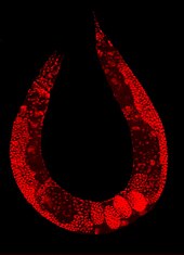
Staining of a Caenorhabditis elegans highlights the nuclei of its cells.

Multicellular organisms are organisms that consist of more than one cell, in contrast to single-celled organisms.

In complex multicellular organisms, cells specialize into different cell types that are adapted to particular functions. In mammals, major cell types include skin cells, muscle cells, neurons, blood cells, fibroblasts, stem cells, and others. Cell types differ both in appearance and function, yet are genetically identical. Cells are able to be of the same genotype but of different cell type due to the differential expression of the genes they contain.

Most distinct cell types arise from a single totipotent cell, called a zygote, that differentiates into hundreds of different cell types during the course of development. Differentiation of cells is driven by different environmental cues (such as cell–cell interaction) and intrinsic differences (such as those caused by the uneven distribution of molecules during division).

Origin of multicellularity

Multicellularity has evolved independently at least 25 times, including in some prokaryotes, like cyanobacteria, myxobacteria, actinomycetes, or Methanosarcina. However, complex multicellular organisms evolved only in six eukaryotic groups: animals, fungi, brown algae, red algae, green algae, and plants. It evolved repeatedly for plants (Chloroplastida), once or twice for animals, once for brown algae, and perhaps several times for fungi, slime molds, and red algae. Multicellularity may have evolved from colonies of interdependent organisms, from cellularization, or from organisms in symbiotic relationships.

The first evidence of multicellularity is from cyanobacteria-like organisms that lived between 3 and 3.5 billion years ago. Other early fossils of multicellular organisms include the contested Grypania spiralis and the fossils of the black shales of the Palaeoproterozoic Francevillian Group Fossil B Formation in Gabon.

The evolution of multicellularity from unicellular ancestors has been replicated in the laboratory, in evolution experiments using predation as the selective pressure.

Origins

The origin of cells has to do with the origin of life, which began the history of life on Earth.

Origin of life

Stromatolites are left behind by cyanobacteria, also called blue-green algae. They are among the oldest fossils of life on Earth. This one-billion-year-old fossil is from Glacier National Park in the United States.

Small molecules needed for life may have been carried to Earth on meteorites, created at deep-sea vents, or synthesized by lightning in a reducing atmosphere. There is little experimental data defining what the first self-replicating forms were. RNA may have been the earliest self-replicating molecule, as it can both store genetic information and catalyze chemical reactions.

Cells emerged around 4 billion years ago. The first cells were most likely heterotrophs. The early cell membranes were probably simpler and more permeable than modern ones, with only a single fatty acid chain per lipid. Lipids spontaneously form bilayered vesicles in water, and could have preceded RNA.

First eukaryotic cells
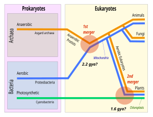
In the theory of symbiogenesis, a merger of an archaean and an aerobic bacterium created the eukaryotes, with aerobic mitochondria, some 2.2 billion years ago. A second merger, 1.6 billion years ago, added chloroplasts, creating the green plants.

Eukaryotic cells were created some 2.2 billion years ago in a process called eukaryogenesis. This is widely agreed to have involved symbiogenesis, in which archaea and bacteria came together to create the first eukaryotic common ancestor. This cell had a new level of complexity and capability, with a nucleus and facultatively aerobic mitochondria. It evolved some 2 billion years ago into a population of single-celled organisms that included the last eukaryotic common ancestor, gaining capabilities along the way, though the sequence of the steps involved has been disputed, and may not have started with symbiogenesis. It featured at least one centriole and cilium, sex (meiosis and syngamy), peroxisomes, and a dormant cyst with a cell wall of chitin and/or cellulose. In turn, the last eukaryotic common ancestor gave rise to the eukaryotes' crown group, containing the ancestors of animals, fungi, plants, and a diverse range of single-celled organisms. The plants were created around 1.6 billion years ago with a second episode of symbiogenesis that added chloroplasts, derived from cyanobacteria.

History of research

Robert Hooke's drawing of cells in cork, 1665

In 1665, Robert Hooke examined a thin slice of cork under his microscope, and saw a structure of small enclosures. He wrote "I could exceeding plainly perceive it to be all perforated and porous, much like a Honey-comb, but that the pores of it were not regular". To further support his theory, Matthias Schleiden and Theodor Schwann both also studied cells of both animal and plants. What they discovered were significant differences between the two types of cells. This put forth the idea that cells were not only fundamental to plants, but animals as well.

Biological membrane

From Wikipedia, the free encyclopedia
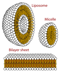
Cross-sectional view of the structures that can be formed by phospholipids in an aqueous solution

A biological membrane, biomembrane or cell membrane is a selectively permeable membrane that separates the interior of a cell from the external environment or creates intracellular compartments by serving as a boundary between one part of the cell and another. Biological membranes, in the form of eukaryotic cell membranes, consist of a phospholipid bilayer with embedded, integral and peripheral proteins used in communication and transportation of chemicals and ions. The bulk of lipids in a cell membrane provides a fluid matrix for proteins to rotate and laterally diffuse for physiological functioning. Proteins are adapted to high membrane fluidity environment of the lipid bilayer with the presence of an annular lipid shell, consisting of lipid molecules bound tightly to the surface of integral membrane proteins. The cell membranes are different from the isolating tissues formed by layers of cells, such as mucous membranes, basement membranes, and serous membranes.

Composition

Asymmetry

A fluid membrane model of the phospholipid bilayer.

The lipid bilayer consists of two layers- an outer leaflet and an inner leaflet. The components of bilayers are distributed unequally between the two surfaces to create asymmetry between the outer and inner surfaces. This asymmetric organization is important for cell functions such as cell signaling. The asymmetry of the biological membrane reflects the different functions of the two leaflets of the membrane. As seen in the fluid membrane model of the phospholipid bilayer, the outer leaflet and inner leaflet of the membrane are asymmetrical in their composition. Certain proteins and lipids rest only on one surface of the membrane and not the other.

• Both the plasma membrane and internal membranes have cytosolic and exoplasmic faces • This orientation is maintained during membrane trafficking – proteins, lipids, glycoconjugates facing the lumen of the ER and Golgi get expressed on the extracellular side of the plasma membrane. In eukaryotic cells, new phospholipids are manufactured by enzymes bound to the part of the endoplasmic reticulum membrane that faces the cytosol. These enzymes, which use free fatty acids as substrates, deposit all newly made phospholipids into the cytosolic half of the bilayer. To enable the membrane as a whole to grow evenly, half of the new phospholipid molecules then have to be transferred to the opposite monolayer. This transfer is catalyzed by enzymes called flippases. In the plasma membrane, flippases transfer specific phospholipids selectively, so that different types become concentrated in each monolayer.

Using selective flippases is not the only way to produce asymmetry in lipid bilayers, however. In particular, a different mechanism operates for glycolipids—the lipids that show the most striking and consistent asymmetric distribution in animal cells.

Lipids

The biological membrane is made up of lipids with hydrophobic tails and hydrophilic heads. The hydrophobic tails are hydrocarbon tails whose length and saturation is important in characterizing the cell. Lipid rafts occur when lipid species and proteins aggregate in domains in the membrane. These help organize membrane components into localized areas that are involved in specific processes, such as signal transduction.

Red blood cells, or erythrocytes, have a unique lipid composition. The bilayer of red blood cells is composed of cholesterol and phospholipids in equal proportions by weight. Erythrocyte membrane plays a crucial role in blood clotting. In the bilayer of red blood cells is phosphatidylserine. This is usually in the cytoplasmic side of the membrane. However, it is flipped to the outer membrane to be used during blood clotting.

Proteins

Phospholipid bilayers contain different proteins. These membrane proteins have various functions and characteristics and catalyze different chemical reactions. Integral proteins span the membranes with different domains on either side. Integral proteins hold strong association with the lipid bilayer and cannot easily become detached. They will dissociate only with chemical treatment that breaks the membrane. Peripheral proteins are unlike integral proteins in that they hold weak interactions with the surface of the bilayer and can easily become dissociated from the membrane. Peripheral proteins are located on only one face of a membrane and create membrane asymmetry.

SOME EXAMPLES OF PLASMA MEMBRANE PROTEINS AND THEIR FUNCTIONS
FUNCTIONAL CLASS PROTEIN EXAMPLE SPECIFIC FUNCTION
Transporters Na+ Pump actively pumps Na+ out of cells and K+ in
Anchors integrins link intracellular actin filaments to extracellular matrix proteins
Receptors platelet-derived growth factor receptor binds extracellular PDGF and, as a consequence, generates intracellular signals that cause the cell to grow and divide
Enzymes adenylyl cyclase catalyzes the production of intracellular signaling molecule cyclic AMP in response to extracellular signals

Oligosaccharides

Oligosaccharides are sugar containing polymers. In the membrane, they can be covalently bound to lipids to form glycolipids or covalently bound to proteins to form glycoproteins. Membranes contain sugar-containing lipid molecules known as glycolipids. In the bilayer, the sugar groups of glycolipids are exposed at the cell surface, where they can form hydrogen bonds. Glycolipids provide the most extreme example of asymmetry in the lipid bilayer. Glycolipids perform a vast number of functions in the biological membrane that are mainly communicative, including cell recognition and cell-cell adhesion. Glycoproteins are integral proteins. They play an important role in the immune response and protection.

Formation

The phospholipid bilayer is formed due to the aggregation of membrane lipids in aqueous solutions. Aggregation is caused by the hydrophobic effect, where hydrophobic ends come into contact with each other and are sequestered away from water. This arrangement maximises hydrogen bonding between hydrophilic heads and water while minimising unfavorable contact between hydrophobic tails and water. The increase in available hydrogen bonding increases the entropy of the system, creating a spontaneous process.

Function

Biological molecules are amphiphilic or amphipathic, i.e. are simultaneously hydrophobic and hydrophilic. The phospholipid bilayer contains charged hydrophilic headgroups, which interact with polar water. The layers also contain hydrophobic tails, which meet with the hydrophobic tails of the complementary layer. The hydrophobic tails are usually fatty acids that differ in lengths. The interactions of lipids, especially the hydrophobic tails, determine the lipid bilayer physical properties such as fluidity.

Membranes in cells typically define enclosed spaces or compartments in which cells may maintain a chemical or biochemical environment that differs from the outside. For example, the membrane around peroxisomes shields the rest of the cell from peroxides, chemicals that can be toxic to the cell, and the cell membrane separates a cell from its surrounding medium. Peroxisomes are one form of vacuole found in the cell that contain by-products of chemical reactions within the cell. Most organelles are defined by such membranes, and are called membrane-bound organelles.

Selective permeability

Probably the most important feature of a biomembrane is that it is a selectively permeable structure. This means that the size, charge, and other chemical properties of the atoms and molecules attempting to cross it will determine whether they succeed in doing so. Selective permeability is essential for effective separation of a cell or organelle from its surroundings. Biological membranes also have certain mechanical or elastic properties that allow them to change shape and move as required.

Generally, small hydrophobic molecules can readily cross phospholipid bilayers by simple diffusion.

Particles that are required for cellular function but are unable to diffuse freely across a membrane enter through a membrane transport protein or are taken in by means of endocytosis, where the membrane allows for a vacuole to join onto it and push its contents into the cell. Many types of specialized plasma membranes can separate cell from external environment: apical, basolateral, presynaptic and postsynaptic ones, membranes of flagella, cilia, microvillus, filopodia and lamellipodia, the sarcolemma of muscle cells, as well as specialized myelin and dendritic spine membranes of neurons. Plasma membranes can also form different types of "supramembrane" structures such as caveolae, postsynaptic density, podosome, invadopodium, desmosome, hemidesmosome, focal adhesion, and cell junctions. These types of membranes differ in lipid and protein composition.

Distinct types of membranes also create intracellular organelles: endosome; smooth and rough endoplasmic reticulum; sarcoplasmic reticulum; Golgi apparatus; lysosome; mitochondrion (inner and outer membranes); nucleus (inner and outer membranes); peroxisome; vacuole; cytoplasmic granules; cell vesicles (phagosome, autophagosome, clathrin-coated vesicles, COPI-coated and COPII-coated vesicles) and secretory vesicles (including synaptosome, acrosomes, melanosomes, and chromaffin granules). Different types of biological membranes have diverse lipid and protein compositions. The content of membranes defines their physical and biological properties. Some components of membranes play a key role in medicine, such as the efflux pumps that pump drugs out of a cell.

Fluidity

The hydrophobic core of the phospholipid bilayer is constantly in motion because of rotations around the bonds of lipid tails. Hydrophobic tails of a bilayer bend and lock together. However, because of hydrogen bonding with water, the hydrophilic head groups exhibit less movement as their rotation and mobility are constrained. This results in increasing viscosity of the lipid bilayer closer to the hydrophilic heads.

Below a transition temperature, a lipid bilayer loses fluidity when the highly mobile lipids exhibits less movement becoming a gel-like solid. The transition temperature depends on such components of the lipid bilayer as the hydrocarbon chain length and the saturation of its fatty acids. Temperature-dependence fluidity constitutes an important physiological attribute for bacteria and cold-blooded organisms. These organisms maintain a constant fluidity by modifying membrane lipid fatty acid composition in accordance with differing temperatures.

In animal cells, membrane fluidity is modulated by the inclusion of the sterol cholesterol. This molecule is present in especially large amounts in the plasma membrane, where it constitutes approximately 20% of the lipids in the membrane by weight. Because cholesterol molecules are short and rigid, they fill the spaces between neighboring phospholipid molecules left by the kinks in their unsaturated hydrocarbon tails. In this way, cholesterol tends to stiffen the bilayer, making it more rigid and less permeable.

For all cells, membrane fluidity is important for many reasons. It enables membrane proteins to diffuse rapidly in the plane of the bilayer and to interact with one another, as is crucial, for example, in cell signaling. It permits membrane lipids and proteins to diffuse from sites where they are inserted into the bilayer after their synthesis to other regions of the cell. It allows membranes to fuse with one another and mix their molecules, and it ensures that membrane molecules are distributed evenly between daughter cells when a cell divides. If biological membranes were not fluid, it is hard to imagine how cells could live, grow, and reproduce.

The fluidity property is at the center of the Helfrich model which allows for calculating the energy cost of an elastic deformation to the membrane.

Antimicrobial peptides

From Wikipedia, the free encyclopedia
https://en.wikipedia.org/wiki/Antimicrobial_peptides
Various structures of antimicrobial peptides

Antimicrobial peptides (AMPs), also called host defence peptides (HDPs) are part of the innate immune response found among all classes of life. Fundamental differences exist between prokaryotic and eukaryotic cells that may represent targets for antimicrobial peptides. These peptides are potent, broad spectrum antimicrobials which demonstrate potential as novel therapeutic agents. Antimicrobial peptides have been demonstrated to kill Gram negative and Gram positive bacteria, enveloped viruses, fungi and even transformed or cancerous cells. Unlike the majority of conventional antibiotics it appears that antimicrobial peptides frequently destabilize biological membranes, can form transmembrane channels, and may also have the ability to enhance immunity by functioning as immunomodulators.

Structure

Antimicrobial peptides from animals, plants and fungi organised by their secondary structure content. Circle size indicates overall molecular weight of each peptide.

Antimicrobial peptides are a unique and diverse group of molecules, which are divided into subgroups on the basis of their amino acid composition and structure. Antimicrobial peptides are generally between 12 and 50 amino acids. These peptides include two or more positively charged residues provided by arginine, lysine or, in acidic environments, histidine, and a large proportion (generally >50%) of hydrophobic residues. The secondary structures of these molecules follow 4 themes, including i) α-helical, ii) β-stranded due to the presence of 2 or more disulfide bonds, iii) β-hairpin or loop due to the presence of a single disulfide bond and/or cyclization of the peptide chain, and iv) extended. Many of these peptides are unstructured in free solution, and fold into their final configuration upon partitioning into biological membranes. The peptides contain hydrophilic amino acid residues aligned along one side and hydrophobic amino acid residues aligned along the opposite side of a helical molecule. This amphipathicity of the antimicrobial peptides allows them to partition into the membrane lipid bilayer. The ability to associate with membranes is a definitive feature of antimicrobial peptides, although membrane permeabilization is not necessary. These peptides have a variety of antimicrobial activities ranging from membrane permeabilization to action on a range of cytoplasmic targets.

Type characteristic AMPs
Anionic peptides rich in glutamic and aspartic acids Maximin H5 from amphibians, dermcidin from humans
Linear cationic α-helical peptides lack in cysteine Cecropins, andropin, moricin, ceratotoxin and melittin from insects, Magainin, dermaseptin, bombinin, brevinin-1, esculentins and buforin II from amphibians, CAP18 from rabbits, LL37 from humans
Cationic peptide enriched for specific amino acid rich in proline, arginine, phenylalanine, glycine, tryptophan abaecin and drosocin, apidaecin, diptericin, and attacin from insects, prophenin from pigs, indolicidin from cattle.
Anionic/cationic peptides forming disulfide bonds contain 1~3 disulfide bond

Activities

The modes of action by Antimicrobial peptides

The modes of action by which antimicrobial peptides kill microbes are varied, and may differ for different bacterial species. Some antimicrobial peptides kill both bacteria and fungi, e.g., psoriasin kills E. coli and several filamentous fungi. The cytoplasmic membrane is a frequent target, but peptides may also interfere with DNA and protein synthesis, protein folding, and cell wall synthesis. The initial contact between the peptide and the target organism is electrostatic, as most bacterial surfaces are anionic, or hydrophobic, such as in the antimicrobial peptide Piscidin. Their amino acid composition, amphipathicity, cationic charge and size allow them to attach to and insert into membrane bilayers to form pores by ‘barrel-stave’, ‘carpet’ or ‘toroidal-pore’ mechanisms. Alternately, they may penetrate into the cell to bind intracellular molecules which are crucial to cell living. Intracellular binding models includes inhibition of cell wall synthesis, alteration of the cytoplasmic membrane, activation of autolysin, inhibition of DNA, RNA, and protein synthesis, and inhibition of certain enzymes. In many cases, the exact mechanism of killing is not known. One emerging technique for the study of such mechanisms is dual polarisation interferometry. In contrast to many conventional antibiotics these peptides appear to be bactericidal instead of bacteriostatic. In general the antimicrobial activity of these peptides is determined by measuring the minimal inhibitory concentration (MIC), which is the lowest concentration of drug that inhibits bacterial growth.

AMPs can possess multiple activities including anti-gram-positive bacterial, anti-gram-negative bacterial, anti-fungal, anti-viral, anti-parasitic, and anti cancer activities. A big AMP functional analysis indicates that among all AMP activities, amphipathicity and charge, two major properties of AMPs, best distinguish between AMPs with and without anti-gram-negative bacterial activities. This implies that being AMPs with anti-gram-negative bacterial activities may prefer or even require strong amphipathicity and net positive charge.

Immunomodulation

In addition to killing bacteria directly they have been demonstrated to have a number of immunomodulatory functions that may be involved in the clearance of infection, including the ability to alter host gene expression, act as chemokines and/or induce chemokine production, inhibiting lipopolysaccharide induced pro-inflammatory cytokine production, promoting wound healing, and modulating the responses of dendritic cells and cells of the adaptive immune response. Animal models indicate that host defense peptides are crucial for both prevention and clearance of infection. It appears as though many peptides initially isolated as and termed "antimicrobial peptides" have been shown to have more significant alternative functions in vivo (e.g. hepcidin). Dusquetide for example is an immunomodulator that acts through p62, a protein involved in toll like receptor based signalling of infection. The peptide is being examined in a Phase III clinical trial by Soligenix (SGNX) to ascertain if it can assist in repair of radiation-induced damage to oral mucosa arising during cancer radiotherapy of the head and neck.

Mechanisms of action

Scanning electron microscopic images (50,000X magnification) displaying the action of an experimental antimicrobial peptide (NN2_0050) on the cell membrane of E. coli (K12 MG1655)  ABOVE: Intact cell membranes in the control group. BELOW: Disrupted cell membranes and leakage of bacterial chromosome (green) in the treated group.

Antimicrobial peptides generally have a net positive charge, allowing them to interact with the negatively charged molecules exposed on bacteria and cancer cell surfaces, such as phospholipid phosphatidylserine, O-glycosylated mucins, sialylated gangliosides, and heparin sulfates. The mechanism of action of these peptides varies widely but can be simplified into two categories: membranolytic and non-membranolytic antimicrobial peptides. The disruption of membranes by membranolytic antimicrobial peptides can be described by four models:

  • Barrel-stave model: The barrel-stave model proposes that AMPs interact with the lipid bilayer of the microbial cell membrane to form transmembrane channels or "barrel staves". These channels are thought to disrupt the membrane's integrity, leading to the death of the microbe.
  • Carpet model: The carpet model proposes that AMPs adsorb onto the lipid bilayer of the microbial cell membrane, forming a dense layer that causes the membrane to become permeabilized. This model suggests that the AMP acts as a "carpet" that covers the surface of the cell, preventing the microbe from functioning properly.
  • Toroidal model: The toroidal model proposes that AMPs interact with the lipid bilayer of the microbial cell membrane to form toroidal structures, which are thought to pinch off sections of the membrane and lead to the formation of vesicles. This process is thought to disrupt the membrane's integrity and cause the death of the microbe.
  • Disordered toroidal-pore model: According to this model, the disordered AMPs wrap around the lipid bilayer and create a pore, which disrupts the membrane's integrity and leads to the death of the microbe. Unlike the toroidal model, which suggests that the AMP creates a stable toroidal structure, the disordered toroidal-pore model suggests that the AMP is flexible and does not form a stable toroidal structure. The peptide-lipid pore complex becomes intrinsically disordered, with the orientation of the peptide not well defined.[21]
Schematic representation of the AMPs mechanisms of action when disrupting membranes.

Several methods have been used to determine the mechanisms of antimicrobial peptide activity. In particular, solid-state NMR studies have provided an atomic-level resolution explanation of membrane disruption by antimicrobial peptides. In more recent years, X-ray crystallography has been used to delineate in atomic detail how the family of plant defensins rupture membranes by identifying key phospholipids in the cell membranes of the pathogen. Human defensins have been thought to act through a similar mechanism, targeting cell membrane lipids as part of their function. In fact human beta-defensin 2 have now been shown to kill the pathogenic fungi Candida albicans through interactions with specific phospholipids. From the computational point of view, Molecular Dynamics simulations can provide detailed information about the structure and dynamics of the peptide-membrane interactions, including the orientation, conformation, and insertion of the peptide in the membrane, as well as specific peptide interactions with lipids, ions and solvent.

Methods Applications
Microscopy to visualize the effects of antimicrobial peptides on microbial cells
Atomic emission spectroscopy to detect loss of intracellular potassium (an indication that bacterial membrane integrity has been compromised)
Fluorescent dyes to measure ability of antimicrobial peptides to permeabilize membrane vesicles
Ion channel formation to assess the formation and stability of an antimicrobial-peptide-induced pore
Circular dichroism and orientated circular dichroism to measure the orientation and secondary structure of an antimicrobial peptide bound to a lipid bilayer
Dual polarization interferometry to measure the different mechanisms of antimicrobial peptides
Solid-state NMR spectroscopy to measure the secondary structure, orientation and penetration of antimicrobial peptides into lipid bilayers in the biologically relevant liquid-crystalline state
Neutron and X-ray diffraction to measure the diffraction patterns of peptide-induced pores within membranes in oriented multilayers or liquids
Molecular dynamics simulations to study the molecular behaviour and search for specific peptide-lipid interactions
Mass spectrometry to measure the proteomic response of microorganisms to antimicrobial peptides

Therapeutic research and use

Antimicrobial peptides have been used as therapeutic agents; their use is generally limited to intravenous administration or topical applications due to their short half-lives. As of January 2018 the following antimicrobial peptides were in clinical use:

Activity beyond antibacterial functions

AMPs have been observed having functions other than bacterial and fungal killing. These activities include antiviral effects, but also roles in host defence such as anticancer functions and roles in neurology. This has led to a movement for re-branding AMPs as "Host-defence peptides" to encompass the broad scope of activities AMPs can have.

Anticancer properties

Some cecropins (e.g. cecropin A, and cecropin B) have anticancer properties and are called anticancer peptides (ACPs). Hybrid ACPs based on Cecropin A have been studied for anticancer properties. The fruit fly Defensin prevents tumour growth, suspected to bind to tumour cells owing to cell membrane modifications common to most cancer cells, such as phosphatidylserine exposure.

Antibiofilm properties

Cecropin A can destroy planktonic and sessile biofilm-forming uropathogenic E. coli (UPEC) cells, either alone or when combined with the antibiotic nalidixic acid, synergistically clearing infection in vivo (in the insect host Galleria mellonella) without off-target cytotoxicity. The multi-target mechanism of action involves outer membrane permeabilization followed by biofilm disruption triggered by the inhibition of efflux pump activity and interactions with extracellular and intracellular nucleic acids.

Other research

Recently there has been some research to identify potential antimicrobial peptides from prokaryotes, aquatic organisms such as fish, and shellfish, and monotremes such as echidnas.

Selectivity

In the competition of bacterial cells and host cells with the antimicrobial peptides, antimicrobial peptides will preferentially interact with the bacterial cell to the mammalian cells, which enables them to kill microorganisms without being significantly toxic to mammalian cells.

With regard to cancer cells, they themselves also secrete human antimicrobial peptides including defensin, and in some cases, they are reported to be more resistant than the surrounding normal cells. Therefore, we cannot conclude that selectivity is always high against cancer cells.

Factors

There are some factors that are closely related to the selectivity property of antimicrobial peptides, among which the cationic property contributes most. Since the surface of the bacterial membranes is more negatively charged than mammalian cells, antimicrobial peptides will show different affinities towards the bacterial membranes and mammalian cell membranes.

In addition, there are also other factors that will affect the selectivity. It's well known that cholesterol is normally widely distributed in the mammalian cell membranes as a membrane stabilizing agent but absent in bacterial cell membranes (except when sequestered by H. pylori); and the presence of these cholesterols will also generally reduce the activities of the antimicrobial peptides, due either to stabilization of the lipid bilayer or to interactions between cholesterol and the peptide. So the cholesterol in mammalian cells will protect the cells from attack by the antimicrobial peptides.

Besides, the transmembrane potential is well known to affect peptide-lipid interactions. There's an inside-negative transmembrane potential existing from the outer leaflet to the inner leaflet of the cell membranes and this inside-negative transmembrane potential will facilitate membrane permeabilization probably by facilitating the insertion of positively charged peptides into membranes. By comparison, the transmembrane potential of bacterial cells is more negative than that of normal mammalian cells, so bacterial membrane will be prone to be attacked by the positively charged antimicrobial peptides.

Similarly, it is also believed that increasing ionic strength, which in general reduces the activity of most antimicrobial peptides, contributes partially to the selectivity of the antimicrobial peptides by weakening the electrostatic interactions required for the initial interaction.

Molecular Basis of Cell Selectivity of Antimicrobial Peptides

Mechanism

The cell membranes of bacteria are rich in acidic phospholipids, such as phosphatidylglycerol and cardiolipin.

In contrast, the outer part of the membranes of plants and mammals is mainly composed of lipids without any net charges since most of the lipids with negatively charged headgroups are principally sequestered into the inner leaflet of the plasma membranes. Thus in the case of mammalian cells, the outer surfaces of the membranes are usually made of zwitterionic phosphatidylcholine and sphingomyelin, even though a small portion of the membrane's outer surfaces contain some negatively charged gangliosides. Therefore, the hydrophobic interaction between the hydrophobic face of amphipathic antimicrobial peptides and the zwitterionic phospholipids on the cell surface of mammalian cell membranes plays a major role in the formation of peptide-cell binding.

Dual polarisation interferometry has been used in vitro to study and quantify the association to headgroup, insertion into the bilayer, pore formation and eventual disruption of the membrane.

Control

A lot of effort has been put into controlling cell selectivity. For example, attempts have been made to modify and optimize the physicochemical parameters of the peptides to control the selectivities, including net charge, helicity, hydrophobicity per residue (H), hydrophobic moment (μ) and the angle subtended by the positively charged polar helix face (Φ). Other mechanisms like the introduction of D-amino acids and fluorinated amino acids in the hydrophobic phase are believed to break the secondary structure and thus reduce hydrophobic interaction with mammalian cells. It has also been found that Pro→Nlys substitution in Pro-containing β-turn antimicrobial peptides was a promising strategy for the design of new small bacterial cell-selective antimicrobial peptides with intracellular mechanisms of action. It has been suggested that direct attachment of magainin to the substrate surface decreased nonspecific cell binding and led to improved detection limit for bacterial cells such as Salmonella and E. coli.

Bacterial resistance

Bacteria use various resistance strategies to avoid antimicrobial peptide killing.

  • Some microorganisms alter net surface charges. Staphylococcus aureus transports D-alanine from the cytoplasm to the surface teichoic acid which reduces the net negative charge by introducing basic amino groups. S. aureus also modifies its anionic membranes via MprF with L-lysine, increasing the positive net charge.
  • The interaction of antimicrobial peptides with membrane targets can be limited by capsule polysaccharide of Klebsiella pneumoniae.
  • Salmonella species reduce the fluidity of their outer membrane by increasing hydrophobic interactions between an increased number of Lipid A acyl tails by adding myristate to Lipid A with 2-hydroxymyristate and forming hepta-acylated Lipid A by adding palmitate. The increased hydrophobic moment is thought to retard or abolish antimicrobial peptide insertion and pore formation. The residues undergo alteration in membrane proteins. In some Gram-negative bacteria, alteration in the production of outer membrane proteins correlates with resistance to killing by antimicrobial peptides.
  • Non-typeable Hemophilus influenzae transports AMPs into the interior of the cell, where they are degraded. Furthermore, H. influenzae remodels its membranes to make it appear as if the bacterium has already been successfully attacked by AMPs, protecting it from being attacked by more AMPs.
  • ATP-binding cassette transporters import antimicrobial peptides and the resistance-nodulation cell-division efflux pump exports antimicrobial peptides. Both transporters have been associated with antimicrobial peptide resistance
  • Bacteria produce proteolytic enzymes, which may degrade antimicrobial peptides leading to their resistance.
  • Outer membrane vesicles produced by Gram-negative bacteria bind the antimicrobial peptides and sequester them away from the cells, thereby protecting the cells. The outer membrane vesicles are also known to contain various proteases, peptidases and other lytic enzymes, which may have a role in degrading the extracellular peptide and nucleic acid molecules, which if allowed to reach to the bacterial cells may be dangerous for the cells.
  • Cyclic-di-GMP signaling had also been involved in the regulation of antimicrobial peptide resistance in Pseudomonas aeruginosa

While these examples show that resistance can evolve naturally, there is increasing concern that using pharmaceutical copies of antimicrobial peptides can make resistance happen more often and faster. In some cases, resistance to these peptides used as a pharmaceutical to treat medical problems can lead to resistance, not only to the medical application of the peptides, but to the physiological function of those peptides.

The ‘Trojan Horse’ approach to solving this problem capitalizes on the innate need for iron by pathogens. “Smuggling” antimicrobials into the pathogen is accomplished by linking them to siderophores for transport. While simple in concept, it has taken many decades of work to accomplish the difficult hurdle of transporting antimicrobials across the cell membranes of pathogens. Lessons learned from the successes and failures of siderophore-conjugate drugs evaluated during the development of novel agents using the ‘Trojan horse’ approach have been reviewed.

Examples

Fruit flies infected by GFP-producing bacteria. Red-eyed flies lacking antimicrobial peptide genes are susceptible to infection, while white-eyed flies have a wild-type immune response.

Antimicrobial peptides are produced by species across the tree of life, including:

Research has increased in recent years to develop artificially-engineered mimics of antimicrobial peptides such as SNAPPs, in part due to the prohibitive cost of producing naturally-derived AMPs. An example of this is the facially cationic peptide C18G, which was designed from the C-terminal domain of human platelet factor IV. Currently, the most widely used antimicrobial peptide is nisin; being the only FDA approved antimicrobial peptide, it is commonly used as an artificial preservative.

Bioinformatics

Several bioinformatic databases exist to catalogue antimicrobial peptides. The Antimicrobial Peptide Database (APD) is the original and model database for antimicrobial peptides (https://aps.unmc.edu). Based on the APD, other databases have also been built, including ADAM (A Database of Anti-Microbial peptides), BioPD (Biologically active Peptide Database), CAMP (Collection of sequences and structures of antimicrobial peptides), DBAASP (Database of Antimicrobial Activity and Structure of Peptides), DRAMP (Data Repository of Antimicrobial Peptides)Welcome To Dramp Database, and LAMP (Linking AMPs).

The Antimicrobial peptide databases may be divided into two categories on the basis of the source of peptides it contains, as specific databases and general databases. These databases have various tools for antimicrobial peptides analysis and prediction. For example, the APD has a widely used calculation interface. It also provides links to many other tools. CAMP contains AMP prediction, feature calculator, BLAST search, ClustalW, VAST, PRATT, Helical wheel etc. In addition, ADAM allows users to search or browse through AMP sequence-structure relationships. Antimicrobial peptides often encompass a wide range of categories such as antifungal, antibacterial, and antituberculosis peptides.

dbAMP: Provides an online platform for exploring antimicrobial peptides with functional activities and physicochemical properties on transcriptome and proteome data. dbAMP is an online resource that addresses various topics such as annotations of antimicrobial peptides (AMPs) including sequence information, antimicrobial activities, post-translational modifications (PTMs), structural visualization, antimicrobial potency, target species with minimum inhibitory concentration (MIC), physicochemical properties, or AMP–protein interactions.

Tools such as PeptideRanker, PeptideLocator, and AntiMPmod allow for the prediction of antimicrobial peptides while others have been developed to predict antifungal and anti-Tuberculosis activities.

Disinfection by-product

From Wikipedia, the free encyclopedia
https://en.wikipedia.org/wiki/Disinfection_by-product

Disinfection by-products (DBPs) are organic and inorganic compounds resulting from chemical reactions between organic and inorganic substances such as contaminates and chemical treatment disinfection agents, respectively, in water during water disinfection processes.

Chlorination disinfection byproducts

Chlorinated disinfection agents such as chlorine and monochloramine are strong oxidizing agents introduced into water in order to destroy pathogenic microbes, to oxidize taste/odor-forming compounds, and to form a disinfectant residual so water can reach the consumer tap safe from microbial contamination. These disinfectants may react with naturally present fulvic and humic acids, amino acids, and other natural organic matter, as well as iodide and bromide ions, to produce a range of DBPs such as the trihalomethanes (THMs), haloacetic acids (HAAs), bromate, and chlorite (which are regulated in the US), and so-called "emerging" DBPs such as halonitromethanes, haloacetonitriles, haloamides, halofuranones, iodo-acids such as iodoacetic acid, iodo-THMs (iodotrihalomethanes), nitrosamines, and others.

Chloramine has become a popular disinfectant in the US, and it has been found to produce N-nitrosodimethylamine (NDMA), which is a possible human carcinogen, as well as highly genotoxic iodinated DBPs, such as iodoacetic acid, when iodide is present in source waters.

Residual chlorine and other disinfectants may also react further within the distribution network – both by further reactions with dissolved natural organic matter and with biofilms present in the pipes. In addition to being highly influenced by the types of organic and inorganic matter in the source water, the different species and concentrations of DBPs vary according to the type of disinfectant used, the dose of disinfectant, the concentration of natural organic matter and bromide/iodide, the time since dosing (i.e. water age), temperature, and pH of the water.

Swimming pools using chlorine have been found to contain trihalomethanes, although generally they are below current EU standard for drinking water (100 micrograms per litre). Concentrations of trihalomethanes (mainly chloroform) of up to 0.43 ppm have been measured. In addition, trichloramine has been detected in the air above swimming pools, and it is suspected in the increased asthma observed in elite swimmers. Trichloramine is formed by the reaction of urea (from urine and sweat) with chlorine and gives the indoor swimming pool its distinctive odor.

Byproducts from non-chlorinated disinfectants

Several powerful oxidizing agents are used in disinfecting and treating drinking water, and many of these also cause the formation of DBPs. Ozone, for example, produces ketones, carboxylic acids, and aldehydes, including formaldehyde. Bromide in source waters can be converted by ozone into bromate, a potent carcinogen that is regulated in the United States, as well as other brominated DBPs.

As regulations are tightened on established DBPs such as THMs and HAAs, drinking water treatment plants may switch to alternative disinfection methods. This change will alter the distribution of classes of DBPs.

Occurrence

DBPs are present in most drinking water supplies that have been subject to chlorination, chloramination, ozonation, or treatment with chlorine dioxide. Many hundreds of DBPs exist in treated drinking water and at least 600 have been identified. The low levels of many of these DBPs, coupled with the analytical costs in testing water samples for them, means that in practice only a handful of DBPs are actually monitored. Increasingly it is recognized that the genotoxicities and cytotoxicities of many of the DBPs not subject to regulatory monitoring, (particularly iodinated, nitrogenous DBPs) are comparatively much higher than those DBPs commonly monitored in the developed world (THMs and HAAs).

In 2021, a new group of DBPs known as halogenated pyridinols was discovered, containing at least 8 previously unknown heterocyclic nitrogenous DBPs. They were found to require low pH treatments of 3.0 to be removed effectively. When their developmental and acute toxicity was tested on zebrafish embryos, it found to be slightly lower than those of halogenated benzoquinones, but dozens of times higher than of commonly known DBPs such as tribromomethane and iodoacetic acid

Health effects

Epidemiological studies have looked at the associations between exposure to DBPs in drinking water with cancers, adverse birth outcomes and birth defects. Meta-analyses and pooled analyses of these studies have demonstrated consistent associations for bladder cancer and for babies being born small for gestational age, but not for congenital anomalies (birth defects). Early-term miscarriages have also been reported in some studies. The exact putative agent remains unknown, however, in the epidemiological studies since the number of DBPs in a water sample are high and exposure surrogates such as monitoring data of a specific by-product (often total trihalomethanes) are used in lieu of more detailed exposure assessment. The World Health Organization has stated that "the risk of death from pathogens is at least 100 to 1000 times greater than the risk of cancer from disinfection by-products (DBPs)" {and} the "risk of illness from pathogens is at least 10 000 to 1 million times greater than the risk of cancer from DBPs".

Regulation and monitoring

The United States Environmental Protection Agency has set Maximum Contaminant Levels (MCLs) for bromate, chlorite, haloacetic acids and total trihalomethanes (TTHMs). In Europe, the level of TTHMs has been set at 100 micrograms per litre, and the level for bromate to 10 micrograms per litre, under the Drinking Water Directive. No guideline values have been set for HAAs in Europe. The World Health Organization has established guidelines for several DBPs, including bromate, bromodichloromethane, chlorate, chlorite, chloroacetic acid, chloroform, cyanogen chloride, dibromoacetonitrile, dibromochloromethane, dichloroacetic acid, dichloroacetonitrile, NDMA, and trichloroacetic acid.

Inequality (mathematics)

From Wikipedia, the free encyclopedia https://en.wikipedia.org/wiki/Inequality...