Search This Blog

Saturday, February 1, 2020

Pain and pleasure

From Wikipedia, the free encyclopedia

Some philosophers, such as Jeremy Bentham, Baruch Spinoza, and Descartes, have hypothesized that the feelings of pain (or suffering) and pleasure are part of a continuum.

There is strong evidence of biological connections between the neurochemical pathways used for the perception of both pain and pleasure, as well as other psychological rewards.

Perception of pain


Sensory input system

From a stimulus-response perspective, the perception of physical pain starts with the nociceptors, a type of physiological receptor that transmits neural signals to the brain when activated. These receptors are commonly found in the skin, membranes, deep fascias, mucosa, connective tissues of visceral organs, ligaments and articular capsules, muscles, tendons, periosteum, and arterial vessels. Once stimuli are received, the various afferent action potentials are triggered and pass along various fibers and axons of these nociceptive nerve cells into the dorsal horn of the spinal cord through the dorsal roots. A neuroanatomical review of the pain pathway, "Afferent pain pathways" by Almeida, describes various specific nociceptive pathways of the spinal cord: spinothalamic tract, spinoreticular tract, spinomesencephalic tract, spinoparabrachial tract, spinohypothalamic tract, spinocervical tract, postsynaptic pathway of the spinal column.

Neural coding and modulation

Activity in many parts of the brain is associated with pain perception. Some of the known parts for the ascending pathway include the thalamus, hypothalamus, midbrain, lentiform nucleus, somatosensory cortices, insular, prefrontal, anterior and parietal cingulum. Then, there are also the descending pathways for the modulation of pain sensation. One of the brainstem regions responsible for this is the periaqueductal gray of the midbrain, which both relieves pain by behavior as well as inhibits the activity of the nociceptive neurons in the dorsal horn of the spinal cord. Other brainstem sites, such as the parabrachial nucleus, the dorsal raphe, locus coeruleus, and the medullary reticular formation also mediate pain relief and use many different neurotransmitters to either facilitate or inhibit activity of the neurons in the dorsal horn. These neurotransmitters include noradrenaline, serotonin, dopamine, histamine, and acetylcholine

Perception of pleasure

Pleasure can be considered from many different perspectives, from physiological (such as the hedonic hotspots that are activated during the experience) to psychological (such as the study of behavioral responses towards reward). Pleasure has also often been compared to, or even defined by many neuroscientists as, a form of alleviation of pain.

Neural coding and modulation

Pleasure has been studied in the systems of taste, olfaction, auditory (musical), visual (art), and sexual activity. Well known hedonic hotspots involved in the processing of pleasure include the nucleus accumbens, posterior ventral pallidum, amygdala, other cortical and subcortical regions. The prefrontal and limbic regions of the neocortex, particularly the orbitofrontal region of the prefrontal cortex, anterior cingulate cortex, and the insular cortex have all been suggested to be pleasure causing substrates in the brain.

Psychology of pain and pleasure (reward-punishment system)

One approach to evaluating the relationship between pain and pleasure is to consider these two systems as a reward-punishment based system. When pleasure is perceived, one associates it with reward. When pain is perceived, one associates with punishment. Evolutionarily, this makes sense, because often, actions that result in pleasure or chemicals that induce pleasure work towards restoring homeostasis in the body. For example, when the body is hungry, the pleasure of rewarding food to one-self restores the body back to a balanced state of replenished energy. Like so, this can also be applied to pain, because the ability to perceive pain enhances both avoidance and defensive mechanisms that were, and still are, necessary for survival.

Opioid and dopamine systems in pain and pleasure

The neural systems to be explored when trying to look for a neurochemical relationship between pain and pleasure are the opioid and dopamine systems. The opioid system is responsible for the actual experience of the sensation, whereas the dopamine system is responsible for the anticipation or expectation of the experience. Opioids work in the modulation of pleasure or pain relief by either blocking neurotransmitter release or by hyperpolarizing neurons by opening up a potassium channel which effectively temporarily blocks the neuron.

Pain and pleasure on a continuum

Arguments for pain and pleasure on a continuum

It has been suggested as early as 4th century BC that pain and pleasure occurs on a continuum. Aristotle claims this antagonistic relationship in his Rhetoric:
"We may lay it down that Pleasure is a movement, a movement by which the soul as a whole is consciously brought into its normal state of being; and that Pain is the opposite."
He describes pain and pleasure very much like a push-pull concept; human beings will move towards something that causes pleasure and will move away from something that causes pain.

Common neuroanatomy

On an anatomical level, it can be shown the source for the modulation of both pain and pleasure originates from neurons in the same locations, including the amygdala, the pallidum, and the nucleus accumbens. Not only have Leknes and Tracey, two leading neuroscientists in the study of pain and pleasure, concluded that pain and reward processing involve many of the same regions of the brain, but also that the functional relationship lies in that pain decreases pleasure and rewards increase analgesia, which is the relief from pain.

Arguments against pain and pleasure on a continuum


Asymmetry between pain and pleasure

Thomas Szasz, the late Professor of Psychiatry Emeritus at the State University of New York Health Science Center in Syracuse, New York, explored how pain and pleasure are not opposites ends of a spectrum in his 1957 book, "Pain and Pleasure -a study of bodily feelings".

Szasz notes that although we often refer to pain and pleasure as opposites in such a way, that this is incorrect; we have receptors for pain, but none in the same way for pleasure; and so it makes sense to ask "where is the pain?" but not "where is the pleasure?". With this vantage point established, the author delves into the topics of metaphorical pain and of legitimacy, of power relations, and of communications, and of myriad others.

Evolutionary hypotheses for the relationship between pain and pleasure

Whether or not pain and pleasure are indeed on a continuum, it still remains scientifically supported that parts of the neural pathways for the two perceptions overlap. There is also scientific evidence that one may have opposing effects on the other. So why would it be evolutionarily advantageous to human beings to develop a relationship between the two perceptions at all?

South African neuroscientists presented evidence that there was a physiological link on a continuum between pain and pleasure in 1980. First, the Neuroscientists, Gillman and Lichtigfeld demonstrated that there were two endogenous endorphin systems, one pain producing and the other pain relieving. A short time later they showed that these two systems might also be involved in addiction, which is initially pursued, presumably for the pleasure generating or pain relieving actions of the addictive substance. Soon after they provided evidence that the endorphins system was involved in sexual pleasure.

Dr. Kringelbach suggests that this relationship between pain and pleasure would be evolutionarily efficient, because it was necessary to know whether or not to avoid or approach something for survival. According to Dr. Norman Doidge, the brain is limited in the sense that it tends to focus on the most used pathways. Therefore, having a common pathway for pain and pleasure could have simplified the way in which human beings have interacted with the environment (Dr. Morten Kringelbach, personal communication, October 24, 2011).

Leknes and Tracey offer two theoretical perspectives to why a relationship could be evolutionarily advantageous.

Opponent process theory

The opponent-process theory is a model that views two components as being pairs that are opposite to each other, such that if one component is experienced, the other component will be repressed. Therefore, an increase in pain should bring about a decrease in pleasure, and a decrease in pain should bring about an increase in pleasure or pain relief. This simple model serves the purpose of explaining the evolutionarily significant role of homeostasis in this relationship. This is evident since both seeking pleasure and avoiding pain are important for survival. Leknes and Tracey provide an example:
"In the face of a large food reward, which can only be obtained at the cost of a small amount of pain, for instance, it would be beneficial if the pleasurable food reduced pain unpleasantness."
They then suggest that perhaps a common currency for which human beings determine the importance of the motivation for each perception can allow them to be weighed against each other in order to make a decision best for survival.

Motivation-decision model

The Motivation-Decision Model, suggested by Fields, is centered around the concept that decision processes are driven by motivations of highest priority. The model predicts that in the case that there is anything more important than pain for survival will cause the human body to mediate pain by activating the descending pain modulation system described earlier. Thus, it is suggested that human beings have developed the unconscious ability to endure pain or sometimes, even relieve pain if it can be more important for survival to gain a larger reward. It may have been more advantageous to link the pain and pleasure perceptions together to be able to reduce pain to gain a reward necessary for fitness, such as childbirth. Like the opponent-process theory, if the body can induce pleasure or pain relief to decrease the effect of pain, it would allow human beings to be able to make the best evolutionary decisions for survival.

Clinical applications


Related diseases

The following neurological and/or mental diseases have been linked to forms of pain or anhedonia: schizophrenia, depression, addiction, cluster headache, chronic pain.

Animal trials

A great deal of what is known about pain and pleasure today primarily comes from studies conducted with rats and primates.

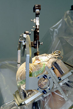
Insertion of electrode during Deep Brain Stimulation surgery using a stereotactic frame
 

Deep brain stimulation

Deep brain stimulation involves the electrical stimulation of deep brain structures by electrodes implanted into the brain. The effects of this neurosurgery has been studied in patients with Parkinson's disease, tremors, dystonia, epilepsy, depression, obsessive-compulsive disorder, Tourette's syndrome, cluster headache and chronic pain. A fine electrode is inserted into the targeted area of the brain and secured to the skull. This is attached to a pulse generator which is implanted elsewhere on the body under the skin. The surgeon then turns the frequency of the electrode to the voltage and frequency desired. Deep brain stimulation has been shown in several studies to both induce pleasure or even addiction as well as ameliorate pain. For chronic pain, lower frequencies (about 5–50 Hz) have produced analgesic effects, whereas higher frequencies (about 120–180 Hz) have alleviated or stopped pyramidal tremors in Parkinson's patients.

There is still further research necessary into how and why exactly DBS works. However, by understanding the relationship between pleasure and pain, procedures like these can be used to treat patients suffering from a high intensity or longevity of pain. So far, DBS has been recognized as a treatment for Parkinson's disease, tremors, and dystonia by the Food and Drug Administration (FDA).
Friday, January 31, 2020

Wirehead (science fiction)

From Wikipedia, the free encyclopedia
 
The wires of an implanted deep brain stimulation (DBS) device are visible as white lines in an X-ray of the skull. Large white areas around maxilla and mandible are metal dentures and are unrelated to the DBS device
 
Artist's conception of two DBS wires/electrodes in the human brain.
 
Enlarged artist's conception of two DBS wires/electrodes with multiple contact points in each wire.

Wirehead is a term used in science fiction works to denote different kinds of interaction between people and technology. The typical wirehead idea is that of a wire going into a human's brain and safe amounts of electricity applied to the wire-conductor to directly interact with the brain, or the specific "pleasure centers" of the brain.

Written fiction


Known Space stories

In Larry Niven's Known Space stories, a wirehead is someone who has been fitted with an electronic brain implant (called a "droud" in the stories) to stimulate the pleasure centres of their brain. In the Known Space universe, wireheading is the most addictive habit known (Louis Wu is the only given example of a recovered addict), and wireheads usually die from neglecting themselves in favour of the ceaseless pleasure. Wireheading is so powerful and easy that it becomes an evolutionary pressure, selecting against that portion of Known Space humanity without self-control. Also in this science fiction there is a device called a "tasp" (similar to transcranial magnetic stimulation) that does not need a surgical implant; the pleasure center of a person's brain is found and remotely stimulated (considered a violation without seeking the person's consent beforehand), an important device in the Ringworld novels

A wirehead's death is central to Niven's Gil 'the Arm' Hamilton story, "Death by Ecstasy", published by Galaxy Magazine in 1969, and a main character in the book Ringworld Engineers is a former wirehead trying to quit. 

Niven's stories explain wireheads by mentioning a study in which experimental rats had electrodes implanted at strategic locations in their brains, so that an applied current would induce a pleasant feeling. If the current could be obtained any time the rats pushed the lever, they would use it over and over, ignoring food and physical necessities until they died. Such experiments were actually conducted by James Olds and Peter Milner in the 1950s, first discovering the locations of such areas, and later showing extremes to which rats would go to obtain the stimulus again.

Mindkiller

Mindkiller, a 1982 sci-fi novel by Spider Robinson set in the late 1980s, explores the social implications of technologies to manipulate the brain, beginning with wireheading, the use of electric current to stimulate the pleasure center of the brain in order to achieve a narcotic high.

Shaper/Mechanist stories

In the Shaper/Mechanist stories of Bruce Sterling, "wirehead" is the Mechanist term for a human who has given up corporeal existence and become an infomorph

The Terminal Man

In The Terminal Man (1972) by Michael Crichton, forty electrodes are implanted into the brain of the character Harold Franklin "Harry" Benson to control seizures. However, his pleasure center is also stimulated, and his body begins producing more seizures to receive the pleasurable sensation.

Film and Television


Brainstorm

In the 1983 film Brainstorm a wireless brain connection machine is made. A character named Hal Abramson abuses the device with a signal of never ending sexual pleasure. 

The Outer Limits 1995 TV series


In The Outer Limits episode named "Awakening", season three, episode 10, a neurologically impaired woman receives a brain implant to help her become more like a typical human.

The Centurions (animated series)

In episode 41, "Zone Dancer" of the 1986The Centurions animated series, the lead character Crystal Kane is accused of "Zone Dancing" (the series' term for computer hacking) and seen using a "droud" to interface her brain with computer networks in what is probably the first animated representation of cyberspace and virtual reality. The story, written by Michael Reaves, weaves a future noir tale of cyberpunk espionage, cloning and private-eye procedural, all set in the universe of the animated series and makes copious references to William Gibson's Neuromancer. There is even a Zone Dancer named Gibson and, in what may be an homage to Larry Niven's Louis Wu, a cyberneticst named Dr. Wu.


House

The title character of the television show House is a physician who suffers from chronic pain. In the episode "Half-Wit", House seeks a medical procedure to stimulate the "pleasure center" of his brain.

Non-fictional examples

In 1924, Dr Hans Berger succeeded in recording the first human electroencephalogram (EEG).

Dr William Grey Walter wrote a paper in 1938 on the EEG ELECTRO-ENCEPHALOGRAPHY, the measurement of electrical activity in the brain using wires of different types.

Dr Wilder Penfield and Dr Herbert Jasper stimulated the brain to find the places where the patients seizures were coming from.

Dr Reginald Bickford in 1944 is reported to have recorded the EEG of psychiatric patients who had had lobotomies.

After the 1949 Nobel prize was awarded to António Egas Moniz for the procedure of lobotomy, a more precise method of destroying brain structures was pursued. In the year 1955 the placing of wires into the mentally ill patient was performed by Dr C.W. Sem Jacobsen. Dr S. Sherwood also performed wire implantation. In the year 1961 five patients had wires implanted to treat their mental illness and a precision leucotomy was performed for favorable results.

In the 1950s there are several doctors who continued to place wires into the human brain. They worked on epileptic and psychiatric patients brains.

Silver and copper electrodes were found to be toxic to brain tissue. Electrodes are encapsulated by fibrous growths as a inflammatory bodily response to a foreign object.

Dr. J. Lawrence Pool wrote "Effects of Electrical Stimulation of the Human Cerebellar Cortex" and described stimulation of a patients brain. April 1943.

Dr. B S Nashold is used as a reference in many medical writings.

Dr. Robert Galbraith Heath placed electrodes in his subjects' brains in the 1950s to try to treat their mental illness. Dr. Heath wrote several papers on his work of stimulating the various regions of the brain.

Dr. Carl Wilhelm Sem-Jacobsen "Depth-electrographic stimulation of the human brain and behavior; from fourteen years of studies and treatment of Parkinson's disease and mental disorders with implanted electrodes,"

José Manuel Rodriguez Delgado also placed electrodes in his patients' brains. He called his inventions a "stimoceiver" and a "chemitrode".
  • 1953 "Induced paroxysmal electrical activity in man recorded simultaneously through subcortical and scalp electrodes"
  • 1955: The patient, a 27-year-old housewife "Stimulation of the amygdaloid nucleus in a schizophrenic patient" by Robert Galbraith Heath
  • 1963: "Electrical self-stimulation of the brain in man" by Robert Galbraith Heath.
  • 1972: A 24-year-old man with temporal lobe epilepsy, identified as patient "B-19". "He was permitted to wear the device for 3 hours at a time: on one occasion he stimulated his septal region 1,200 times, on another occasion 1,500 times, and on a third occasion 900 times. He protested each time the unit was taken from him, pleading to self-stimulate just a few more times..."
  • 1986: A 48-year-old woman with chronic pain. "The patient self-stimulated throughout the day, neglecting personal hygiene and family commitments."
  • 1986: To treat patients suffering from pain due to cancer Dr Young and Dr Brechner made a study of electrical stimulation of the brain.
  • 2012: Cathy Hutchinson who is paralyzed had one hundred electrodes placed on the surface of her brain. With this brain–computer interface she is able to control a variety of devices.
  • 2013: A 49-year-old, right-handed woman had multiple electrodes placed in her brain for epilepsy. She reported an orgasmic ecstasy following the stimulation of the left hippocampus.
  • 2016: The New England Journal of Medicine describes a growing do-it-yourself (DIY) medical engineering culture that includes DIY transcranial direct-current stimulation
  • 2019:"Electronic implants studied for treatment of drug addiction" In China, doctors are treating addiction with brain implants aimed to stimulate the nucleus accumbens.

Brain implant

From Wikipedia, the free encyclopedia
 
A laboratory rat with a brain implant used to record neuronal activity
 
Brain implants, often referred to as neural implants, are technological devices that connect directly to a biological subject's brain – usually placed on the surface of the brain, or attached to the brain's cortex. A common purpose of modern brain implants and the focus of much current research is establishing a biomedical prosthesis circumventing areas in the brain that have become dysfunctional after a stroke or other head injuries. This includes sensory substitution, e.g., in vision. Other brain implants are used in animal experiments simply to record brain activity for scientific reasons. Some brain implants involve creating interfaces between neural systems and computer chips. This work is part of a wider research field called brain-computer interfaces. (Brain-computer interface research also includes technology such as EEG arrays that allow interface between mind and machine but do not require direct implantation of a device.) 

Neural implants such as deep brain stimulation and Vagus nerve stimulation are increasingly becoming routine for patients with Parkinson's disease and clinical depression, respectively.

Purpose

Brain implants electrically stimulate, block or record (or both record and stimulate simultaneously) signals from single neurons or groups of neurons (biological neural networks) in the brain. The blocking technique is called intra-abdominal vagal blocking. This can only be done where the functional associations of these neurons are approximately known. Because of the complexity of neural processing and the lack of access to action potential related signals using neuroimaging techniques, the application of brain implants has been seriously limited until recent advances in neurophysiology and computer processing power. Much research is also being done on the surface chemistry of neural implants in effort to design products which minimize all negative effects that an active implant can have on the brain, and that the body can have on the function of the implant. Researchers are also exploring a range of delivery systems, such as using veins, to deliver these implants without brain surgery; by leaving the skull sealed shut, patients could receive their neural implants without running as great a risk of seizures, strokes, or permanent neural impairments, all of which can be caused by open-brain surgery.

Research and applications

Research in sensory substitution has made significant progress since 1970. Especially in vision, due to the knowledge of the working of the visual system, eye implants (often involving some brain implants or monitoring) have been applied with demonstrated success. For hearing, cochlear implants are used to stimulate the auditory nerve directly. The vestibulocochlear nerve is part of the peripheral nervous system, but the interface is similar to that of true brain implants.

Multiple projects have demonstrated success at recording from the brains of animals for long periods of time. As early as 1976, researchers at the NIH led by Edward Schmidt made action potential recordings of signals from rhesus monkey motor cortexes using immovable "hatpin" electrodes, including recording from single neurons for over 30 days, and consistent recordings for greater than three years from the best electrodes. 

The "hatpin" electrodes were made of pure iridium and insulated with Parylene, materials that are currently used in the Cyberkinetics implementation of the Utah array. These same electrodes, or derivations thereof using the same biocompatible electrode materials, are currently used in visual prosthetics laboratories, laboratories studying the neural basis of learning, and motor prosthetics approaches other than the Cyberkinetics probes.

Schematic of the "Utah" Electrode Array
 
Other laboratory groups produce their own implants to provide unique capabilities not available from the commercial products.

Breakthroughs include studies of the process of functional brain re-wiring throughout the learning of a sensory discrimination, control of physical devices by rat brains, monkeys over robotic arms, remote control of mechanical devices by monkeys and humans, remote control over the movements of roaches, the first reported use of the Utah Array in a human for bidirectional signalling. Currently a number of groups are conducting preliminary motor prosthetic implants in humans. These studies are presently limited to several months by the longevity of the implants. The array now forms the sensor component of the Braingate

Much research is also being done on the surface chemistry of neural implants in effort to design products which minimize all negative effects that an active implant can have on the brain, and that the body can have on the function of the implant. 

Another type of neural implant that is being experimented on is Prosthetic Neuronal Memory Silicon Chips, which imitate the signal processing done by functioning neurons that allows peoples' brains to create long-term memories. 

In 2016, scientists at the University of Illinois at Urbana–Champaign announced development of tiny brain sensors for use postoperative monitoring, which melt away when they are no longer needed.

In 2016, scientists out of the University of Melbourne published proof-of-concept data related to a discovery for Stentrode, a device implanted via the jugular vein, demonstrated the potential for a neural recording device to be engineered onto a stent and implanted into a blood vessel in the brain, without the need for open brain surgery. The technology platform is being developed for patients with paralysis to facilitate control of external devices such as robotic limbs, computers and exoskeletons by translating brain activity. It may ultimately help diagnose and treat a range of brain pathologies, such as epilepsy and Parkinson’s disease.

Military

DARPA has announced its interest in developing "cyborg insects" to transmit data from sensors implanted into the insect during the pupal stage. The insect's motion would be controlled from a Micro-Electro-Mechanical System (MEMS) and could conceivably survey an environment or detect explosives and gas. Similarly, DARPA is developing a neural implant to remotely control the movement of sharks. The shark's unique senses would then be exploited to provide data feedback in relation to enemy ship movement or underwater explosives.

In 2006, researchers at Cornell University invented a new surgical procedure to implant artificial structures into insects during their metamorphic development. The first insect cyborgs, moths with integrated electronics in their thorax, were demonstrated by the same researchers. The initial success of the techniques has resulted in increased research and the creation of a program called Hybrid-Insect-MEMS, HI-MEMS. Its goal, according to DARPA's Microsystems Technology Office, is to develop "tightly coupled machine-insect interfaces by placing micro-mechanical systems inside the insects during the early stages of metamorphosis".

The use of neural implants has recently been attempted, with success, on cockroaches. Surgically applied electrodes were put on the insect, which were remotely controlled by a human. The results, although sometimes different, basically showed that the cockroach could be controlled by the impulses it received through the electrodes. DARPA is now funding this research because of its obvious beneficial applications to the military and other areas.

In 2009 at the Institute of Electrical and Electronics Engineers (IEEE) Micro-electronic mechanical systems (MEMS) conference in Italy, researchers demonstrated the first "wireless" flying-beetle cyborg. Engineers at the University of California at Berkeley pioneered the design of a "remote controlled beetle", funded by the DARPA HI-MEMS Program. This was followed later that year by the demonstration of wireless control of a "lift-assisted" moth-cyborg.

Eventually researchers plan to develop HI-MEMS for dragonflies, bees, rats and pigeons. For the HI-MEMS cybernetic bug to be considered a success, it must fly 100 metres (330 ft) from a starting point, guided via computer into a controlled landing within 5 metres (16 ft) of a specific end point. Once landed, the cybernetic bug must remain in place.

In 2012, DARPA provided seed funding to Dr. Thomas Oxley, a neurointerventionist at Mount Sinai Hospital in New York City, for a technology that became known as Stentrode. Oxley’s group in Australia was the only non-US-based funded by DARPA as part of the Reliable Neural Interface Technology (RE-NET) program. This technology is the first to attempt to provide neural implants through a minimally invasive surgical procedure that does not require cutting into the skull. That is, an electrode array built onto a self-expanding stent, implanted into the brain via cerebral angiography. This pathway can provide safe, easy access and capture a strong signal for a number of indications beyond addressing paralysis, and is currently in clinical trials in patients with severe paralysis seeking to regain the ability to communicate. 

In 2015 it was reported that scientists from the Perception and Recognition Neuro-technologies Laboratory at the Southern Federal University in Rostov-on-Don suggested using rats with microchips planted in their brains to detect explosive devices.

In 2016 it was reported that American engineers are developing a system that would transform locusts into "remote controlled explosive detectors" with electrodes in their brains beaming information about dangerous substances back to their operators.


Rehabilitation

Neurostimulators have been in use since 1997 to ease the symptoms of such diseases as epilepsy, Parkinson's disease, dystonia and recently depression.

Current brain implants are made from a variety of materials such as tungsten, silicon, platinum-iridium, or even stainless steel. Future brain implants may make use of more exotic materials such as nanoscale carbon fibers (nanotubes), and polycarbonate urethane.

Brain implants are also being explored by DARPA as part of the Reliable Neural-Interface Technology (RE-NET) program launched in 2010 to directly address the need for high-performance neural interfaces to control the dexterous functions made possible by DARPA’s advanced prosthetic limbs. The goal is to provide high-bandwidth, intuitive control interface for these limbs, they will not achieve their full potential to improve quality of life for wounded troops.

Historical research

In 1870, Eduard Hitzig and Gustav Fritsch demonstrated that electrical stimulation of the brains of dogs could produce movements. Robert Bartholow showed the same to be true for humans in 1874. By the start of the 20th century, Fedor Krause began to systematically map human brain areas, using patients that had undergone brain surgery.

Prominent research was conducted in the 1950s. Robert G. Heath experimented with aggressive mental patients, aiming to influence his subjects' moods through electrical stimulation.

Yale University physiologist Jose Delgado demonstrated limited control of animal and human subjects' behaviours using electronic stimulation. He invented the stimoceiver or transdermal stimulator, a device implanted in the brain to transmit electrical impulses that modify basic behaviours such as aggression or sensations of pleasure.

Delgado was later to write a popular book on mind control, called Physical Control of the Mind, where he stated: "the feasibility of remote control of activities in several species of animals has been demonstrated [...] The ultimate objective of this research is to provide an understanding of the mechanisms involved in the directional control of animals and to provide practical systems suitable for human application."

In the 1950s, the CIA also funded research into mind control techniques, through programs such as MKULTRA. Perhaps because he received funding for some research through the US Office of Naval Research, it has been suggested (but not proven) that Delgado also received backing through the CIA. He denied this claim in a 2005 article in Scientific American describing it only as a speculation by conspiracy-theorists. He stated that his research was only progressively scientifically motivated to understand how the brain works.

Recent advances in neurotechnologies and neuroimaging, along with an increased understanding of neurocircuitry, are factors contributing to the rapid rise in the use of neurostimulation therapies to treat an increasingly wide range of neurologic and psychiatric disorders. Electrical stimulation technologies are evolving after remaining fairly stagnant for the past 30 years, moving toward potential closed-loop therapeutic control systems with the ability to deliver stimulation with higher spatial resolution to provide continuous customized neuromodulation for optimal clinical outcomes.

Concerns and ethical considerations

Ethical questions raised include who are good candidates to receive neural implants and what are good and bad uses of neural implants. Whilst deep brain stimulation is increasingly becoming routine for patients with Parkinson's disease, there may be some behavioural side effects. Reports in the literature describe the possibility of apathy, hallucinations, compulsive gambling, hypersexuality, cognitive dysfunction, and depression. However, these may be temporary and related to correct placement and calibration of the stimulator and so are potentially reversible.

Some transhumanists, such as Raymond Kurzweil and Kevin Warwick, see brain implants as part of a next step for humans in progress and evolution, whereas others, especially bioconservatives, view them as unnatural, with humankind losing essential human qualities. It raises controversy similar to other forms of human enhancement. For instance, it is argued that implants would technically change people into cybernetic organisms (cyborgs). It's also expected that all research will comply to the Declaration of Helsinki. Yet further, the usual legal duties apply such as information to the person wearing implants and that the implants are voluntary, with (very) few exceptions.

Other concerns involve vulnerabilities of neural implants to cybercrime or intrusive surveillance as neural implants could be hacked, misused or misdesigned.

Sadja states that "one's private thoughts are important to protect" and doesn't consider it a good idea to just charge the government or any company with protecting them. Walter Glannon, a neuroethicist of the University of Calgary notes that "there is a risk of the microchips being hacked by third parties" and that "this could interfere with the user's intention to perform actions, violate privacy by extracting information from the chip".

Synaptogenesis

From Wikipedia, the free encyclopedia
https://en.wikipedia.org/wiki/Synaptogenesis

Synaptogenesis is the formation of synapses between neurons in the nervous system. Although it occurs throughout a healthy person's lifespan, an explosion of synapse formation occurs during early brain development, known as exuberant synaptogenesis. Synaptogenesis is particularly important during an individual's critical period, during which there is a certain degree of synaptic pruning due to competition for neural growth factors by neurons and synapses. Processes that are not used, or inhibited during their critical period will fail to develop normally later on in life.

Formation of the neuromuscular junction


Function

The neuromuscular junction (NMJ) is the most well-characterized synapse in that it provides a simple and accessible structure that allows for easy manipulation and observation. The synapse itself is composed of three cells: the motor neuron, the myofiber, and the Schwann cell. In a normally functioning synapse, a signal will cause the motor neuron to depolarize, by releasing the neurotransmitter acetylcholine (ACh). Acetylcholine travels across the synaptic cleft where it reaches acetylcholine receptors (AChR) on the plasma membrane of the myofiber, the sarcolemma. As the AChRs open ion channels, the membrane depolarizes, causing muscle contraction. The entire synapse is covered in a myelin sheath provided by the Schwann cell to insulate and encapsulate the junction. Another important part of the neuromuscular system and central nervous system are the astrocytes. While originally they were thought to have only functioned as support for the neurons, they play an important role in functional plasticity of synapses.

Origin and movement of cells

During development, each of the three germ layer cell types arises from different regions of the growing embryo. The individual myoblasts originate in the mesoderm and fuse to form a multi-nucleated myotube. During or shortly after myotube formation, motoneurons from the neural tube form preliminary contacts with the myotube. The Schwann cells arise from the neural crest and are led by the axons to their destination. Upon reaching it, they form a loose, unmyelinated covering over the innervating axons. The movement of the axons (and subsequently the Schwann cells) is guided by the growth cone, a filamentous projection of the axon that actively searches for neurotrophins released by the myotube.

The specific patterning of synapse development at the neuromuscular junction shows that the majority of muscles are innervated at their midpoints. Although it may seem that the axons specifically target the midpoint of the myotube, several factors reveal that this is not a valid claim. It appears that after the initial axonal contact, the newly formed myotube proceeds to grow symmetrically from that point of innervation. Coupled with the fact that AChR density is the result of axonal contact instead of the cause, the structural patterns of muscle fibers can be attributed to both myotatic growth as well as axonal innervation.

The preliminary contact formed between the motoneuron and the myotube generates synaptic transmission almost immediately, but the signal produced is very weak. There is evidence that Schwann cells may facilitate these preliminary signals by increasing the amount of spontaneous neurotransmitter release through small molecule signals. After about a week, a fully functional synapse is formed following several types of differentiation in both the post-synaptic muscle cell and the pre-synaptic motoneuron. This pioneer axon is of crucial importance because the new axons that follow have a high propensity for forming contacts with well-established synapses.

Post-synaptic differentiation

The most noticeable difference in the myotube following contact with the motoneuron is the increased concentration of AChR in the plasma membrane of the myotube in the synapse. This increased amount of AChR allows for more effective transmission of synaptic signals, which in turn leads to a more-developed synapse. The density of AChR is > 10,000/μm2 and approximately 10/μm2 around the edge. This high concentration of AChR in the synapse is achieved through clustering of AChR, up-regulation of the AChR gene transcription in the post-synaptic nuclei, and down-regulation of the AChR gene in the non-synaptic nuclei. The signals that initiate post-synaptic differentiation may be neurotransmitters released directly from the axon to the myotube, or they may arise from changes activated in the extracellular matrix of the synaptic cleft.

Clustering

AChR experiences multimerization within the post-synaptic membrane largely due to the signaling molecule Agrin. The axon of the motoneuron releases agrin, a proteoglycan that initiates a cascade that eventually leads to AChR association. Agrin binds to a muscle-specific kinase (MuSK) receptor in the post-synaptic membrane, and this in turn leads to downstream activation of the cytoplasmic protein Rapsyn. Rapsyn contains domains that allow for AChR association and multimerization, and it is directly responsible for AChR clustering in the post-synaptic membrane: rapsyn-deficient mutant mice fail to form AChR clusters.

Synapse-specific transcription

The increased concentration of AChR is not simply due to a rearrangement of pre-existing synaptic components. The axon also provides signals that regulate gene expression within the myonuclei directly beneath the synapse. This signaling provides for localized up-regulation of transcription of AChR genes and consequent increase in local AChR concentration. The two signaling molecules released by the axon are calcitonin gene-related peptide (CGRP) and neuregulin, which trigger a series of kinases that eventually lead to transcriptional activation of the AChR genes.

Extrasynaptic repression

Repression of the AChR gene in the non-synaptic nuclei is an activity-dependent process involving the electrical signal generated by the newly formed synapse. Reduced concentration of AChR in the extrasynaptic membrane in addition to increased concentration in the post-synaptic membrane helps ensure the fidelity of signals sent by the axon by localizing AChR to the synapse. Because the synapse begins receiving inputs almost immediately after the motoneuron comes into contact with the myotube, the axon quickly generates an action potential and releases ACh. The depolarization caused by AChR induces muscle contraction and simultaneously initiates repression of AChR gene transcription across the entire muscle membrane. Note that this affects gene transcription at a distance: the receptors that are embedded within the post-synaptic membrane are not susceptible to repression.

Pre-synaptic differentiation

Although the mechanisms regulating pre-synaptic differentiation are unknown, the changes exhibited at the developing axon terminal are well characterized. The pre-synaptic axon shows an increase in synaptic volume and area, an increase of synaptic vesicles, clustering of vesicles at the active zone, and polarization of the pre-synaptic membrane. These changes are thought to be mediated by neurotrophin and cell adhesion molecule release from muscle cells, thereby emphasizing the importance of communication between the motoneuron and the myotube during synaptogenesis. Like post-synaptic differentiation, pre-synaptic differentiation is thought to be due to a combination of changes in gene expression and a redistribution of pre-existing synaptic components. Evidence for this can be seen in the up-regulation of genes expressing vesicle proteins shortly after synapse formation as well as their localization at the synaptic terminal.

Synaptic maturation

Immature synapses are multiply innervated at birth, due the high propensity for new axons to innervate at a pre-existing synapse. As the synapse matures, the synapses segregate and eventually all axonal inputs except for one retract in a process called synapse elimination. Furthermore, the post-synaptic end plate grows deeper and creates folds through invagination to increase the surface area available for neurotransmitter reception. At birth, Schwann cells form loose, unmyelinated covers over groups of synapses, but as the synapse matures, Schwann cells become dedicated to a single synapse and form a myelinated cap over the entire neuromuscular junction.

Synapse elimination

The process of synaptic pruning known as synapse elimination is a presumably activity-dependent process that involves competition between axons. Hypothetically, a synapse strong enough to produce an action potential will trigger the myonuclei directly across from the axon to release synaptotrophins that will strengthen and maintain well-established synapses. This synaptic strengthening is not conferred upon the weaker synapses, thereby starving them out. It has also been suggested that in addition to the synaptotrophins released to the synapse exhibiting strong activity, the depolarization of the post-synaptic membrane causes release of synaptotoxins that ward off weaker axons.

Synapse formation specificity

A remarkable aspect of synaptogenesis is the fact that motoneurons are able to distinguish between fast and slow-twitch muscle fibers; fast-twitch muscle fibers are innervated by "fast" motoneurons, and slow-twitch muscle fibers are innervated by "slow" motoneurons. There are two hypothesized paths by which the axons of motoneurons achieve this specificity, one in which the axons actively recognize the muscles that they innervate and make selective decisions based on inputs, and another that calls for more indeterminate innervation of muscle fibers. In the selective paths, the axons recognize the fiber type, either by factors or signals released specifically by the fast or slow-twitch muscle fibers. In addition, selectivity can be traced to the lateral position that the axons are predeterminately arranged in order to link them to the muscle fiber that they will eventually innervate. The hypothesized non-selective pathways indicate that the axons are guided to their destinations by the matrix through which they travel. Essentially, a path is laid out for the axon and the axon itself is not involved in the decision-making process. Finally, the axons may non-specifically innervate muscle fibers and cause the muscles to acquire the characteristics of the axon that innervates them. In this path, a "fast" motoneuron can convert any muscle fiber into a fast-twitch muscle fiber. There is evidence for both selective and non-selective paths in synapse formation specificity, leading to the conclusion that the process is a combination of several factors.

Central nervous system synapse formation

Although the study of synaptogenesis within the central nervous system (CNS) is much more recent than that of the NMJ, there is promise of relating the information learned at the NMJ to synapses within the CNS. Many similar structures and basic functions exist between the two types of neuronal connections. At the most basic level, the CNS synapse and the NMJ both have a nerve terminal that is separated from the postsynaptic membrane by a cleft containing specialized extracellular material. Both structures exhibit localized vesicles at the active sites, clustered receptors at the post-synaptic membrane, and glial cells that encapsulate the entire synaptic cleft. In terms of synaptogenesis, both synapses exhibit differentiation of the pre- and post-synaptic membranes following initial contact between the two cells. This includes the clustering of receptors, localized up-regulation of protein synthesis at the active sites, and neuronal pruning through synapse elimination.

Despite these similarities in structure, there is a fundamental difference between the two connections. The CNS synapse is strictly neuronal and does not involve muscle fibers: for this reason the CNS uses different neurotransmitter molecules and receptors. More importantly, neurons within the CNS often receive multiple inputs that must be processed and integrated for successful transfer of information. Muscle fibers are innervated by a single input and operate in an all or none fashion. Coupled with the plasticity that is characteristic of the CNS neuronal connections, it is easy to see how increasingly complex CNS circuits can become.

Factors regulating synaptogenesis in the CNS


Signaling

The main method of synaptic signaling in the NMJ is through use of the neurotransmitter acetylcholine and its receptor. The CNS homolog is glutamate and its receptors, and one of special significance is the N-methyl-D-aspartate (NMDA) receptor. It has been shown that activation of NMDA receptors initiates synaptogenesis through activation of downstream products. The heightened level of NMDA receptor activity during development allows for increased influx of calcium, which acts as a secondary signal. Eventually, immediate early genes (IEG) are activated by transcription factors and the proteins required for neuronal differentiation are translated. The NMDA receptor function is associated with the estrogen receptor in hippocampal neurons. Experiments conducted with estradiol show that exposure to the estrogen significantly increases synaptic density and protein concentration.

Synaptic signaling during synaptogenesis is not only activity-dependent, but is also dependent on the environment in which the neurons are located. For instance, brain-derived neurotrophic factor (BDNF) is produced by the brain and regulates several functions within the developing synapse, including enhancement of transmitter release, increased concentration of vesicles, and cholesterol biosynthesis. Cholesterol is essential to synaptogenesis because the lipid rafts that it forms provide a scaffold upon which numerous signaling interactions can occur. BDNF-null mutants show significant defects in neuronal growth and synapse formation. Aside from neurotrophins, cell-adhesion molecules are also essential to synaptogenesis. Often the binding of pre-synaptic cell-adhesion molecules with their post-synaptic partners triggers specializations that facilitate synaptogenesis. Indeed, a defect in genes encoding neuroligin, a cell-adhesion molecule found in the post-synaptic membrane, has been linked to cases of autism and mental retardation. Finally, many of these signaling processes can be regulated by matrix metalloproteinases (MMPs) as the targets of many MMPs are these specific cell-adhesion molecules.

Morphology

The special structure found in the CNS that allows for multiple inputs is the dendritic spine, the highly dynamic site of excitatory synapses. This morphological dynamism is due to the specific regulation of the actin cytoskeleton, which in turn allows for regulation of synapse formation. Dendritic spines exhibit three main morphologies: filopodia, thin spines, and mushroom spines. The filopodia play a role in synaptogenesis through initiation of contact with axons of other neurons. Filopodia of new neurons tend to associate with multiply synapsed axons, while the filopodia of mature neurons tend to sites devoid of other partners. The dynamism of spines allows for the conversion of filopodia into the mushroom spines that are the primary sites of glutamate receptors and synaptic transmission.

Environmental enrichment

Rats raised with environmental enrichment have 25% more synapses than controls. This effect occurs whether a more stimulating environment is experienced immediately following birth, after weaning, or during maturity. Stimulation effects not only synaptogenesis upon pyramidal neurons but also stellate ones.

Contributions of the Wnt protein family

The (Wnt) family, includes several embryonic morphogens that contribute to early pattern formation in the developing embryo. Recently data have emerged showing that the Wnt protein family has roles in the later development of synapse formation and plasticity. Wnt contribution to synaptogenesis has been verified in both the central nervous system and the neuromuscular junction.

Central nervous system

Wnt family members contribute to synapse formation in the cerebellum by inducing presynaptic and postsynaptic terminal formation. This brain region contains three main neuronal cell types- Purkinje cells, granule cells and mossy fiber cells. Wnt-3 expression contributes to Purkinje cell neurite outgrowth and synapse formation. Granule cells express Wnt-7a to promote axon spreading and branching in their synaptic partner, mossy fiber cells. Retrograde secretion of Wnt-7a to mossy fiber cells causes growth cone enlargement by spreading microtubules. Furthermore, Wnt-7a retrograde signaling recruits synaptic vesicles and presynaptic proteins to the synaptic active zone. Wnt-5a performs a similar function on postsynaptic granule cells; this Wnt stimulates receptor assembly and clustering of the scaffolding protein PSD-95.

In the hippocampus Wnts in conjunction with cell electrical activity promote synapse formation. Wnt7b is expressed in maturing dendrites, and the expression of the Wnt receptor Frizzled (Fz), increases highly with synapse formation in the hippocampus. NMDA glutamate receptor activation increases Wnt2 expression. Long term potentiation (LTP) due to NMDA activation and subsequent Wnt expression leads to Fz-5 localization at the postsynaptic active zone. Furthermore, Wnt7a and Wnt2 signaling after NMDA receptor mediated LTP leads to increased dendritic arborization and regulates activity induced synaptic plasticity. Blocking Wnt expression in the hippocampus mitigates these activity dependent effects by reducing dendritic arborization and subsequently, synaptic complexity.

Neuromuscular junction

Similar mechanisms of action by Wnts in the central nervous system are observed in the neuromuscular junction (NMJ) as well. In the Drosophila NMJ mutations in the Wnt5 receptor Derailed (drl) reduce the number of and density of synaptic active zones. The major neurotransmitter in this system is glutamate. Wnt is needed to localize glutamatergic receptors on postsynaptic muscle cells. As a result, Wnt mutations diminish evoked currents on the postsynaptic muscle.

In the vertebrate NMJ, motor neuron expression of Wnt-11r contributes to acetylcholine receptor (AChR) clustering in the postsynaptic density of muscle cells. Wnt-3 is expressed by muscle fibers and is secreted retrogradely onto motor neurons. In motor neurons, Wnt-3 works with Agrin to promote growth cone enlargement, axon branching and synaptic vesicle clustering.

Inequality (mathematics)

From Wikipedia, the free encyclopedia https://en.wikipedia.org/wiki/Inequality...