Search This Blog

Wednesday, April 9, 2025

Molecular phylogenetics

From Wikipedia, the free encyclopedia

Molecular phylogenetics (/məˈlɛkjʊlər ˌfləˈnɛtɪks, mɒ-, m-/) is the branch of phylogeny that analyzes genetic, hereditary molecular differences, predominantly in DNA sequences, to gain information on an organism's evolutionary relationships. From these analyses, it is possible to determine the processes by which diversity among species has been achieved. The result of a molecular phylogenetic analysis is expressed in a phylogenetic tree. Molecular phylogenetics is one aspect of molecular systematics, a broader term that also includes the use of molecular data in taxonomy and biogeography.

Molecular phylogenetics and molecular evolution correlate. Molecular evolution is the process of selective changes (mutations) at a molecular level (genes, proteins, etc.) throughout various branches in the tree of life (evolution). Molecular phylogenetics makes inferences of the evolutionary relationships that arise due to molecular evolution and results in the construction of a phylogenetic tree.

History

The theoretical frameworks for molecular systematics were laid in the 1960s in the works of Emile Zuckerkandl, Emanuel Margoliash, Linus Pauling, and Walter M. Fitch. Applications of molecular systematics were pioneered by Charles G. Sibley (birds), Herbert C. Dessauer (herpetology), and Morris Goodman (primates), followed by Allan C. Wilson, Robert K. Selander, and John C. Avise (who studied various groups). Work with protein electrophoresis began around 1956. Although the results were not quantitative and did not initially improve on morphological classification, they provided tantalizing hints that long-held notions of the classifications of birds, for example, needed substantial revision. In the period of 1974–1986, DNA–DNA hybridization was the dominant technique used to measure genetic difference.

Theoretical background

Early attempts at molecular systematics were also termed chemotaxonomy and made use of proteins, enzymes, carbohydrates, and other molecules that were separated and characterized using techniques such as chromatography. These have been replaced in recent times largely by DNA sequencing, which produces the exact sequences of nucleotides or bases in either DNA or RNA segments extracted using different techniques. In general, these are considered superior for evolutionary studies, since the actions of evolution are ultimately reflected in the genetic sequences. At present, it is still a long and expensive process to sequence the entire DNA of an organism (its genome). However, it is quite feasible to determine the sequence of a defined area of a particular chromosome. Typical molecular systematic analyses require the sequencing of around 1000 base pairs. At any location within such a sequence, the bases found in a given position may vary between organisms. The particular sequence found in a given organism is referred to as its haplotype. In principle, since there are four base types, with 1000 base pairs, we could have 41000 distinct haplotypes. However, for organisms within a particular species or in a group of related species, it has been found empirically that only a minority of sites show any variation at all, and most of the variations that are found are correlated, so that the number of distinct haplotypes that are found is relatively small.

In a phylogenetic tree, numerous groupings (clades) exist. A clade may be defined as a group of organisms having a common ancestor throughout evolution. This figure illustrates how a clade in a phylogenetic tree may be expressed.

In a molecular systematic analysis, the haplotypes are determined for a defined area of genetic material; a substantial sample of individuals of the target species or other taxon is used; however, many current studies are based on single individuals. Haplotypes of individuals of closely related, yet different, taxa are also determined. Finally, haplotypes from a smaller number of individuals from a definitely different taxon are determined: these are referred to as an outgroup. The base sequences for the haplotypes are then compared. In the simplest case, the difference between two haplotypes is assessed by counting the number of locations where they have different bases: this is referred to as the number of substitutions (other kinds of differences between haplotypes can also occur, for example, the insertion of a section of nucleic acid in one haplotype that is not present in another). The difference between organisms is usually re-expressed as a percentage divergence, by dividing the number of substitutions by the number of base pairs analysed: the hope is that this measure will be independent of the location and length of the section of DNA that is sequenced.

An older and superseded approach was to determine the divergences between the genotypes of individuals by DNA–DNA hybridization. The advantage claimed for using hybridization rather than gene sequencing was that it was based on the entire genotype, rather than on particular sections of DNA. Modern sequence comparison techniques overcome this objection by the use of multiple sequences.

Once the divergences between all pairs of samples have been determined, the resulting triangular matrix of differences is submitted to some form of statistical cluster analysis, and the resulting dendrogram is examined in order to see whether the samples cluster in the way that would be expected from current ideas about the taxonomy of the group. Any group of haplotypes that are all more similar to one another than any of them is to any other haplotype may be said to constitute a clade, which may be visually represented as the figure displayed on the right demonstrates. Statistical techniques such as bootstrapping and jackknifing help in providing reliability estimates for the positions of haplotypes within the evolutionary trees.

Techniques and applications

Every living organism contains deoxyribonucleic acid (DNA), ribonucleic acid (RNA), and proteins. In general, closely related organisms have a high degree of similarity in the molecular structure of these substances, while the molecules of organisms distantly related often show a pattern of dissimilarity. Conserved sequences, such as mitochondrial DNA, are expected to accumulate mutations over time, and assuming a constant rate of mutation, provide a molecular clock for dating divergence. Molecular phylogeny uses such data to build a "relationship tree" that shows the probable evolution of various organisms. With the invention of Sanger sequencing in 1977, it became possible to isolate and identify these molecular structures. High-throughput sequencing may also be used to obtain the transcriptome of an organism, allowing inference of phylogenetic relationships using transcriptomic data.

The most common approach is the comparison of homologous sequences for genes using sequence alignment techniques to identify similarity. Another application of molecular phylogeny is in DNA barcoding, wherein the species of an individual organism is identified using small sections of mitochondrial DNA or chloroplast DNA. Another application of the techniques that make this possible can be seen in the very limited field of human genetics, such as the ever-more-popular use of genetic testing to determine a child's paternity, as well as the emergence of a new branch of criminal forensics focused on evidence known as genetic fingerprinting.

Molecular phylogenetic analysis

There are several methods available for performing a molecular phylogenetic analysis. One method, including a comprehensive step-by-step protocol on constructing a phylogenetic tree, including DNA/Amino Acid contiguous sequence assembly, multiple sequence alignment, model-test (testing best-fitting substitution models), and phylogeny reconstruction using Maximum Likelihood and Bayesian Inference, is available at Nature Protocol.

Another molecular phylogenetic analysis technique has been described by Pevsner and shall be summarized in the sentences to follow (Pevsner, 2015). A phylogenetic analysis typically consists of five major steps. The first stage comprises sequence acquisition. The following step consists of performing a multiple sequence alignment, which is the fundamental basis of constructing a phylogenetic tree. The third stage includes different models of DNA and amino acid substitution. Several models of substitution exist. A few examples include Hamming distance, the Jukes and Cantor one-parameter model, and the Kimura two-parameter model (see Models of DNA evolution). The fourth stage consists of various methods of tree building, including distance-based and character-based methods. The normalized Hamming distance and the Jukes-Cantor correction formulas provide the degree of divergence and the probability that a nucleotide changes to another, respectively. Common tree-building methods include unweighted pair group method using arithmetic mean (UPGMA) and Neighbor joining, which are distance-based methods, Maximum parsimony, which is a character-based method, and Maximum likelihood estimation and Bayesian inference, which are character-based/model-based methods. UPGMA is a simple method; however, it is less accurate than the neighbor-joining approach. Finally, the last step comprises evaluating the trees. This assessment of accuracy is composed of consistency, efficiency, and robustness.

Five Stages of Molecular Phylogenetic Analysis

MEGA (molecular evolutionary genetics analysis) is an analysis software that is user-friendly and free to download and use. This software is capable of analyzing both distance-based and character-based tree methodologies. MEGA also contains several options one may choose to utilize, such as heuristic approaches and bootstrapping. Bootstrapping is an approach that is commonly used to measure the robustness of topology in a phylogenetic tree, which demonstrates the percentage each clade is supported after numerous replicates. In general, a value greater than 70% is considered significant. The flow chart displayed on the right visually demonstrates the order of the five stages of Pevsner's molecular phylogenetic analysis technique that have been described.

Limitations

Molecular systematics is an essentially cladistic approach: it assumes that classification must correspond to phylogenetic descent, and that all valid taxa must be monophyletic. This is a limitation when attempting to determine the optimal tree(s), which often involves bisecting and reconnecting portions of the phylogenetic tree(s).

The recent discovery of extensive horizontal gene transfer among organisms provides a significant complication to molecular systematics, indicating that different genes within the same organism can have different phylogenies. HGTs can be detected and excluded using a number of phylogenetic methods (see Inferring horizontal gene transfer § Explicit phylogenetic methods).

In addition, molecular phylogenies are sensitive to the assumptions and models that go into making them. Firstly, sequences must be aligned; then, issues such as long-branch attraction, saturation, and taxon sampling problems must be addressed. This means that strikingly different results can be obtained by applying different models to the same dataset. The tree-building method also brings with it specific assumptions about tree topology, evolution speeds, and sampling. The simplistic UPGMA assumes a rooted tree and a uniform molecular clock, both of which can be incorrect.

Carcinogen

From Wikipedia, the free encyclopedia
https://en.wikipedia.org/wiki/Carcinogen
 
A carcinogen (/kɑːrˈsɪnəən/) is any agent that promotes the development of cancer. Carcinogens can include synthetic chemicals, naturally occurring substances, physical agents such as ionizing and non-ionizing radiation, and biologic agents such as viruses and bacteria. Most carcinogens act by creating mutations in DNA that disrupt a cell's normal processes for regulating growth, leading to uncontrolled cellular proliferation. This occurs when the cell's DNA repair processes fail to identify DNA damage allowing the defect to be passed down to daughter cells. The damage accumulates over time. This is typically a multi-step process during which the regulatory mechanisms within the cell are gradually dismantled allowing for unchecked cellular division.

The specific mechanisms for carcinogenic activity is unique to each agent and cell type. Carcinogens can be broadly categorized, however, as activation-dependent and activation-independent which relate to the agent's ability to engage directly with DNA. Activation-dependent agents are relatively inert in their original form, but are bioactivated in the body into metabolites or intermediaries capable of damaging human DNA. These are also known as "indirect-acting" carcinogens. Examples of activation-dependent carcinogens include polycyclic aromatic hydrocarbons (PAHs), heterocyclic aromatic amines, and mycotoxins. Activation-independent carcinogens, or "direct-acting" carcinogens, are those that are capable of directly damaging DNA without any modification to their molecular structure. These agents typically include electrophilic groups that react readily with the net negative charge of DNA molecules. Examples of activation-independent carcinogens include ultraviolet light, ionizing radiation and alkylating agents.

The time from exposure to a carcinogen to the development of cancer is known as the latency period. For most solid tumors in humans the latency period is between 10 and 40 years depending on cancer type. For blood cancers, the latency period may be as short as two. Due to prolonged latency periods identification of carcinogens can be challenging.

A number of organizations review and evaluate the cumulative scientific evidence regarding the potential carcinogenicity of specific substances. Foremost among these is the International Agency for Research on Cancer (IARC). IARC routinely publishes monographs in which specific substances are evaluated for their potential carcinogenicity to humans and subsequently categorized into one of four groupings: Group 1: Carcinogenic to humans, Group 2A: Probably carcinogenic to humans, Group 2B: Possibly carcinogenic to humans and Group 3: Not classifiable as to its carcinogenicity to humans. Other organizations that evaluate the carcinogenicity of substances include the National Toxicology Program of the US Public Health Service, NIOSH, the American Conference of Governmental Industrial Hygienists and others.

There are numerous sources of exposures to carcinogens including ultraviolet radiation from the sun, radon gas emitted in residential basements, environmental contaminants such as chlordecone, cigarette smoke and ingestion of some types of foods such as alcohol and processed meats. Occupational exposures represent a major source of carcinogens with an estimated 666,000 annual fatalities worldwide attributable to work related cancers. According to NIOSH, 3-6% of cancers worldwide are due to occupational exposures. Well established occupational carcinogens include vinyl chloride and hemangiosarcoma of the liver, benzene and leukemia, aniline dyes and bladder cancer, asbestos and mesothelioma, polycyclic aromatic hydrocarbons and scrotal cancer among chimney sweeps to name a few.

Radiation

Ionizing Radiation

CERCLA identifies all radionuclides as carcinogens, although the nature of the emitted radiation (alpha, beta, gamma, or neutron and the radioactive strength), its consequent capacity to cause ionization in tissues, and the magnitude of radiation exposure, determine the potential hazard. Carcinogenicity of radiation depends on the type of radiation, type of exposure, and penetration. For example, alpha radiation has low penetration and is not a hazard outside the body, but emitters are carcinogenic when inhaled or ingested. For example, Thorotrast, a (incidentally radioactive) suspension previously used as a contrast medium in x-ray diagnostics, is a potent human carcinogen known because of its retention within various organs and persistent emission of alpha particles. Low-level ionizing radiation may induce irreparable DNA damage (leading to replicational and transcriptional errors needed for neoplasia or may trigger viral interactions) leading to pre-mature aging and cancer.

Non-ionizing radiation

Not all types of electromagnetic radiation are carcinogenic. Low-energy waves on the electromagnetic spectrum including radio waves, microwaves, infrared radiation and visible light are thought not to be, because they have insufficient energy to break chemical bonds. Evidence for carcinogenic effects of non-ionizing radiation is generally inconclusive, though there are some documented cases of radar technicians with prolonged high exposure experiencing significantly higher cancer incidence.

Higher-energy radiation, including ultraviolet radiation (present in sunlight) generally is carcinogenic, if received in sufficient doses. For most people, ultraviolet radiations from sunlight is the most common cause of skin cancer. In Australia, where people with pale skin are often exposed to strong sunlight, melanoma is the most common cancer diagnosed in people aged 15–44 years.

Substances or foods irradiated with electrons or electromagnetic radiation (such as microwave, X-ray or gamma) are not carcinogenic. In contrast, non-electromagnetic neutron radiation produced inside nuclear reactors can produce secondary radiation through nuclear transmutation.

Common carcinogens associated with food

Alcohol

Alcohol is a carcinogen of the head and neck, esophagus, liver, colon and rectum, and breast. It has a synergistic effect with tobacco smoke in the development of head and neck cancers. In the United States approximately 6% of cancers and 4% of cancer deaths are attributable to alcohol use.

Processed meats

Chemicals used in processed and cured meat such as some brands of bacon, sausages and ham may produce carcinogens. For example, nitrites used as food preservatives in cured meat such as bacon have also been noted as being carcinogenic with demographic links, but not causation, to colon cancer.

Meats cooked at high temperatures

Cooking food at high temperatures, for example grilling or barbecuing meats, may also lead to the formation of minute quantities of many potent carcinogens that are comparable to those found in cigarette smoke (i.e., benzo[a]pyrene). Charring of food looks like coking and tobacco pyrolysis, and produces carcinogens. There are several carcinogenic pyrolysis products, such as polynuclear aromatic hydrocarbons, which are converted by human enzymes into epoxides, which attach permanently to DNA. Pre-cooking meats in a microwave oven for 2–3 minutes before grilling shortens the time on the hot pan, and removes heterocyclic amine (HCA) precursors, which can help minimize the formation of these carcinogens.

Acrylamide in foods

Frying, grilling or broiling food at high temperatures, especially starchy foods, until a toasted crust is formed generates acrylamides. This discovery in 2002 led to international health concerns. Subsequent research has however found that it is not likely that the acrylamides in burnt or well-cooked food cause cancer in humans; Cancer Research UK categorizes the idea that burnt food causes cancer as a "myth".

Biologic Agents

Several biologic agents are known carcinogens.

Aflatoxin B1, a toxin produced by the fungus Aspergillus flavus which is a common contaminant of stored grains and nuts is a known cause of hepatocellular cancer. The bacteria H. Pylori is known to cause stomach cancer and MALT lymphoma. Hepatitis B and C are associated with the development of hepatocellular cancer. HPV is the primary cause of cervical cancer.

Cigarette smoke

Tobacco smoke contains at least 70 known carcinogens and is implicated in the development of numerous types of cancers including cancers of the lung, larynx, esophagus, stomach, kidney, pancreas, liver, bladder, cervix, colon, rectum and blood. Potent carcinogens found in cigarette smoke include polycyclic aromatic hydrocarbons (PAH, such as benzo(a)pyrene), benzene, and nitrosamine.

Occupational carcinogens

Given that populations of workers are more likely to have consistent, often high level exposures to chemicals rarely encountered in normal life, much of the evidence for the carcinogenicity of specific agents is derived from studies of workers.

Selected carcinogens

Carcinogen Associated cancer sites or types Occupational uses or sources
Arsenic and its compounds
  • Smelting byproduct
  • Component of:
    • Alloys
    • Electrical and semiconductor devices
    • Medications (e.g. melarsoprol)
    • Herbicides
    • Fungicides
    • Animal dips
    • Drinking water from contaminated aquifers.
Asbestos

Not in widespread use, but found in:

  • Constructions
    • Roofing papers
    • Floor tiles
  • Fire-resistant textiles
  • Friction linings (brake pads) (only outside Europe)
    • Replacement friction linings for automobiles still may contain asbestos
Benzene
Beryllium and its compounds
  • Lung
  • Lightweight alloys
    • Aerospace applications
    • Nuclear reactors
Cadmium and its compounds
Hexavalent chromium(VI) compounds
  • Lung
  • Paints
  • Pigments
  • Preservatives
Nitrosamines
  • Lung
  • Esophagus
  • Liver
Ethylene oxide
  • Leukemia
Nickel
  • Nickel plating
  • Ferrous alloys
  • Ceramics
  • Batteries
  • Stainless-steel welding byproduct
Radon and its decay products
  • Lung
  • Uranium decay
    • Quarries and mines
    • Cellars and poorly ventilated places
Vinyl chloride
Shift work that involves

circadian disruption


Involuntary smoking (Passive smoking)
  • Lung

Radium-226, Radium-224,
Plutonium-238, Plutonium-239
and other alpha particle
emitters with high atomic weight
Unless otherwise specified, ref is:[34]

Others

Mechanisms of carcinogenicity

Carcinogens can be classified as genotoxic or nongenotoxic. Genotoxins cause irreversible genetic damage or mutations by binding to DNA. Genotoxins include chemical agents like N-nitroso-N-methylurea (NMU) or non-chemical agents such as ultraviolet light and ionizing radiation. Certain viruses can also act as carcinogens by interacting with DNA.

Nongenotoxins do not directly affect DNA but act in other ways to promote growth. These include hormones and some organic compounds.

Classification

Approximate equivalences
between classification schemes
IARC GHS NTP ACGIH EU
Group 1 Cat. 1A Known A1 Cat. 1A
Group 2A Cat. 1B Reasonably
suspected
A2 Cat. 1B
Group 2B
Cat. 2   A3 Cat. 2
Group 3
  A4  
Group 4 A5

International Agency for Research on Cancer

The International Agency for Research on Cancer (IARC) is an intergovernmental agency established in 1965, which forms part of the World Health Organization of the United Nations. It is based in Lyon, France. Since 1971 it has published a series of Monographs on the Evaluation of Carcinogenic Risks to Humans that have been highly influential in the classification of possible carcinogens.

  • Group 1: the agent (mixture) is carcinogenic to humans. The exposure circumstance entails exposures that are carcinogenic to humans.
  • Group 2A: the agent (mixture) is most likely (product more likely to be) carcinogenic to humans. The exposure circumstance entails exposures that are probably carcinogenic to humans.
  • Group 2B: the agent (mixture) is possibly (chance of product being) carcinogenic to humans. The exposure circumstance entails exposures that are possibly carcinogenic to humans.
  • Group 3: the agent (mixture or exposure circumstance) is not classifiable as to its carcinogenicity to humans.
  • Group 4: the agent (mixture) is most likely not carcinogenic to humans.

Globally Harmonized System

The Globally Harmonized System of Classification and Labelling of Chemicals (GHS) is a United Nations initiative to attempt to harmonize the different systems of assessing chemical risk which currently exist (as of March 2009) around the world. It classifies carcinogens into two categories, of which the first may be divided again into subcategories if so desired by the competent regulatory authority:

  • Category 1: known or presumed to have carcinogenic potential for humans
    • Category 1A: the assessment is based primarily on human evidence
    • Category 1B: the assessment is based primarily on animal evidence
  • Category 2: suspected human carcinogens

U.S. National Toxicology Program

The National Toxicology Program of the U.S. Department of Health and Human Services is mandated to produce a biennial Report on Carcinogens. As of August 2024, the latest edition was the 15th report (2021). It classifies carcinogens into two groups:

  • Known to be a human carcinogen
  • Reasonably anticipated to be a human carcinogen

American Conference of Governmental Industrial Hygienists

The American Conference of Governmental Industrial Hygienists (ACGIH) is a private organization best known for its publication of threshold limit values (TLVs) for occupational exposure and monographs on workplace chemical hazards. It assesses carcinogenicity as part of a wider assessment of the occupational hazards of chemicals.

  • Group A1: Confirmed human carcinogen
  • Group A2: Suspected human carcinogen
  • Group A3: Confirmed animal carcinogen with unknown relevance to humans
  • Group A4: Not classifiable as a human carcinogen
  • Group A5: Not suspected as a human carcinogen

European Union

The European Union classification of carcinogens is contained in the Regulation (EC) No 1272/2008. It consists of three categories:

  • Category 1A: Carcinogenic
  • Category 1B: May cause cancer
  • Category 2: Suspected of causing cancer

The former European Union classification of carcinogens was contained in the Dangerous Substances Directive and the Dangerous Preparations Directive. It also consisted of three categories:

  • Category 1: Substances known to be carcinogenic to humans.
  • Category 2: Substances which should be regarded as if they are carcinogenic to humans.
  • Category 3: Substances which cause concern for humans, owing to possible carcinogenic effects but in respect of which the available information is not adequate for making a satisfactory assessment.

This assessment scheme is being phased out in favor of the GHS scheme (see above), to which it is very close in category definitions.

Safe Work Australia

Under a previous name, the NOHSC, in 1999 Safe Work Australia published the Approved Criteria for Classifying Hazardous Substances [NOHSC:1008(1999)]. Section 4.76 of this document outlines the criteria for classifying carcinogens as approved by the Australian government. This classification consists of three categories:

  • Category 1: Substances known to be carcinogenic to humans.
  • Category 2: Substances that should be regarded as if they were carcinogenic to humans.
  • Category 3: Substances that have possible carcinogenic effects in humans but about which there is insufficient information to make an assessment.

Major carcinogens implicated in the four most common cancers worldwide

In this section, the carcinogens implicated as the main causative agents of the four most common cancers worldwide are briefly described. These four cancers are lung, breast, colon, and stomach cancers. Together they account for about 41% of worldwide cancer incidence and 42% of cancer deaths (for more detailed information on the carcinogens implicated in these and other cancers, see references).

Lung cancer

Lung cancer (pulmonary carcinoma) is the most common cancer in the world, both in terms of cases (1.6 million cases; 12.7% of total cancer cases) and deaths (1.4 million deaths; 18.2% of total cancer deaths). Lung cancer is largely caused by tobacco smoke. Risk estimates for lung cancer in the United States indicate that tobacco smoke is responsible for 90% of lung cancers. Other factors are implicated in lung cancer, and these factors can interact synergistically with smoking so that total attributable risk adds up to more than 100%. These factors include occupational exposure to carcinogens (about 9-15%), radon (10%) and outdoor air pollution (1-2%).

Tobacco smoke is a complex mixture of more than 5,300 identified chemicals. The most important carcinogens in tobacco smoke have been determined by a "Margin of Exposure" approach. Using this approach, the most important tumorigenic compounds in tobacco smoke were, in order of importance, acrolein, formaldehyde, acrylonitrile, 1,3-butadiene, cadmium, acetaldehyde, ethylene oxide, and isoprene. Most of these compounds cause DNA damage by forming DNA adducts or by inducing other alterations in DNA.[citation needed] DNA damages are subject to error-prone DNA repair or can cause replication errors. Such errors in repair or replication can result in mutations in tumor suppressor genes or oncogenes leading to cancer.

Breast cancer

Breast cancer is the second most common cancer [(1.4 million cases, 10.9%), but ranks 5th as cause of death (458,000, 6.1%)]. Increased risk of breast cancer is associated with persistently elevated blood levels of estrogen. Estrogen appears to contribute to breast carcinogenesis by three processes; (1) the metabolism of estrogen to genotoxic, mutagenic carcinogens, (2) the stimulation of tissue growth, and (3) the repression of phase II detoxification enzymes that metabolize ROS leading to increased oxidative DNA damage.

The major estrogen in humans, estradiol, can be metabolized to quinone derivatives that form adducts with DNA. These derivatives can cause depurination, the removal of bases from the phosphodiester backbone of DNA, followed by inaccurate repair or replication of the apurinic site leading to mutation and eventually cancer. This genotoxic mechanism may interact in synergy with estrogen receptor-mediated, persistent cell proliferation to ultimately cause breast cancer. Genetic background, dietary practices and environmental factors also likely contribute to the incidence of DNA damage and breast cancer risk.

Consumption of alcohol has also been linked to an increased risk for breast cancer.

Colon cancer

Colorectal cancer is the third most common cancer [1.2 million cases (9.4%), 608,000 deaths (8.0%)]. Tobacco smoke may be responsible for up to 20% of colorectal cancers in the United States. In addition, substantial evidence implicates bile acids as an important factor in colon cancer. Twelve studies (summarized in Bernstein et al.) indicate that the bile acids deoxycholic acid (DCA) or lithocholic acid (LCA) induce production of DNA-damaging reactive oxygen species or reactive nitrogen species in human or animal colon cells. Furthermore, 14 studies showed that DCA and LCA induce DNA damage in colon cells. Also 27 studies reported that bile acids cause programmed cell death (apoptosis).

Increased apoptosis can result in selective survival of cells that are resistant to induction of apoptosis.[52] Colon cells with reduced ability to undergo apoptosis in response to DNA damage would tend to accumulate mutations, and such cells may give rise to colon cancer. Epidemiologic studies have found that fecal bile acid concentrations are increased in populations with a high incidence of colon cancer. Dietary increases in total fat or saturated fat result in elevated DCA and LCA in feces and elevated exposure of the colon epithelium to these bile acids. When the bile acid DCA was added to the standard diet of wild-type mice invasive colon cancer was induced in 56% of the mice after 8 to 10 months. Overall, the available evidence indicates that DCA and LCA are centrally important DNA-damaging carcinogens in colon cancer.

Stomach cancer

Stomach cancer is the fourth most common cancer [990,000 cases (7.8%), 738,000 deaths (9.7%)]. Helicobacter pylori infection is the main causative factor in stomach cancer. Chronic gastritis (inflammation) caused by H. pylori is often long-standing if not treated. Infection of gastric epithelial cells with H. pylori results in increased production of reactive oxygen species (ROS). ROS cause oxidative DNA damage including the major base alteration 8-hydroxydeoxyguanosine (8-OHdG). 8-OHdG resulting from ROS is increased in chronic gastritis. The altered DNA base can cause errors during DNA replication that have mutagenic and carcinogenic potential. Thus H. pylori-induced ROS appear to be the major carcinogens in stomach cancer because they cause oxidative DNA damage leading to carcinogenic mutations.

Diet is also thought to be a contributing factor in stomach cancer: in Japan, where very salty pickled foods are popular, the incidence of stomach cancer is high. Preserved meat such as bacon, sausages, and ham increases the risk, while a diet rich in fresh fruit, vegetables, peas, beans, grains, nuts, seeds, herbs, and spices will reduce the risk. The risk also increases with age.

Gastritis

From Wikipedia, the free encyclopedia
 
Gastritis
Micrograph showing gastritis. H&E stain.
SpecialtyGastroenterology
SymptomsUpper abdominal pain, nausea, vomiting, bloating, loss of appetite, heartburn
ComplicationsBleeding, stomach ulcers, stomach tumors, pernicious anemia
DurationShort or long term
CausesHelicobacter pylori, NSAIDs, alcohol, tobacco, cocaine, viruses, parasites, autoimmune
Diagnostic methodEndoscopy, upper gastrointestinal series, blood tests, stool tests
Differential diagnosisMyocardial infarction, inflammation of the pancreas, gallbladder problems, peptic ulcer disease
TreatmentAntacids, H2 blockers, proton pump inhibitors, antibiotics, sucralfate, bismuth subsalicylate, antiemetics
Frequency~50% of people
Deaths50,000 (2015)

Gastritis is the inflammation of the lining of the stomach. It may occur as a short episode or may be of a long duration. There may be no symptoms but, when symptoms are present, the most common is upper abdominal pain (see dyspepsia). Other possible symptoms include nausea and vomiting, bloating, loss of appetite and heartburn. Complications may include stomach bleeding, stomach ulcers, and stomach tumors. When due to autoimmune problems, low red blood cells due to not enough vitamin B12 may occur, a condition known as pernicious anemia.

Common causes include infection with Helicobacter pylori and use of nonsteroidal anti-inflammatory drugs (NSAIDs). When caused by H. pylori this is now termed Helicobacter pylori induced gastritis, and included as a listed disease in ICD11. Less common causes include alcohol, smoking, cocaine, severe illness, autoimmune problems, radiation therapy and Crohn's disease. Endoscopy, a type of X-ray known as an upper gastrointestinal series, blood tests, and stool tests may help with diagnosis. Other conditions with similar symptoms include inflammation of the pancreas, gallbladder problems, and peptic ulcer disease.

Prevention is by avoiding things that cause the disease. Treatment includes medications such as antacids, H2 blockers, or proton pump inhibitors. During an acute attack drinking viscous lidocaine may help. If gastritis is due to NSAIDs these may be stopped. If H. pylori is present it may be treated with a combination of antibiotics such as amoxicillin and clarithromycin. For those with pernicious anemia, vitamin B12 supplements are recommended either by mouth or by injection. People are usually advised to avoid foods that bother them.

Gastritis is believed to affect about half of people worldwide. In 2013 there were approximately 90 million new cases of the condition. As people get older the disease becomes more common. It, along with a similar condition in the first part of the intestines known as duodenitis, resulted in 50,000 deaths in 2015. H. pylori was first discovered in 1981 by Barry Marshall and Robin Warren.

Signs and symptoms

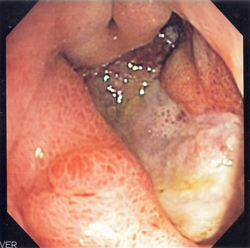
A peptic ulcer may accompany gastritis. Endoscopic image.

Many people with gastritis experience no symptoms at all. However, upper central abdominal pain is the most common symptom; the pain may be dull, vague, burning, aching, gnawing, sore, or sharp. Pain is usually located in the upper central portion of the abdomen, but it may occur anywhere from the upper left portion of the abdomen around to the back.

Other signs and symptoms may include the following:

Causes

There are two categories of gastritis depending on the cause of the disease. There is erosive gastritis, for which the common causes are stress, alcohol, some drugs, such as aspirin and other nonsteroidal anti-inflammatory drugs (NSAIDs), and Crohn's disease. And, there is non-erosive gastritis, for which the most common cause is a Helicobacter pylori infection. 

Helicobacter pylori

Helicobacter pylori colonizes the stomachs of more than half of the world's population, and the infection continues to play a key role in the pathogenesis of a number of gastroduodenal diseases. Colonization of the gastric mucosa with Helicobacter pylori results in the development of chronic gastritis in infected individuals and, in a subset of patients, chronic gastritis progresses to complications (e.g., ulcer disease, stomach cancers, and some distinct extragastric disorders). Gastritis caused by H. pylori infection is termed Helicobacter pylori induced gastritis, and listed as a disease in ICD11. More than 80% of individuals infected with the bacterium are asymptomatic and it has been postulated that it may play an important role in the natural stomach ecology.

Critical illness

Gastritis may also develop after major surgery or traumatic injury ("Cushing ulcer"), burns ("Curling ulcer"), or severe infections. Gastritis may also occur in those who have had weight loss surgery resulting in the banding or reconstruction of the digestive tract.

Diet

Evidence does not support a role for specific foods, including spicy foods and coffee, in the development of peptic ulcers. People are usually advised to avoid foods that bother them. There is little specific advice on diet published by authoritative sources. The National Health Service of the United Kingdom advises avoiding spicy, acidic or fried foods which may irritate the stomach.

Pathophysiology

Acute

Early acute superficial gastritis: Marked neutrophilic infiltrates appear in the mucous neck region and lamina with a pit microabscess. This case was caused by Helicobacter pylori.

Acute erosive gastritis typically involves discrete foci of surface necrosis due to damage to mucosal defenses. NSAIDs inhibit cyclooxygenase-1, or COX-1, an enzyme responsible for the biosynthesis of eicosanoids in the stomach, which increases the possibility of peptic ulcers forming. Also, NSAIDs, such as aspirin, reduce a substance that protects the stomach called prostaglandin. These drugs used in a short period are not typically dangerous. However, regular use can lead to gastritis. Additionally, severe physiologic stress from sepsis, hypoxia, trauma, or surgery is also a common etiology for acute erosive gastritis, resulting in "stress ulcers". This form of gastritis can occur in more than 5% of hospitalized patients.

Also, alcohol consumption does not cause chronic gastritis. It does, however, erode the mucosal lining of the stomach; low doses of alcohol stimulate hydrochloric acid secretion. High doses of alcohol do not stimulate secretion of acid.

Chronic

Chronic gastritis refers to a wide range of problems of the gastric issues. The immune system makes proteins and antibodies that fight infections in the body to maintain a homeostatic condition. In some disorders the body targets the stomach as if it were a foreign protein or pathogen; it makes antibodies against, severely damages, and may even destroy the stomach or its lining. In some cases bile, normally used to aid digestion in the small intestine, will enter through the pyloric valve of the stomach if it has been removed during surgery or does not work properly, also leading to gastritis. Gastritis may also be caused by other medical conditions, including HIV/AIDS, Crohn's disease, certain connective tissue disorders, and liver or kidney failure. Since 1992, chronic gastritis lesions are classified according to the Sydney system.

Metaplasia

Mucous gland metaplasia, the reversible replacement of differentiated cells, occurs in the setting of severe damage of the gastric glands, which then waste away (atrophic gastritis) and are progressively replaced by mucous glands. Gastric ulcers may develop; it is unclear if they are the causes or the consequences. Intestinal metaplasia typically begins in response to chronic mucosal injury in the antrum and may extend to the body. Gastric mucosa cells change to resemble intestinal mucosa and may even assume absorptive characteristics. Intestinal metaplasia is classified histologically as complete or incomplete. With complete metaplasia, gastric mucosa is completely transformed into small-bowel mucosa, both histologically and functionally, with the ability to absorb nutrients and secrete peptides. In incomplete metaplasia, the epithelium assumes a histologic appearance closer to that of the large intestine and frequently exhibits dysplasia.

Diagnosis

Updated Sydney System for visual classification of gastritis on histopathology.

Often, a diagnosis can be made based on patients' description of their symptoms. Other methods which may be used to verify gastritis include:

  • Blood tests:
  • Urinalysis
  • Stool sample, to look for blood in the stool
  • X-rays
  • Endoscopy, to check for stomach lining inflammation and mucous erosion
  • Stomach biopsy, to test for gastritis and other conditions

The OLGA staging frame of chronic gastritis on histopathology. Atrophy is scored as the percentage of atrophic glands and scored on a four-tiered scale. No atrophy (0%) = score 0; mild atrophy (1–30%) = score 1; moderate atrophy (31–60%) = score 2; severe atrophy (>60%) = score 3. These scores (0–3) are used in the OLGA staging assessment in each 10 compartment:


Corpus
No atrophy
(score 0)
Mild atrophy
(score 1)
Moderate atrophy
(score 2)
Severe atrophy
(score 3)
Antrum
(including
incisura
angularis)
No atrophy (score 0) Stage 0 Stage I Stage II Stage II
Mild atrophy (score 1) Stage I Stage I Stage II Stage III
Moderate atrophy (score 2) Stage II Stage II Stage III Stage IV
Severe atrophy (score 3) Stage III Stage III Stage IV Stage IV

Treatment

Antacids are a common treatment for mild to medium gastritis. When antacids do not provide enough relief, medications such as H2 blockers and proton-pump inhibitors that help reduce the amount of acid are often prescribed.

Cytoprotective agents are designed to help protect the tissues that line the stomach and small intestine. They include the medications sucralfate and misoprostol. If NSAIDs are being taken regularly, one of these medications to protect the stomach may also be taken. Another cytoprotective agent is bismuth subsalicylate.

Several regimens are used to treat H. pylori infection. Most use a combination of two antibiotics and a proton pump inhibitor. Sometimes bismuth is added to the regimen.

History

In 1,000 A.D, Avicenna first gave the description of stomach cancer. In 1728, German physician Georg Ernst Stahl first coined the term "gastritis". Italian anatomical pathologist Giovanni Battista Morgagni further described the characteristics of gastric inflammation. He described the characteristics of erosive or ulcerative gastritis and erosive gastritis. Between 1808 and 1831, French physician François-Joseph-Victor Broussais gathered information from the autopsies of dead French soldiers. He described chronic gastritis as "Gastritide" and erroneously believed that gastritis was the cause of ascites, typhoid fever, and meningitis. In 1854, Charles Handfield Jones and Wilson Fox described the microscopic changes of stomach inner lining in gastritis which existed in diffuse and segmental forms. In 1855, Baron Carl von Rokitansky first described hypertrophic gastritis. In 1859, British physician, William Brinton first described about acute, subacute, and chronic gastritis. In 1870, Samuel Fenwick noted that pernicious anemia causes glandular atrophy in gastritis. German surgeon Georg Ernst Konjetzny noticed that both gastric ulcer and gastric cancer are the results of gastric inflammation. Shields Warren and Willam A. Meissner described the intestinal metaplasia of the stomach as a feature of chronic gastritis.

Psychology

From Wikipedia, the free encyclopedia https://en.wikipedia.org/wiki/Psychology   Psychology is the scientific study of mind ...