Search This Blog

Friday, September 29, 2023

Astronomical coordinate systems

From Wikipedia, the free encyclopedia
 
Orientation of astronomical coordinates
A star's   galactic,   ecliptic, and   equatorial coordinates, as projected on the celestial sphere. Ecliptic and equatorial coordinates share the   March equinox as the primary direction, and galactic coordinates are referred to the   galactic center. The origin of coordinates (the "center of the sphere") is ambiguous; see celestial sphere for more information.

Astronomical (or celestial) coordinate systems are organized arrangements for specifying positions of satellites, planets, stars, galaxies, and other celestial objects relative to physical reference points available to a situated observer (e.g. the true horizon and north to an observer on Earth's surface). Coordinate systems in astronomy can specify an object's position in three-dimensional space or plot merely its direction on a celestial sphere, if the object's distance is unknown or trivial.

Spherical coordinates, projected on the celestial sphere, are analogous to the geographic coordinate system used on the surface of Earth. These differ in their choice of fundamental plane, which divides the celestial sphere into two equal hemispheres along a great circle. Rectangular coordinates, in appropriate units, have the same fundamental (x, y) plane and primary (x-axis) direction, such as an axis of rotation. Each coordinate system is named after its choice of fundamental plane.

Coordinate systems

The following table lists the common coordinate systems in use by the astronomical community. The fundamental plane divides the celestial sphere into two equal hemispheres and defines the baseline for the latitudinal coordinates, similar to the equator in the geographic coordinate system. The poles are located at ±90° from the fundamental plane. The primary direction is the starting point of the longitudinal coordinates. The origin is the zero distance point, the "center of the celestial sphere", although the definition of celestial sphere is ambiguous about the definition of its center point.

Coordinate system Center point
(origin)
Fundamental plane
(0° latitude)
Poles Coordinates Primary direction
(0° longitude)
Latitude Longitude
Horizontal (also called alt-az or el-az) Observer Horizon Zenith, nadir Altitude (a) or elevation Azimuth (A) North or south point of horizon
Equatorial Center of the Earth (geocentric), or Sun (heliocentric) Celestial equator Celestial poles Declination (δ) Right ascension (α)
or hour angle (h)
March equinox
Ecliptic Ecliptic Ecliptic poles Ecliptic latitude (β) Ecliptic longitude (λ)
Galactic Center of the Sun Galactic plane Galactic poles Galactic latitude (b) Galactic longitude (l) Galactic Center
Supergalactic
Supergalactic plane Supergalactic poles Supergalactic latitude (SGB) Supergalactic longitude (SGL) Intersection of supergalactic plane and galactic plane

Horizontal system

The horizontal, or altitude-azimuth, system is based on the position of the observer on Earth, which revolves around its own axis once per sidereal day (23 hours, 56 minutes and 4.091 seconds) in relation to the star background. The positioning of a celestial object by the horizontal system varies with time, but is a useful coordinate system for locating and tracking objects for observers on Earth. It is based on the position of stars relative to an observer's ideal horizon.

Equatorial system

The equatorial coordinate system is centered at Earth's center, but fixed relative to the celestial poles and the March equinox. The coordinates are based on the location of stars relative to Earth's equator if it were projected out to an infinite distance. The equatorial describes the sky as seen from the Solar System, and modern star maps almost exclusively use equatorial coordinates.

The equatorial system is the normal coordinate system for most professional and many amateur astronomers having an equatorial mount that follows the movement of the sky during the night. Celestial objects are found by adjusting the telescope's or other instrument's scales so that they match the equatorial coordinates of the selected object to observe.

Popular choices of pole and equator are the older B1950 and the modern J2000 systems, but a pole and equator "of date" can also be used, meaning one appropriate to the date under consideration, such as when a measurement of the position of a planet or spacecraft is made. There are also subdivisions into "mean of date" coordinates, which average out or ignore nutation, and "true of date," which include nutation.

Ecliptic system

The fundamental plane is the plane of the Earth's orbit, called the ecliptic plane. There are two principal variants of the ecliptic coordinate system: geocentric ecliptic coordinates centered on the Earth and heliocentric ecliptic coordinates centered on the center of mass of the Solar System.

The geocentric ecliptic system was the principal coordinate system for ancient astronomy and is still useful for computing the apparent motions of the Sun, Moon, and planets.

The heliocentric ecliptic system describes the planets' orbital movement around the Sun, and centers on the barycenter of the Solar System (i.e. very close to the center of the Sun). The system is primarily used for computing the positions of planets and other Solar System bodies, as well as defining their orbital elements.

Galactic system

The galactic coordinate system uses the approximate plane of the Milky Way Galaxy as its fundamental plane. The Solar System is still the center of the coordinate system, and the zero point is defined as the direction towards the Galactic Center. Galactic latitude resembles the elevation above the galactic plane and galactic longitude determines direction relative to the center of the galaxy.

Supergalactic system

The supergalactic coordinate system corresponds to a fundamental plane that contains a higher than average number of local galaxies in the sky as seen from Earth.

Converting coordinates

Conversions between the various coordinate systems are given. See the notes before using these equations.

Notation

Hour angle ↔ right ascension

Equatorial ↔ ecliptic

The classical equations, derived from spherical trigonometry, for the longitudinal coordinate are presented to the right of a bracket; dividing the first equation by the second gives the convenient tangent equation seen on the left. The rotation matrix equivalent is given beneath each case. This division is ambiguous because tan has a period of 180° (π) whereas cos and sin have periods of 360° (2π).

Equatorial ↔ horizontal

Azimuth (A) is measured from the south point, turning positive to the west. Zenith distance, the angular distance along the great circle from the zenith to a celestial object, is simply the complementary angle of the altitude: 90° − a.

In solving the tan(A) equation for A, in order to avoid the ambiguity of the arctangent, use of the two-argument arctangent, denoted arctan(x,y), is recommended. The two-argument arctangent computes the arctangent of y/x, and accounts for the quadrant in which it is being computed. Thus, consistent with the convention of azimuth being measured from the south and opening positive to the west,

,

where

.

If the above formula produces a negative value for A, it can be rendered positive by simply adding 360°.

[a]

Again, in solving the tan(h) equation for h, use of the two-argument arctangent that accounts for the quadrant is recommended. Thus, again consistent with the convention of azimuth being measured from the south and opening positive to the west,

,

where

Equatorial ↔ galactic

These equations are for converting equatorial coordinates to Galactic coordinates.

are the equatorial coordinates of the North Galactic Pole and is the Galactic longitude of the North Celestial Pole. Referred to J2000.0 the values of these quantities are:

If the equatorial coordinates are referred to another equinox, they must be precessed to their place at J2000.0 before applying these formulae.

These equations convert to equatorial coordinates referred to B2000.0.

Notes on conversion

  • Angles in the degrees ( ° ), minutes ( ′ ), and seconds ( ″ ) of sexagesimal measure must be converted to decimal before calculations are performed. Whether they are converted to decimal degrees or radians depends upon the particular calculating machine or program. Negative angles must be carefully handled; –10° 20′ 30″ must be converted as −10° −20′ −30″.
  • Angles in the hours ( h ), minutes ( m ), and seconds ( s ) of time measure must be converted to decimal degrees or radians before calculations are performed. 1h = 15°; 1m = 15′; 1s = 15″
  • Angles greater than 360° (2π) or less than 0° may need to be reduced to the range 0°−360° (0–2π) depending upon the particular calculating machine or program.
  • The cosine of a latitude (declination, ecliptic and Galactic latitude, and altitude) are never negative by definition, since the latitude varies between −90° and +90°.
  • Inverse trigonometric functions arcsine, arccosine and arctangent are quadrant-ambiguous, and results should be carefully evaluated. Use of the second arctangent function (denoted in computing as atn2(y,x) or atan2(y,x), which calculates the arctangent of y/x using the sign of both arguments to determine the right quadrant) is recommended when calculating longitude/right ascension/azimuth. An equation which finds the sine, followed by the arcsin function, is recommended when calculating latitude/declination/altitude.
  • Azimuth (A) is referred here to the south point of the horizon, the common astronomical reckoning. An object on the meridian to the south of the observer has A = h = 0° with this usage. However, n Astropy's AltAz, in the Large Binocular Telescope FITS file convention, in XEphem, in the IAU library Standards of Fundamental Astronomy and Section B of the Astronomical Almanac for example, the azimuth is East of North. In navigation and some other disciplines, azimuth is figured from the north.
  • The equations for altitude (a) do not account for atmospheric refraction.
  • The equations for horizontal coordinates do not account for diurnal parallax, that is, the small offset in the position of a celestial object caused by the position of the observer on the Earth's surface. This effect is significant for the Moon, less so for the planets, minute for stars or more distant objects.
  • Observer's longitude (λo) here is measured positively westward from the prime meridian; this is contrary to current IAU standards.

Neuroinflammation

From Wikipedia, the free encyclopedia
https://en.wikipedia.org/wiki/Neuroinflammation

Neuroinflammation is inflammation of the nervous tissue. It may be initiated in response to a variety of cues, including infection, traumatic brain injury, toxic metabolites, or autoimmunity. In the central nervous system (CNS), including the brain and spinal cord, microglia are the resident innate immune cells that are activated in response to these cues. The CNS is typically an immunologically privileged site because peripheral immune cells are generally blocked by the blood–brain barrier (BBB), a specialized structure composed of astrocytes and endothelial cells. However, circulating peripheral immune cells may surpass a compromised BBB and encounter neurons and glial cells expressing major histocompatibility complex molecules, perpetuating the immune response. Although the response is initiated to protect the central nervous system from the infectious agent, the effect may be toxic and widespread inflammation as well as further migration of leukocytes through the blood–brain barrier may occur.

Causes

Neuroinflammation is widely regarded as chronic, as opposed to acute, inflammation of the central nervous system. Acute inflammation usually follows injury to the central nervous system immediately, and is characterized by inflammatory molecules, endothelial cell activation, platelet deposition, and tissue edema. Chronic inflammation is the sustained activation of glial cells and recruitment of other immune cells into the brain. It is chronic inflammation that is typically associated with neurodegenerative diseases. Common causes of chronic neuroinflammation include:

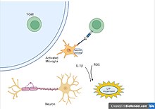
The initiation of neuroinflammation in the body. (Created with BioRender.com)

Viruses, bacteria, and other infectious agents activate the body’s defense systems and cause immune cells to protect the designed area from the damage. Some of these foreign pathogens can trigger a strong inflammatory response that can compromise the integrity of the blood-brain barrier and thus change the flow of inflammation in nearby tissue. The location along with the type of infection can determine what type of inflammatory response is activated and whether specific cytokines or immune cells will act.

Neuroimmune response

Glial cells

Microglia are recognized as the innate immune cells of the central nervous system. Microglia actively survey their environment and change their cell morphology significantly in response to neural injury. Acute inflammation in the brain is typically characterized by rapid activation of microglia. During this period, there is no peripheral immune response. Over time, however, chronic inflammation causes the degradation of tissue and of the blood–brain barrier. During this time, microglia generate reactive oxygen species and release signals to recruit peripheral immune cells for an inflammatory response.

Astrocytes are glial cells that are the most abundant cells in the brain. They are involved in maintenance and support of neurons and compose a significant component of the blood–brain barrier. After insult to the brain, such as traumatic brain injury, astrocytes may become activated in response to signals released by injured neurons or activated microglia. Once activated, astrocytes may release various growth factors and undergo morphological changes. For example, after injury, astrocytes form the glial scar composed of a proteoglycan matrix that hinders axonal regeneration. However, more recent studies revealed that glia scar is not detrimental, but is in fact beneficial for axonal regeneration.

Cytokines

Cytokines are a class of proteins regulating inflammation, cell signaling, and various cell processes such as growth and survival. Chemokines are a subset of cytokines that regulate cell migration, such as attracting immune cells to a site of infection or injury. Various cell types in the brain may produce cytokines and chemokines such as microglia, astrocytes, endothelial cells, and other glial cells. Physiologically, chemokines and cytokines function as neuromodulators that regulate inflammation and development. In the healthy brain, cells secrete cytokines to produce a local inflammatory environment to recruit microglia and clear the infection or injury. However, in neuroinflammation, cells may have sustained release of cytokines and chemokines which may compromise the blood–brain barrier. Peripheral immune cells are called to the site of injury via these cytokines and may now migrate across the compromised blood brain barrier into the brain. Common cytokines produced in response to brain injury include: interleukin-6 (IL-6), which is produced during astrogliosis, and interleukin-1 beta (IL-1β) and tumor necrosis factor alpha (TNF-α), which can induce neuronal cytotoxicity. Although the pro-inflammatory cytokines may cause cell death and secondary tissue damage, they are necessary to repair the damaged tissue. For example, TNF-α causes neurotoxicity at early stages of neuroinflammation, but contributes to tissue growth at later stages of inflammation.

Peripheral immune response

The blood–brain barrier is a structure composed of endothelial cells and astrocytes that forms a barrier between the brain and circulating blood. Physiologically, this enables the brain to be protected from potentially toxic molecules and cells in the blood. Astrocytes form tight junctions, and therefore may strictly regulate what may pass the blood–brain barrier and enter the interstitial space. After injury and sustained release of inflammatory factors such as chemokines, the blood–brain barrier may be compromised, becoming permeable to circulating blood components and peripheral immune cells. Cells involved in the innate and adaptive immune responses, such as macrophages, T cells, and B cells, may then enter into the brain. This exacerbates the inflammatory environment of the brain and contributes to chronic neuroinflammation and neurodegeneration.

Traumatic brain injury

Traumatic brain injury (TBI) is brain trauma caused by significant force to the head. Following TBI, there are both reparative and degenerative mechanisms that lead to an inflammatory environment. Within minutes of injury, pro-inflammatory cytokines are released. The pro-inflammatory cytokine Il-1β is one such cytokine that exacerbates the tissue damage caused by TBI. TBI may cause significant damage to vital components to the brain, including the blood–brain barrier. Il-1β causes DNA fragmentation and apoptosis, and together with TNF-α may cause damage to the blood–brain barrier and infiltration of leukocytes. Increased density of activated immune cells have been found in the human brain after concussion.

As the most abundant immune cells in the brain, Microglia are important to the brain’s defense against injury. The major caveat of these cells comes from the fact that their ability to promote recovery mechanism with anti-inflammatory factors, is inhibited by their secondary ability to make a large amount of pro-inflammatory cytokines. This can result in sustained brain damage as anti-inflammatory factors decrease in amount when more pro-inflammatory cytokines are produced in excess by microglia. The cytokines produced by microglia, astrocytes, and other immune cells, activate glial cells further increasing the number of pro-inflammatory factors that further prevent neurological systems from recovering. The dual nature of microglia is one example of why neuroinflammation can be helpful or hurtful under specific conditions.

Role of Neuroinflammation in the Pathophysiology of TBI. (Created with BioRender.com)

Spinal cord injury

Spinal Cord Injury (SCI) can be divided into three separate phases. The primary or acute phase occurs from seconds to minutes after injury, the secondary phase occurs from minutes to weeks after injury, and the chronic phase occurs from months to years following injury. A primary SCI is caused by spinal cord compression or transection, leading to glutamate excitotoxicity, sodium and calcium ion imbalances, and free radical damage. Neurodegeneration via apoptosis and demyelination of neuronal cells causes inflammation at the injury site. This leads to a secondary SCI, whose symptoms include edema, cavitation of spinal parenchyma, reactive gliosis, and potentially permanent loss of function.

During the SCI induced inflammatory response, several pro-inflammatory cytokines including interleukin 1β (IL-1β), inducible Nitric Oxide Synthase (iNOS), Interferon-γ (IFN-γ), IL-6, IL-23, and tumor necrosis factor α (TNFα) are secreted, activating local microglia and attracting various immune cells such as naive bone-marrow derived macrophages. These activated microglia and macrophages play a role in the pathogenesis of SCI.

Upon infiltration of the injury site's epicenter, macrophages will undergo phenotype switching from an M2 phenotype to an M1-like phenotype. The M2 phenotype is associated with anti-inflammatory factors such as IL-10, IL-4, and IL-13 and contributes to wound healing and tissue repair. However, the M1-like phenotype is associated with pro-inflammatory cytokines and reactive oxygen species that contribute to increased damage and inflammation. Factors such as myelin debris, which is formed by the injury at the damage site, has been shown to induce the phenotype shift from M2 to M1. A decreased population of M2 macrophages and an increased population of M1 macrophages is associated with chronic inflammation. Short term inflammation is important in clearing cell debris from the site of injury, but it is this chronic, long-term inflammation that will lead to further cell death and damage radiating from the site of injury.

Aging

Aging is often associated with cognitive impairment and increased propensity for developing neurodegenerative diseases, such as Alzheimer's disease. Elevated inflammatory markers seemed to accelerate the brain aging process In the aged brain alone, without any evident disease, there are chronically increased levels of pro-inflammatory cytokines and reduced levels of anti-inflammatory cytokines. The homeostatic imbalance between anti-inflammatory and pro-inflammatory cytokines in aging is one factor that increases the risk for neurodegenerative disease. Additionally, there is an increased number of activated microglia in aged brains, which have increased expression of major histocompatibility complex II (MHC II), ionized calcium binding adaptor-1 (IBA1), CD86, ED1 macrophage antigen, CD4, and leukocyte common antigen. These activated microglia decrease the ability for neurons to undergo long term potentiation (LTP) in the hippocampus and thereby reduce the ability to form memories.

Impairment of neuron LTP by activated Microglia. (Created with BioRender.com)

As one of the major cytokines responsible for maintaining inflammatory balance, IL-6 can also be used as a biological marker to observe the correlation between age and neuroinflammation. The same levels of IL-6 observed in the brain after injury, have also been found in the elderly and indicate the potential for cognitive impairment to develop. The unnecessary upregulation of IL-6 in the elderly population is a result of dysfunctional mediation by glial cells that can lead to the priming of glial cells and result in a more sensitive neuroinflammatory response.

Role in neurodegenerative disease

Alzheimer's disease

Alzheimer's disease (AD) has historically been characterized by two major hallmarks: neurofibrillary tangles and amyloid-beta plaques. Neurofibrillary tangles are insoluble aggregates of tau proteins, and amyloid-beta plaques are extracellular deposits of the amyloid-beta protein. Current thinking in AD pathology goes beyond these two typical hallmarks to suggest that a significant portion of neurodegeneration in Alzheimer's is due to neuroinflammation. Activated microglia are seen in abundance in post-mortem AD brains. Current thought is that inflammatory cytokine-activated microglia cannot phagocytose amyloid-beta, which may contribute to plaque accumulation as opposed to clearance. Additionally, the inflammatory cytokine IL-1β is upregulated in AD and is associated with decreases of synaptophysin and consequent synaptic loss. Further evidence that inflammation is associated with disease progression in AD is that individuals who take non-steroidal anti-inflammatory drugs (NSAIDs) regularly have been associated with a 67% of protection against the onset of AD (relative to the placebo group) in a four-year follow-up assessment. Elevated inflammatory markers showed an association with accelerated brain aging, which might explain the link to neurodegeneration in AD-related brain regions.

Parkinson's disease

The leading hypothesis of Parkinson's disease progression includes neuroinflammation as a major component. This hypothesis stipulates that Stage 1 of Parkinson's disease begins in the gut, as evidenced by a large number of cases that begin with constipation. The inflammatory response in the gut may play a role in alpha-synuclein (α-Syn) aggregation and misfolding, a characteristic of Parkinson's disease pathology. If there is a balance between good bacteria and bad bacteria in the gut, the bacteria may remain contained to the gut. However, dysbiosis of good bacteria and bad bacteria may cause a “leaky” gut, creating an inflammatory response. This response aids α-Syn misfolding and transfer across neurons, as the protein works its way up to the CNS. The brainstem is vulnerable to inflammation, which would explain Stage 2, including sleep disturbances and depression. In Stage 3 of the hypothesis, the inflammation affects the substantia nigra, the dopamine producing cells of the brain, beginning the characteristic motor deficits of Parkinson's disease. Stage 4 of Parkinson's disease includes deficits caused by inflammation in key regions of the brain that regulate executive function and memory. As evidence supporting this hypothesis, patients in Stage 3 (motor deficits) that are not experiencing cognitive deficits already show that there is neuroinflammation of the cortex. This suggests that neuroinflammation may be a precursor to the deficits seen in Parkinson's disease.

Amyotrophic lateral sclerosis

Unlike other neurodegenerative diseases, the exact pathophysiology of amyotrophic lateral sclerosis (ALS) is still far from being fully uncovered. Several hypotheses have been proposed to explain the development and progression of this lethal disease, by which neuroinflammation is one of the above. It is characterised by the activation of microglia and astrocytes, T lymphocyte infiltration, and the production of pro-inflammatory cytokines. Features of neuroinflammation were observed in the brain of living ALS patients, post-mortem CNS samples, and mouse models of ALS. Multiple evidence has described the mechanism of how microglial and astrocyte activation can promote disease progression (reviewed by). Replacement of mSOD1 microglia and astrocytes with the wild-type forms delayed motor neuron (MN) degeneration and extended the lifespan of ALS mice. Infiltration of T cells was reported in both early and late stages of ALS. Among all T cells, CD4+ T cells has drawn the most attention by being a neuroprotective agent during MN loss. T regulatory (Treg) cells is also a safeguard against neuroinflammation, demonstrated by the evidence of inverse correlation of the number of Treg cells and disease progression/ severity. Apart from the three phenotypes discussed, peripheral macrophages/ monocytes and the complement system are also suggested to be contributed to disease pathogenesis. Activation and invasion of peripheral monocytes observed in the spinal cord of ALS patients and mice may lead to MN loss. Expression of several complement components are reported to be upregulated in the samples isolated from ALS patients and transgenic rodent models. Further studies are required to elucidate their roles in ALS.

Multiple sclerosis

Multiple sclerosis is the most common disabling neurological disease of young adults. It is characterized by demyelination and neurodegeneration, which contribute to the common symptoms of cognitive deficits, limb weakness, and fatigue. In multiple sclerosis, inflammatory cytokines disrupt the blood–brain barrier and allow for the migration of peripheral immune cells into the central nervous system. When they have migrated into the central nervous system, B cells and plasma cells produce antibodies against the myelin sheath that insulates neurons, degrading the myelin and slowing conduction in the neurons. Additionally, T cells may enter through the blood–brain barrier, be activated by local antigen presenting cells, and attack the myelin sheath. This has the same effect of degrading the myelin and slowing conduction. As in other neurodegenerative diseases, activated microglia produce inflammatory cytokines that contribute to widespread inflammation. It has been shown that inhibiting microglia decreases the severity of multiple sclerosis.

Role as a therapeutic target

Drug therapy

Because neuroinflammation has been associated with a variety of neurodegenerative diseases, there is increasing interest to determine whether reducing inflammation will reverse neurodegeneration. Inhibiting inflammatory cytokines, such as IL-1β, decreases neuronal loss seen in neurodegenerative diseases. Current treatments for multiple sclerosis include interferon-B, Glatiramer acetate, and Mitoxantrone, which function by reducing or inhibiting T Cell activation, but have the side effect of systemic immunosuppression  In Alzheimer's disease, the use of non-steroidal anti-inflammatory drugs decreases the risk of developing the disease. Current treatments for Alzheimer's disease include NSAIDs and glucocorticoids. NSAIDs function by blocking conversion of prostaglandin H2 into other prostaglandins (PGs) and thromboxane (TX). Prostoglandins and thromboxane act as inflammatory mediators and increase microvascular permeability.

Exercise

Exercise is a promising mechanism of prevention and treatment for various diseases characterized by neuroinflammation. Aerobic exercise is used widely to reduce inflammation in the periphery by activating protective systems in the body that stabilize internal environment. Exercise has been shown to decrease proliferation of microglia in the brain, decrease hippocampal expression of immune-related genes and reduce expression of inflammatory cytokines such as TNF-α.

The neuroprotective and anti-inflammatory effects of exercise on cognitive diseases. (Created with BioRender.com)

Exercise can help protect the mind and body by maintaining the brain’s internal environment, focusing on recruiting anti-inflammatory cytokines, and activating cellular processes that proactively protect against damage while also initiating recovery mechanisms. The ability of physical activity to stimulate immune defenses against neuroinflammation-related diseases has been observed in recent clinical studies. The application of various exercises under a range of different conditions resulted in higher neurological metabolism, stronger protection against free radicals, and stronger neuroplasticity against neurological diseases. The resulting increase in brain function was due to the induced change in gene expression, increase in trophic factors, and reduction in pro-inflammatory cytokines.

Ventricular fibrillation

From Wikipedia, the free encyclopedia
https://en.wikipedia.org/wiki/Ventricular_fibrillation

Ventricular fibrillation
12-lead ECG showing ventricular fibrillation
SpecialtyCardiology
SymptomsCardiac arrest with loss of consciousness and no pulse
CausesCoronary heart disease (including myocardial infarction), valvular heart disease, cardiomyopathy, Brugada syndrome, electric shock, long QT syndrome, intracranial hemorrhage
Diagnostic methodElectrocardiogram
Differential diagnosisTorsades de pointes
TreatmentCardiopulmonary resuscitation (CPR) with defibrillation
PrognosisSurvival rate 17% (out of hospital), 46% (in hospital)
Frequency~10% of people in cardiac arrest

Ventricular fibrillation (V-fib or VF) is an abnormal heart rhythm in which the ventricles of the heart quiver. It is due to disorganized electrical activity. Ventricular fibrillation results in cardiac arrest with loss of consciousness and no pulse. This is followed by sudden cardiac death in the absence of treatment. Ventricular fibrillation is initially found in about 10% of people with cardiac arrest.

Ventricular fibrillation can occur due to coronary heart disease, valvular heart disease, cardiomyopathy, Brugada syndrome, long QT syndrome, electric shock, or intracranial hemorrhage. Diagnosis is by an electrocardiogram (ECG) showing irregular unformed QRS complexes without any clear P waves. An important differential diagnosis is torsades de pointes.

Treatment is with cardiopulmonary resuscitation (CPR) and defibrillation. Biphasic defibrillation may be better than monophasic. The medication epinephrine or amiodarone may be given if initial treatments are not effective. Rates of survival among those who are out of hospital when the arrhythmia is detected is about 17% while in hospital it is about 46%.

Signs and symptoms

Ventricular fibrillation is a cause of cardiac arrest. The ventricular muscle twitches randomly rather than contracting in a coordinated fashion (from the apex of the heart to the outflow of the ventricles), and so the ventricles fail to pump blood around the body – because of this, it is classified as a cardiac arrest rhythm, and patients in V-fib should be treated with cardiopulmonary resuscitation (CPR) and prompt defibrillation. Left untreated, ventricular fibrillation is rapidly fatal as the vital organs of the body, including the heart, are starved of oxygen, and as a result patients in this rhythm will not be conscious or responsive to stimuli. Prior to cardiac arrest, patients may complain of varying symptoms depending on the underlying cause. Patients may exhibit signs of agonal breathing, which to a layperson can look like normal spontaneous breathing, but is a sign of hypoperfusion of the brainstem.

It has an appearance on electrocardiography of irregular electrical activity with no discernable pattern. It may be described as "coarse" or "fine" depending on its amplitude, or as progressing from coarse to fine V-fib. Coarse V-fib may be more responsive to defibrillation, while fine V-fib can mimic the appearance of asystole on a defibrillator or cardiac monitor set to a low gain. Some clinicians may attempt to defibrillate fine V-fib in the hope that it can be reverted to a cardiac rhythm compatible with life, whereas others will deliver CPR and sometimes drugs as described in the advanced cardiac life support protocols in an attempt to increase its amplitude and the odds of successful defibrillation.

Causes

Ventricular fibrillation has been described as "chaotic asynchronous fractionated activity of the heart" (Moe et al. 1964). A more complete definition is that ventricular fibrillation is a "turbulent, disorganized electrical activity of the heart in such a way that the recorded electrocardiographic deflections continuously change in shape, magnitude and direction".

Ventricular fibrillation most commonly occurs within diseased hearts, and, in the vast majority of cases, is a manifestation of underlying ischemic heart disease. Ventricular fibrillation is also seen in those with cardiomyopathy, myocarditis, and other heart pathologies. In addition, it is seen with electrolyte imbalance, overdoses of cardiotoxic drugs, and following near drowning or major trauma. It is also notable that ventricular fibrillation occurs where there is no discernible heart pathology or other evident cause, the so-called idiopathic ventricular fibrillation.

Idiopathic ventricular fibrillation occurs with a reputed incidence of approximately 1% of all cases of out-of-hospital arrest, as well as 3–9% of the cases of ventricular fibrillation unrelated to myocardial infarction, and 14% of all ventricular fibrillation resuscitations in patients under the age of 40. It follows then that, on the basis of the fact that ventricular fibrillation itself is common, idiopathic ventricular fibrillation accounts for an appreciable mortality. Recently described syndromes such as the Brugada Syndrome may give clues to the underlying mechanism of ventricular arrhythmias. In the Brugada syndrome, changes may be found in the resting ECG with evidence of right bundle branch block (RBBB) and ST elevation in the chest leads V1-V3, with an underlying propensity to sudden cardiac death.

The relevance of this is that theories of the underlying pathophysiology and electrophysiology must account for the occurrence of fibrillation in the apparent "healthy" heart. It is evident that there are mechanisms at work that we do not fully appreciate and understand. Investigators are exploring new techniques of detecting and understanding the underlying mechanisms of sudden cardiac death in these patients without pathological evidence of underlying heart disease.

Familial conditions that predispose individuals to developing ventricular fibrillation and sudden cardiac death are often the result of gene mutations that affect cellular transmembrane ion channels. For example, in Brugada Syndrome, sodium channels are affected. In certain forms of long QT syndrome, the potassium inward rectifier channel is affected.

In 1899, it was also found that ventricular fibrillation was, typically, the ultimate cause of death when the electric chair was used.

Pathophysiology

Fine ventricular fibrillation as seen on a rhythm strip
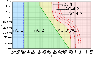
Log-log graph of the effect of alternating current I of duration T passing from left hand to feet as defined in IEC publication 60479–1.
AC-1: imperceptible
AC-2: perceptible but no muscle reaction
AC-3: muscle contraction with reversible effects
AC-4: possible irreversible effects
AC-4.1: up to 5% probability of ventricular fibrillation
AC-4.2: 5-50% probability of fibrillation
AC-4.3: over 50% probability of fibrillation

Abnormal automaticity

Automaticity is a measure of the propensity of a fiber to initiate an impulse spontaneously. The product of a hypoxic myocardium can be hyperirritable myocardial cells. These may then act as pacemakers. The ventricles are then being stimulated by more than one pacemaker. Scar and dying tissue is inexcitable, but around these areas usually lies a penumbra of hypoxic tissue that is excitable. Ventricular excitability may generate re-entry ventricular arrhythmia.

Most myocardial cells with an associated increased propensity to arrhythmia development have an associated loss of membrane potential. That is, the maximum diastolic potential is less negative and therefore exists closer to the threshold potential. Cellular depolarisation can be due to a raised external concentration of potassium ions K+, a decreased intracellular concentration of sodium ions Na+, increased permeability to Na+, or a decreased permeability to K+. The ionic basic automaticity is the net gain of an intracellular positive charge during diastole in the presence of a voltage-dependent channel activated by potentials negative to –50 to –60 mV.

Myocardial cells are exposed to different environments. Normal cells may be exposed to hyperkalaemia; abnormal cells may be perfused by normal environment. For example, with a healed myocardial infarction, abnormal cells can be exposed to an abnormal environment such as with a myocardial infarction with myocardial ischaemia. In conditions such as myocardial ischaemia, possible mechanism of arrhythmia generation include the resulting decreased internal K+ concentration, the increased external K+ concentration, norepinephrine release and acidosis. When myocardial cell are exposed to hyperkalemia, the maximum diastolic potential is depolarized as a result of the alteration of Ik1 potassium current, whose intensity and direction is strictly dependent on intracellular and extracellular potassium concentrations. With Ik1 suppressed, an hyperpolarizing effect is lost and therefore there can be activation of funny current even in myocardial cells (which is normally suppressed by the hyperpolarizing effect of coexisting potassium currents). This can lead to the instauration of automaticity in ischemic tissue.

Re-entry

The role of re-entry or circus motion was demonstrated separately by G. R. Mines and W. E. Garrey. Mines created a ring of excitable tissue by cutting the atria out of the ray fish. Garrey cut out a similar ring from the turtle ventricle. They were both able to show that, if a ring of excitable tissue was stimulated at a single point, the subsequent waves of depolarisation would pass around the ring. The waves eventually meet and cancel each other out, but, if an area of transient block occurred with a refractory period that blocked one wavefront and subsequently allowed the other to proceed retrogradely over the other path, then a self-sustaining circus movement phenomenon would result. For this to happen, however, it is necessary that there be some form of non-uniformity. In practice, this may be an area of ischemic or infarcted myocardium, or underlying scar tissue.

It is possible to think of the advancing wave of depolarisation as a dipole with a head and a tail. The length of the refractory period and the time taken for the dipole to travel a certain distance—the propagation velocity—will determine whether such a circumstance will arise for re-entry to occur. Factors that promote re-entry would include a slow-propagation velocity, a short refractory period with a sufficient size of ring of conduction tissue. These would enable a dipole to reach an area that had been refractory and is now able to be depolarised with continuation of the wavefront.

In clinical practice, therefore, factors that would lead to the right conditions to favour such re-entry mechanisms include increased heart size through hypertrophy or dilatation, drugs which alter the length of the refractory period and areas of cardiac disease. Therefore, the substrate of ventricular fibrillation is transient or permanent conduction block. Block due either to areas of damaged or refractory tissue leads to areas of myocardium for initiation and perpetuation of fibrillation through the phenomenon of re-entry.

Triggered activity

Triggered activity can occur due to the presence of afterdepolarisations. These are depolarising oscillations in the membrane voltage induced by preceding action potentials. These can occur before or after full repolarisation of the fiber and as such are termed either early (EADs) or delayed afterdepolarisations (DADs). All afterdepolarisations may not reach threshold potential, but, if they do, they can trigger another afterdepolarisation, and thus self-perpetuate.

Power spectrum

Ventricular fibrillation as seen in lead II

The distribution of frequency and power of a waveform can be expressed as a power spectrum in which the contribution of different waveform frequencies to the waveform under analysis is measured. This can be expressed as either the dominant or peak frequency, i.e., the frequency with the greatest power or the median frequency, which divides the spectrum in two halves.

Frequency analysis has many other uses in medicine and in cardiology, including analysis of heart rate variability and assessment of cardiac function, as well as in imaging and acoustics.

Histopathology

Micrograph showing myofibre break-up with squared nuclei, a morphologic correlate of ventricular fibrillation. H&E stain.

Myofibre break-up, abbreviated MFB, is associated with ventricular fibrillation leading to death. Histomorphologically, MFB is characterized by fractures of the cardiac myofibres perpendicular to their long axis, with squaring of the myofibre nuclei.

Treatment

Defibrillation is the definitive treatment of ventricular fibrillation, whereby an electrical current is applied to the ventricular mass either directly or externally through pads or paddles, with the aim of depolarising enough of the myocardium for coordinated contractions to occur again. The use of this is often dictated around the world by Advanced Cardiac Life Support or Advanced Life Support algorithms, which is taught to medical practitioners including doctors, nurses and paramedics and also advocates the use of drugs, predominantly epinephrine, after every second unsuccessful attempt at defibrillation, as well as cardiopulmonary resuscitation (CPR) between defibrillation attempts. Though ALS/ACLS algorithms encourage the use of drugs, they state first and foremost that defibrillation should not be delayed for any other intervention and that adequate cardiopulmonary resuscitation be delivered with minimal interruption.

The precordial thump is a manoeuver promoted as a mechanical alternative to defibrillation. Some advanced life support algorithms advocate its use once and only in the case of witnessed and monitored V-fib arrests as the likelihood of it successfully cardioverting a patient are small and this diminishes quickly in the first minute of onset.

People who survive a "V-fib arrest" and who make a good recovery are often considered for an implantable cardioverter-defibrillator, which can quickly deliver this same life-saving defibrillation should another episode of ventricular fibrillation occur outside a hospital environment.

Epidemiology

Sudden cardiac arrest is the leading cause of death in the industrialised world. It exacts a significant mortality with approximately 70,000 to 90,000 sudden cardiac deaths each year in the United Kingdom, and survival rates are only 2%. The majority of these deaths are due to ventricular fibrillation secondary to myocardial infarction, or "heart attack". During ventricular fibrillation, cardiac output drops to zero, and, unless remedied promptly, death usually ensues within minutes.

History

Lyman Brewer suggests that the first recorded account of ventricular fibrillation dates as far back as 1500 BC, and can be found in the Ebers papyrus of ancient Egypt. An extract, recorded 3500 years ago, states: "When the heart is diseased, its work is imperfectly performed: the vessels proceeding from the heart become inactive, so that you cannot feel them … if the heart trembles, has little power and sinks, the disease is advanced and death is near." A book authored by Jo Miles suggests that it may even go back farther. Tests done on frozen remains found in the Himalayas seemed fairly conclusive that the first known case of ventricular fibrillation dates back to at least 2500 BC.

Whether this is a description of ventricular fibrillation is debatable. The next recorded description occurs 3000 years later and is recorded by Vesalius, who described the appearance of "worm-like" movements of the heart in animals prior to death.

The significance and clinical importance of these observations and descriptions possibly of ventricular fibrillation were not recognised until John Erichsen in 1842 described ventricular fibrillation following the ligation of a coronary artery (Erichsen JE 1842). Subsequent to this in 1850, fibrillation was described by Ludwig and Hoffa when they demonstrated the provocation of ventricular fibrillation in an animal by applying a "Faradic" (electrical) current to the heart.

In 1874, Edmé Félix Alfred Vulpian coined the term mouvement fibrillaire, a term that he seems to have used to describe both atrial and ventricular fibrillation. John A. MacWilliam, a physiologist who had trained under Ludwig and who subsequently became Professor of Physiology at the University of Aberdeen, gave an accurate description of the arrhythmia in 1887. This definition still holds today, and is interesting in the fact that his studies and description predate the use of electrocardiography. His description is as follows: "The ventricular muscle is thrown into a state of irregular arrhythmic contraction, whilst there is a great fall in the arterial blood pressure, the ventricles become dilated with blood as the rapid quivering movement of their walls is insufficient to expel their contents; the muscular action partakes of the nature of a rapid incoordinate twitching of the muscular tissue … The cardiac pump is thrown out of gear, and the last of its vital energy is dissipated in the violent and the prolonged turmoil of fruitless activity in the ventricular walls." MacWilliam spent many years working on ventricular fibrillation and was one of the first to show that ventricular fibrillation could be terminated by a series of induction shocks through the heart.

The first electrocardiogram recording of ventricular fibrillation was by August Hoffman in a paper published in 1912. At this time, two other researchers, George Ralph Mines and Garrey, working separately, produced work demonstrating the phenomenon of circus movement and re-entry as possible substrates for the generation of arrhythmias. This work was also accompanied by Lewis, who performed further outstanding work into the concept of "circus movement."

Later milestones include the work by W. J. Kerr and W. L. Bender in 1922, who produced an electrocardiogram showing ventricular tachycardia evolving into ventricular fibrillation. The re-entry mechanism was also advocated by DeBoer, who showed that ventricular fibrillation could be induced in late systole with a single shock to a frog heart. The concept of "R on T ectopics" was further brought out by Katz in 1928. This was called the "vulnerable period" by Wiggers and Wegria in 1940, who brought to attention the concept of the danger of premature ventricular beats occurring on a T wave.

Another definition of VF was produced by Wiggers in 1940. He described ventricular fibrillation as "an incoordinate type of contraction which, despite a high metabolic rate of the myocardium, produces no useful beats. As a result, the arterial pressure falls abruptly to very low levels, and death results within six to eight minutes from anemia [ischemia] of the brain and spinal cord".

Spontaneous conversion of ventricular fibrillation to a more benign rhythm is rare in all but small animals. Defibrillation is the process that converts ventricular fibrillation to a more benign rhythm. This is usually by application of an electric shock to the myocardium and is discussed in detail in the relevant article.

Operator (computer programming)

From Wikipedia, the free encyclopedia https://en.wikipedia.org/wiki/Operator_(computer_programmin...