Search This Blog

Friday, September 29, 2023

Neuroinflammation

From Wikipedia, the free encyclopedia
https://en.wikipedia.org/wiki/Neuroinflammation

Neuroinflammation is inflammation of the nervous tissue. It may be initiated in response to a variety of cues, including infection, traumatic brain injury, toxic metabolites, or autoimmunity. In the central nervous system (CNS), including the brain and spinal cord, microglia are the resident innate immune cells that are activated in response to these cues. The CNS is typically an immunologically privileged site because peripheral immune cells are generally blocked by the blood–brain barrier (BBB), a specialized structure composed of astrocytes and endothelial cells. However, circulating peripheral immune cells may surpass a compromised BBB and encounter neurons and glial cells expressing major histocompatibility complex molecules, perpetuating the immune response. Although the response is initiated to protect the central nervous system from the infectious agent, the effect may be toxic and widespread inflammation as well as further migration of leukocytes through the blood–brain barrier may occur.

Causes

Neuroinflammation is widely regarded as chronic, as opposed to acute, inflammation of the central nervous system. Acute inflammation usually follows injury to the central nervous system immediately, and is characterized by inflammatory molecules, endothelial cell activation, platelet deposition, and tissue edema. Chronic inflammation is the sustained activation of glial cells and recruitment of other immune cells into the brain. It is chronic inflammation that is typically associated with neurodegenerative diseases. Common causes of chronic neuroinflammation include:

The initiation of neuroinflammation in the body. (Created with BioRender.com)

Viruses, bacteria, and other infectious agents activate the body’s defense systems and cause immune cells to protect the designed area from the damage. Some of these foreign pathogens can trigger a strong inflammatory response that can compromise the integrity of the blood-brain barrier and thus change the flow of inflammation in nearby tissue. The location along with the type of infection can determine what type of inflammatory response is activated and whether specific cytokines or immune cells will act.

Neuroimmune response

Glial cells

Microglia are recognized as the innate immune cells of the central nervous system. Microglia actively survey their environment and change their cell morphology significantly in response to neural injury. Acute inflammation in the brain is typically characterized by rapid activation of microglia. During this period, there is no peripheral immune response. Over time, however, chronic inflammation causes the degradation of tissue and of the blood–brain barrier. During this time, microglia generate reactive oxygen species and release signals to recruit peripheral immune cells for an inflammatory response.

Astrocytes are glial cells that are the most abundant cells in the brain. They are involved in maintenance and support of neurons and compose a significant component of the blood–brain barrier. After insult to the brain, such as traumatic brain injury, astrocytes may become activated in response to signals released by injured neurons or activated microglia. Once activated, astrocytes may release various growth factors and undergo morphological changes. For example, after injury, astrocytes form the glial scar composed of a proteoglycan matrix that hinders axonal regeneration. However, more recent studies revealed that glia scar is not detrimental, but is in fact beneficial for axonal regeneration.

Cytokines

Cytokines are a class of proteins regulating inflammation, cell signaling, and various cell processes such as growth and survival. Chemokines are a subset of cytokines that regulate cell migration, such as attracting immune cells to a site of infection or injury. Various cell types in the brain may produce cytokines and chemokines such as microglia, astrocytes, endothelial cells, and other glial cells. Physiologically, chemokines and cytokines function as neuromodulators that regulate inflammation and development. In the healthy brain, cells secrete cytokines to produce a local inflammatory environment to recruit microglia and clear the infection or injury. However, in neuroinflammation, cells may have sustained release of cytokines and chemokines which may compromise the blood–brain barrier. Peripheral immune cells are called to the site of injury via these cytokines and may now migrate across the compromised blood brain barrier into the brain. Common cytokines produced in response to brain injury include: interleukin-6 (IL-6), which is produced during astrogliosis, and interleukin-1 beta (IL-1β) and tumor necrosis factor alpha (TNF-α), which can induce neuronal cytotoxicity. Although the pro-inflammatory cytokines may cause cell death and secondary tissue damage, they are necessary to repair the damaged tissue. For example, TNF-α causes neurotoxicity at early stages of neuroinflammation, but contributes to tissue growth at later stages of inflammation.

Peripheral immune response

The blood–brain barrier is a structure composed of endothelial cells and astrocytes that forms a barrier between the brain and circulating blood. Physiologically, this enables the brain to be protected from potentially toxic molecules and cells in the blood. Astrocytes form tight junctions, and therefore may strictly regulate what may pass the blood–brain barrier and enter the interstitial space. After injury and sustained release of inflammatory factors such as chemokines, the blood–brain barrier may be compromised, becoming permeable to circulating blood components and peripheral immune cells. Cells involved in the innate and adaptive immune responses, such as macrophages, T cells, and B cells, may then enter into the brain. This exacerbates the inflammatory environment of the brain and contributes to chronic neuroinflammation and neurodegeneration.

Traumatic brain injury

Traumatic brain injury (TBI) is brain trauma caused by significant force to the head. Following TBI, there are both reparative and degenerative mechanisms that lead to an inflammatory environment. Within minutes of injury, pro-inflammatory cytokines are released. The pro-inflammatory cytokine Il-1β is one such cytokine that exacerbates the tissue damage caused by TBI. TBI may cause significant damage to vital components to the brain, including the blood–brain barrier. Il-1β causes DNA fragmentation and apoptosis, and together with TNF-α may cause damage to the blood–brain barrier and infiltration of leukocytes. Increased density of activated immune cells have been found in the human brain after concussion.

As the most abundant immune cells in the brain, Microglia are important to the brain’s defense against injury. The major caveat of these cells comes from the fact that their ability to promote recovery mechanism with anti-inflammatory factors, is inhibited by their secondary ability to make a large amount of pro-inflammatory cytokines. This can result in sustained brain damage as anti-inflammatory factors decrease in amount when more pro-inflammatory cytokines are produced in excess by microglia. The cytokines produced by microglia, astrocytes, and other immune cells, activate glial cells further increasing the number of pro-inflammatory factors that further prevent neurological systems from recovering. The dual nature of microglia is one example of why neuroinflammation can be helpful or hurtful under specific conditions.

Role of Neuroinflammation in the Pathophysiology of TBI. (Created with BioRender.com)

Spinal cord injury

Spinal Cord Injury (SCI) can be divided into three separate phases. The primary or acute phase occurs from seconds to minutes after injury, the secondary phase occurs from minutes to weeks after injury, and the chronic phase occurs from months to years following injury. A primary SCI is caused by spinal cord compression or transection, leading to glutamate excitotoxicity, sodium and calcium ion imbalances, and free radical damage. Neurodegeneration via apoptosis and demyelination of neuronal cells causes inflammation at the injury site. This leads to a secondary SCI, whose symptoms include edema, cavitation of spinal parenchyma, reactive gliosis, and potentially permanent loss of function.

During the SCI induced inflammatory response, several pro-inflammatory cytokines including interleukin 1β (IL-1β), inducible Nitric Oxide Synthase (iNOS), Interferon-γ (IFN-γ), IL-6, IL-23, and tumor necrosis factor α (TNFα) are secreted, activating local microglia and attracting various immune cells such as naive bone-marrow derived macrophages. These activated microglia and macrophages play a role in the pathogenesis of SCI.

Upon infiltration of the injury site's epicenter, macrophages will undergo phenotype switching from an M2 phenotype to an M1-like phenotype. The M2 phenotype is associated with anti-inflammatory factors such as IL-10, IL-4, and IL-13 and contributes to wound healing and tissue repair. However, the M1-like phenotype is associated with pro-inflammatory cytokines and reactive oxygen species that contribute to increased damage and inflammation. Factors such as myelin debris, which is formed by the injury at the damage site, has been shown to induce the phenotype shift from M2 to M1. A decreased population of M2 macrophages and an increased population of M1 macrophages is associated with chronic inflammation. Short term inflammation is important in clearing cell debris from the site of injury, but it is this chronic, long-term inflammation that will lead to further cell death and damage radiating from the site of injury.

Aging

Aging is often associated with cognitive impairment and increased propensity for developing neurodegenerative diseases, such as Alzheimer's disease. Elevated inflammatory markers seemed to accelerate the brain aging process In the aged brain alone, without any evident disease, there are chronically increased levels of pro-inflammatory cytokines and reduced levels of anti-inflammatory cytokines. The homeostatic imbalance between anti-inflammatory and pro-inflammatory cytokines in aging is one factor that increases the risk for neurodegenerative disease. Additionally, there is an increased number of activated microglia in aged brains, which have increased expression of major histocompatibility complex II (MHC II), ionized calcium binding adaptor-1 (IBA1), CD86, ED1 macrophage antigen, CD4, and leukocyte common antigen. These activated microglia decrease the ability for neurons to undergo long term potentiation (LTP) in the hippocampus and thereby reduce the ability to form memories.
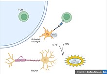
Impairment of neuron LTP by activated Microglia. (Created with BioRender.com)

As one of the major cytokines responsible for maintaining inflammatory balance, IL-6 can also be used as a biological marker to observe the correlation between age and neuroinflammation. The same levels of IL-6 observed in the brain after injury, have also been found in the elderly and indicate the potential for cognitive impairment to develop. The unnecessary upregulation of IL-6 in the elderly population is a result of dysfunctional mediation by glial cells that can lead to the priming of glial cells and result in a more sensitive neuroinflammatory response.

Role in neurodegenerative disease

Alzheimer's disease

Alzheimer's disease (AD) has historically been characterized by two major hallmarks: neurofibrillary tangles and amyloid-beta plaques. Neurofibrillary tangles are insoluble aggregates of tau proteins, and amyloid-beta plaques are extracellular deposits of the amyloid-beta protein. Current thinking in AD pathology goes beyond these two typical hallmarks to suggest that a significant portion of neurodegeneration in Alzheimer's is due to neuroinflammation. Activated microglia are seen in abundance in post-mortem AD brains. Current thought is that inflammatory cytokine-activated microglia cannot phagocytose amyloid-beta, which may contribute to plaque accumulation as opposed to clearance. Additionally, the inflammatory cytokine IL-1β is upregulated in AD and is associated with decreases of synaptophysin and consequent synaptic loss. Further evidence that inflammation is associated with disease progression in AD is that individuals who take non-steroidal anti-inflammatory drugs (NSAIDs) regularly have been associated with a 67% of protection against the onset of AD (relative to the placebo group) in a four-year follow-up assessment. Elevated inflammatory markers showed an association with accelerated brain aging, which might explain the link to neurodegeneration in AD-related brain regions.

Parkinson's disease

The leading hypothesis of Parkinson's disease progression includes neuroinflammation as a major component. This hypothesis stipulates that Stage 1 of Parkinson's disease begins in the gut, as evidenced by a large number of cases that begin with constipation. The inflammatory response in the gut may play a role in alpha-synuclein (α-Syn) aggregation and misfolding, a characteristic of Parkinson's disease pathology. If there is a balance between good bacteria and bad bacteria in the gut, the bacteria may remain contained to the gut. However, dysbiosis of good bacteria and bad bacteria may cause a “leaky” gut, creating an inflammatory response. This response aids α-Syn misfolding and transfer across neurons, as the protein works its way up to the CNS. The brainstem is vulnerable to inflammation, which would explain Stage 2, including sleep disturbances and depression. In Stage 3 of the hypothesis, the inflammation affects the substantia nigra, the dopamine producing cells of the brain, beginning the characteristic motor deficits of Parkinson's disease. Stage 4 of Parkinson's disease includes deficits caused by inflammation in key regions of the brain that regulate executive function and memory. As evidence supporting this hypothesis, patients in Stage 3 (motor deficits) that are not experiencing cognitive deficits already show that there is neuroinflammation of the cortex. This suggests that neuroinflammation may be a precursor to the deficits seen in Parkinson's disease.

Amyotrophic lateral sclerosis

Unlike other neurodegenerative diseases, the exact pathophysiology of amyotrophic lateral sclerosis (ALS) is still far from being fully uncovered. Several hypotheses have been proposed to explain the development and progression of this lethal disease, by which neuroinflammation is one of the above. It is characterised by the activation of microglia and astrocytes, T lymphocyte infiltration, and the production of pro-inflammatory cytokines. Features of neuroinflammation were observed in the brain of living ALS patients, post-mortem CNS samples, and mouse models of ALS. Multiple evidence has described the mechanism of how microglial and astrocyte activation can promote disease progression (reviewed by). Replacement of mSOD1 microglia and astrocytes with the wild-type forms delayed motor neuron (MN) degeneration and extended the lifespan of ALS mice. Infiltration of T cells was reported in both early and late stages of ALS. Among all T cells, CD4+ T cells has drawn the most attention by being a neuroprotective agent during MN loss. T regulatory (Treg) cells is also a safeguard against neuroinflammation, demonstrated by the evidence of inverse correlation of the number of Treg cells and disease progression/ severity. Apart from the three phenotypes discussed, peripheral macrophages/ monocytes and the complement system are also suggested to be contributed to disease pathogenesis. Activation and invasion of peripheral monocytes observed in the spinal cord of ALS patients and mice may lead to MN loss. Expression of several complement components are reported to be upregulated in the samples isolated from ALS patients and transgenic rodent models. Further studies are required to elucidate their roles in ALS.

Multiple sclerosis

Multiple sclerosis is the most common disabling neurological disease of young adults. It is characterized by demyelination and neurodegeneration, which contribute to the common symptoms of cognitive deficits, limb weakness, and fatigue. In multiple sclerosis, inflammatory cytokines disrupt the blood–brain barrier and allow for the migration of peripheral immune cells into the central nervous system. When they have migrated into the central nervous system, B cells and plasma cells produce antibodies against the myelin sheath that insulates neurons, degrading the myelin and slowing conduction in the neurons. Additionally, T cells may enter through the blood–brain barrier, be activated by local antigen presenting cells, and attack the myelin sheath. This has the same effect of degrading the myelin and slowing conduction. As in other neurodegenerative diseases, activated microglia produce inflammatory cytokines that contribute to widespread inflammation. It has been shown that inhibiting microglia decreases the severity of multiple sclerosis.

Role as a therapeutic target

Drug therapy

Because neuroinflammation has been associated with a variety of neurodegenerative diseases, there is increasing interest to determine whether reducing inflammation will reverse neurodegeneration. Inhibiting inflammatory cytokines, such as IL-1β, decreases neuronal loss seen in neurodegenerative diseases. Current treatments for multiple sclerosis include interferon-B, Glatiramer acetate, and Mitoxantrone, which function by reducing or inhibiting T Cell activation, but have the side effect of systemic immunosuppression  In Alzheimer's disease, the use of non-steroidal anti-inflammatory drugs decreases the risk of developing the disease. Current treatments for Alzheimer's disease include NSAIDs and glucocorticoids. NSAIDs function by blocking conversion of prostaglandin H2 into other prostaglandins (PGs) and thromboxane (TX). Prostoglandins and thromboxane act as inflammatory mediators and increase microvascular permeability.

Exercise

Exercise is a promising mechanism of prevention and treatment for various diseases characterized by neuroinflammation. Aerobic exercise is used widely to reduce inflammation in the periphery by activating protective systems in the body that stabilize internal environment. Exercise has been shown to decrease proliferation of microglia in the brain, decrease hippocampal expression of immune-related genes and reduce expression of inflammatory cytokines such as TNF-α.

The neuroprotective and anti-inflammatory effects of exercise on cognitive diseases. (Created with BioRender.com)

Exercise can help protect the mind and body by maintaining the brain’s internal environment, focusing on recruiting anti-inflammatory cytokines, and activating cellular processes that proactively protect against damage while also initiating recovery mechanisms. The ability of physical activity to stimulate immune defenses against neuroinflammation-related diseases has been observed in recent clinical studies. The application of various exercises under a range of different conditions resulted in higher neurological metabolism, stronger protection against free radicals, and stronger neuroplasticity against neurological diseases. The resulting increase in brain function was due to the induced change in gene expression, increase in trophic factors, and reduction in pro-inflammatory cytokines.

Arrhythmia

From Wikipedia, the free encyclopedia
https://en.wikipedia.org/wiki/Arrhythmia

Arrhythmia
Other namesCardiac arrhythmia, cardiac dysrhythmia, irregular heartbeat, heart arrhythmia
Ventricular fibrillation (VF) showing disorganized electrical activity producing a spiked tracing on an electrocardiogram (ECG)
SpecialtyCardiology
SymptomsPalpitations, lightheadedness, passing out, shortness of breath, chest pain[1]
ComplicationsStroke, heart failure
Usual onsetOlder age
TypesExtra beats, supraventricular tachycardias, ventricular arrhythmias, bradyarrhythmias
CausesProblems with the electrical conduction system of the heart
Diagnostic methodElectrocardiogram, Holter monitor
TreatmentMedications, medical procedures (pacemaker), surgery
FrequencyMillions

Arrhythmias, also known as cardiac arrhythmias, heart arrhythmias, or dysrhythmias, are irregularities in the heartbeat, including when it is too fast or too slow. A resting heart rate that is too fast – above 100 beats per minute in adults – is called tachycardia, and a resting heart rate that is too slow – below 60 beats per minute – is called bradycardia. Some types of arrhythmias have no symptoms. Symptoms, when present, may include palpitations or feeling a pause between heartbeats. In more serious cases, there may be lightheadedness, passing out, shortness of breath or chest pain. While most cases of arrhythmia are not serious, some predispose a person to complications such as stroke or heart failure. Others may result in sudden death.

Arrhythmias are often categorized into four groups: extra beats, supraventricular tachycardias, ventricular arrhythmias and bradyarrhythmias. Extra beats include premature atrial contractions, premature ventricular contractions and premature junctional contractions. Supraventricular tachycardias include atrial fibrillation, atrial flutter and paroxysmal supraventricular tachycardia. Ventricular arrhythmias include ventricular fibrillation and ventricular tachycardia. Bradyarrhythmias are due to sinus node dysfunction or atrioventricular conduction disturbances. Arrhythmias are due to problems with the electrical conduction system of the heart. A number of tests can help with diagnosis, including an electrocardiogram (ECG) and Holter monitor.

Many arrhythmias can be effectively treated. Treatments may include medications, medical procedures such as inserting a pacemaker, and surgery. Medications for a fast heart rate may include beta blockers, or antiarrhythmic agents such as procainamide, which attempt to restore a normal heart rhythm. This latter group may have more significant side effects, especially if taken for a long period of time. Pacemakers are often used for slow heart rates. Those with an irregular heartbeat are often treated with blood thinners to reduce the risk of complications. Those who have severe symptoms from an arrhythmia or are medically unstable may receive urgent treatment with a controlled electric shock in the form of cardioversion or defibrillation.

Arrhythmia affects millions of people. In Europe and North America, as of 2014, atrial fibrillation affects about 2% to 3% of the population. Atrial fibrillation and atrial flutter resulted in 112,000 deaths in 2013, up from 29,000 in 1990. However, in most recent cases concerning the SARS-CoV‑2 pandemic, cardiac arrhythmias are commonly developed and associated with high morbidity and mortality among patients hospitalized with the COVID-19 infection, due to the infection's ability to cause myocardial injury. Sudden cardiac death is the cause of about half of deaths due to cardiovascular disease and about 15% of all deaths globally. About 80% of sudden cardiac death is the result of ventricular arrhythmias. Arrhythmias may occur at any age but are more common among older people. Arrhythmias may also occur in children; however, the normal range for the heart rate varies with age.

Classification

Broad classification of arrhythmias according to region of heart required to sustain the rhythm

Arrhythmia may be classified by rate (tachycardia, bradycardia), mechanism (automaticity, re-entry, triggered) or duration (isolated premature beats; couplets; runs, that is 3 or more beats; non-sustained = less than 30 seconds or sustained= over 30 seconds).

Arrhythmias are also classified by site of origin:

Atrial arrhythmia

Junctional arrhythmia

Ventricular arrhythmia

Heart blocks

These are also known as AV blocks, because the vast majority of them arise from pathology at the atrioventricular node. They are the most common causes of bradycardia:

First, second, and third-degree blocks also can occur at the level of the sinoatrial junction. This is referred to as sinoatrial block typically manifesting with various degrees and patterns of sinus bradycardia.

Sudden arrhythmic death syndrome

Sudden arrhythmic death syndrome (SADS), is a term used as part of sudden unexpected death syndrome to describe sudden death because of cardiac arrest occasioned by an arrhythmia in the presence or absence of any structural heart disease on autopsy. The most common cause of sudden death in the US is coronary artery disease specifically because of poor oxygenation of the heart muscle, that is myocardial ischemia or a heart attack Approximately 180,000 to 250,000 people die suddenly of this cause every year in the US. SADS may occur from other causes. There are many inherited conditions and heart diseases that can affect young people which can subsequently cause sudden death without advance symptoms.

Causes of SADS in young people include viral myocarditis, long QT syndrome, Brugada syndrome, Catecholaminergic polymorphic ventricular tachycardia, hypertrophic cardiomyopathy and arrhythmogenic right ventricular dysplasia.

Fetal arrhythmia

Arrhythmias may also occur in the fetus. The normal heart rate of the fetus is between 110 and 160 beats per minute. Any rhythm beyond these limits is abnormal and classed as a fetal arrhythmia. These are mainly the result of premature atrial contractions, usually give no symptoms, and have little consequence. However, around one percent of these will be the result of significant structural damage to the heart.

Signs and symptoms

The term cardiac arrhythmia covers a very large number of very different conditions.

The most common symptom of arrhythmia is an awareness of an abnormal heartbeat, called palpitations. These may be infrequent, frequent, or continuous. Some of these arrhythmias are harmless (though distracting for patients) but some of them predispose to adverse outcomes.

Some arrhythmias do not cause symptoms and are not associated with increased mortality. However, some asymptomatic arrhythmias are associated with adverse events. Examples include a higher risk of blood clotting within the heart and a higher risk of insufficient blood being transported to the heart because of a weak heartbeat. Other increased risks are of embolization and stroke, heart failure, and sudden cardiac death.

If an arrhythmia results in a heartbeat that is too fast, too slow, or too weak to supply the body's needs, this manifests as lower blood pressure and may cause lightheadedness, dizziness, syncope or brain death due to insufficient supply of blood to the brain.

Some types of arrhythmia result in cardiac arrest, or sudden death.

Medical assessment of the abnormality using an electrocardiogram is one way to diagnose and assess the risk of any given arrhythmia.

Mechanism

Cardiac arrhythmia are caused by one of two major mechanism. The first of arrhythmia is a result of enhanced or abnormal impulse formation originating at the pacemaker or the His-Purkinje network. The second is due to re-entry conduction disturbances.

Diagnostic

Cardiac arrhythmia is often first detected by simple but nonspecific means: auscultation of the heartbeat with a stethoscope, or feeling for peripheral pulses. These cannot usually diagnose specific arrhythmia but can give a general indication of the heart rate and whether it is regular or irregular. Not all the electrical impulses of the heart produce audible or palpable beats; in many cardiac arrhythmias, the premature or abnormal beats do not produce an effective pumping action and are experienced as "skipped" beats.

The simplest specific diagnostic test for assessment of heart rhythm is the electrocardiogram (abbreviated ECG or EKG). A Holter monitor is an EKG recorded over a 24-hour period, to detect arrhythmias that may happen briefly and unpredictably throughout the day.

A more advanced study of the heart's electrical activity can be performed to assess the source of the aberrant heart beats. This can be accomplished in an electrophysiology study, an endovascular procedure that uses a catheter to "listen" to the electrical activity from within the heart, additionally if the source of the arrhythmias is found, often the abnormal cells can be ablated and the arrhythmia can be permanently corrected. Transesophageal atrial stimulation (TAS) instead uses an electrode inserted through the esophagus to a part where the distance to the posterior wall of the left atrium is only approximately 5–6 mm (remaining constant in people of different age and weight). Transesophageal atrial stimulation can differentiate between atrial flutter, AV nodal reentrant tachycardia and orthodromic atrioventricular reentrant tachycardia. It can also evaluate the risk in people with Wolff–Parkinson–White syndrome, as well as terminate supraventricular tachycardia caused by re-entry.

Differential diagnosis

Normal electrical activity

Each heartbeat originates as an electrical impulse from a small area of tissue in the right atrium of the heart called the sinus node or sinoatrial node (SA node). The impulse initially causes both atria to contract, then activates the atrioventricular node (AV node), which is normally the only electrical connection between the atria and the ventricles (main pumping chambers). The impulse then spreads through both ventricles via the bundle of His and the Purkinje fibers causing a synchronized contraction of the heart muscle and, thus, the pulse.

In adults, the normal resting heart rate ranges from 60 to 90 beats per minute. The resting heart rate in children is much faster. In athletes, however, the resting heart rate can be as slow as 40 beats per minute, and be considered normal.

The term sinus arrhythmia refers to a normal phenomenon of alternating mild acceleration and slowing of the heart rate that occurs with breathing in and out respectively. It is usually quite pronounced in children and steadily decreases with age. This can also be present during meditation breathing exercises that involve deep inhaling and breath holding patterns.

Bradycardias

Normal sinus rhythm, with solid black arrows pointing to normal P waves representative of normal sinus node function, followed by a pause in sinus node activity (resulting in a transient loss of heartbeats). Note that the P wave that disrupts the pause (indicated by the dashed arrow) does not look like the previous (normal) P waves – this last P wave is arising from a different part of the atrium, representing an escape rhythm.

A slow rhythm (less than 60 beats/min) is labelled bradycardia. This may be caused by a slowed signal from the sinus node (sinus bradycardia), by a pause in the normal activity of the sinus node (sinus arrest), or by blocking of the electrical impulse on its way from the atria to the ventricles (AV block or heart block). Heart block comes in varying degrees and severity. It may be caused by reversible poisoning of the AV node (with drugs that impair conduction) or by irreversible damage to the node. Bradycardias may also be present in the normally functioning heart of endurance athletes or other well-conditioned persons. Bradycardia may also occur in some types of seizures.

Tachycardias

In adults and children over 15, resting heart rate faster than 100 beats per minute is labeled tachycardia. Tachycardia may result in palpitation; however, tachycardia is not necessarily an arrhythmia. Increased heart rate is a normal response to physical exercise or emotional stress. This is mediated by the sympathetic nervous system on the sinus node and called sinus tachycardia. Other conditions that increase sympathetic nervous system activity in the heart include ingested or injected substances, such as caffeine or amphetamines, and an overactive thyroid gland (hyperthyroidism) or anemia.

Tachycardia that is not sinus tachycardia usually results from the addition of abnormal impulses to the normal cardiac cycle. Abnormal impulses can begin by one of three mechanisms: automaticity, re-entry, or triggered activity. A specialized form of re-entry which is both common and problematic is termed fibrillation.

Although the term "tachycardia" has been known for over 160 years, bases for the classification of arrhythmias are still being discussed.

Heart defects

Congenital heart defects are structural or electrical pathway problems in the heart that are present at birth. Anyone can be affected by this because overall health does not play a role in the problem. Problems with the electrical pathway of the heart can cause very fast or even deadly arrhythmias. Wolff–Parkinson–White syndrome is due to an extra pathway in the heart that is made up of electrical muscle tissue. This tissue allows the electrical impulse, which stimulates the heartbeat, to happen very rapidly. Right ventricular outflow tract tachycardia is the most common type of ventricular tachycardia in otherwise healthy individuals. This defect is due to an electrical node in the right ventricle just before the pulmonary artery. When the node is stimulated, the patient will go into ventricular tachycardia, which does not allow the heart to fill with blood before beating again. Long QT syndrome is another complex problem in the heart and has been labeled as an independent factor in mortality. There are multiple methods of treatment for these including cardiac ablations, medication treatment, or lifestyle changes to have less stress and exercise.

Automaticity

Automaticity refers to a cardiac muscle cell firing off an impulse on its own. All of the cells in the heart have the ability to initiate an action potential; however, only some of these cells are designed to routinely trigger heartbeats. These cells are found in the conduction system of the heart and include the SA node, AV node, Bundle of His, and Purkinje fibers. The sinoatrial node is a single specialized location in the atrium that has a higher automaticity (a faster pacemaker) than the rest of the heart and, therefore, is usually responsible for setting the heart rate and initiating each heartbeat.

Any part of the heart that initiates an impulse without waiting for the sinoatrial node is called an ectopic focus and is, by definition, a pathological phenomenon. This may cause a single premature beat now and then, or, if the ectopic focus fires more often than the sinoatrial node, it can produce a sustained abnormal rhythm. Rhythms produced by an ectopic focus in the atria, or by the atrioventricular node, are the least dangerous dysrhythmias; but they can still produce a decrease in the heart's pumping efficiency because the signal reaches the various parts of the heart muscle with different timing than usual and can be responsible for poorly coordinated contraction.

Conditions that increase automaticity include sympathetic nervous system stimulation and hypoxia. The resulting heart rhythm depends on where the first signal begins: If it is the sinoatrial node, the rhythm remains normal but rapid; if it is an ectopic focus, many types of dysrhythmia may ensue.

Re-entry

Re-entrant arrhythmias occur when an electrical impulse recurrently travels in a tight circle within the heart, rather than moving from one end of the heart to the other and then stopping.

Every cardiac cell can transmit impulses of excitation in every direction but will do so only once within a short time. Normally, the action potential impulse will spread through the heart quickly enough that each cell will respond only once. However, if there is some essential heterogeneity of refractory period or if conduction is abnormally slow in some areas (for example in heart damage) so the myocardial cells are unable to activate the fast sodium channel, part of the impulse will arrive late and potentially be treated as a new impulse. Depending on the timing, this can produce a sustained abnormal circuit rhythm.

As a sort of re-entry, vortices of excitation in the myocardium (autowave vortices) are considered to be the main mechanism of life-threatening cardiac arrhythmias. In particular, the autowave reverberator is common in the thin walls of the atria, sometimes resulting in atrial flutter. Re-entry is also responsible for most paroxysmal supraventricular tachycardia, and dangerous ventricular tachycardia. These types of re-entry circuits are different from WPW syndromes, which utilize abnormal conduction pathways.

Although omega-3 fatty acids from fish oil can be protective against arrhythmias, they can facilitate re-entrant arrhythmias.

Fibrillation

When an entire chamber of the heart is involved in multiple micro-re-entry circuits and is, therefore, quivering with chaotic electrical impulses, it is said to be in fibrillation.

Fibrillation can affect the atrium (atrial fibrillation) or the ventricle (ventricular fibrillation): ventricular fibrillation is imminently life-threatening.

  • Atrial fibrillation affects the upper chambers of the heart, known as the atria. Atrial fibrillation may be due to serious underlying medical conditions and should be evaluated by a physician. It is not typically a medical emergency.
  • Ventricular fibrillation occurs in the ventricles (lower chambers) of the heart; it is always a medical emergency. If left untreated, ventricular fibrillation (VF, or V-fib) can lead to death within minutes. When a heart goes into V-fib, effective pumping of the blood stops. V-fib is considered a form of cardiac arrest. An affected individual will not survive unless cardiopulmonary resuscitation (CPR) and defibrillation are provided immediately.

CPR can prolong the survival of the brain in the lack of a normal pulse, but defibrillation is the only intervention that can restore a healthy heart rhythm. Defibrillation is performed by applying an electric shock to the heart, which resets the cells, permitting a normal beat to re-establish itself.

Triggered beats

Triggered beats occur when problems at the level of the ion channels in individual heart cells result in abnormal propagation of electrical activity and can lead to a sustained abnormal rhythm. They are relatively rare and can result from the action of anti-arrhythmic drugs, or after depolarizations.

Management

The method of cardiac rhythm management depends firstly on whether the affected person is stable or unstable. Treatments may include physical maneuvers, medications, electricity conversion, or electro- or cryo-cautery.

In the United States, people admitted to the hospital with cardiac arrhythmia and conduction disorders with and without complications were admitted to the intensive care unit more than half the time in 2011.

Physical maneuvers

Several physical acts can increase parasympathetic nervous supply to the heart, resulting in blocking of electrical conduction through the AV node. This can slow down or stop several arrhythmias that originate above or at the AV node (see main article: supraventricular tachycardias). Parasympathetic nervous supply to the heart is via the vagus nerve, and these maneuvers are collectively known as vagal maneuvers.

Antiarrhythmic drugs

There are many classes of antiarrhythmic medications, with different mechanisms of action and many different individual drugs within these classes. Although the goal of drug therapy is to prevent arrhythmia, nearly every antiarrhythmic drug has the potential to act as a pro-arrhythmic, and so must be carefully selected and used under medical supervision.

Other drugs

Several groups of drugs slow conduction through the heart, without actually preventing an arrhythmia. These drugs can be used to "rate control" a fast rhythm and make it physically tolerable for the patient.

Some arrhythmias promote blood clotting within the heart and increase the risk of embolus and stroke. Anticoagulant medications such as warfarin and heparins, and anti-platelet drugs such as aspirin can reduce the risk of clotting.

Electricity

Arrhythmias may also be treated electrically, by applying a shock across the heart – either externally to the chest wall, or internally to the heart via implanted electrodes.

Cardioversion is either achieved pharmacologically or via the application of a shock synchronized to the underlying heartbeat. It is used for the treatment of supraventricular tachycardias. In elective cardioversion, the recipient is usually sedated or lightly anesthetized for the procedure.

Defibrillation differs in that the shock is not synchronized. It is needed for the chaotic rhythm of ventricular fibrillation and is also used for pulseless ventricular tachycardia. Often, more electricity is required for defibrillation than for cardioversion. In most defibrillation, the recipient has lost consciousness so there is no need for sedation.

Defibrillation or cardioversion may be accomplished by an implantable cardioverter-defibrillator (ICD).

Electrical treatment of arrhythmias also includes cardiac pacing. Temporary pacing may be necessary for reversible causes of very slow heartbeats, or bradycardia (for example, from drug overdose or myocardial infarction). A permanent pacemaker may be placed in situations where the bradycardia is not expected to recover.

Electrical cautery

Some cardiologists further sub-specialize into electrophysiology. In specialized catheter laboratories, they use fine probes inserted through the blood vessels to map electrical activity from within the heart. This allows abnormal areas of conduction to be located very accurately and subsequently destroyed by heat, cold, electrical, or laser probes in a process called catheter ablation.

This procedure may be completely curative for some forms of arrhythmia, but for others, the success rate remains disappointing. AV nodal reentrant tachycardia is often curable by ablating one of the pathways in the AV node (usually the slow pathway). Atrial fibrillation can also be treated, by performing a pulmonary vein isolation, but the results are less reliable.

Research

Arrhythmias due to medications have been reported since the 1920s with the use of quinine. In the 1960s and 1970s problems with antihistamines and antipsychotics were discovered. It was not until the 1980s that the underlying issue, QTc prolongation was determined.

Planetary boundary layer

From Wikipedia, the free encyclopedia
Depiction of where the planetary boundary layer lies on a sunny day.

In meteorology, the planetary boundary layer (PBL), also known as the atmospheric boundary layer (ABL) or peplosphere, is the lowest part of the atmosphere and its behaviour is directly influenced by its contact with a planetary surface. On Earth it usually responds to changes in surface radiative forcing in an hour or less. In this layer physical quantities such as flow velocity, temperature, and moisture display rapid fluctuations (turbulence) and vertical mixing is strong. Above the PBL is the "free atmosphere", where the wind is approximately geostrophic (parallel to the isobars), while within the PBL the wind is affected by surface drag and turns across the isobars (see Ekman layer for more detail).

Cause of surface wind gradient

The difference in the amount of aerosols below and above the boundary layer is easy to see in this aerial photograph. Light pollution from the city of Berlin is strongly scattered below the layer, but above the layer it mostly propagates out into space.

Typically, due to aerodynamic drag, there is a wind gradient in the wind flow ~100 meters above the Earth's surface—the surface layer of the planetary boundary layer. Wind speed increases with increasing height above the ground, starting from zero due to the no-slip condition. Flow near the surface encounters obstacles that reduce the wind speed, and introduce random vertical and horizontal velocity components at right angles to the main direction of flow. This turbulence causes vertical mixing between the air moving horizontally at one level and the air at those levels immediately above and below it, which is important in dispersion of pollutants and in soil erosion.

The reduction in velocity near the surface is a function of surface roughness, so wind velocity profiles are quite different for different terrain types. Rough, irregular ground, and man-made obstructions on the ground can reduce the geostrophic wind speed by 40% to 50%. Over open water or ice, the reduction may be only 20% to 30%. These effects are taken into account when siting wind turbines.

For engineering purposes, the wind gradient is modeled as a simple shear exhibiting a vertical velocity profile varying according to a power law with a constant exponential coefficient based on surface type. The height above ground where surface friction has a negligible effect on wind speed is called the "gradient height" and the wind speed above this height is assumed to be a constant called the "gradient wind speed". For example, typical values for the predicted gradient height are 457 m for large cities, 366 m for suburbs, 274 m for open terrain, and 213 m for open sea.

Although the power law exponent approximation is convenient, it has no theoretical basis. When the temperature profile is adiabatic, the wind speed should vary logarithmically with height. Measurements over open terrain in 1961 showed good agreement with the logarithmic fit up to 100 m or so (within the surface layer), with near constant average wind speed up through 1000 m.

The shearing of the wind is usually three-dimensional, that is, there is also a change in direction between the 'free' pressure gradient-driven geostrophic wind and the wind close to the ground. This is related to the Ekman spiral effect. The cross-isobar angle of the diverted ageostrophic flow near the surface ranges from 10° over open water, to 30° over rough hilly terrain, and can increase to 40°-50° over land at night when the wind speed is very low.

After sundown the wind gradient near the surface increases, with the increasing stability. Atmospheric stability occurring at night with radiative cooling tends to vertically constrain turbulent eddies, thus increasing the wind gradient. The magnitude of the wind gradient is largely influenced by the weather, principally atmospheric stability and the height of any convective boundary layer or capping inversion. This effect is even larger over the sea, where there is much less diurnal variation of the height of the boundary layer than over land. In the convective boundary layer, strong mixing diminishes vertical wind gradient.

Nocturnal and diurnal conditions

The planetary boundary layer is different between day and night. During the day inversion layers formed during the night are broken up as a consequence of the turbulent rise of heated air. The boundary layer stabilises "shortly before sunset" and remains so during the night. All this make up a daily cycle. During winter and cloudy days the breakup of the nighttime layering is incomplete and atmospheric conditions established in previous days can persist. The breakup of the nighttime boundary layer structure is fast on sunny days. The driving force is convective cells with narrow updraft areas and large areas of gentle downdraft. These cells exceed 200–500 m in diameter.

Constituent layers

A shelf cloud at the leading edge of a thunderstorm complex on the South Side of Chicago that extends from the Hyde Park community area to over the Regents Park twin towers and out over Lake Michigan

As Navier–Stokes equations suggest, the planetary boundary layer turbulence is produced in the layer with the largest velocity gradients that is at the very surface proximity. This layer – conventionally called a surface layer – constitutes about 10% of the total PBL depth. Above the surface layer the PBL turbulence gradually dissipates, losing its kinetic energy to friction as well as converting the kinetic to potential energy in a density stratified flow. The balance between the rate of the turbulent kinetic energy production and its dissipation determines the planetary boundary layer depth. The PBL depth varies broadly. At a given wind speed, e.g. 8 m/s, and so at a given rate of the turbulence production, a PBL in wintertime Arctic could be as shallow as 50 m, a nocturnal PBL in mid-latitudes could be typically 300 m in thickness, and a tropical PBL in the trade-wind zone could grow to its full theoretical depth of 2000 m. The PBL depth can be 4000 m or higher in late afternoon over desert.

In addition to the surface layer, the planetary boundary layer also comprises the PBL core (between 0.1 and 0.7 of the PBL depth) and the PBL top or entrainment layer or capping inversion layer (between 0.7 and 1 of the PBL depth). Four main external factors determine the PBL depth and its mean vertical structure:

  1. the free atmosphere wind speed;
  2. the surface heat (more exactly buoyancy) balance;
  3. the free atmosphere density stratification;
  4. the free atmosphere vertical wind shear or baroclinicity.

Principal types

Convective planetary boundary layer (CBL)

A convective planetary boundary layer is a type of planetary boundary layer where positive buoyancy flux at the surface creates a thermal instability and thus generates additional or even major turbulence. (This is also known as having CAPE or convective available potential energy; see atmospheric convection.) A convective boundary layer is typical in tropical and mid-latitudes during daytime. Solar heating assisted by the heat released from the water vapor condensation could create so strong convective turbulence that the Free convective layer comprises the entire troposphere up to the tropopause (the boundary in the Earth's atmosphere between the troposphere and the stratosphere), which is at 10 km to 18 km in the Intertropical convergence zone).

Stably stratified planetary boundary layer (SBL)

Interactions between the carbon (green), water (blue) and heat (red) cycles in the coupled land–ABL system. As the atmospheric boundary layer decreases in height due to subsidence, it experiences an increase in temperature, a reduction in moisture, and a depletion of CO2. This implies a reaction of the land surface ecosystem that will evapotranspire (evaporation from the soil and transpiration from plants) more, to compensate for this loss of moisture in the lower layer, but gradually causing a drying of the soil. (Source: Combe, M., Vilà-Guerau de Arellano, J., Ouwersloot, H. G., Jacobs, C. M. J., and Peters, W.: Two perspectives on the coupled carbon, water and energy exchange in the planetary boundary layer, Biogeosciences, 12, 103–123, .https://doi.org/10.5194/bg-12-103-2015, 2015)

The SBL is a PBL when negative buoyancy flux at the surface damps the turbulence; see Convective inhibition. An SBL is solely driven by the wind shear turbulence and hence the SBL cannot exist without the free atmosphere wind. An SBL is typical in nighttime at all locations and even in daytime in places where the Earth's surface is colder than the air above. An SBL plays a particularly important role in high latitudes where it is often prolonged (days to months), resulting in very cold air temperatures.

Physical laws and equations of motion, which govern the planetary boundary layer dynamics and microphysics, are strongly non-linear and considerably influenced by properties of the Earth's surface and evolution of processes in the free atmosphere. To deal with this complexity, the whole array of turbulence modelling has been proposed. However, they are often not accurate enough to meet practical requirements. Significant improvements are expected from application of a large eddy simulation technique to problems related to the PBL.

Perhaps the most important processes, which are critically dependent on the correct representation of the PBL in the atmospheric models (Atmospheric Model Intercomparison Project), are turbulent transport of moisture (evapotranspiration) and pollutants (air pollutants). Clouds in the boundary layer influence trade winds, the hydrological cycle, and energy exchange.

Inequality (mathematics)

From Wikipedia, the free encyclopedia https://en.wikipedia.org/wiki/Inequality...