Search This Blog

Saturday, October 26, 2019

Electrical muscle stimulation

From Wikipedia, the free encyclopedia
 
Athlete squatting with four-channel, electrical muscle stimulation machine for training, attached through self-adhesive pads to her quadriceps.
 
Electrical muscle stimulation (EMS), also known as neuromuscular electrical stimulation (NMES) or electromyostimulation, is the elicitation of muscle contraction using electric impulses. EMS has received an increasing amount of attention in the last few years for many reasons: it can be utilized as a strength training tool for healthy subjects and athletes; it could be used as a rehabilitation and preventive tool for partially or totally immobilized patients; it could be utilized as a testing tool for evaluating the neural and/or muscular function in vivo; it could be used as a post-exercise recovery tool for athletes. The impulses are generated by a device and are delivered through electrodes on the skin near to the muscles being stimulated. The electrodes are generally pads that adhere to the skin. The impulses mimic the action potential that comes from the central nervous system, causing the muscles to contract. The use of EMS has been cited by sports scientists as a complementary technique for sports training, and published research is available on the results obtained. In the United States, EMS devices are regulated by the U.S. Food and Drug Administration (FDA).

A number of reviews have looked at the devices.

Uses

Active recovery session
Athlete recovering with four-channel, electrical muscle stimulation machine attached through self-adhesive pads to her hamstrings
 
Electrical muscle stimulation can be used as a training, therapeutic, or cosmetic tool.

Physical therapy

In medicine, EMS is used for rehabilitation purposes, for instance in physical therapy in the prevention muscle atrophy due to inactivity or neuromuscular imbalance, which can occur for example after musculoskeletal injuries (damage to bones, joints, muscles, ligaments and tendons). This is distinct from transcutaneous electrical nerve stimulation (TENS), in which an electric current is used for pain therapy. 

In EMS training few muscular groups are targeted at the same time, for specific training goals.

Weight loss

The FDA rejects certification of devices that claim weight reduction. EMS devices cause a calorie burning that is marginal at best: calories are burnt in significant amount only when most of the body is involved in physical exercise: several muscles, the heart and the respiratory system are all engaged at once. However, some authors imply that EMS can lead to exercise, since people toning their muscles with electrical stimulation are more likely afterwards to participate in sporting activities as the body becomes ready, fit, willing and able to take on physical activity.

Effects

"Strength training by NMES does promote neural and muscular adaptations that are complementary to the well-known effects of voluntary resistance training". This statement is part of the editorial summary of a 2010 world congress of researchers on the subject. Additional studies on practical applications, which came after that congress, pointed out important factors that make the difference between effective and ineffective EMS. This in retrospect explains why in the past some researchers and practitioners obtained results that others could not reproduce. Also, as published by reputable universities, EMS causes adaptation, i.e. training, of muscle fibers. Because of the characteristics of skeletal muscle fibers, different types of fibers can be activated to differing degrees by different types of EMS, and the modifications induced depend on the pattern of EMS activity. These patterns, referred to as protocols or programs, will cause a different response from contraction of different fiber types. Some programs will improve fatigue resistance, i.e. endurance, others will increase force production.

History

Luigi Galvani (1761) provided the first scientific evidence that current can activate muscle. During the 19th and 20th centuries, researchers studied and documented the exact electrical properties that generate muscle movement. It was discovered that the body functions induced by electrical stimulation caused long-term changes in the muscles. In the 1960s, Soviet sport scientists applied EMS in the training of elite athletes, claiming 40% force gains. In the 1970s, these studies were shared during conferences with the Western sport establishments. However, results were conflicting, perhaps because the mechanisms in which EMS acted were poorly understood. Recent medical physiology research pinpointed the mechanisms by which electrical stimulation causes adaptation of cells of muscles, blood vessels and nerves.

Society and culture

United States regulation

The U.S. Food and Drug Administration (FDA) certifies and releases EMS devices into two broad categories: over-the counter devices (OTC), and prescription devices. OTC devices are marketable only for muscle toning; prescription devices can be purchased only with a medical prescription for therapy. Prescription devices should be used under supervision of an authorized practitioner, for the following uses:
  • Relaxation of muscle spasms;
  • Prevention or retardation of disuse atrophy;
  • Increasing local blood circulation;
  • Muscle re-education;
  • Immediate post-surgical stimulation of calf muscles to prevent venous thrombosis;
  • Maintaining or increasing range of motion.
The FDA mandates that manuals prominently display contraindication, warnings, precautions and adverse reactions, including: no use for wearer of pacemaker; no use on vital parts, such as carotid sinus nerves, across the chest, or across the brain; caution in the use during pregnancy, menstruation, and other particular conditions that may be affected by muscle contractions; potential adverse effects include skin irritations and burns.

Only FDA-certified devices can be lawfully sold in the US without medical prescription. These can be found at the corresponding FDA webpage for certified devices. The FTC has cracked down on consumer EMS devices that made unsubstantiated claims; many have been removed from the market, some have obtained FDA certification.

Devices

Non-professional devices target home-market consumers with wearable units in which EMS circuitry is contained in belt-like garments (ab toning belts) or other clothing items.

The Relax-A-Cizor was one brand of device manufactured by the U.S. company Relaxacizor, Inc.

From the 1950s, the company marketed the device for use in weight loss and fitness. Electrodes from the device were attached to the skin and caused muscle contractions by way of electrical currents. The device caused 40 muscular contractions per minute in the muscles affected by the motor nerve points in the area of each pad. The directions for use recommended use of the device at least 30 minutes daily for each figure placement area, and suggested that the user might use it for longer periods if they wished. The device was offered in a number of different models which were powered either by battery or household current.

Relax-A-Cizors had from 1 to 6 channels. Two pads (or electrodes) were connected by wires to each channel. The user applied from 2 to 12 pads to various parts of their body. For each channel there was a dial which purported to control the intensity of the electrical current flowing into the user's body between the two pads connected to that channel.

As of 1970, the device was manufactured in Chicago, Illinois, by Eastwood Industries, Inc., a wholly owned subsidiary of Relaxacizor, Inc., and was then distributed throughout the country at the direction of Relaxacizor, Inc., or Relaxacizor Sales, Inc.

The device was banned by the United States Food and Drug Administration in 1970 as it was deemed to be potentially unhealthy and dangerous to the users. The case went to court, and the United States District Court for the Central District of California held that the Relax-A-Cizor was a "device" within the meaning of 21 U.S.C. § 321 (h) because it was intended to affect the structure and functions of the body as a girth reducer and exerciser, and upheld the FDA's assertions that the device was potentially hazardous to health.

The FDA informed owners of Relax-A-Cizors that second-hand sale of Relax-A-Cizors was illegal, and recommended that they should destroy the devices or render them inoperable.

Slendertone is another brand name. As of 2015 the company's Slendertone Flex product had been approved by the U.S. Food and Drug Administration for over-the-counter sale for toning, strengthening and firming abdominal muscles.

Motor neuron

From Wikipedia, the free encyclopedia
 
Motor neuron
Medulla oblongata - posterior - cn xii - very high mag.jpg
Micrograph of the hypoglossal nucleus showing motor neurons with their characteristic coarse Nissl substance ("tigroid" cytoplasm). H&E-LFB stain.
Details
LocationVentral horn of the spinal cord, some cranial nerve nuclei
ShapeProjection neuron
FunctionExcitatory projection (to NMJ)
NeurotransmitterUMN to LMN: glutamate; LMN to NMJ: ACh
Presynaptic connectionsPrimary motor cortex via the Corticospinal tract
Postsynaptic connectionsMuscle fibers and other neurons
Identifiers
MeSHD009046
NeuroLex IDnifext_103
TAA14.2.00.021
FMA83617

A motor neuron (or motoneuron) is a neuron whose cell body is located in the motor cortex, brainstem or the spinal cord, and whose axon (fiber) projects to the spinal cord or outside of the spinal cord to directly or indirectly control effector organs, mainly muscles and glands. There are two types of motor neuron – upper motor neurons and lower motor neurons. Axons from upper motor neurons synapse onto interneurons in the spinal cord and occasionally directly onto lower motor neurons. The axons from the lower motor neurons are efferent nerve fibers that carry signals from the spinal cord to the effectors. Types of lower motor neurons are alpha motor neurons, beta motor neurons, and gamma motor neurons.

A single motor neuron may innervate many muscle fibres and a muscle fibre can undergo many action potentials in the time taken for a single muscle twitch. Innervation takes place at a neuromuscular junction and twitches can become superimposed as a result of summation or a tetanic contraction. Individual twitches can become indistinguishable, and tension rises smoothly eventually reaching a plateau.

Development

Motor neurons begin to develop early in embryonic development, and motor function continues to develop well into childhood. In the neural tube cells are specified to either the rostral-caudal axis or ventral-dorsal axis. The axons of motor neurons begin to appear in the fourth week of development from the ventral region of the ventral-dorsal axis (the basal plate). This homeodomain is known as the motor neural progenitor domain (pMN). Transcription factors here include Pax6, OLIG2, Nkx-6.1, and Nkx-6.2, which are regulated by sonic hedgehog (Shh). The OLIG2 gene being the most important due to its role in promoting Ngn2 expression, a gene that causes cell cycle exiting as well as promoting further transcription factors associated with motor neuron development.

Further specification of motor neurons occurs when retinoic acid, fibroblast growth factor, Wnts, and TGFb, are integrated into the various Hox transcription factors. There are 13 Hox transcription factors and along with the signals, determine whether a motor neuron will be more rostral or caudal in character. In the spinal column, Hox 4-11 sort motor neurons to one of the five motor columns.

Motor columns of spinal cord 
Motor column Location in spinal cord Target
Median motor column Present entire length Axial muscles
Hypaxial motor column Thoracic region Body wall muscles
Preganglionic motor column Thoracic region Sympathetic ganglion
Lateral motor column Brachial and lumbar region (both regions are further divided into medial and lateral domains) Muscles of the limbs
Phrenic motor column Cervical region Diaphragm

Anatomy and physiology

Spinal cord tracts
 
Location of lower motor neurons in spinal cord

Upper motor neurons

Upper motor neurons originate in the motor cortex located in the precentral gyrus. The cells that make up the primary motor cortex are Betz cells, which are a type of pyramidal cell. The axons of these cells descend from the cortex to form the corticospinal tract. Corticomotorneurons project from the primary cortex directly onto motor neurons in the ventral horn of the spinal cord. Their axons synapse on the spinal motor neurons of multiple muscles as well as on spinal interneurons. They are unique to primates and it has been suggested that their function is the adaptive control of the hands including the relatively independent control of individual fingers. Corticomotorneurons have so far only been found in the primary motor cortex and not in secondary motor areas.

Nerve tracts

Nerve tracts are bundles of axons as white matter, that carry action potentials to their effectors. In the spinal cord these descending tracts carry impulses from different regions. These tracts also serve as the place of origin for lower motor neurons. There are seven major descending motor tracts to be found in the spinal cord:
  • Lateral corticospinal tract
  • Rubrospinal tract
  • Lateral reticulospinal tract
  • Vestibulospinal tract
  • Medial reticulospinal tract
  • Tectospinal tract
  • Anterior corticospinal tract

Lower motor neurons

Lower motor neurons are those that originate in the spinal cord and directly or indirectly innervate effector targets. The target of these neurons varies, but in the somatic nervous system the target will be some sort of muscle fiber. There are three primary categories lower motor neurons, which can be further divided in sub-categories.

According to their targets, motor neurons are classified into three broad categories:
  • Somatic motor neurons
  • Special visceral motor neurons
  • General visceral motor neurons

Somatic motor neurons

Somatic motor neurons originate in the central nervous system, project their axons to skeletal muscles  (such as the muscles of the limbs, abdominal, and intercostal muscles), which are involved in locomotion. The three types of these neurons are the alpha efferent neurons, beta efferent neurons, and gamma efferent neurons. They are called efferent to indicate the flow of information from the central nervous system (CNS) to the periphery.
  • Alpha motor neurons innervate extrafusal muscle fibers, which are the main force-generating component of a muscle. Their cell bodies are in the ventral horn of the spinal cord and they are sometimes called ventral horn cells. A single motor neuron may synapse with 150 muscle fibers on average. The motor neuron and all of the muscle fibers to which it connects is a motor unit. Motor units are split up into 3 categories: Main Article: Motor Unit
    • Slow (S) motor units stimulate small muscle fibers, which contract very slowly and provide small amounts of energy but are very resistant to fatigue, so they are used to sustain muscular contraction, such as keeping the body upright. They gain their energy via oxidative means and hence require oxygen. They are also called red fibers.
    • Fast fatiguing (FF) motor units stimulate larger muscle groups, which apply large amounts of force but fatigue very quickly. They are used for tasks that require large brief bursts on energy, such as jumping or running. They gain their energy via glycolytic means and hence don't require oxygen. They are called white fibers.
    • Fast fatigue-resistant motor units stimulate moderate-sized muscles groups that don't react as fast as the FF motor units, but can be sustained much longer (as implied by the name) and provide more force than S motor units. These use both oxidative and glycolytic means to gain energy.
In addition to voluntary skeletal muscle contraction, alpha motor neurons also contribute to muscle tone, the continuous force generated by noncontracting muscle to oppose stretching. When a muscle is stretched, sensory neurons within the muscle spindle detect the degree of stretch and send a signal to the CNS. The CNS activates alpha motor neurons in the spinal cord, which cause extrafusal muscle fibers to contract and thereby resist further stretching. This process is also called the stretch reflex.
  • Beta motor neurons innervate intrafusal muscle fibers of muscle spindles, with collaterals to extrafusal fibres. There are two types of beta motor neurons: Slow Contracting- These innervate extrafusal fibers. Fast Contracting- These innervate intrafusal fibers.
  • Gamma motor neurons innervate intrafusal muscle fibers found within the muscle spindle. They regulate the sensitivity of the spindle to muscle stretching. With activation of gamma neurons, intrafusal muscle fibers contract so that only a small stretch is required to activate spindle sensory neurons and the stretch reflex. There are two types of gamma motor neurons: Dynamic- These focus on Bag1 fibers and enhance dynamic sensitivity. Static- These focus on Bag2 fibers and enhance stretch sensitivity.
  • Regulatory factors of lower motor neurons
    • Size Principle – this relates to the soma of the motor neuron. This restricts larger neurons to receive a larger excitatory signal in order to stimulate the muscle fibers it innervates. By reducing unnecessary muscle fiber recruitment, the body is able to optimize energy consumption.
    • Persistent Inward Current (PIC) – recent animal study research has shown that constant flow of ions such as calcium and sodium through channels in the soma and dendrites influence the synaptic input. An alternate way to think of this is that the post-synaptic neuron is being primed before receiving an impulse.
    • After Hyper-polarization (AHP) – we are all familiar with the idea of hyperpolarization after an action potential. A trend has been identified that shows slow motor neurons to have more intense AHPs for a longer duration. One way to remember this is that slow muscle fibers can contract for longer, so it makes sense that their corresponding motor neurons fire at a slower rate.

Special visceral motor neurons

These are also known as branchial motor neurons, which are involved in facial expression, mastication, phonation, and swallowing. Associated cranial nerves are the oculomotor, abducens, trochlear, and hypoglossal nerves.
Branch of NS Position Neurotransmitter
Somatic n/a Acetylcholine
Parasympathetic Preganglionic Acetylcholine
Parasympathetic Ganglionic Acetylcholine
Sympathetic Preganglionic Acetylcholine
Sympathetic Ganglionic Norepinephrine*
*Except fibers to sweat glands and certain blood vessels
Motor neuron neurotransmitters

General visceral motor neurons

These motor neurons indirectly innervate cardiac muscle and smooth muscles of the viscera ( the muscles of the arteries): they synapse onto neurons located in ganglia of the autonomic nervous system (sympathetic and parasympathetic), located in the peripheral nervous system (PNS), which themselves directly innervate visceral muscles (and also some gland cells). 

In consequence, the motor command of skeletal and branchial muscles is monosynaptic involving only one motor neuron, either somatic or branchial, which synapses onto the muscle. Comparatively, the command of visceral muscles is disynaptic involving two neurons: the general visceral motor neuron, located in the CNS, synapses onto a ganglionic neuron, located in the PNS, which synapses onto the muscle. 

All vertebrate motor neurons are cholinergic, that is, they release the neurotransmitter acetylcholine. Parasympathetic ganglionic neurons are also cholinergic, whereas most sympathetic ganglionic neurons are noradrenergic, that is, they release the neurotransmitter noradrenaline. (see Table)

Neuromuscular junctions

A single motor neuron may innervate many muscle fibres and a muscle fibre can undergo many action potentials in the time taken for a single muscle twitch. As a result, if an action potential arrives before a twitch has completed, the twitches can superimpose on one another, either through summation or a tetanic contraction. In summation, the muscle is stimulated repetitively such that additional action potentials coming from the somatic nervous system arrive before the end of the twitch. The twitches thus superimpose on one another, leading to a force greater than that of a single twitch. A tetanic contraction is caused by constant, very high frequency stimulation - the action potentials come at such a rapid rate that individual twitches are indistinguishable, and tension rises smoothly eventually reaching a plateau.

The interface between a motor neuron and muscle fiber is a specialized synapse called the neuromuscular junction. Upon adequate stimulation, the motor neuron releases a flood of acetylcholine (Ach) neurotransmitters from the axon terminals from synaptic vesicles bind with the plasma membrane. The acetylcholine molecules bind to postsynaptic receptors found within the motor end plate. Once two acetylcholine receptors have been bound, an ion channel is opened and sodium ions are allowed to flow into the cell. The influx of sodium into the cell causes depolarization and triggers a muscle action potential. T tubules of the sarcolemma are then stimulated to elicit calcium ion release from the sarcoplasmic reticulum. It is this chemical release that causes the target muscle fiber to contract.

In invertebrates, depending on the neurotransmitter released and the type of receptor it binds, the response in the muscle fiber could be either excitatory or inhibitory. For vertebrates, however, the response of a muscle fiber to a neurotransmitter can only be excitatory, in other words, contractile. Muscle relaxation and inhibition of muscle contraction in vertebrates is obtained only by inhibition of the motor neuron itself. This is how muscle relaxants work by acting on the motor neurons that innervate muscles (by decreasing their electrophysiological activity) or on cholinergic neuromuscular junctions, rather than on the muscles themselves.

Diabetic neuropathy

From Wikipedia, the free encyclopedia
 
Diabetic neuropathy
SpecialtyEndocrinology

Diabetic neuropathies are nerve damaging disorders associated with diabetes mellitus. These conditions are thought to result from a diabetic microvascular injury involving small blood vessels that supply nerves (vasa nervorum) in addition to macrovascular conditions that can accumulate in diabetic neuropathy. Relatively common conditions which may be associated with diabetic neuropathy include third, fourth, or sixth cranial nerve palsy; mononeuropathy; mononeuropathy multiplex; diabetic amyotrophy; a painful polyneuropathy; autonomic neuropathy; and thoracoabdominal neuropathy.

Signs and symptoms

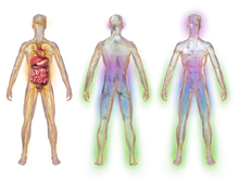
Illustration depicting areas affected by diabetic neuropathy
 
Diabetic neuropathy affects all peripheral nerves including sensory neurons, motor neurons, but rarely affects the autonomic nervous system. Therefore, diabetic neuropathy can affect all organs and systems, as all are innervated. There are several distinct syndromes based on the organ systems and members affected, but these are by no means exclusive. A patient can have sensorimotor and autonomic neuropathy or any other combination. Signs and symptoms vary depending on the nerve(s) affected and may include symptoms other than those listed. Symptoms usually develop gradually over years. 

Symptoms may include the following:

Pathogenesis

The following factors are thought to be involved in the development of diabetic neuropathy:

Microvascular disease

Vascular and neural diseases are closely related and intertwined. Blood vessels depend on normal nerve function, and nerves depend on adequate blood flow. The first pathological change in the small blood vessels is narrowing of the blood vessels. As the disease progresses, neuronal dysfunction correlates closely with the development of blood vessel abnormalities, such as capillary basement membrane thickening and endothelial hyperplasia, which contribute to diminished oxygen tension and hypoxia. Neuronal ischemia is a well-established characteristic of diabetic neuropathy. Blood vessel opening agents (e.g., ACE inhibitors, α1-antagonists) can lead to substantial improvements in neuronal blood flow, with corresponding improvements in nerve conduction velocities. Thus, small blood vessel dysfunction occurs early in diabetes, parallels the progression of neural dysfunction, and may be sufficient to support the severity of structural, functional, and clinical changes observed in diabetic neuropathy.

Advanced glycated end products

Elevated levels of glucose within cells cause a non-enzymatic covalent bonding with proteins, which alters their structure and inhibits their function. Some of these glycated proteins have been implicated in the pathology of diabetic neuropathy and other long-term complications of diabetes.

Polyol pathway

Also called the sorbitol/aldose reductase pathway, the polyol pathway appears to be implicated in diabetic complications, especially in microvascular damage to the retina, kidney, and nerves.

Sensorimotor polyneuropathy

Longer nerve fibers are affected to a greater degree than shorter ones because nerve conduction velocity is slowed in proportion to a nerve's length. In this syndrome, decreased sensation and loss of reflexes occurs first in the toes on each foot, then extends upward. It is usually described as a glove-stocking distribution of numbness, sensory loss, dysesthesia and night time pain. The pain can feel like burning, pricking sensation, achy or dull. A pins and needles sensation is common. Loss of proprioception, the sense of where a limb is in space, is affected early. These patients cannot feel when they are stepping on a foreign body, like a splinter, or when they are developing a callous from an ill-fitting shoe. Consequently, they are at risk of developing ulcers and infections on the feet and legs, which can lead to amputation. Similarly, these patients can get multiple fractures of the knee, ankle or foot, and develop a Charcot joint. Loss of motor function results in dorsiflexion, contractures of the toes, loss of the interosseous muscle function that leads to contraction of the digits, so-called hammer toes. These contractures occur not only in the foot but also in the hand where the loss of the musculature makes the hand appear gaunt and skeletal. The loss of muscular function is progressive.

Autonomic neuropathy

The autonomic nervous system is composed of nerves serving the heart, lungs, blood vessels, bone, adipose tissue, sweat glands, gastrointestinal system and genitourinary system. Autonomic neuropathy can affect any of these organ systems. The most commonly recognized autonomic dysfunction in diabetics is orthostatic hypotension, or becoming dizzy and possibly fainting when standing up due to a sudden drop in blood pressure. In the case of diabetic autonomic neuropathy, it is due to the failure of the heart and arteries to appropriately adjust heart rate and vascular tone to keep blood continually and fully flowing to the brain. This symptom is usually accompanied by a loss of respiratory sinus arrhythmia – the usual change in heart rate seen with normal breathing. These two findings suggest autonomic neuropathy.

GI tract manifestations include gastroparesis, nausea, bloating, and diarrhea. Because many diabetics take oral medication for their diabetes, absorption of these medicines is greatly affected by the delayed gastric emptying. This can lead to hypoglycemia when an oral diabetic agent is taken before a meal and does not get absorbed until hours, or sometimes days later when there is normal or low blood sugar already. Sluggish movement of the small intestine can cause bacterial overgrowth, made worse by the presence of hyperglycemia. This leads to bloating, gas and diarrhea.

Urinary symptoms include urinary frequency, urgency, incontinence and retention. Again, because of the retention of urine, urinary tract infections are frequent. Urinary retention can lead to bladder diverticula, stones, reflux nephropathy.

Cranial neuropathy

When cranial nerves are affected, neuropathies of the oculomotor nerve (cranial nerve #3 or CNIII) are most common. The oculomotor nerve controls all the muscles that move the eye except for the lateral rectus and superior oblique muscles. It also serves to constrict the pupil and open the eyelid. The onset of a diabetic third nerve palsy is usually abrupt, beginning with frontal or pain around the eye and then double vision. All the oculomotor muscles innervated by the third nerve may be affected, but those that control pupil size are usually well-preserved early on. This is because the parasympathetic nerve fibers within CNIII that influence pupillary size are found on the periphery of the nerve (in terms of a cross-sectional view), which makes them less susceptible to ischemic damage (as they are closer to the vascular supply). The sixth nerve, the abducens nerve, which innervates the lateral rectus muscle of the eye (moves the eye laterally), is also commonly affected but fourth nerve, the trochlear nerve, (innervates the superior oblique muscle, which moves the eye downward) involvement is unusual. Damage to a specific nerve of the thoracic or lumbar spinal nerves can occur and may lead to painful syndromes that mimic a heart attack, gallbladder inflammation, or appendicitis. Diabetics have a higher incidence of entrapment neuropathies, such as carpal tunnel syndrome.

Diagnosis

Diabetic peripheral neuropathy is the most likely diagnosis for someone with diabetes who has pain in a leg or foot, although it may also be caused by vitamin B12 deficiency or osteoarthritis. A 2010 review in the Journal of the American Medical Association's "Rational Clinical Examination Series" evaluated the usefulness of the clinical examination in diagnosing diabetic peripheral neuropathy. While the physician typically assesses the appearance of the feet, presence of ulceration, and ankle reflexes, the most useful physical examination findings for large fiber neuropathy are an abnormally decreased vibration perception to a 128-Hz tuning fork (likelihood ratio (LR) range, 16–35) or pressure sensation with a 5.07 Semmes-Weinstein monofilament (LR range, 11–16). Normal results on vibration testing (LR range, 0.33–0.51) or monofilament (LR range, 0.09–0.54) make large fiber peripheral neuropathy from diabetes less likely. Combinations of signs do not perform better than these 2 individual findings. Nerve conduction tests may show reduced functioning of the peripheral nerves, but seldom correlate with the severity of diabetic peripheral neuropathy and are not appropriate as routine tests for the condition.

Classification

Diabetic neuropathy encompasses a series of different neuropathic syndromes which can be schematized in the following way:
  • Focal and multifocal neuropathies:
    • Mononeuropathy
    • Amyotrophy, radiculopathy
    • Multiple lesions "mononeuritis multiplex"
    • Entrapment (e.g. median, ulnar, peroneal)
  • Symmetrical neuropathies:
    • Acute sensory
    • Autonomic
    • Distal symmetrical polyneuropathy (DSPN), the diabetic type of which is also known as diabetic peripheral neuropathy (DPN) (most common presentation)

Prevention

Prevention is by good blood sugar control and exercise.

Treatment

Except for tight glucose control, treatments are for reducing pain and other symptoms.

Medication options for pain control include antiepileptic drugs (AEDs), serotonin-norepinephrine reuptake inhibitors (SNRIs), tricyclic antidepressants (TCAs), and capsaicin cream. About 10% of people who use capsaicin cream have a large benefit.

A systematic review concluded that "tricyclic antidepressants and traditional anticonvulsants are better for short term pain relief than newer generation anticonvulsants." A further analysis of previous studies showed that the agents carbamazepine, venlafaxine, duloxetine, and amitriptyline were more effective than placebo, but that comparative effectiveness between each agent is unclear.

The only three medications approved by the United States' Food and Drug Administration for diabetic peripheral neuropathy (DPN) are the antidepressant duloxetine, the anticonvulsant pregabalin, and the long-acting opioid tapentadol ER. Before trying a systemic medication, some doctors recommend treating localized diabetic peripheral neuropathy with lidocaine patches.

Antiepileptic drugs

Multiple guidelines from medical organizations such as the American Association of Clinical Endocrinologists, American Academy of Neurology, European Federation of Neurological Societies, and the National Institute of Clinical Excellence recommend AEDs, such as pregabalin, as first-line treatment for painful diabetic neuropathy. Pregabalin is supported by low-quality evidence as more effective than placebo for reducing diabetic neuropathic pain but its effect is small. Studies have reached differing conclusions about whether gabapentin relieves pain more effectively than placebo. Available evidence is insufficient to determine if zonisamide or carbamazepine are effective for diabetic neuropathy. The first metabolite of carbamazepine, known as oxcarbazepine, appears to have a small beneficial effect on pain. A 2014 systematic review and network meta-analysis concluded topiramate, valproic acid, lacosamide, and lamotrigine are ineffective for pain from diabetic peripheral neuropathy. The most common side effects associated with AED use include sleepiness, dizziness, and nausea.

Serotonin-norepinephrine reuptake inhibitors

As above, the serotonin-norepinephrine reuptake inhibitors (SNRIs) duloxetine and venlafaxine are recommended in multiple medical guidelines as first or second-line therapy for DPN. A 2017 systematic review and meta-analysis of randomized controlled trials concluded there is moderate quality evidence that duloxetine and venlafaxine each provide a large benefit in reducing diabetic neuropathic pain. Common side effects include dizziness, nausea, and sleepiness.

Selective serotonin reuptake inhibitor

SSRIs include fluoxetine, paroxetine, sertraline, and citalopram have been found to be no more efficacious than placebo in several controlled trials and therefore are not recommended to treat painful diabetic neuropathy. Side effects are rarely serious and do not cause any permanent disabilities. They cause sedation and weight gain, which can worsen a diabetic person's glycemic control. They can be used at dosages that also relieve the symptoms of depression, a common comorbidity of diabetic neuropathy.

Tricyclic antidepressants

TCAs include imipramine, amitriptyline, desipramine, and nortriptyline. They are generally regarded as first or second-line treatment for DPN. Of the TCAs, imipramine has been the best studied. These medications are effective at decreasing painful symptoms but suffer from multiple side effects that are dose-dependent. One notable side effect is cardiac toxicity, which can lead to fatal abnormal heart rhythms. Additional common side effects include dry mouth, difficulty sleeping, and sedation. At low dosages used for neuropathy, toxicity is rare, but if symptoms warrant higher doses, complications are more common. Among the TCAs, amitriptyline is most widely used for this condition, but desipramine and nortriptyline have fewer side effects.

Opioids

Typical opioid medications, such as oxycodone, appear to be no more effective than placebo. In contrast, low-quality evidence supports a moderate benefit from the use of atypical opioids (e.g., tramadol and tapentadol), which also have SNRI properties. Opioid medications are recommended as second or third-line treatment for DPN.

Topical agents

Capsaicin applied to the skin in a 0.075% concentration has not been found to be more effective than placebo for treating pain associated with diabetic neuropathy. There is insufficient evidence to draw conclusions for more concentrated forms of capsaicin, clonidine, or lidocaine applied to the skin.

Other

Low-quality evidence supports a moderate-large beneficial effect of botulinum toxin injections. Dextromethorphan does not appear to be effective in treating diabetic neuropathic pain. There is insufficient evidence to draw firm conclusions for the utility of the cannabinoids nabilone and nabiximols. There are some in vitro studies indicating the beneficial effect of erythropoietin on the diabetic neuropathy; however, one nerve conduction study in mild-moderate diabetic individuals showed that erythropoietin alone or in combination with gabapentin does not have any beneficial effect on progression of diabetic neuropathy.

Medical devices

Monochromatic infrared photo energy treatment (MIRE) has been shown to be an effective therapy in reducing and often eliminating pain associated with diabetic neuropathy. The studied wavelength of 890 nm is able to penetrate into the subcutaneous tissue where it acts upon a specialized part of the cell called the cytochrome C. The infrared light energy prompts the cytochrome C to release nitric oxide into the cells. The nitric oxide in turn promotes vasodilation which results in increased blood flow that helps nourish damaged nerve cells. Once the nutrient rich blood is able to reach the affected areas (typically the feet, lower legs and hands) it promotes the regeneration of nerve tissues and helps reduce inflammation thereby reducing and/or eliminating pain in the area.

Physical therapy

Physical therapy may help reduce dependency on pain relieving drug therapies. Certain physiotherapy techniques can help alleviate symptoms brought on from diabetic neuropathy such as deep pain in the feet and legs, tingling or burning sensation in extremities, muscle cramps, muscle weakness, sexual dysfunction, and diabetic foot.

Transcutaneous electrical nerve stimulation (TENS) and interferential current (IFC) use a painless electric current and the physiological effects from low frequency electrical stimulation to relieve stiffness, improve mobility, relieve neuropathic pain, reduce oedema, and heal resistant foot ulcers.

Gait training, posture training, and teaching these patients the basic principles of off-loading can help prevent and/or stabilize foot complications such as foot ulcers. Off-loading techniques can include the use of mobility aids (e.g. crutches) or foot splints. Gait re-training would also be beneficial for individuals who have lost limbs, due to diabetic neuropathy, and now wear a prosthesis.

Exercise programs, along with manual therapy, will help to prevent muscle contractures, spasms and atrophy. These programs may include general muscle stretching to maintain muscle length and a person’s range of motion. General muscle strengthening exercises will help to maintain muscle strength and reduce muscle wasting. Aerobic exercise such as swimming and using a stationary bicycle can help peripheral neuropathy, but activities that place excessive pressure on the feet (e.g. walking long distances, running) may be contraindicated.

Heat, therapeutic ultrasound, hot wax are also useful for treating diabetic neuropathy. Pelvic floor muscle exercises can improve sexual dysfunction caused by neuropathy.

Tight glucose control

Treatment of early manifestations of sensorimotor polyneuropathy involves improving glycemic control. Tight control of blood glucose can reverse the changes of diabetic neuropathy, but only if the neuropathy and diabetes are recent in onset. Conversely, painful symptoms of neuropathy in uncontrolled diabetics tend to subside as the disease and numbness progress.

Prognosis

The mechanisms of diabetic neuropathy are poorly understood. At present, treatment alleviates pain and can control some associated symptoms, but the process is generally progressive.

As a complication, there is an increased risk of injury to the feet because of loss of sensation. Small infections can progress to ulceration and this may require amputation.

Epidemiology

Globally diabetic neuropathy affects approximately 132 million people as of 2010 (1.9% of the population).

Diabetes is the leading known cause of neuropathy in developed countries, and neuropathy is the most common complication and greatest source of morbidity and mortality in diabetes. It is estimated that neuropathy affects 25% of people with diabetes. Diabetic neuropathy is implicated in 50–75% of nontraumatic amputations.

The main risk factor for diabetic neuropathy is hyperglycemia. In the DCCT (Diabetes Control and Complications Trial, 1995) study, the annual incidence of neuropathy was 2% per year but dropped to 0.56% with intensive treatment of Type 1 diabetics. The progression of neuropathy is dependent on the degree of glycemic control in both Type 1 and Type 2 diabetes. Duration of diabetes, age, cigarette smoking, hypertension, height, and hyperlipidemia are also risk factors for diabetic neuropathy.

Lie group

From Wikipedia, the free encyclopedia https://en.wikipedia.org/wiki/Lie_group In mathematics , a Lie gro...