Search This Blog

Monday, May 20, 2019

Adrenal insufficiency

From Wikipedia, the free encyclopedia

Adrenal insufficiency
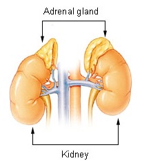
Illu adrenal gland.jpg
Adrenal gland
SpecialtyEndocrinology

Adrenal insufficiency is a condition in which the adrenal glands do not produce adequate amounts of steroid hormones, primarily cortisol; but may also include impaired production of aldosterone (a mineralocorticoid), which regulates sodium conservation, potassium secretion, and water retention. Craving for salt or salty foods due to the urinary losses of sodium is common.

Addison's disease and congenital adrenal hyperplasia can manifest as adrenal insufficiency. If not treated, adrenal insufficiency may result in severe abdominal pains, vomiting, profound muscle weakness and fatigue, depression, extremely low blood pressure (hypotension), weight loss, kidney failure, changes in mood and personality, and shock (adrenal crisis). An adrenal crisis often occurs if the body is subjected to stress, such as an accident, injury, surgery, or severe infection; death may quickly follow.

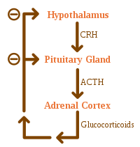
Adrenal insufficiency can also occur when the hypothalamus or the pituitary gland does not make adequate amounts of the hormones that assist in regulating adrenal function. This is called secondary or tertiary adrenal insufficiency and is caused by lack of production of ACTH in the pituitary or lack of CRH in the hypothalamus, respectively.

Types

There are three major types of adrenal insufficiency.

Signs and symptoms

Signs and symptoms include: hypoglycemia, dehydration, weight loss, and disorientation. Additional signs and symptoms include weakness, tiredness, dizziness, low blood pressure that falls further when standing (orthostatic hypotension), cardiovascular collapse, muscle aches, nausea, vomiting, and diarrhea. These problems may develop gradually and insidiously. Addison's disease can present with tanning of the skin that may be patchy or even all over the body. Characteristic sites of tanning are skin creases (e.g. of the hands) and the inside of the cheek (buccal mucosa). Goitre and vitiligo may also be present. Eosinophilia may also occur.

Causes

Causes of acute adrenal insufficiency are mainly sudden withdrawal of long-term corticosteroid therapy, Waterhouse–Friderichsen syndrome, and stress in people with underlying chronic adrenal insufficiency. The latter is termed critical illness–related corticosteroid insufficiency

For chronic adrenal insufficiency, the major contributors are autoimmune adrenalitis (Addison's Disease), tuberculosis, AIDS, and metastatic disease. Minor causes of chronic adrenal insufficiency are systemic amyloidosis, fungal infections, hemochromatosis, and sarcoidosis.

Autoimmune adrenalitis may be part of Type 2 autoimmune polyglandular syndrome, which can include type 1 diabetes, hyperthyroidism, and autoimmune thyroid disease (also known as autoimmune thyroiditis, Hashimoto's thyroiditis, and Hashimoto's disease). Hypogonadism may also present with this syndrome. Other diseases that are more common in people with autoimmune adrenalitis include premature ovarian failure, celiac disease, and autoimmune gastritis with pernicious anemia.

Adrenoleukodystrophy can also cause adrenal insufficiency.

Adrenal insufficiency can also result when a patient has a craniopharyngioma, which is a histologically benign tumor that can damage the pituitary gland and so cause the adrenal glands not to function. This would be an example of secondary adrenal insufficiency syndrome.

Causes of adrenal insufficiency can be categorized by the mechanism through which they cause the adrenal glands to produce insufficient cortisol. These are adrenal dysgenesis (the gland has not formed adequately during development), impaired steroidogenesis (the gland is present but is biochemically unable to produce cortisol) or adrenal destruction (disease processes leading to glandular damage).

Corticosteroid withdrawal

Use of high-dose steroids for more than a week begins to produce suppression of the person's adrenal glands because the exogenous glucocorticoids suppress release of hypothalamic corticotropin-releasing hormone (CRH) and pituitary adrenocorticotropic hormone (ACTH). With prolonged suppression, the adrenal glands atrophy (physically shrink), and can take months to recover full function after discontinuation of the exogenous glucocorticoid. During this recovery time, the person is vulnerable to adrenal insufficiency during times of stress, such as illness, due to both adrenal atrophy and suppression of CRH and ACTH release. Use of steroids joint injections may also result in adrenal suppression after discontinuation.

Adrenal dysgenesis

All causes in this category are genetic, and generally very rare. These include mutations to the SF1 transcription factor, congenital adrenal hypoplasia due to DAX-1 gene mutations and mutations to the ACTH receptor gene (or related genes, such as in the Triple A or Allgrove syndrome). DAX-1 mutations may cluster in a syndrome with glycerol kinase deficiency with a number of other symptoms when DAX-1 is deleted together with a number of other genes.

Impaired steroidogenesis

To form cortisol, the adrenal gland requires cholesterol, which is then converted biochemically into steroid hormones. Interruptions in the delivery of cholesterol include Smith–Lemli–Opitz syndrome and abetalipoproteinemia

Of the synthesis problems, congenital adrenal hyperplasia is the most common (in various forms: 21-hydroxylase, 17α-hydroxylase, 11β-hydroxylase and 3β-hydroxysteroid dehydrogenase), lipoid CAH due to deficiency of StAR and mitochondrial DNA mutations. Some medications interfere with steroid synthesis enzymes (e.g. ketoconazole), while others accelerate the normal breakdown of hormones by the liver (e.g. rifampicin, phenytoin).

Adrenal destruction

Autoimmune adrenalitis is the most common cause of Addison's disease in the industrialised world. Autoimmune destruction of the adrenal cortex is caused by an immune reaction against the enzyme 21-hydroxylase (a phenomenon first described in 1992). This may be isolated or in the context of autoimmune polyendocrine syndrome (APS type 1 or 2), in which other hormone-producing organs, such as the thyroid and pancreas, may also be affected.

Adrenal destruction is also a feature of adrenoleukodystrophy (ALD), and when the adrenal glands are involved in metastasis (seeding of cancer cells from elsewhere in the body, especially lung), hemorrhage (e.g. in Waterhouse–Friderichsen syndrome or antiphospholipid syndrome), particular infections (tuberculosis, histoplasmosis, coccidioidomycosis), or the deposition of abnormal protein in amyloidosis.

Pathophysiology

Hyponatremia can be caused by glucocorticoid deficiency. Low levels of glucocorticoids leads to systemic hypotension (one of the effects of cortisol is to increase peripheral resistance), which results in a decrease in stretch of the arterial baroreceptors of the carotid sinus and the aortic arch. This removes the tonic vagal and glossopharyngeal inhibition on the central release of ADH: high levels of ADH will ensue, which will subsequently lead to increase in water retention and hyponatremia. 

Differently from mineralocorticoid deficiency, glucocorticoid deficiency does not cause a negative sodium balance (in fact a positive sodium balance may occur).

Diagnosis

The best diagnostic tool to confirm adrenal insufficiency is the ACTH stimulation test; however, if a patient is suspected to be suffering from an acute adrenal crisis, immediate treatment with IV corticosteroids is imperative and should not be delayed for any testing, as the patient's health can deteriorate rapidly and result in death without replacing the corticosteroids. 

Dexamethasone should be used as the corticosteroid if the plan is to do the ACTH stimulation test at a later time as it is the only corticosteroid that will not affect the test results.

If not performed during crisis, then labs to be run should include: random cortisol, serum ACTH, aldosterone, renin, potassium and sodium. A CT of the adrenal glands can be used to check for structural abnormalities of the adrenal glands. An MRI of the pituitary can be used to check for structural abnormalities of the pituitary. However, in order to check the functionality of the Hypothalamic Pituitary Adrenal (HPA) Axis the entire axis must be tested by way of ACTH stimulation test, CRH stimulation test and perhaps an Insulin Tolerance Test (ITT). In order to check for Addison’s Disease, the auto-immune type of primary adrenal insufficiency, labs should be drawn to check 21-hydroxylase autoantibodies.

Effects

Type Hypothalamus
(tertiary)1
Pituitary
(secondary)
Adrenal glands
(primary)7
Underlying causes Abrupt steroid withdrawal, Tumor of the hypothalamus (adenoma), antibodies, environment (i.e. toxins), head injury Tumor of the pituitary (adenoma), antibodies, environment, head injury, surgical removal6, Sheehan's syndrome Tumor of the adrenal (adenoma), stress, antibodies, environment, Addison's disease, trauma, surgical removal (resection), miliary tuberculosis of the adrenal
CRH low high2 high
ACTH low low high
DHEA low low high
DHEA-S low low high
Cortisol low3 low3 low4
Aldosterone low normal low
Renin low low high
Sodium (Na) low low low
Potassium (K) low normal high
1 Automatically includes diagnosis of secondary (hypopituitarism)
2 Only if CRH production in the hypothalamus is intact
3 Value doubles or more in stimulation
4 Value less than doubles in stimulation
5 Most common, does not include all possible causes
6 Usually because of very large tumor (macroadenoma)
7 Includes Addison's disease

Treatment

(To balance sodium, potassium and increase water retention)

Cushing's disease

From Wikipedia, the free encyclopedia

Cushing's disease
Other namesCushing disease, tertiary or secondary hypercortisolism, tertiary or secondary hypercorticism, Itsenko-Cushing disease
SpecialtyEndocrinology

Cushing's disease is one cause of Cushing's syndrome characterised by increased secretion of adrenocorticotropic hormone (ACTH) from the anterior pituitary (secondary hypercortisolism). This is most often as a result of a pituitary adenoma (specifically pituitary basophilism) or due to excess production of hypothalamus CRH (corticotropin releasing hormone) (tertiary hypercortisolism/hypercorticism) that stimulates the synthesis of cortisol by the adrenal glands. Pituitary adenomas are responsible for 80% of endogenous Cushing's syndrome, when excluding Cushing's syndrome from exogenously administered corticosteroids.

This should not be confused with ectopic Cushing syndrome or exogenous steroid use.

Signs and symptoms

The symptoms of Cushing's disease are similar to those seen in other causes of Cushing's syndrome. Patients with Cushing's disease usually present with one or more signs and symptoms secondary to the presence of excess cortisol or ACTH. Although uncommon, some patients with Cushing's disease have large pituitary tumors (macroadenomas). In addition to the severe hormonal effects related to increased blood cortisol levels, the large tumor can compress adjacent structures. These tumors can compress the nerves that carry information from the eyes, causing a decrease in peripheral vision. Glaucoma and cataracts also may occur in Cushing's syndrome. In children, the two main symptoms are obesity and decreased linear growth.

The clinical diagnosis must be based on the presence of one or more of the symptoms listed below, because the syndrome itself has no true pathognomonic signs or symptoms. The most common symptoms seen in male patients are purple striae, muscle atrophy, osteoporosis, and kidney stones.

Common

Common signs and symptoms of Cushing's disease include the following:
  • weight gain
  • high blood pressure
  • poor short-term memory
  • irritability
  • excess hair growth (women)
  • Impaired immunological function
  • red, ruddy face
  • extra fat around neck, "Buffalo Hump"
  • moon face
  • fatigue
  • red stretch marks
  • poor concentration
  • irregular menstruation

Less common

The less-common signs and symptoms of Cushing's disease include the following: 
  • insomnia
  • recurrent infection
  • thin skin and stretch marks
  • easy bruising
  • weak bones
  • acne
  • balding (women)
  • depression
  • hip and shoulder weakness
  • swelling of feet/legs
  • diabetes mellitus
  • erectile dysfunction

Diagnosis

Diagnosis is made first by diagnosing Cushing's syndrome, which can be difficult to do clinically since the most characteristic symptoms only occur in a minority of patients. Some of the biochemical diagnostic tests used include salivary and blood serum cortisol testing, 24-hour urinary free cortisol (UFC) testing, the dexamethasone suppression test (DST), and bilateral inferior petrosal sinus sampling (IPSS or BIPSS for bilateral IPSS). No single test is perfect and multiple tests should always be used to achieve a proper diagnosis. Diagnosing Cushing's disease is a multidisciplinary process involving doctors, endocrinologists, radiologists, surgeons, and chemical pathologists.

ACTH blood test

Once Cushing's syndrome has been diagnosed, the first step towards finding the cause is measuring plasma adrenocorticotropic hormone (ACTH) concentration. A concentration consistently below 1.1 pmol/L is classified as corticotropin-independent and does not lead to a diagnosis of Cushing's disease. In such cases, the next step is adrenal imaging with CT. If plasma corticotropin concentrations are consistently above 3.3 pmol/L, then corticotropin-dependent Cushing's syndrome is most likely. Any intermediate values need to be cautiously interpreted and a corticotropin-releasing hormone (CRH) test is advised in order to confirm corticotropin dependency. If corticotropin-dependent Cushing's syndrome is determined then the next step is to distinguish between Cushing's disease and ectopic corticotropin syndrome. This is done via a combination of techniques including CRH, high-dose DST, BIPSS, and pituitary MRI.

Dexamethasone suppression test

Two dexamethasone suppression tests (DSTs) are generally used, the overnight test and the 48 hour test. For both tests, a plasma cortisol level above 50 nmol/L is indicative of Cushing's disease. However, 3–8% of patients with Cushing's disease will test negative due to a retention of dexamethasone suppression abilities. For non-Cushing or healthy patients, the false-positive rate is 30%. The 48-h DST is advantageous since it is more specific and can be done by outpatients upon proper instruction. In the high-dose 48-h DST, 2 mg of dexamethasone is given every 6 hours for 48 hours or a single dose of 8 mg is given. This test is not needed if the 48-h low-dose DST has shown suppression of cortisol by over 30%. These tests are based on the glucocorticoid sensitivity of pituitary adenomas compared to non-pituitary tumors.

ACTH stimulation test

An ACTH stimulation test involving administration of corticotropin-releasing hormone (CRH) or another agent can differentiate this condition from ectopic ACTH secretion. In a patient with Cushing's disease, the tumor cells will be stimulated to release corticotropin and elevated plasma corticotropin levels will be detected. This rarely occurs with ectopic corticotropin syndrome and thus is quite useful for distinguishing between the two conditions. If ectopic, the plasma ACTH and cortisol levels should remain unchanged; if this is pituitary related, levels of both would rise. The CRH test uses recombinant human or bovine-sequence CRH, which is administered via a 100μg intravenous bolus dose. The sensitivity of the CRH test for detecting Cushing's disease is 93% when plasma levels are measured after fifteen and thirty minutes. However, this test is used only as a last resort due to its high cost and complexity.

Imaging

A CT or MRI of the pituitary may also show the ACTH secreting tumor if present. However, in 40% of Cushing's disease patients MRI is unable to detect a tumor. In one study of 261 patients with confirmed pituitary Cushing's disease, only 48% of pituitary lesions were identified using MRI prior to surgery. The average size of tumor, both those that were identified on MRI and those that were only discovered during surgery, was 6 mm.

Inferior petrosal sinus sampling

IPSS (inferior petrosal sinus sampling) or BIPSS (bilateral IPSS) is a more accurate but invasive test used to differentiate pituitary from ectopic or adrenal Cushing's syndrome. A corticotropin gradient sample via BIPSS is required to confirm diagnosis when pituitary MRI imaging and biochemical diagnostic tests have been inconclusive. A basal central:peripheral ratio of over 3:1 when CRH is administered is indicative of Cushing’s disease. This test has been the gold standard for distinguishing between Cushing's disease and ectopic corticotropin syndrome. The BIPSS has a sensitivity and specificity of 94% for Cushing's disease but it is usually used as a last resort due to its invasiveness, rare but serious complications, and the expertise required to perform it.

Urinary free cortisol test

Another diagnostic test used is the urinary free cortisol (UFC) test, which measures the excess cortisol excreted by the kidneys into the urine. Results of 4x higher cortisol levels than normal are likely to be Cushing's disease. This test should be repeated three times in order to exclude any normally occurring periods of hypercortisolism. The UFC test has a specificity of 81% and thus has a high rate of false-positives that are due to pseudo-Cushing states, sleep apnea, polycystic ovary syndrome, familial glucocorticoid resistance, and hyperthyroidism.

Late night (midnight) salivary cortisol test

The late night or midnight salivary cortisol test has been gaining support due to its ease of collection and stability at room temperature, therefore it can be assigned to outpatients. The test measures free circulating cortisol and has both a sensitivity and specificity of 95–98%. This test is especially useful for diagnosing children.

Treatment

The first-line treatment of Cushing's disease is surgical resection of ACTH-secreting pituitary adenoma; this surgery involves removal of the tumor via transsphenoidal surgery (TSS). There are two possible options for access to sphenoidal sinus including of endonosal approach (through the nostril) or sublabial approach (through an incision under the upper lip); many factors such as the size of nostril, the size of the lesion, and the preferences of the surgeon cause the selection of one access route over the other. Some tumors do not contain a discrete border between tumor and pituitary gland; therefore, careful sectioning through pituitary gland may be required to identify the location of tumor. The probability of successful resection is higher in patients where the tumor was identified at initial surgery in comparison to patients where no tumor was found initially; the overall remission rates in patients with microadenomas undergoing TSS are in range of 65%–90%, and the remission rate in patients with macroadenomas are lower than 65%. patients with persistent disease after initial surgery are treated with repeated pituitary surgery as soon as the active persistent disease is evident; however, reoperation has lower success rate and increases the risk of pituitary insufficiency.

Pituitary radiation therapy is another option for treatment of postoperative persisting hypercortisolemia following unsuccessful transsphenoidal surgery. External-beam pituitary RT is more effective treatment for pediatric CD in children with cure rates of 80%-88%. Hypopituitarism specifically growth hormone deficiency has been reported as the only most common late morbidity of this treatment; GHD has been reported in 36% and 68% of the patients undergoing post pituitary RT for Cushing's disease.

Bilateral adrenalectomy is another treatment which provides immediate reduction of cortisol level and control of hypercortisolism. However, it requires education of patients, because lifelong glucocorticoid and mineralocorticoid replacement therapy is needed for these patients. One of the major complications of this treatment is progression of Nelson's syndrome which is caused by enhance level of tumor growth and ACTH secretion post adrenalectomy in 8%–29% of patients with CD.

During post surgical recovery, patients collect 24-hour urine sample and blood sample for detecting the level of cortisol with the purpose of cure test; level of cortisol near the detection limit assay, corresponds to cure. Hormonal replacement such as steroid is given to patients because of steroid withdrawal. After the completion of collecting urine and blood samples, patients are asked to switch to glucocorticoid such as prednisone to decrease symptoms associated with adrenal withdrawal. Miitotaine is also used.

A study of 3,525 cases of TSS for Cushing's disease in the nationally representative sample of US hospitals between 1993 and 2002 was conducted and revealed the following results: the in-hospital mortality rate was 0.7%; the complication rate was 42.1%. Diabetes insipidus (15%), fluid and electrolyte abnormalities (12.5%), and neurological deficits (5.6%) were the most common complications reported. The analyses of the study show that complications were more likely in patients with pre-operative comorbidities. Patients older than 64 years were more likely to have an adverse outcome and prolonged hospital stay. Women were 0.3 times less likely to have adverse outcomes in comparison to men.

Epidemiology

Cases of Cushing's disease are rare, and little epidemiological data is available on the disease. An 18-year study conducted on the population of Vizcaya, Spain reported a 0.004% prevalence of Cushing's disease. The average incidence of newly diagnosed cases was 2.4 cases per million inhabitants per year. The disease is often diagnosed 3–6 years after the onset of illness. Several studies have shown that Cushing's disease is more prevalent in women than men at a ratio of 3–6:1, respectively. Moreover, most women affected were between the ages of 50 and 60 years. The prevalence of hypertension, and abnormalities in glucose metabolism are major predictors of mortality and morbidity in untreated cases of the disease. The mortality rate of Cushing's disease was reported to be 10–11%, with the majority of deaths due to vascular disease. Women aged 45–70 years have a significantly higher mortality rate than men. Moreover, the disease shows a progressive increase with time. Reasons for the trend are unknown, but better diagnostic tools, and a higher incidence rate are two possible explanations.

History

The disease associated with this increased secretion of cortisol was described by the American neurosurgeon Harvey Cushing in 1912, after he was presented with a unique case of the disease in 1910 a 23-year-old woman called Minnie G. whose symptoms included painful obesity, amenorrhea, hypertrichosis (abnormal hair growth), underdevelopment of secondary sexual characteristics, hydrocephalus and cerebral tension. This combination of symptoms was not yet described by any medical disorder at the time. However, Cushing was confident that Minnie's symptoms were due to dysfunction of the pituitary gland, and resembled those associated with an adrenal tumor. Given this conviction, and his knowledge of the three anterior pituitary cell types, Cushing hypothesized that if acidophil hyperpituitarism (excess secretion from the acidophil cells) caused acromegaly, then an excess of basophil cells must be involved in another pituitary disorder that involves sexual dysfunction (amenorrhea in females and erectile dysfunction in males) and could explain Minnie's symptoms. Experimental evidence and case reports by Cushing led to his publication in 1932 on pituitary basophilism as the cause of Cushing's disease. In this publication, the clinical symptoms of the disease, named after Cushing, were described. Out of the 12 cases with hypercortisolism described in Cushing's monograph on the pituitary body, 67% died within a few years after symptom presentation, whereas Minnie G. survived for more than 40 years after symptom presentation, despite the fact that she did not receive any treatments for a pituitary tumor. The prolonged survival made Minnie's case unique at the time. The reason behind this survival remains a mystery, since an autopsy of Minnie was refused after her death. However, the most likely explanation, proposed by J. Aidan Carney and based on statistical evidence, was that the basophil adenoma Minnie might have harbored underwent partial infarction, leading to symptom regression. The other hypothesis was that Minnie might have suffered from Primary Pigmented Nodular Adrenocortical Disease (PPNAD), which when associated with Cushing's syndrome (Carney complex) can infrequently cause spontaneous symptom regression of the latter.

In 1924, the Soviet neurologist Nikolai Itsenko reported two patients with pituitary adenoma. The resulting excessive adrenocorticotropic hormone secretion led to the production of large amounts of cortisol by the adrenal glands. Considering this impact, the name of Itsenko was added to the title in some East European and Asian countries, and the disease is called Itsenko-Kushing disease.

Sunday, May 19, 2019

Addison's disease

From Wikipedia, the free encyclopedia

Addison's disease
Other namesAddison disease, chronic adrenal insufficiency, hypocortisolism, hypoadrenalism, primary adrenal insufficiency
Addisons hyperpigmentation.jpg
Classic darkening of the skin due to increased pigment as seen in Addison's disease
SpecialtyEndocrinology
SymptomsAbdominal pain, weakness, weight loss, darkening of the skin
ComplicationsAdrenal crisis
Usual onsetMiddle-aged females
CausesProblems with the adrenal gland
Diagnostic methodBlood tests, urine tests, medical imaging
TreatmentCorticosteroid such as hydrocortisone and fludrocortisone
Frequency0.9–1.4 per 10,000 people (developed world)

Addison's disease, also known as primary adrenal insufficiency and hypocortisolism, is a long-term endocrine disorder in which the adrenal glands do not produce enough steroid hormones. Symptoms generally come on slowly and may include abdominal pain, weakness, and weight loss. Darkening of the skin in certain areas may also occur. Under certain circumstances, an adrenal crisis may occur with low blood pressure, vomiting, lower back pain, and loss of consciousness. An adrenal crisis can be triggered by stress, such as from an injury, surgery, or infection.

Addison's disease arises from problems with the adrenal gland such that not enough of the steroid hormone cortisol and possibly aldosterone are produced, most often due to damage by the body's own immune system in the developed world and tuberculosis in the developing world. Other causes include certain medications, sepsis, and bleeding into both adrenal glands. Secondary adrenal insufficiency is caused by not enough adrenocorticotropic hormone (ACTH) (produced by the pituitary gland) or CRH (produced by the hypothalamus). Despite this distinction, adrenal crises can happen in all forms of adrenal insufficiency. Addison's disease is generally diagnosed by blood tests, urine tests, and medical imaging.

Treatment involves replacing the absent hormones. This involves taking a corticosteroid such as hydrocortisone and fludrocortisone. These medications are usually taken by mouth. Lifelong, continuous steroid replacement therapy is required, with regular follow-up treatment and monitoring for other health problems. A high-salt diet may also be useful in some people. If symptoms worsen, an injection of corticosteroid is recommended and people should carry a dose with them. Often, large amounts of intravenous fluids with the sugar dextrose are also required. Without treatment, an adrenal crisis can result in death.

Addison's disease affects about 0.9 to 1.4 per 10,000 people in the developed world. It occurs most frequently in middle-aged females. Secondary adrenal insufficiency is more common. Long-term outcomes with treatment are typically good. It is named after Thomas Addison, a graduate of the University of Edinburgh Medical School, who first described the condition in 1855. The adjective "addisonian" is used to describe features of the condition, as well as people with Addison's disease.

Signs and symptoms

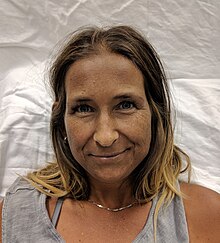
A Caucasian woman with Addison's disease
 
Legs of a Caucasian woman with Addison's disease
 
The symptoms of Addison's disease develop gradually and may become established before they are recognized. They can be nonspecific and are potentially attributable to other medical conditions. 

The signs and symptoms include fatigue; lightheadedness upon standing or difficulty standing, muscle weakness, fever, weight loss, anxiety, nausea, vomiting, diarrhea, headache, sweating, changes in mood or personality, and joint and muscle pains. Some people have cravings for salt or salty foods due to the loss of sodium through their urine. Hyperpigmentation of the skin may be seen, particularly when the person lives in a sunny area, as well as darkening of the palmar crease, sites of friction, recent scars, the vermilion border of the lips, and genital skin. These skin changes are not encountered in secondary and tertiary hypoadrenalism.

On physical examination, these clinical signs may be noticed:
  • Low blood pressure with or without orthostatic hypotension (blood pressure that decreases with standing)
  • Darkening (hyperpigmentation) of the skin, including areas not exposed to the sun. Characteristic sites of darkening are skin creases (e.g., of the hands), nipple, and the inside of the cheek (buccal mucosa); also, old scars may darken. This occurs because melanocyte-stimulating hormone (MSH) and ACTH share the same precursor molecule, pro-opiomelanocortin (POMC). After production in the anterior pituitary gland, POMC gets cleaved into gamma-MSH, ACTH, and beta-lipotropin. The subunit ACTH undergoes further cleavage to produce alpha-MSH, the most important MSH for skin pigmentation. In secondary and tertiary forms of adrenal insufficiency, skin darkening does not occur, as ACTH is not overproduced.
Addison's disease is associated with the development of other autoimmune diseases, such as type I diabetes, thyroid disease (Hashimoto's thyroiditis), celiac disease, or vitiligo. Addison's disease may be the only manifestation of undiagnosed celiac disease. Both diseases share the same genetic risk factors (HLA-DQ2 and HLA-DQ8 haplotypes).

The presence of Addison's in addition to mucocutaneous candidiasis, hypoparathyroidism, or both, is called autoimmune polyendocrine syndrome type 1. The presence of Addison's in addition to autoimmune thyroid disease, type 1 diabetes, or both, is called autoimmune polyendocrine syndrome type 2.

Addisonian crisis

An "Addisonian crisis" or "adrenal crisis" is a constellation of symptoms that indicates severe adrenal insufficiency. This may be the result of either previously undiagnosed Addison's disease, a disease process suddenly affecting adrenal function (such as adrenal hemorrhage), or an intercurrent problem (e.g., infection, trauma) in someone known to have Addison's disease. It is a medical emergency and potentially life-threatening situation requiring immediate emergency treatment. 

Characteristic symptoms are:

Causes

The negative feedback loop for glucocorticoids
 
Causes of adrenal insufficiency can be categorized by the mechanism through which they cause the adrenal glands to produce insufficient cortisol. These are adrenal dysgenesis (the gland has not formed adequately during development), impaired steroidogenesis (the gland is present but is biochemically unable to produce cortisol) or adrenal destruction (disease processes leading to glandular damage).

Adrenal destruction

Autoimmune adrenalitis is the most common cause of Addison's disease in the industrialized world. Autoimmune destruction of the adrenal cortex is caused by an immune reaction against the enzyme 21-hydroxylase (a phenomenon first described in 1992). This may be isolated or in the context of autoimmune polyendocrine syndrome (APS type 1 or 2), in which other hormone-producing organs, such as the thyroid and pancreas, may also be affected.

Adrenal destruction is also a feature of adrenoleukodystrophy, and when the adrenal glands are involved in metastasis (seeding of cancer cells from elsewhere in the body, especially lung), hemorrhage (e.g., in Waterhouse–Friderichsen syndrome or antiphospholipid syndrome), particular infections (tuberculosis, histoplasmosis, coccidioidomycosis), or the deposition of abnormal protein in amyloidosis.

Adrenal dysgenesis

All causes in this category are genetic, and generally very rare. These include mutations to the SF1 transcription factor, congenital adrenal hypoplasia due to DAX-1 gene mutations and mutations to the ACTH receptor gene (or related genes, such as in the Triple-A or Allgrove syndrome). DAX-1 mutations may cluster in a syndrome with glycerol kinase deficiency with a number of other symptoms when DAX-1 is deleted together with a number of other genes.

Impaired steroidogenesis

To form cortisol, the adrenal gland requires cholesterol, which is then converted biochemically into steroid hormones. Interruptions in the delivery of cholesterol include Smith–Lemli–Opitz syndrome and abetalipoproteinemia

Of the synthesis problems, congenital adrenal hyperplasia is the most common (in various forms: 21-hydroxylase, 17α-hydroxylase, 11β-hydroxylase and 3β-hydroxysteroid dehydrogenase), lipoid CAH due to deficiency of StAR and mitochondrial DNA mutations. Some medications interfere with steroid synthesis enzymes (e.g., ketoconazole), while others accelerate the normal breakdown of hormones by the liver (e.g., rifampicin, phenytoin).

Diagnosis

Suggestive features

Routine laboratory investigations may show:

Testing

In suspected cases of Addison's disease, demonstration of low adrenal hormone levels even after appropriate stimulation (called the ACTH stimulation test or synacthen test) with synthetic pituitary ACTH hormone tetracosactide is needed for the diagnosis. Two tests are performed, the short and the long test. Dexamethasone does not cross-react with the assay and can be administered concomitantly during testing. 

The short test compares blood cortisol levels before and after 250 micrograms of tetracosactide (intramuscular or intravenous) is given. If, one hour later, plasma cortisol exceeds 170 nmol/l and has risen by at least 330 nmol/l to at least 690 nmol/l, adrenal failure is excluded. If the short test is abnormal, the long test is used to differentiate between primary adrenal insufficiency and secondary adrenocortical insufficiency. 

The long test uses 1 mg tetracosactide (intramuscular). Blood is taken 1, 4, 8, and 24 hr later. Normal plasma cortisol level should reach 1000 nmol/l by 4 hr. In primary Addison's disease, the cortisol level is reduced at all stages, whereas in secondary corticoadrenal insufficiency, a delayed but normal response is seen. 

Other tests may be performed to distinguish between various causes of hypoadrenalism, including renin and adrenocorticotropic hormone levels, as well as medical imaging - usually in the form of ultrasound, computed tomography or magnetic resonance imaging

Adrenoleukodystrophy, and the milder form, adrenomyeloneuropathy, cause adrenal insufficiency combined with neurological symptoms. These diseases are estimated to be the cause of adrenal insufficiency in about 35% of diagnosed male with idiopathic Addison’s disease, and should be considered in the differential diagnosis of any male with adrenal insufficiency. Diagnosis is made by a blood test to detect very long chain fatty acids.

Treatment

Maintenance

Treatment for Addison's disease involves replacing the missing cortisol, sometimes in the form of hydrocortisone tablets, or prednisone tablets in a dosing regimen that mimics the physiological concentrations of cortisol. Alternatively, one-quarter as much prednisolone may be used for equal glucocorticoid effect as hydrocortisone. Treatment is usually lifelong. In addition, many people require fludrocortisone as replacement for the missing aldosterone. 

People with Addison's are often advised to carry information on them (e.g., in the form of a MedicAlert bracelet or information card) for the attention of emergency medical services personnel who might need to attend to their needs. It is also recommended that a needle, syringe, and injectable form of cortisol be carried for emergencies. People with Addison's disease are advised to increase their medication during periods of illness or when undergoing surgery or dental treatment. Immediate medical attention is needed when severe infections, vomiting, or diarrhea occur, as these conditions can precipitate an Addisonian crisis. A person who is vomiting may require injections of hydrocortisone instead.

Crisis

Standard therapy involves intravenous injections of glucocorticoids and large volumes of intravenous saline solution with dextrose (glucose). This treatment usually brings rapid improvement. If intravenous access is not immediately available, intramuscular injection of glucocorticoids can be used. When the person can take fluids and medications by mouth, the amount of glucocorticoids is decreased until a maintenance dose is reached. If aldosterone is deficient, maintenance therapy also includes oral doses of fludrocortisone acetate.

Prognosis

Outcomes are typically good when treated. Most can expect to live relatively normal lives. Someone with the disease should be observant of symptoms of an "Addison's crisis" while the body is strained, as in rigorous exercise or being sick, the latter often needing emergency treatment with intravenous injections to treat the crisis.

Individuals with Addison's disease have more than a doubled mortality rate. Furthermore, individuals with Addison's disease and diabetes mellitus have an almost 4 time increase in mortality compared to individuals with only diabetes.

Epidemiology

The frequency rate of Addison's disease in the human population is sometimes estimated at roughly one in 100,000. Some put the number closer to 40–144 cases per million population (1/25,000–1/7,000). Addison's can affect persons of any age, sex, or ethnicity, but it typically presents in adults between 30 and 50 years of age. Research has shown no significant predispositions based on ethnicity.

History

Discovery

Addison’s disease is named after Thomas Addison, the British physician who first described the condition in On the Constitutional and Local Effects of Disease of the Suprarenal Capsules (1855). He originally described it as "melasma suprarenale," but later physicians gave it the medical eponym "Addison's disease" in recognition of Addison's discovery.

All six of the original people under Addison's care had tuberculosis of the adrenal glands. While the six under Addison in 1855 all had adrenal tuberculosis, the term "Addison's disease" does not imply an underlying disease process. 

The condition was initially considered a form of anemia associated with the adrenal glands. Because little was known at the time about the adrenal glands (then called "Supra-Renal Capsules"), Addison’s monograph describing the condition was an isolated insight. As the adrenal function became better known, Addison’s monograph became known as an important medical contribution and a classic example of careful medical observation.

Other animals

Hypoadrenocorticism is uncommon in dogs, and rare in cats. Individual cases have been reported in a grey seal, a red panda, a flying fox, and a sloth.

In dogs, hypoadrenocorticism has been diagnosed in many breeds. Vague symptoms which wax and wane can cause delay in recognition of the presence of the disease. Female dogs appear more affected than male dogs, though this may not be the case in all breeds. The disease is most often diagnosed in dogs which are young to middle aged, but it can occur at any age from 4 months to 14 years. Treatment of hypoadrenocorticism must replace the hormones (cortisol and aldosterone) which the dog cannot produce itself. This is achieved either by daily treatment with fludrocortisone, or monthly injections with desoxycorticosterone pivalate (DOCP) and daily treatment with a glucocorticoid, such as prednisone. Several follow-up blood tests are required so that the dose can be adjusted until the dog is receiving the correct amount of treatment, because the medications used in the therapy of hypoadrenocorticism can cause excessive thirst and urination if not prescribed at the lowest effective dose. In anticipation of stressful situations, such as staying in a boarding kennel, dogs require an increased dose of prednisone. Lifelong treatment is required, but the prognosis for dogs with hypoadrenocorticism is very good.

Marxism

From Wikipedia, the free encyclopedia Marxism is a political philosophy and method of socioeconomic analysis. It uses a dialectical and ...