Search This Blog

Thursday, August 21, 2014

Bioinformatics

Bioinformatics

From Wikipedia, the free encyclopedia
For the journal, see Bioinformatics (journal).
Map of the human X chromosome (from the NCBI website). Assembly of the human genome is one of the greatest achievements of bioinformatics.

Bioinformatics Listeni/ˌb.ˌɪnfərˈmætɪks/ is an interdisciplinary scientific field that develops methods and software tools for storing, retrieving, organizing and analyzing biological data. As an interdisciplinary field, bioinformatics combines computer science, statistics, mathematics and engineering to study and process biological data.

Information systems such as databases and ontologies are used to store and organize biological data. Analyzing biological data to produce meaningful information involves writing and running software programs that use algorithms from graph theory, artificial intelligence, soft computing, data mining, image processing, and computer simulation. The algorithms in turn depend on theoretical foundations such as discrete mathematics, control theory, system theory, information theory, and statistics.

Bioinformatics is similar but distinct science from biological computation and computational biology. Biological computation uses bioengineering and biology to build biological computers, whereas bioinformatics uses computation to better understand biology. Bioinformatics and computational biology have similar aims and approaches, but differ in scale: bioinformatics organizes and analyzes basic biological data, whereas computational biology builds theoretical models of biological systems, just as mathematical biology does with mathematical models.

Commonly used software tools and technologies in the field include Java, C#, XML, Perl, C, C++, Python, R, SQL, CUDA, MATLAB, and spreadsheet applications.[1][2][3]

Introduction

Bioinformatics has become an important part of many areas of biology. In experimental molecular biology, bioinformatics techniques such as image and signal processing allow extraction of useful results from large amounts of raw data. In the field of genetics and genomics, it aids in sequencing and annotating genomes and their observed mutations. It plays a role in the textual mining of biological literature and the development of biological and gene ontologies to organize and query biological data. It plays a role in the analysis of gene and protein expression and regulation.
Bioinformatics tools aid in the comparison of genetic and genomic data and more generally in the understanding of evolutionary aspects of molecular biology. At a more integrative level, it helps analyze and catalogue the biological pathways and networks that are an important part of systems biology. In structural biology, it aids in the simulation and modeling of DNA, RNA, and protein structures as well as molecular interactions.

 History

Paulien Hogeweg coined the term "Bioinformatics" in 1970 to refer to the study of information processes in biotic systems.[4][5][6] This definition placed bioinformatics as a field parallel to biophysics (the study of physical processes in biological systems) or biochemistry (the study of chemical processes in biological systems).[4]

Sequences. Computers became essential in molecular biology when protein sequences became available after Frederick Sanger determined the sequence of insulin in the early 1950s. Comparing multiple sequences manually turned out to be impractical. A pioneer in the field was Margaret Oakley Dayhoff, who has been hailed by David Lipman, director of the National Center for Biotechnology Information, as the "mother and father of bioinformatics."[7] Dayhoff compiled one of the first protein sequence databases, initially published as books[8] and pioneered methods of sequence alignment and molecular evolution.[9] Another early contributor to bioinformatics was Elvin A. Kabat, who pioneered biological sequence analysis in 1970 with his comprehensive volumes of antibody sequences released with Tai Te Wu between 1980 and 1991.[10]

Genomes. As whole genome sequences became available, again with the pioneering work of Frederick Sanger,[11] the term bioinformatics was re-discovered to refer to the creation of databases such as GenBank in 1982. With the public availability of data tools for their analysis were quickly developed and described in journals such as Nucleic Acids Research which published specialized issues on bioinformatics tools as early as 1982.

Goals

In order to study how normal cellular activities are altered in different disease states, the biological data must be combined to form a comprehensive picture of these activities. Therefore, the field of bioinformatics has evolved such that the most pressing task now involves the analysis and interpretation of various types of data. This includes nucleotide and amino acid sequences, protein domains, and protein structures.[12] The actual process of analyzing and interpreting data is referred to as computational biology. Important sub-disciplines within bioinformatics and computational biology include:
  • the development and implementation of computer programs that enable efficient access to, use and management of, various types of information.
  • the development of new algorithms (mathematical formulas) and statistical measures with which to assess relationships among members of large data sets. For example, there are methods to locate a gene within a sequence, to predict protein structure and/or function, and to cluster protein sequences into families of related sequences.
The primary goal of bioinformatics is to increase the understanding of biological processes. What sets it apart from other approaches, however, is its focus on developing and applying computationally intensive techniques to achieve this goal. Examples include: pattern recognition, data mining, machine learning algorithms, and visualization. Major research efforts in the field include sequence alignment, gene finding, genome assembly, drug design, drug discovery, protein structure alignment, protein structure prediction, prediction of gene expression and protein–protein interactions, genome-wide association studies, and the modeling of evolution.

Bioinformatics now entails the creation and advancement of databases, algorithms, computational and statistical techniques, and theory to solve formal and practical problems arising from the management and analysis of biological data.

Over the past few decades rapid developments in genomic and other molecular research technologies and developments in information technologies have combined to produce a tremendous amount of information related to molecular biology. Bioinformatics is the name given to these mathematical and computing approaches used to glean understanding of biological processes.

Approaches

Common activities in bioinformatics include mapping and analyzing DNA and protein sequences, aligning DNA and protein sequences to compare them, and creating and viewing 3-D models of protein structures.

There are two fundamental ways of modelling a Biological system (e.g., living cell) both coming under Bioinformatic approaches.
  • Static
    • Sequences – Proteins, Nucleic acids and Peptides
    • Interaction data among the above entities including microarray data and Networks of proteins, metabolites
  • Dynamic
    • Structures – Proteins, Nucleic acids, Ligands (including metabolites and drugs) and Peptides (structures studied with bioinformatics tools are not considered static anymore and their dynamics is often the core of the structural studies)
    • Systems Biology comes under this category including reaction fluxes and variable concentrations of metabolites
    • Multi-Agent Based modelling approaches capturing cellular events such as signalling, transcription and reaction dynamics
A broad sub-category under bioinformatics is structural bioinformatics.

Sequence analysis

 
The sequences of different genes or proteins may be aligned side-by-side to measure their similarity. This alignment compares protein sequences containing WPP domains.

Since the Phage Φ-X174 was sequenced in 1977,[13] the DNA sequences of thousands of organisms have been decoded and stored in databases. This sequence information is analyzed to determine genes that encode polypeptides (proteins), RNA genes, regulatory sequences, structural motifs, and repetitive sequences. A comparison of genes within a species or between different species can show similarities between protein functions, or relations between species (the use of molecular systematics to construct phylogenetic trees). With the growing amount of data, it long ago became impractical to analyze DNA sequences manually. Today, computer programs such as BLAST are used daily to search sequences from more than 260 000 organisms, containing over 190 billion nucleotides.[14] These programs can compensate for mutations (exchanged, deleted or inserted bases) in the DNA sequence, to identify sequences that are related, but not identical. A variant of this sequence alignment is used in the sequencing process itself. The so-called shotgun sequencing technique (which was used, for example, by The Institute for Genomic Research to sequence the first bacterial genome, Haemophilus influenzae)[15] does not produce entire chromosomes. Instead it generates the sequences of many thousands of small DNA fragments (ranging from 35 to 900 nucleotides long, depending on the sequencing technology). The ends of these fragments overlap and, when aligned properly by a genome assembly program, can be used to reconstruct the complete genome. Shotgun sequencing yields sequence data quickly, but the task of assembling the fragments can be quite complicated for larger genomes. For a genome as large as the human genome, it may take many days of CPU time on large-memory, multiprocessor computers to assemble the fragments, and the resulting assembly will usually contain numerous gaps that have to be filled in later. Shotgun sequencing is the method of choice for virtually all genomes sequenced today, and genome assembly algorithms are a critical area of bioinformatics research.

Another aspect of bioinformatics in sequence analysis is annotation. This involves computational gene finding to search for protein-coding genes, RNA genes, and other functional sequences within a genome. Not all of the nucleotides within a genome are part of genes. Within the genomes of higher organisms, large parts of the DNA do not serve any obvious purpose. This so-called junk DNA may, however, contain unrecognized functional elements. Bioinformatics helps to bridge the gap between genome and proteome projects — for example, in the use of DNA sequences for protein identification.
 

Genome annotation

In the context of genomics, annotation is the process of marking the genes and other biological features in a DNA sequence. The first genome annotation software system was designed in 1995 by Owen White, who was part of the team at The Institute for Genomic Research that sequenced and analyzed the first genome of a free-living organism to be decoded, the bacterium Haemophilus influenzae. White built a software system to find the genes (fragments of genomic sequence that encode proteins), the transfer RNAs, and to make initial assignments of function to those genes. Most current genome annotation systems work similarly, but the programs available for analysis of genomic DNA, such as the GeneMark program trained and used to find protein-coding genes in Haemophilus influenzae, are constantly changing and improving.

Computational evolutionary biology

Evolutionary biology is the study of the origin and descent of species, as well as their change over time. Informatics has assisted evolutionary biologists by enabling researchers to:
  • trace the evolution of a large number of organisms by measuring changes in their DNA, rather than through physical taxonomy or physiological observations alone,
  • more recently, compare entire genomes, which permits the study of more complex evolutionary events, such as gene duplication, horizontal gene transfer, and the prediction of factors important in bacterial speciation,
  • build complex computational models of populations to predict the outcome of the system over time[16]
  • track and share information on an increasingly large number of species and organisms
Future work endeavours to reconstruct the now more complex tree of life.

The area of research within computer science that uses genetic algorithms is sometimes confused with computational evolutionary biology, but the two areas are not necessarily related.

Comparative genomics

The core of comparative genome analysis is the establishment of the correspondence between genes (orthology analysis) or other genomic features in different organisms. It is these intergenomic maps that make it possible to trace the evolutionary processes responsible for the divergence of two genomes. A multitude of evolutionary events acting at various organizational levels shape genome evolution. At the lowest level, point mutations affect individual nucleotides. At a higher level, large chromosomal segments undergo duplication, lateral transfer, inversion, transposition, deletion and insertion. Ultimately, whole genomes are involved in processes of hybridization, polyploidization and endosymbiosis, often leading to rapid speciation. The complexity of genome evolution poses many exciting challenges to developers of mathematical models and algorithms, who have recourse to a spectra of algorithmic, statistical and mathematical techniques, ranging from exact, heuristics, fixed parameter and approximation algorithms for problems based on parsimony models to Markov Chain Monte Carlo algorithms for Bayesian analysis of problems based on probabilistic models.
Many of these studies are based on the homology detection and protein families computation.

Genetics of Disease

With the advent of next-generation sequencing we are obtaining enough sequence data to map the genes of complex diseases such as infertility,[17] breast cancer [18] or Alzheimer's Disease.[19] Genome-wide association studies are essential to pinpoint the mutations for such complex diseases.[20]

Analysis of mutations in cancer

In cancer, the genomes of affected cells are rearranged in complex or even unpredictable ways. Massive sequencing efforts are used to identify previously unknown point mutations in a variety of genes in cancer. Bioinformaticians continue to produce specialized automated systems to manage the sheer volume of sequence data produced, and they create new algorithms and software to compare the sequencing results to the growing collection of human genome sequences and germline polymorphisms. New physical detection technologies are employed, such as oligonucleotide microarrays to identify chromosomal gains and losses (called comparative genomic hybridization), and single-nucleotide polymorphism arrays to detect known point mutations. These detection methods simultaneously measure several hundred thousand sites throughout the genome, and when used in high-throughput to measure thousands of samples, generate terabytes of data per experiment. Again the massive amounts and new types of data generate new opportunities for bioinformaticians.
The data is often found to contain considerable variability, or noise, and thus Hidden Markov model and change-point analysis methods are being developed to infer real copy number changes.

Another type of data that requires novel informatics development is the analysis of lesions found to be recurrent among many tumors.

Gene and protein expression

Analysis of gene expression

The expression of many genes can be determined by measuring mRNA levels with multiple techniques including microarrays, expressed cDNA sequence tag (EST) sequencing, serial analysis of gene expression (SAGE) tag sequencing, massively parallel signature sequencing (MPSS), RNA-Seq, also known as "Whole Transcriptome Shotgun Sequencing" (WTSS), or various applications of multiplexed in-situ hybridization. All of these techniques are extremely noise-prone and/or subject to bias in the biological measurement, and a major research area in computational biology involves developing statistical tools to separate signal from noise in high-throughput gene expression studies.
Such studies are often used to determine the genes implicated in a disorder: one might compare microarray data from cancerous epithelial cells to data from non-cancerous cells to determine the transcripts that are up-regulated and down-regulated in a particular population of cancer cells.

Analysis of protein expression

Protein microarrays and high throughput (HT) mass spectrometry (MS) can provide a snapshot of the proteins present in a biological sample. Bioinformatics is very much involved in making sense of protein microarray and HT MS data; the former approach faces similar problems as with microarrays targeted at mRNA, the latter involves the problem of matching large amounts of mass data against predicted masses from protein sequence databases, and the complicated statistical analysis of samples where multiple, but incomplete peptides from each protein are detected.

Analysis of regulation

Regulation is the complex orchestration of events starting with an extracellular signal such as a hormone and leading to an increase or decrease in the activity of one or more proteins.
Bioinformatics techniques have been applied to explore various steps in this process. For example, promoter analysis involves the identification and study of sequence motifs in the DNA surrounding the coding region of a gene. These motifs influence the extent to which that region is transcribed into mRNA. Expression data can be used to infer gene regulation: one might compare microarray data from a wide variety of states of an organism to form hypotheses about the genes involved in each state. In a single-cell organism, one might compare stages of the cell cycle, along with various stress conditions (heat shock, starvation, etc.). One can then apply clustering algorithms to that expression data to determine which genes are co-expressed. For example, the upstream regions (promoters) of co-expressed genes can be searched for over-represented regulatory elements. Examples of clustering algorithms applied in gene clustering are k-means clustering, self-organizing maps (SOMs), hierarchical clustering, and consensus clustering methods such as the Bi-CoPaM. The later, namely Bi-CoPaM, has been actually proposed to address various issues specific to gene discovery problems such as consistent co-expression of genes over multiple microarray datasets.[21][22]

Structural bioinformatics

Prediction of protein structure

Protein structure prediction is another important application of bioinformatics. The amino acid sequence of a protein, the so-called primary structure, can be easily determined from the sequence on the gene that codes for it. In the vast majority of cases, this primary structure uniquely determines a structure in its native environment. (Of course, there are exceptions, such as the bovine spongiform encephalopathy – a.k.a. Mad Cow Diseaseprion.) Knowledge of this structure is vital in understanding the function of the protein. For lack of better terms, structural information is usually classified as one of secondary, tertiary and quaternary structure. A viable general solution to such predictions remains an open problem. Most efforts have so far been directed towards heuristics that work most of the time.
One of the key ideas in bioinformatics is the notion of homology. In the genomic branch of bioinformatics, homology is used to predict the function of a gene: if the sequence of gene A, whose function is known, is homologous to the sequence of gene B, whose function is unknown, one could infer that B may share A's function. In the structural branch of bioinformatics, homology is used to determine which parts of a protein are important in structure formation and interaction with other proteins. In a technique called homology modeling, this information is used to predict the structure of a protein once the structure of a homologous protein is known. This currently remains the only way to predict protein structures reliably.

One example of this is the similar protein homology between hemoglobin in humans and the hemoglobin in legumes (leghemoglobin). Both serve the same purpose of transporting oxygen in the organism. Though both of these proteins have completely different amino acid sequences, their protein structures are virtually identical, which reflects their near identical purposes.

Other techniques for predicting protein structure include protein threading and de novo (from scratch) physics-based modeling.

Network and systems biology

Network analysis seeks to understand the relationships within biological networks such as metabolic or protein-protein interaction networks. Although biological networks can be constructed from a single type of molecule or entity (such as genes), network biology often attempts to integrate many different data types, such as proteins, small molecules, gene expression data, and others, which are all connected physically and/or functionally.
Systems biology involves the use of computer simulations of cellular subsystems (such as the networks of metabolites and enzymes which comprise metabolism, signal transduction pathways and gene regulatory networks) to both analyze and visualize the complex connections of these cellular processes. Artificial life or virtual evolution attempts to understand evolutionary processes via the computer simulation of simple (artificial) life forms.

Molecular interaction networks

Interactions between proteins are frequently visualized and analyzed using networks. This network is made up of protein-protein interactions from Treponema pallidum, the causative agent of syphilis and other diseases.
Tens of thousands of three-dimensional protein structures have been determined by X-ray crystallography and protein nuclear magnetic resonance spectroscopy (protein NMR) and a central question in structural bioinformatics is whether it is practical to predict possible protein–protein interactions only based on these 3D shapes, without performing protein–protein interaction experiments. A variety of methods have been developed to tackle the protein–protein docking problem, though it seems that there is still much work to be done in this field.
Other interactions encountered in the field include Protein–ligand (including drug) and protein–peptide. Molecular dynamic simulation of movement of atoms about rotatable bonds is the fundamental principle behind computational algorithms, termed docking algorithms, for studying molecular interactions.

Others

Literature analysis

The growth in the number of published literature makes it virtually impossible to read every paper, resulting in disjointed sub-fields of research. Literature analysis aims to employ computational and statistical linguistics to mine this growing library of text resources. For example:
  • abbreviation recognition – identify the long-form and abbreviation of biological terms,
  • named entity recognition – recognizing biological terms such as gene names
  • protein-protein interaction – identify which proteins interact with which proteins from text
The area of research draws from statistics and computational linguistics.

High-throughput image analysis

Computational technologies are used to accelerate or fully automate the processing, quantification and analysis of large amounts of high-information-content biomedical imagery. Modern image analysis systems augment an observer's ability to make measurements from a large or complex set of images, by improving accuracy, objectivity, or speed. A fully developed analysis system may completely replace the observer. Although these systems are not unique to biomedical imagery, biomedical imaging is becoming more important for both diagnostics and research. Some examples are:
  • high-throughput and high-fidelity quantification and sub-cellular localization (high-content screening, cytohistopathology, Bioimage informatics)
  • morphometrics
  • clinical image analysis and visualization
  • determining the real-time air-flow patterns in breathing lungs of living animals
  • quantifying occlusion size in real-time imagery from the development of and recovery during arterial injury
  • making behavioral observations from extended video recordings of laboratory animals
  • infrared measurements for metabolic activity determination
  • inferring clone overlaps in DNA mapping, e.g. the Sulston score

High-throughput single cell data analysis

Computational techniques are used to analyse high-throughput, low-measurement single cell data, such as that obtained from flow cytometry. These methods typically involve finding populations of cells that are relevant to a particular disease state or experimental condition.

Biodiversity Informatics

Biodiversity informatics deals with the collection and analysis of biodiversity data, such as taxonomic databases, or microbiome data. Examples of such analyses include phylogenetics, niche modelling, species richness mapping, or species identification tools.

Databases

Databases are essential for bioinformatics research and applications. There is a huge number of available databases covering almost everything from DNA and protein sequences, molecular structures, to phenotypes and biodiversity. Databases generally fall into one of three types. Some contain data resulting directly from empirical methods such as gene knockouts. Others consist of predicted data, and most contain data from both sources. There are meta-databases that incorporate data compiled from multiple other databases. Some others are specialized, such as those specific to an organism. These databases vary in their format, way of accession and whether they are public or not. Some of the most commonly used databases are listed below. For a more comprehensive list, please check the link at the beginning of the subsection. Please keep in mind that this is a quick sampling and generally most computation data is supported by wet lab data as well.

Software and tools

Software tools for bioinformatics range from simple command-line tools, to more complex graphical programs and standalone web-services available from various bioinformatics companies or public institutions.

Open-source bioinformatics software

Many free and open-source software tools have existed and continued to grow since the 1980s.[23] The combination of a continued need for new algorithms for the analysis of emerging types of biological readouts, the potential for innovative in silico experiments, and freely available open code bases have helped to create opportunities for all research groups to contribute to both bioinformatics and the range of open-source software available, regardless of their funding arrangements. The open source tools often act as incubators of ideas, or community-supported plug-ins in commercial applications. They may also provide de facto standards and shared object models for assisting with the challenge of bioinformation integration.

The range of open-source software packages includes titles such as Bioconductor, BioPerl, Biopython, BioJava, BioRuby, Bioclipse, EMBOSS, .NET Bio, Taverna workbench, and UGENE. In order to maintain this tradition and create further opportunities, the non-profit Open Bioinformatics Foundation[23] have supported the annual Bioinformatics Open Source Conference (BOSC) since 2000.[24]

Web services in bioinformatics

SOAP- and REST-based interfaces have been developed for a wide variety of bioinformatics applications allowing an application running on one computer in one part of the world to use algorithms, data and computing resources on servers in other parts of the world. The main advantages derive from the fact that end users do not have to deal with software and database maintenance overheads.

Basic bioinformatics services are classified by the EBI into three categories: SSS (Sequence Search Services), MSA (Multiple Sequence Alignment), and BSA (Biological Sequence Analysis).[25] The availability of these service-oriented bioinformatics resources demonstrate the applicability of web-based bioinformatics solutions, and range from a collection of standalone tools with a common data format under a single, standalone or web-based interface, to integrative, distributed and extensible bioinformatics workflow management systems.

Bioinformatics workflow management systems

A Bioinformatics workflow management system is a specialized form of a workflow management system designed specifically to compose and execute a series of computational or data manipulation steps, or a workflow, in a Bioinformatics application. Such systems are designed to
  • provide an easy-to-use environment for individual application scientists themselves to create their own workflows
  • provide interactive tools for the scientists enabling them to execute their workflows and view their results in real-time
  • simplify the process of sharing and reusing workflows between the scientists.
  • enable scientists to track the provenance of the workflow execution results and the workflow creation steps.
Some of the platforms giving this service: Galaxy, Kepler, Taverna, UGENE, Anduril.

Education platforms

Software platforms designed to teach bioinformatics concepts and methods include Rosalind and online courses offered through the Swiss Institute of Bioinformatics Training Portal.

Conferences

There are several large conferences that are concerned with bioinformatics. Some of the most notable examples are Intelligent Systems for Molecular Biology (ISMB), European Conference on Computational Biology (ECCB), Research in Computational Molecular Biology (RECOMB) and American Society of Mass Spectrometry (ASMS).

‘Elementary particles may be thought of as small black holes’

‘Elementary particles may be thought of as small black holes’

Shubashree Desikan
Original link:  http://www.thehindu.com/sci-tech/science/elementary-particles-may-be-thought-of-as-small-black-holes-says-ashoke-sen-string-theory-expert/article6335341.ece 

Ashoke Sen
Special Arrangement Ashoke Sen
Ashoke Sen, String Theory expert from Harish Chandra Research Institute, Allahabad, is one of the three recipients of the Dirac Medal awarded by International Centre for Theoretical Physics (ICTP) this year. Professor Sen, who receives this prize for his work on black holes and symmetries of string theory, communicates his thoughts in an e-mail interview with Shubashree Desikan.
 
How do you feel on receiving this prize?

I certainly feel very honoured and happy to receive this prize. This prize is particularly important for me because this is given by ICTP which has played a significant role in the development of science in the developing countries. My own association with ICTP goes back to about 30 years.
 
Can you explain your work on symmetries of string theory?

My work on symmetries of string theory is on what is known as strong-weak coupling duality or S-duality.

A symmetry refers to a transformation under which an object looks the same. For example a square has 90 degrees rotational symmetry; if we rotate it by 90 degrees about its centre, it looks the same. Before my work, it had been suspected for a while by a few people (Montonen and Olive; Osborn; Duff and his collaborators; Font, Quevedo, Lust and Rey; myself; Schwarz and others) that string theory, and its close cousin, quantum field theories, had some unusual symmetries which are not easily visible. However because such symmetries were not easily visible, it was hard to decide if they are really there, and very few people took this idea seriously.

In my work in 1994, I showed how one can do concrete calculations to test whether such symmetries are really there and I worked out an explicit example which gave non-trivial evidence for such a symmetry. Later this technique was used in many other cases, and led to the discovery of many such symmetries. Eventually based on these results string theorists came to realise that what people thought earlier as different theories are all different ways of describing the same underlying theory. This unified all string theories into one theory.
 
The citation also mentions your work on black holes, what was this work?

My work on black holes was on the connection between black holes and elementary particles.
Normally we think of elementary particles as tiny objects. On the other hand, black holes can come in all sizes but normally we think of them as big objects from which even light cannot escape. My work indicated that if we consider smaller and smaller black holes, at some stage the properties of black holes become indistinguishable from those of elementary particles. Thus elementary particles may be thought of as small black holes and vice versa.

This work was preceded and followed by many important developments. The suggestion that black holes may behave like elementary particles had been there before my work — notably by 't Hooft and a little later by Susskind and his collaborators. But the calculations based on black holes and elementary particles did not match, [People] attributed it to the fact that the calculations for black holes were not reliable when the black holes are small.

As a result one could neither verify nor refute their proposal reliably. My contribution (in 1995) was to identify a specific system with large amount of symmetry that allowed us to do this calculation reliably. The results from black holes indeed matched those of the elementary particles in that system, giving concrete evidence that small black holes indeed describe elementary particles.
 
But string theory often faces a lot of flak as being a theory that can never be probed by experiment. So what is the justification for studying it?

String theory tries to give a unified description of all particles and forces operating between them.
One of the main successes of string theory is that it has been able to unify the general theory of relativity, which describes gravity, and quantum mechanics.

Unfortunately a direct test of string theory requires colliding extremely high energy particles and observing the result of this collision. It is impossible to achieve this with present technology.

This problem is not unique to string theory.

Any direct experimental test of quantum nature of gravity will require such high energy collisions. Given that such energies are not available today, we have two choices: either give up attempts to find a quantum theory of gravity or try to use existing knowledge to do the best we can. String theory follows the second path.

Requiring that the theory is mathematically consistent has led to many new results in mathematics. 
Without these mathematical relations, string theory would fail to be a consistent theory.
But so far all such new relations found in string theory have proved to be correct, providing further evidence for the underlying consistency of the theory.

The Smallest Possible Scale in the Universe

Original link:  https://medium.com/starts-with-a-bang/the-smallest-possible-scale-in-the-universe-9e79497b9945

Good ideas start with a question. Great ideas start with a question that comes back to you. One such question that has haunted scientists and philosophers for thousands of years is whether there is a smallest unit of length, a shortest distance below which we cannot resolve structures. Can we forever look closer and ever closer into space, time, and matter? Or is there a fundamental limit, and if so, what is it, and what is it that dictates its nature?


Image credit: The Mona Lisa, by Sanghyuk Moon.

I picture our foreign ancestors sitting in their cave watching the world in amazement, wondering what the stones, the trees and they themselves are made of — and then starving to death. Luckily, those smart enough to hunt down the occasional bear eventually gave rise to a human civilization that was sheltered enough from the harshness of life to let the survivors get back to watching and wondering what we are made of. Science and philosophy in earnest is only a few thousand years old, but the question whether there is smallest unit has been a driving force in our studies of the natural world for all of recorded history.

The Ancient Greeks invented atomism: the idea that there is an ultimate and smallest element of matter that everything is made of dates back to Democritus of Abdera. Zeno’s famous paradoxa sought to shed light on the possibility of infinite divisibility. The question returned in the modern age with the advent of quantum mechanics, with Heisenberg’s uncertainty principle fundamentally limiting the precision to which we can measure. It became only more pressing with the divergences inherent to quantum field theory, due to the necessary inclusion of infinitely short distances.


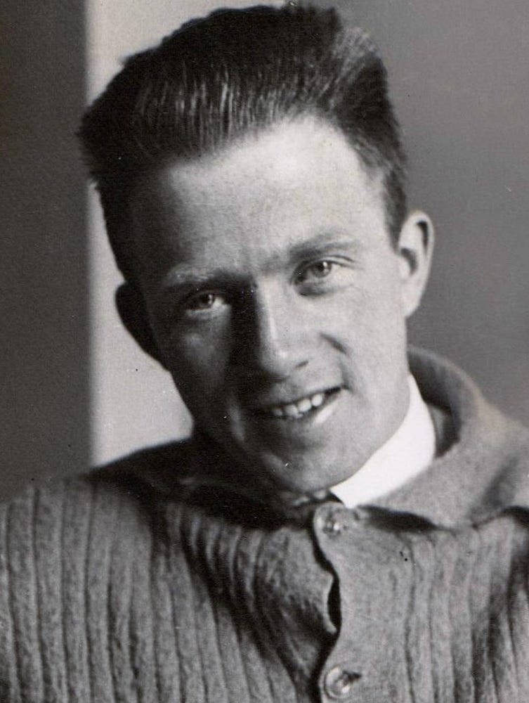
Image credit: Friedrich Hund, 1926, via creative commons 3.0.

It was in fact Heisenberg who first suggested that the divergences in quantum field theory might be cured by the existence of a fundamentally minimal length, and he introduced it by making position operators non-commuting among themselves. Just as the non-commutativity of momentum and position operators leads to an uncertainty principle, the non-commutativity of position operators limits how well distances can be measured.

Image credit: A generalized uncertainty relation, via https://blogger.googleusercontent.com/img/b/R29vZ2xl/AVvXsEhaZfXIF7oBxyGhcQI9jEbJ-1eLd8laPqujvVszzKIMAjJ2ZUp5Ji88Y1SUBkT6r6h08A2URvMgs0u5pXmucMPxPO70t0YITw9jtEQUIMdGUD9CE2GpI7WZYdjXNOZzOoBytiZ7Lq8Olys/s200/gup4.jpg.

Heisenberg’s main worry, which the minimal length was supposed to deal with, was the non-renormalizability of Fermi’s theory of beta-decay. This theory, however, turned out to be only an approximation to the renormalizable electro-weak interaction, so he had to worry no more.
Heisenberg’s idea was forgotten for some decades, then picked up again and eventually grew into the area of non-commutative geometries. Meanwhile, the problem of quantizing gravity appeared on stage and with it, again, non-renormalizability.

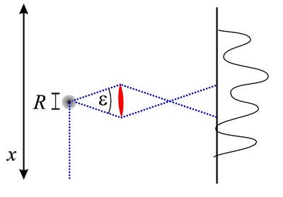
Image credit: A schematic diagram of a Heisenberg microscope, via https://blogger.googleusercontent.com/img/b/R29vZ2xl/AVvXsEhMT45R2Sh3G6P2mxWTo8ZssAF-mdQU0SaZxBaDdfJvBwu_yoSz0f3_CI4uQE_rfCaYhHzOR7tKfJw3Fq3kWLq_0KLA3rPFdcwHVsg_z2VRAX9K0zT_mPwZYWxyhYeZyHOpMLs3w54QLA4/s400/heisenberg_microscope.jpg.

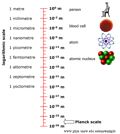
In the mid-1960s, Alden Mead reinvestigated Heisenberg’s microscope, the argument that lead to the uncertainty principle, with (non-quantized) gravity taken into account. He showed that gravity amplifies the uncertainty inherent to position so that it becomes impossible to measure distances below the Planck length: about 10^-33 cm. Mead’s argument was forgotten, then rediscovered in the 1990s by string theorists who had noticed that using strings to prevent divergences (by avoiding point-interactions) also implies a finite resolution, if in a technically somewhat different way than Mead’s.

Image credit: © School of Physics UNSW.

Since then, the idea that the Planck length may be a fundamental length beyond which there is nothing new to find, ever, appeared in other approaches towards quantum gravity, such as Loop Quantum Gravity and Asymptotically Safe Gravity. It has also been studied as an effective theory by modifying quantum field theory to include a minimal length from scratch, and often runs under the name “generalized uncertainty”.

One of the main difficulties with these theories is that a minimal length, if interpreted as the length of a ruler, would not be invariant under Lorentz-transformations due to length contraction. In other words, the idea of a “minimum length” would suddenly imply that different observers (i.e., people moving at different velocities) would measure different fundamental minimum lengths from one another! This problem is easy to overcome in momentum space, where it is a maximal energy that has to be made Lorentz-invariant, because momentum space is not translationally invariant. But in position space, one either has to break Lorentz-invariance or deform it and give up locality, which has observable consequences, and not always desired ones. Personally, I think it is a mistake to interpret the minimal length as the length of a ruler (a component of a Lorentz-vector), and it should instead be interpreted as a Lorentz-invariant scalar to begin with, but opinions on that matter differ.

The science and history of the physical idea of a minimal length has now been covered in a recent book by Amit Hagar.

Image credit: Amit Hagar’s book, “Discrete or Continuous? The Quest for a Fundamental Length in Modern Physics,” via Amazon.

Amit is a philosopher but he certainly knows his math and physics. Indeed, I suspect the book would be quite hard to understand for a reader without at least some background knowledge in those two subjects. Amit has made a considerable effort to address the topic of a fundamental length from as many perspectives as possible, and he covers a lot of scientific history and philosophical 
considerations that I had not previously been aware of. The book is also noteworthy for including a chapter on quantum gravity phenomenology.

My only complaint about the book is its title, because the question of “discrete vs. continuous” is not the same as the question of “finite vs. infinite resolution.” One can have a continuous structure and yet be unable to resolve it beyond some limit, such as would be the case when the limit makes itself noticeable as a blur rather than a discretization. On the other hand, one can have a discrete structure that does not prevent arbitrarily sharp resolution, which can happen when localization on a single base-point of the discrete structure is possible.

(Amit’s book is admittedly quite pricey, so let me add that he said should sales numbers reach 500, Cambridge University Press will put a considerably less expensive paperback version on offer. So tell your library to get a copy and let’s hope we’ll make it to 500 so it becomes affordable for more of the interested readers.)

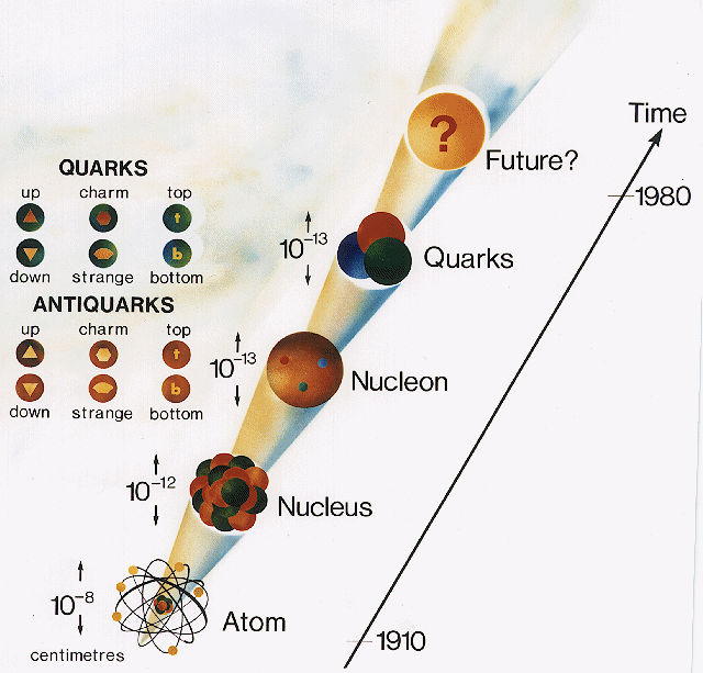
Image credit: Volker Crede, via http://hadron.physics.fsu.edu/~crede/quarks.html.

Every once in a while I think that there maybe is no fundamentally smallest unit of length; that all these arguments for its existence are wrong. I like to think that we can look infinitely close into structures and will never find a final theory, turtles upon turtles, or that structures are ultimately self-similar and repeat. Alas, it is hard to make sense of the romantic idea of universes in universes in universes mathematically, not that I didn’t try, and so the minimal length keeps coming back to me.

Many (if not most) endeavors to find observational evidence for quantum gravity today look for manifestations of a minimal length in one way or the other, such as modifications of the dispersion relation, modifications of the commutation-relations, or Bekenstein’s tabletop search for quantum gravity. The question of whether there is a smallest possible scale in the Universe is today a very active area of research. We’ve come a long way, but we’re still out to answer the same questions that people asked themselves thousands of years ago. Although we’ve certainly made a lot of progress, the ultimate answer is still beyond our abilities to resolve.

This post was written by Sabine Hossenfelder, assistant professor of physics at Nordita. You can read her (more technical) paper on a fundamental minimum length here, and follow her tweets at @skdh.

Cheminformatics

Cheminformatics

From Wikipedia, the free encyclopedia
 
Cheminformatics (also known as chemoinformatics, chemioinformatics and chemical informatics) is the use of computer and informational techniques applied to a range of problems in the field of chemistry. These in silico techniques are used in, for example, pharmaceutical companies in the process of drug discovery. These methods can also be used in chemical and allied industries in various other forms.

History

The term chemoinformatics was defined by F.K. Brown [1][2] in 1998:
Chemoinformatics is the mixing of those information resources to transform data into information and information into knowledge for the intended purpose of making better decisions faster in the area of drug lead identification and optimization.
Since then, both spellings have been used, and some have evolved to be established as Cheminformatics,[3] while European Academia settled in 2006 for Chemoinformatics.[4] The recent establishment of the Journal of Cheminformatics is a strong push towards the shorter variant.

Basics

Cheminformatics combines the scientific working fields of chemistry, computer science and information science for example in the areas of topology, chemical graph theory, information retrieval and data mining in the chemical space.[5][6][7][8]Cheminformatics can also be applied to data analysis for various industries like paper and pulp, dyes and such allied industries.

Applications

Storage and retrieval

The primary application of cheminformatics is in the storage, indexing and search of information relating to compounds. The efficient search of such stored information includes topics that are dealt with in computer science as data mining, information retrieval, information extraction and machine learning. Related research topics include:

File formats

The in silico representation of chemical structures uses specialized formats such as the XML-based Chemical Markup Language or SMILES. These representations are often used for storage in large chemical databases. While some formats are suited for visual representations in 2 or 3 dimensions, others are more suited for studying physical interactions, modeling and docking studies.

Virtual libraries

Chemical data can pertain to real or virtual molecules. Virtual libraries of compounds may be generated in various ways to explore chemical space and hypothesize novel compounds with desired properties.

Virtual libraries of classes of compounds (drugs, natural products, diversity-oriented synthetic products) were recently generated using the FOG (fragment optimized growth) algorithm. [9] This was done by using cheminformatic tools to train transition probabilities of a Markov chain on authentic classes of compounds, and then using the Markov chain to generate novel compounds that were similar to the training database.

Virtual screening

In contrast to high-throughput screening, virtual screening involves computationally screening in silico libraries of compounds, by means of various methods such as docking, to identify members likely to possess desired properties such as biological activity against a given target. In some cases, combinatorial chemistry is used in the development of the library to increase the efficiency in mining the chemical space. More commonly, a diverse library of small molecules or natural products is screened.

Quantitative structure-activity relationship (QSAR)

This is the calculation of quantitative structure-activity relationship and quantitative structure property relationship values, used to predict the activity of compounds from their structures. In this context there is also a strong relationship to Chemometrics. Chemical expert systems are also relevant, since they represent parts of chemical knowledge as an in silico representation.

Alle Menschen werden Brüder

Alle Menschen werden Brüder
Freude, schöner Götterfunken
Tochter aus Elysium,
Wir betreten feuertrunken,
Himmlische, dein Heiligtum!
Deine Zauber binden wieder
Was die Mode streng geteilt;
Alle Menschen werden Brüder,
Wo dein sanfter Flügel weilt.

Wem der große Wurf gelungen,
Eines Freundes Freund zu sein;
Wer ein holdes Weib errungen,
Mische seinen Jubel ein!
Ja, wer auch nur eine Seele
Sein nennt auf dem Erdenrund!
Und wer's nie gekonnt, der stehle
Weinend sich aus diesem Bund!

Freude trinken alle Wesen
An den Brüsten der Natur;
Alle Guten, alle Bösen
Folgen ihrer Rosenspur.
Küsse gab sie uns und Reben,
Einen Freund, geprüft im Tod;
Wollust ward dem Wurm gegeben,
und der Cherub steht vor Gott.

Froh,
wie seine Sonnen fliegen
Durch des Himmels prächt'gen Plan,
Laufet, Brüder, eure Bahn,
Freudig, wie ein Held zum Siegen.

Seid umschlungen, Millionen!
Diesen Kuß der ganzen Welt!
Brüder, über'm Sternenzelt
Muß ein lieber Vater wohnen.
Ihr stürzt nieder, Millionen?
Ahnest du den Schöpfer, Welt?
Such' ihn über'm Sternenzelt!
Über Sternen muß er wohnen.

One-nanometer synthetic molecular machine successfully observed and touched

One-nanometer synthetic molecular machine successfully observed and touched

(Nanowerk News) A research group led by Professor Hiroyuki Noji, Department of Applied Chemistry, Graduate School of Engineering, University of Tokyo, successfully observed and touched the rotational motion of a 1-nm synthetic molecular machine through the application of a single-molecule capturing and manipulation technique using optical microscopy and a bead probe (single-molecule motion capturing), which allows visualization of molecular mechanical motion.
single-molecule motion capturing and manipulation of 1-nm synthetic molecular machine by optical microscopy
Schematic diagram illustrating single-molecule motion capturing and manipulation of 1-nm synthetic molecular machine by optical microscopy using a bead probe (single-molecule motion capturing).
1 Molecule motion capturing is a technique originally invented for studying the functions of energy-conversion molecules in vivo (biomolecular machines). It had been hoped that this technique would be available for the measurement of artificial molecular machines (synthetic molecular machines) because of its broad applicability such as “seeing and touching” a single biomolecular machine as well as measuring the direction of its movement. However, it had been difficult to apply this technique to synthetic molecular machines because they are about 1 nm in size, which is one-tenth the size of biomolecular machines, which are about 10 nm in size.
Recently, Professor Noji’s group has further improved the single-molecule motion capturing technique, using a bead of 200 nm in diameter which is visible by optical microscopy. With this technique, the rotational motion of a double-decker porphyrin (DD) of 1 nm in size was observed. The conventional method was improved by dealing with issues such as the low efficiency of the immobilization reaction due to the small size of synthetic molecular machines and undesirable interaction between the bead and the substrate.
These improvements broadened its applicability. Furthermore, the group successfully manipulated the motion of the DD molecule by applying an external force to the bead. Since the smallest bio- and synthetic molecular machines are 1 nm in size, this method will enable visualizing the movements of any molecular machine. This is currently the only method available to evaluate the performance of a single synthetic molecular machine through “seeing and touching” it, and to verify whether synthetic molecular motors are generating their own locomotive power, which is one of the goals being pursued with synthetic molecular machines. Suppose that, for example, a light-driven synthetic molecular motor is created in the future, and that it can be connected to a biomolecular motor, then it may be feasible to develop custom-made energy conversion technology that can manipulate various light-related chemical reactions.
This research was jointly carried out with project researcher Tomohiro Ikeda and Takahiro Tsukahara, Department of Applied Chemistry, Graduate School of Engineering, University of Tokyo, Professor Ryota Iino, Molecular Machine Designing Laboratory, Okazaki Institute for Integrative Bioscience, National Institute of Natural Sciences, and Masayuki Takeuchi, group leader, Organic Materials Group, Polymer Materials Unit, National Institute for Materials Science. This study was funded by the CREST program offered by Strategic Basic Research Programs, Japan Science and Technology Agency.
The results of this research will be soon published in the scientific journal, Angewandte Chemie International Edition, issued by the German Chemical Society. Since this paper was regarded as significant by the society, it was selected as a “hot paper” and will be highlighted on the front and back covers of the issue.
Source: NIMS
If you liked this article, please give it a quick review on reddit or StumbleUpon. Thanks!

Read more: One-nanometer synthetic molecular machine successfully observed and touched http://www.nanowerk.com/nanotechnology-news/newsid=37028.php#ixzz3B2BfK02R
Follow us: @nanowerk on Twitter

Molecular Shuttle Improves Photocatalytic Hydrogen Production

Molecular Shuttle Improves Photocatalytic Hydrogen Production

Molecular shuttle speeds up photocatalytic hydrogen production
Molecular shuttle speeds up photocatalytic hydrogen production. (Credit: UK Department for Business, Innovation and Skills)

An LMU team affiliated with the Nanosystems Initiative Munich (NIM) has achieved a breakthrough in light-driven generation of hydrogen with semiconductor nanocrystals by using a molecular shuttle to enhance charge-carrier transport.

SEE ALSO: One-Atom Thick Catalyst May Help Produce Cheap Hydrogen
(Credit: C. Hohmann / Nanosystems Initiative Munich)

In their latest experiments with semiconductor nanocrystals as light absorbers, physicists led by Professor Jochen Feldmann (LMU Munich), in collaboration with a team of chemists under the direction of Professor Andrey Rogach (City University of Hong Kong), have succeeded in significantly increasing the yield of hydrogen produced by the photocatalytic splitting of water. The crucial innovation, reported in the latest issue of the journal Nature Materials (see footnote), is the use of a so-called molecular shuttle to markedly improve the mobility of charge carriers in their reaction system.

The basic principle behind photocatalysis seems to be quite simple. When a quantum of light (a “photon”) with sufficient energy excites a semiconductor nanocrystal, it produces a negative charge (electron) and a positive charge (hole). Photocatalytic synthesis of hydrogen gas from water requires the transfer of electrons to the hydrogen, while the holes interact with the oxygen or are scavenged by other molecules.

However, before any of this can happen, the photogenerated electrons and holes must be quickly separated from each other. If the semiconducting nanocrystals are decorated with nanoparticles of a metal catalyst—such as the precious metal platinum—the electron can rapidly transfer to the metal and hydrogen production ensues. But unless the positively charged holes are effectively removed, they will accumulate, eventually bringing H2 synthesis to a halt.

One problem for an efficient removal of holes is the need of polar molecules being attached to the nanocrystals as surface ligands in order to make the nanocrystals water-soluble. By doing so, however, the resulting “ligand forest” of the attached polar molecules makes it difficult for the holes to interact with water or larger scavenger molecules.

One can compare this to the problem of delivering airline passengers to their final destination. Spatial constraints obviously make it impossible for the aircraft to convey its passengers directly to their hotels in town. Instead, smaller and more maneuverable carriers, such as the shuttle buses, are used for the short last stage of the trip. In a similar way, the research teams in Munich and Hong Kong hit on the idea of using one of the smallest constituents of their system—the hydroxyl ion formed by the dissociation of water—to penetrate the ligand forest, collect the holes from the surface of the crystals and transport them to a larger acceptor molecule. Moreover, the concentration of this molecular shuttle in the system can be easily controlled by altering the pH of the solution. Indeed, raising the pH of the solution drastically increases the rate of hydrogen production.

“I was amazed the first time I tried it. As soon as I increased the pH I could see, with the naked eye, bubbles of hydrogen rising to the surface.” says Thomas Simon, a PhD student at Professor Feldmann’s chair.

The new system also has other advantages. First of all, its long-term stability could be markedly improved. Furthermore, it turns out that the costly platinum catalyst can be replaced by nickel, a far less expensive metal. “The discovery of this new mechanism could lead to entirely new approaches to the photocatalytic production of hydrogen.” adds Dr. Jacek Stolarczyk, who heads the Photocatalysis group at the chair of Photonics and Optoelectronics (PhOG) at LMU.

Chair holder Professor Jochen Feldmann, who also serves as Director of the NIM Cluster of Excellence, emphasizes the crucial role of the close collaboration between the different research groups involved in the project: “Our work could only be successful by being a product of an interdisciplinary team, and with the generous support by the NIM cluster and the Bavarian Research Network ’Solar Technologies go Hybrid’ (SolTech).”

Cryogenics

From Wikipedia, the free encyclopedia https://en.wikipedia.org/wiki/Cryogenics...