Search This Blog

Friday, July 13, 2018

Neuropsychopharmacology

From Wikipedia, the free encyclopedia

Neuropsychopharmacology, an interdisciplinary science related to psychopharmacology (how drugs affect the mind) and fundamental neuroscience, is the study of the neural mechanisms that drugs act upon to influence behavior. It entails research of mechanisms of neuropathology, pharmacodynamics (drug action), psychiatric illness, and states of consciousness. These studies are instigated at the detailed level involving neurotransmission/receptor activity, bio-chemical processes, and neural circuitry. Neuropsychopharmacology supersedes psychopharmacology in the areas of "how" and "why", and additionally addresses other issues of brain function. Accordingly, the clinical aspect of the field includes psychiatric (psychoactive) as well as neurologic (non-psychoactive) pharmacology-based treatments. Developments in neuropsychopharmacology may directly impact the studies of anxiety disorders, affective disorders, psychotic disorders, degenerative disorders, eating behavior, and sleep behavior.

History

Drugs such as opium, alcohol, and certain plants have been used for millennia by humans to ease suffering or change awareness, but until the modern scientific era nobody knew how these substances worked. The first half of the 20th century saw psychology and psychiatry as largely phenomenological, in that behaviors or themes which were observed in patients could often be correlated to a limited variety of factors such as childhood experience, inherited tendencies, or injury to specific brain areas. Models of mental function and dysfunction were based on such observations. Indeed, the behavioral branch of psychology dispensed altogether with what actually happened inside the brain, regarding most mental dysfunction as what could be dubbed as "software" errors. In the same era, the nervous system was progressively being studied at the microscopic and chemical level, but there was virtually no mutual benefit with clinical fields—until several developments after World War II began to bring them together. Neuropsychopharmacology may be regarded to have begun in the earlier 1950s with the discovery of drugs such as MAO inhibitors, tricyclic antidepressants, thorazine and lithium which showed some clinical specificity for mental illnesses such as depression and schizophrenia.[1] Until that time, treatments that actually targeted these complex illnesses were practically non-existent. The prominent methods which could directly affect brain circuitry and neurotransmitter levels were the pre-frontal lobotomy, and electroconvulsive therapy, the latter of which was conducted without muscle relaxants which often caused the patient great physical injury.

The field now known as neuropsychopharmacology has resulted from the growth and extension of many previously isolated fields which have met at the core of psychiatric medicine, and engages a broad range of professionals from psychiatrists to researchers in genetics and chemistry. The use of the term has gained popularity since 1990 with the founding of several journals and institutions such as the Hungarian College of Neuropsychopharmacology.[1] This rapidly maturing field shows some degree of flux, as research hypotheses are often restructured based on new information.

Overview

An implicit premise in neuropsychopharmacology with regard to the psychological aspects is that all states of mind, including both normal and drug-induced altered states, and diseases involving mental or cognitive dysfunction, have a neuro-chemical basis at the fundamental level, and certain circuit pathways in the central nervous system at a higher level. Thus the understanding of nerve cells or neurons in the brain is central to understanding the mind. It is reasoned that the mechanisms involved can be elucidated through modern clinical and research methods such as genetic manipulation in animal subjects, imaging techniques such as functional magnetic resonance imaging (fMRI), and in vitro studies using selective binding agents on live tissue cultures. These allow neural activity to be monitored and measured in response to a variety of test conditions. Other important observational tools include radiological imaging[2] such as positron emission tomography (PET) and single-photon emission computed tomography (SPECT). These imaging techniques are extremely sensitive and can image tiny molecular concentrations on the order of 10−10 M such as found with extrastriatal D1 receptor for dopamine.

One of the ultimate goals is to devise and develop prescriptions of treatment for a variety of neuro-pathological conditions and psychiatric disorders. More profoundly, though, the knowledge gained may provide insight into the very nature of human thought, mental abilities like learning and memory, and perhaps consciousness itself. A direct product of neuropsychopharmacological research is the knowledge base required to develop drugs which act on very specific receptors within a neurotransmitter system. These "hyperselective-action" drugs would allow the direct targeting of specific sites of relevant neural activity, thereby maximizing the efficacy (or technically the potency) of the drug within the clinical target and minimizing adverse effects.

The groundwork is currently being paved for the next generation of pharmacological treatments which will improve quality of life with increasing efficiency. For example, contrary to previous thought, it is now known that the adult brain does to some extent grow new neurons—the study of which, in addition to neurotrophic factors, may hold hope for neuro-degenerative diseases like Alzheimer's, Parkinson's, ALS, and types of chorea. All of the proteins involved in neurotransmission are a small fraction of the more than 100,000 proteins in the brain. Thus there are many proteins which are not even in the direct path of signal transduction, any of which may still be a target for specific therapy. At present, novel pharmacological approaches to diseases or conditions are reported at a rate of almost one per week.[3]

Neurotransmission

So far as we know, everything we perceive, feel, think, know, and do are a result of neurons firing and resetting. When a cell in the brain fires, small chemical and electrical swings called the action potential may affect the firing of as many as a thousand other neurons in a process called neurotransmission. In this way signals are generated and carried through networks of neurons, the bulk electrical effect of which can be measured directly on the scalp by an EEG device.
By the last decade of the 20th century, the essential knowledge of all the central features of neurotransmission had been gained.[4] These features are:
The more recent advances involve understanding at the organic molecular level; biochemical action of the endogenous ligands, enzymes, receptor proteins, etc. The critical changes affecting cell firing occur when the signalling neurotransmitters from one neuron, acting as ligands, bind to receptors of another neuron. Many neurotransmitter systems and receptors are well known, and research continues toward the identification and characterization of a large number of very specific sub-types of receptors. For the six more important neurotransmitters Glu, GABA, Ach, NE, DA, and 5HT (listed at neurotransmitter) there are at least 29 major subtypes of receptor. Further "sub-subtypes" exist together with variants, totalling in the hundreds for just these 6 transmitters. - (see serotonin receptor for example.) It is often found that receptor subtypes have differentiated function, which in principle opens up the possibility of refined intentional control over brain function.

It has previously been known that ultimate control over the membrane voltage or potential of a nerve cell, and thus the firing of the cell, resides with the trans-membrane ion channels which control the membrane currents via the ions K+, Na+, and Ca++, and of lesser importance Mg++ and Cl. The concentration differences between the inside and outside of the cell determine the membrane voltage.

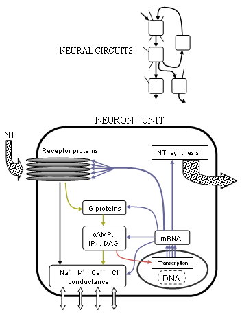
Abstract simplified diagram showing overlap between neurotransmission and metabolic activity. Neurotransmitters bind to receptors which cause changes to ion channels (black, yellow), metabotropic receptors also affect DNA transcription (red), transcription is responsible for all cell proteins including enzymes which manufacture neurotransmitters (blue).

Precisely how these currents are controlled has become much clearer with the advances in receptor structure and G-protein-coupled processes. Many receptors are found to be pentameric clusters of five trans-membrane proteins (not necessarily the same) or receptor subunits, each a chain of many amino acids. Transmitters typically bind at the junction between two of these proteins, on the parts that protrude from the cell membrane. If the receptor is of the ionotropic type, a central pore or channel in the middle of the proteins will be mechanically moved to allow certain ions to flow through, thus altering the ion concentration difference. If the receptor is of the metabotropic type, G-proteins will cause metabolism inside the cell that may eventually change other ion channels. Researchers are better understanding precisely how these changes occur based on the protein structure shapes and chemical properties.

The scope of this activity has been stretched even further to the very blueprint of life since the clarification of the mechanism underlying gene transcription. The synthesis of cellular proteins from nuclear DNA has the same fundamental machinery[5] for all cells; the exploration of which now has a firm basis thanks to the Human Genome Project which has enumerated the entire human DNA sequence, although many of the estimated 35,000 genes remain to be identified. The complete neurotransmission process extends to the genetic level. Gene expression determines protein structures through type II RNA polymerase. So enzymes which synthesize or breakdown neurotransmitters, receptors, and ion channels are each made from mRNA via the DNA transcription of their respective gene or genes. But neurotransmission, in addition to controlling ion channels either directly or otherwise through metabotropic processes, also actually modulates gene expression. This is most prominently achieved through modification of the transcription initiation process by a variety of transcription factors produced from receptor activity.

Aside from the important pharmacological possibilities of gene expression pathways, the correspondence of a gene with its protein allows the important analytical tool of gene knockout. Living specimens can be created using homolog recombination in which a specific gene cannot be expressed. The organism will then be deficient in the associated protein which may be a specific receptor. This method avoids chemical blockade which can produce confusing or ambiguous secondary effects so that the effects of a lack of receptor can be studied in a purer sense.

Drugs

The inception of many classes of drugs is in principle straightforward: any chemical that can enhance or diminish the action of a target protein could be investigated further for such use. The trick is to find such a chemical that is receptor-specific (cf. "dirty drug") and safe to consume. The 2005 Physicians' Desk Reference lists twice the number of prescription drugs as the 1990 version.[6] Many people by now are familiar with "selective serotonin reuptake inhibitors", or SSRIs which exemplify modern pharmaceuticals. These SSRI anti-depressant drugs, such as Paxil and Prozac, selectively and therefore primarily inhibit the transport of serotonin which prolongs the activity in the synapse. There are numerous categories of selective drugs, and transport blockage is only one mode of action. The FDA has approved drugs which selectively act on each of the major neurotransmitters such as NE reuptake inhibitor antidepressants, DA blocker anti-psychotics, and GABA agonist tranquilizers (benzodiazepines).

New endogenous chemicals are continually identified. Specific receptors have been found for the drugs THC (cannabis) and GHB,[7] with endogenous transmitters anandamide and GHB. Another recent major discovery occurred in 1999 when orexin, or hypocretin, was found to have a role in arousal, since the lack of orexin receptors mirrors the condition of narcolepsy. Orexin agonism may explain the anti-narcoleptic action of the drug modafinil which was already being used only a year prior.

The next step, which major pharmaceutical companies are currently working hard to develop, are receptor subtype-specific drugs and other specific agents. An example is the push for better anti-anxiety agents (anxiolytics) based on GABAA(α2) agonists, CRF1 blockers, and 5HT2c blockers.[8] Another is the proposal of new routes of exploration for anti-psychotics such as glycine reuptake inhibitors.[9] Although the capabilities exist for receptor-specific drugs, a shortcoming of drug therapy is the lack of ability to provide anatomical specificity. By altering receptor function in one part of the brain, abnormal activity can be induced in other parts of the brain due to the same type of receptor changes. A common example is the effect of D2 altering drugs (neuroleptics) which can help schizophrenia, but cause a variety of dyskinesias by their action on motor cortex.

Modern studies are revealing details of mechanisms of damage to the nervous system such as apoptosis (programmed cell death) and free-radical disruption. PCP has been found to cause cell death in striatopallidal cells and abnormal vacuolization in hippocampal and other neurons. The hallucinogen persisting perception disorder (HPPD), also known as post-psychedelic perception disorder, has been observed in patients as long as 26 years after LSD use. The plausible cause of HPPD is damage to the inhibitory GABA circuit in the visual pathway (GABA agonists such as midazolam can decrease some effects of LSD intoxication). The damage may be the result of an excitotoxic response of 5HT2 interneurons. [Note: the vast majority of LSD users do not experience HPPD. Its manifestation may be equally dependent on individual brain chemistry as on the drug use itself.] As for MDMA, aside from persistent losses of 5HT and SERT, long-lasting reduction of serotonergic axons and terminals is found from short-term use, and regrowth may be of compromised function.

Neural circuits

It is a not-so-recent discovery that many functions of the brain are localized to associated areas like motor and speech ability. Functional associations of brain anatomy are now being complemented with clinical, behavioral, and genetic correlates of receptor action, completing the knowledge of neural signalling (see also: Human Cognome Project). The signal paths of neurons are hyper-organized beyond the cellular scale into often complex neural circuit pathways. Knowledge of these pathways is perhaps the easiest to interpret, being most recognizable from a systems analysis point of view, as may be seen in the following abstracts.

Progress has been made on central mechanisms of hallucination believed to be common to psychedelic drugs and psychosis. It is likely the effect of partial agonistic action on the serotonin system. The 5HT2A receptor and possibly the 5HT1C are involved by releasing glutamate in the frontal cortex, while simultaneously in the locus coeruleus sensory information is promoted and spontaneous activity decreases. One hypothesis suggests that in the frontal cortex, 5HT2A promotes late asynchronous excitatory post-synaptic potentials, a process antagonized by serotonin itself through 5HT1 which may explain why SSRI's and other serotonin-affecting drugs do not normally cause a patient to hallucinate.

Diagram of neural circuit which regulates melatonin production via actual circuit pathways. Green light in the eye inhibits pineal production of melatonin (Inhibitory connections shown in red). Also shown:reaction sequence for melatonin synthesis.

Circadian rhythm, or sleep/wake cycling, is centered in the suprachiasmatic nucleus (SCN) within the hypothalamus, and is marked by melatonin levels 2000-4,000% higher during sleep than in the day. A circuit is known to start with melanopsin cells in the eye which stimulate the SCN through glutamate neurons of the hypothalamic tract. GABA-ergic neurons from the SCN inhibit the paraventricular nucleus, which signals the superior cervical ganglion (SCG) through sympathetic fibers. The output of the SCG, stimulates NE receptors (β) in the pineal gland which produces N-acetyltransferase, causing production of melatonin from serotonin. Inhibitory melatonin receptors in the SCN then provide a positive feedback pathway. Therefore, light inhibits the production of melatonin which "entrains" the 24-hour cycle of SCN activity.[10][11] The SCN also receives signals from other parts of the brain, and its (approximately) 24-hour cycle does not only depend on light patterns. In fact, sectioned tissue from the SCN will exhibit daily cycle in vitro for many days. Additionally, (not shown in diagram), the basal nucleus provides GABA-ergic inhibitory input to the pre-optic anterior hypothalamus (PAH). When adenosine builds up from the metabolism of ATP throughout the day, it binds to adenosine receptors, inhibiting the basal nucleus. The PAH is then activated, generating slow-wave sleep activity. Caffeine is known to block adenosine receptors, thereby inhibiting sleep among other things.

Research

Research in neuropsychopharmacology comes from a wide range of activities in neuroscience and clinical research. This has motivated organizations such as the American College of Neuropsychopharmacology (ACNP), the European College of Neuropsychopharmacology (ECNP), and the Collegium Internationale Neuro-psychopharmacologicum (CINP) to be established as a measure of focus. The ECNP publishes European Neuropsychopharmacology, and as part of the Reed Elsevier Group, the ACNP publishes the journal Neuropsychopharmacology, and the CINP publishes the journal International Journal of Neuropsychopharmacology with Cambridge University Press. In 2002, the most recent comprehensive collected work of the ACNP, "Neuropsychopharmacology: The Fifth Generation of Progress" was compiled. It is one measure of the current state of knowledge, and might be said to represent a landmark in the century-long goal to establish the basic neuro-biological principles which govern the actions of the brain.

Many other journals exist which contain relevant information such as Neuroscience. Some of them are listed at Brown University Library.
Polydendrocytes

From Wikipedia, the free encyclopedia
Polydendrocytes (also known as NG2 cells, NG2 glia, or oligodendrocyte progenitor cells) are process-bearing glial cells (neuroglia) in the mammalian central nervous system (CNS) that are identified by the expression of the NG2 chondroitin sulfate proteoglycan (CSPG4)  and the alpha receptor for platelet-derived growth factor (PDGFRA). They are distinct from other cell populations such as neurons, astrocytes, oligodendrocytes, microglia, and neural stem cells and are recognized as the fourth major glial cell type in the mammalian CNS.[citation needed] Studies have implicated polydendrocytes in many cellular and physiological processes. Polydendrocytes in the postnatal mouse CNS and those grown in culture generate oligodendrocytes, and thus they are often equated with oligodendrocyte progenitor cells (OPCs). Under some culture conditions, polydendrocytes give rise to astrocytes. A subpopulation of polydendrocytes in the gray matter of the embryonic CNS also generates protoplasmic astrocytes. In addition, polydendrocytes express receptors for various neurotransmitters and undergo membrane depolarization when they receive synaptic inputs from neurons.

History

It had been known since the early 1900s that astrocytes, oligodendrocytes, and microglia make up the major glial cell populations in the mammalian CNS. The presence of another glial cell population had escaped recognition because of the lack of a suitable marker to identify them in tissue sections. The notion that there exists a population of glial progenitor cells in the developing and mature CNS began to be entertained in the late 1980s by several independent groups. In one series of studies on the development and origin of oligodendrocytes in the rodent CNS, a population of immature cells that appeared to be precursors to oligodendrocytes was identified by the expression of the GD3 ganglioside.[3][4]

In a separate series of studies, cells from perinatal rat optic nerves that expressed the A2B5 ganglioside were shown to differentiate into oligodendrocytes in culture.[5] Subsequently, A2B5+ cells from other CNS regions and from adult CNS were also shown to generate oligodendrocytes. Based on the observation that these cells require PDGF for their proliferation and expansion, the expression of the alpha receptor for platelet-derived growth factor (Pdgfra) was used to search for the in vivo correlates of the A2B5+ cells, which led to the discovery of a unique population of Pdgfra+ cells in the CNS whose appearance and distribution were consistent with those of developing oligodendrocytes.[6]

Independently, Stallcup and colleagues generated an antiserum that recognized a group of rat brain tumor cell lines, which exhibited properties that were intermediate between those of typical neurons and glial cells. Biochemical studies showed that the antiserum recognized a chondroitin sulfate proteoglycan with a core glycoprotein of 300 kDa,[7] and the antigen was named NG2 (nerve/glial antigen 2).[8][9] NG2 was found to be expressed on A2B5+ oligodendrocyte precursor cells isolated from the perinatal rat CNS tissues and on process-bearing cells in the CNS in vivo.[7][10] Comparison of NG2 and Pdgfra expression revealed that NG2 and Pdgfra are expressed on the same population of cells in the CNS.[11] These cells represent 2-9% of all the cells and remain proliferative in the mature CNS.[12]

Origin and fate

Origin

In the embryonic spinal cord, a major source of polydendrocytes is the ventral ventricular zone of the pMN domain marked by the expression of the transcription factors Olig1 and Olig2 and the p3 domain that expresses Nkx2.2, which are induced by the morphogen Shh (sonic hedgehog). Some polydendrocytes also arise from the dorsal ventricular zone. In the forebrain, three regionally distinct sources have been shown to generate polydendrocytes sequentially: an early ventral source from the medial ganglionic eminence marked by Nkx2.1, followed by progenitor cells in the lateral ganglionic eminence marked by Gsh2, and finally the dorsal neocortical germinal zone marked by Emx1.[13] After the committed progenitor cells exit the germinal zones, they begin to express NG2 and Pdgfra and expand by local proliferation and migration and eventually occupy the entire CNS parenchyma. Polydendrocytes continue to exist in the adult CNS and retain their proliferative ability throughout life.

Fate

The fate of polydendrocytes has been highly debated.[14] Using Cre-Lox recombination-mediated genetic fate mapping, several labs have reported the fate of polydendrocytes using different Cre driver and reporter mouse lines;[15][16][17][18] reviewed in reference.[19] The consensus of these studies is that polydendrocytes generate predominantly oligodendrocytes in both gray and white matter. The rate at which they generate oligodendrocytes declines with age and is greater in white matter than in gray matter. These studies revealed that up to 30% of the oligodendrocytes that exist in the adult corpus callosum are generated de novo from polydendrocytes over a period of 2 months. It is not known whether all polydendrocytes eventually generate oligodendrocytes while self-renewing its population or whether some remain as polydendrocytes throughout the life of the animal and never differentiate into oligodendrocytes.

Using NG2cre mice, it was shown that polydendrocytes in the prenatal and perinatal gray matter of the ventral forebrain and spinal cord generate protoplasmic astrocytes in addition to oligodendrocytes. However, contrary to the prediction from optic nerve cultures, polydendrocytes in white matter do not generate astrocytes. When the oligodendrocyte transcription factor Olig2 is deleted specifically in polydendrocytes, there is a region- and age-dependent switch in the fate of polydendrocytes from oligodendrocytes to astrocytes.[20]

Although controversy still continues about the neuronal fate of polydendrocytes, the consensus from a number of recent genetic fate mapping studies described above seems to be that polydendrocytes do not generate a significant number of neurons under normal conditions, and that they are distinct from neural stem cells that reside in the subventricular zone.[21]

Functions

In remyelination

Polydendrocytes retain the ability to proliferate in adulthood and comprise 70-90% of the proliferating cell population in the mature CNS.[12][22] Under conditions in the developing and mature CNS where a reduction in the normal number of oligodendrocytes or myelin occurs, polydendrocytes react promptly by undergoing increased proliferation. In acute or chronic demyelinated lesions created in the rodent CNS by chemical agents such as lysolecithin or cuprizone, polydendrocytes proliferate in response to demyelination, and the proliferated cells differentiate into remyelinating oligodendrocytes.[23][24] Similarly, polydendrocyte proliferation occurs in other types of injury that are accompanied by loss of myelin, such as spinal cord injury.[25]

If polydendrocytes were capable of giving rise to myelinating oligodendrocytes, one would expect complete remyelination of pathologically demyelinated lesions such as those seen in multiple sclerosis (MS). However, complete myelin regeneration is usually not observed clinically or in chronic experimental models. Possible explanations for remyelination failure include depletion of polydendrocytes over time, failure to recruit polydendrocytes to the demyelinated lesion, and failure of recruited polydendrocytes to differentiate into mature oligodendrocytes.[25]

Numerous factors have been shown to regulate polydendrocyte proliferation, migration, and differentiation [25][25] (reviewed in [26][27][28]). In fresh MS lesions, clusters of HNK-1+ oligodendrocytes have been observed,[29] which suggests that under favorable conditions polydendrocytes expand around demyelinated lesions and generate new oligodendrocytes. In chronic MS lesions where remyelination is incomplete, there is evidence that there are oligodendrocytes with processes extending toward demyelinated axons, but they do not seem to be able to generate new myelin.[30] The mechanisms that regulate differentiation of polydendrocytes into myelinating oligodendrocytes are an actively investigated area of research.

Another unanswered question is whether the polydendrocyte pool eventually becomes depleted after they are used to generate remyelinating cells. Clonal analysis of isolated polydendrocytes in the normal mouse forebrain suggests that in the adult, most clones originating from single polydendrocytes consist of either a heterogeneous population containing both oligodendrocytes and polydendrocytes or consist exclusively of polydendrocytes, suggesting that polydendrocytes in the adult CNS are able to self-renew and are not depleted under normal conditions.[31] However, it is not known whether this dynamic is altered in response to demyelinating lesions.

Neuron-polydendrocyte interactions

There is substantial evidence that indicates a functional interaction between polydendrocytes and neurons.

Node of Ranvier

Polydendrocytes extend their processes to the nodes of Ranvier [32] and together with astrocyte processes make up the nodal glial complex. Since the nodes of Ranvier contain a high density of voltage-dependent sodium channels and allow regenerative action potentials to be generated, it is speculated that this location allows polydendrocytes to sense and possibly respond to neuronal activity

Neuron-polydendrocyte synapse

Studies have shown that neurons form synapses with polydendrocytes in both gray matter [33] and white matter.[32][34] Polydendrocytes express the AMPA type glutamate receptors and GABAA receptors and undergo small membrane depolarizations when stimulated by glutamate or GABA that is vesicularly released from presynaptic terminals. Electron microscopy revealed polydendrocyte membranes apposed to neuronal presynaptic terminals filled with synaptic vesicles. Polydendrocytes lose their ability to respond to synaptic inputs from neurons as they differentiate into mature oligodendrocytes.[35][36]

Polydendrocytes can undergo cell division while maintaining synaptic inputs from neurons.[37] These observations suggest that cells that receive neuronal synaptic inputs and those that differentiate into oligodendrocytes are not mutually exclusive cell populations but that the same population of polydendrocytes can receive synaptic inputs and generate myelinating oligodendrocytes. The functional significance of the neuron-polydendrocyte synapses remains to be elucidated.

Neuroglia

From Wikipedia, the free encyclopedia

Neuroglia
Glial Cell Types.png
Illustration of the four different types of glial cells found in the central nervous system: ependymal cells (light pink), astrocytes (green), microglial cells (dark red), and oligodendrocytes (light blue).
Details
PrecursorNeuroectoderm for macroglia, and hematopoietic stem cells for microglia
SystemNervous system
Identifiers
MeSHD009457
TAA14.0.00.005
THH2.00.06.2.00001
FMA54541
Anatomical terms of microanatomy
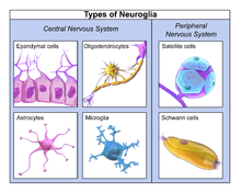
Neuroglia, also called glial cells or simply glia, are non-neuronal cells in the central nervous system (brain and spinal cord) and the peripheral nervous system. They maintain homeostasis, form myelin, and provide support and protection for neurons. In the central nervous system, glial cells include oligodendrocytes, astrocytes, ependymal cells, and microglia, and in the peripheral nervous system glial cells include Schwann cells and satellite cells. They have four main functions: (1) To surround neurons and hold them in place (2) To supply nutrients and oxygen to neurons (3) To insulate one neuron from another (4) To destroy pathogens and remove dead neurons. They also play a role in neurotransmission and synaptic connections, and in physiological processes like breathing.

Neuroglia, also called glial cells or simply glia, are non-neuronal cells in the central nervous system (brain and spinal cord) and the peripheral nervous system. They maintain homeostasis, form myelin, and provide support and protection for neurons. In the central nervous system, glial cells include oligodendrocytes, astrocytes, ependymal cells, and microglia, and in the peripheral nervous system glial cells include Schwann cells and satellite cells. They have four main functions: (1) To surround neurons and hold them in place (2) To supply nutrients and oxygen to neurons (3) To insulate one neuron from another (4) To destroy pathogens and remove dead neurons. They also play a role in neurotransmission and synaptic connections, and in physiological processes like breathing.

Glia were discovered in 1856, by the pathologist Rudolf Virchow in his search for a "connective tissue" in the brain. The term derives from Greek γλία and γλοία "glue"(/ˈɡlə/ or /ˈɡlə/), and suggests the original impression that they were the glue of the nervous system.

Types

Neuroglia of the brain shown by Golgi's method
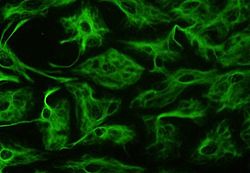
Astrocytes can be identified in culture because, unlike other mature glia, they express glial fibrillary acidic protein (GFAP)
Glial cells in a rat brain stained with an antibody against GFAP
Different types of neuroglia

Macroglia

Derived from ectodermal tissue.

Location Name Description
CNS Astrocytes The most abundant type of macroglial cell in the CNS,[6] astrocytes (also called astroglia) have numerous projections that link neurons to their blood supply while forming the blood-brain barrier. They regulate the external chemical environment of neurons by removing excess potassium ions, and recycling neurotransmitters released during synaptic transmission. Astrocytes may regulate vasoconstriction and vasodilation by producing substances such as arachidonic acid, whose metabolites are vasoactive.
Astrocytes signal each other using ATP. The gap junctions (also known as electrical synapses) between astrocytes allow the messenger molecule IP3 to diffuse from one astrocyte to another. IP3 activates calcium channels on cellular organelles, releasing calcium into the cytoplasm. This calcium may stimulate the production of more IP3 and cause release of ATP through channels in the membrane made of pannexins. The net effect is a calcium wave that propagates from cell to cell. Extracellular release of ATP, and consequent activation of purinergic receptors on other astrocytes, may also mediate calcium waves in some cases.
In general, there are two types of astrocytes, protoplasmic and fibrous, similar in function but distinct in morphology and distribution. Protoplasmic astrocytes have short, thick, highly branched processes and are typically found in gray matter. Fibrous astrocytes have long, thin, less branched processes and are more commonly found in white matter.
It has recently been shown that astrocyte activity is linked to blood flow in the brain, and that this is what is actually being measured in fMRI.[7] They also have been involved in neuronal circuits playing an inhibitory role after sensing changes in extracellular calcium.[8]
CNS Oligodendrocytes Oligodendrocytes are cells that coat axons in the central nervous system (CNS) with their cell membrane, forming a specialized membrane differentiation called myelin, producing the myelin sheath. The myelin sheath provides insulation to the axon that allows electrical signals to propagate more efficiently.[9]
CNS Ependymal cells Ependymal cells, also named ependymocytes, line the spinal cord and the ventricular system of the brain. These cells are involved in the creation and secretion of cerebrospinal fluid (CSF) and beat their cilia to help circulate the CSF and make up the blood-CSF barrier. They are also thought to act as neural stem cells.[10]
CNS Radial glia Radial glia cells arise from neuroepithelial cells after the onset of neurogenesis. Their differentiation abilities are more restricted than those of neuroepithelial cells. In the developing nervous system, radial glia function both as neuronal progenitors and as a scaffold upon which newborn neurons migrate. In the mature brain, the cerebellum and retina retain characteristic radial glial cells. In the cerebellum, these are Bergmann glia, which regulate synaptic plasticity. In the retina, the radial Müller cell is the glial cell that spans the thickness of the retina and, in addition to astroglial cells,[11] participates in a bidirectional communication with neurons.[12]
PNS Schwann cells Similar in function to oligodendrocytes, Schwann cells provide myelination to axons in the peripheral nervous system (PNS). They also have phagocytotic activity and clear cellular debris that allows for regrowth of PNS neurons.[13]
PNS Satellite cells Satellite glial cells are small cells that surround neurons in sensory, sympathetic, and parasympathetic ganglia.[14] These cells help regulate the external chemical environment. Like astrocytes, they are interconnected by gap junctions and respond to ATP by elevating intracellular concentration of calcium ions. They are highly sensitive to injury and inflammation, and appear to contribute to pathological states, such as chronic pain.[15]
PNS Enteric glial cells Are found in the intrinsic ganglia of the digestive system. They are thought to have many roles in the enteric system, some related to homeostasis and muscular digestive processes.[16]

Microglia

Microglia are specialized macrophages capable of phagocytosis that protect neurons of the central nervous system.[17] They are derived from the earliest wave of mononuclear cells that originate in yolk sac blood islands early in development, and colonize the brain shortly after the neural precursors begin to differentiate.[18]

These cells are found in all regions of the brain and spinal cord. Microglial cells are small relative to macroglial cells, with changing shapes and oblong nuclei. They are mobile within the brain and multiply when the brain is damaged. In the healthy central nervous system, microglia processes constantly sample all aspects of their environment (neurons, macroglia and blood vessels). In a healthy brain, microglia direct the immune response to brain damage and play an important role in the inflammation that accompanies the damage. Many diseases and disorders are associated with deficient microglia, such as Alzheimer's disease, Parkinson's disease, and ALS.

Other

Pituicytes from the posterior pituitary are glia cells with characteristics in common to astrocytes.[19] Tanycytes in the median eminence of the hypothalamus are a type of ependymal cell that descend from radial glia and line the base of the third ventricle.[20]

Total number

In general, neuroglial cells are smaller than neurons; there are about 86 billion neurons and 85 billion "non-neuronal" (glia) cells in the human male brain. Glial cells make up about half the total volume of the brain and spinal cord.[21][22]^ The ratio varies from one part of the brain to another. The glia/neuron ratio in the cerebral cortex is 3.72 (60.84 billion glia (72%); 16.34 billion neurons), while that of the cerebellum is only 0.23 (16.04 billion glia; 69.03 billion neurons). The ratio in the cerebral cortex gray matter is 1.48, with 3.76 for the gray and white matter combined.[21] The ratio of the basal ganglia, diencephalon and brainstem combined is 11.35.[21]

Most cerebral cortex glia are oligodendrocytes (75.6%); astrocytes account for 17.3% and microglia for 6.5%.[23]

Development

23-week fetal brain culture astrocyte

Most glia are derived from ectodermal tissue of the developing embryo, in particular the neural tube and crest. The exception is microglia, which are derived from hemopoietic stem cells. In the adult, microglia are largely a self-renewing population and are distinct from macrophages and monocytes, which infiltrate the injured and diseased CNS.

In the central nervous system, glia develop from the ventricular zone of the neural tube. These glia include the oligodendrocytes, ependymal cells, and astrocytes. In the peripheral nervous system, glia derive from the neural crest. These PNS glia include Schwann cells in nerves and satellite glial cells in ganglia.

Current research involving glial cells in the human cochlea proposes that these cells are the common precursor to both mature Schwann cells and satellite glial cells. Additionally, the peripheral glial cells located along the peripheral processes expressed NGFR, indicating a phenotype distinct from the peripheral glial cells located along the central processes.[24]

Capacity to divide

Glia retain the ability to undergo cell division in adulthood, whereas most neurons cannot. The view is based on the general deficiency of the mature nervous system in replacing neurons after an injury, such as a stroke or trauma, while very often there is a profound proliferation of glia, or gliosis near or at the site of damage. However, detailed studies found no evidence that 'mature' glia, such as astrocytes or oligodendrocytes, retain the ability of mitosis. Only the resident oligodendrocyte precursor cells seem to keep this ability after the nervous system matures. On the other hand, there are a few regions in the mature nervous system, such as the dentate gyrus of the hippocampus and the subventricular zone, where generation of new neurons can be observed.[25]

Glial cells are known to be capable of mitosis. By contrast, scientific understanding of whether neurons are permanently post-mitotic,[26] or capable of mitosis,[27][28][29] is still developing. In the past, glia had been considered[by whom?] to lack certain features of neurons. For example, glial cells were not believed to have chemical synapses or to release transmitters. They were considered to be the passive bystanders of neural transmission. However, recent studies have shown this to be untrue.[30]

Functions

Some glial cells function primarily as the physical support for neurons. Others regulate the internal environment of the brain, especially the fluid surrounding neurons and their synapses, and nutrify neurons. During early embryogenesis, glial cells direct the migration of neurons and produce molecules that modify the growth of axons and dendrites.

Neuron repair and development

Glia are also crucial in the development of the nervous system and in processes such as synaptic plasticity and synaptogenesis. Glia have a role in the regulation of repair of neurons after injury. In the central nervous system (CNS), glia suppress repair. Glial cells known as astrocytes enlarge and proliferate to form a scar and produce inhibitory molecules that inhibit regrowth of a damaged or severed axon. In the peripheral nervous system (PNS), glial cells known as Schwann cells promote repair. After axonal injury, Schwann cells regress to an earlier developmental state to encourage regrowth of the axon. This difference between the CNS and the PNS, raises hopes for the regeneration of nervous tissue in the CNS. For example, a spinal cord may be able to be repaired following injury or severance. Schwann cells are also known as neuri-lemmocytes. These cells envelop nerve fibers of the PNS by winding repeatedly around a nerve fiber with the nucleus inside of it. This process creates a myelin sheath, which not only aids in conductivity but also assists in the regeneration of damaged fibers.

Myelin sheath creation

Oligodendrocytes are another type of glial cell of the CNS. These dendrocytes resemble an octopus bulbous body and contain up to fifteen arm-like processes. Each “arm” reaches out to a nerve fiber and spirals around it, creating a myelin sheath. This myelin sheath insulates the nerve fiber from the extracellular fluid as well as speeds up the signal conduction in the nerve fiber.[31]

Neurotransmission

Recent research indicates that glial cells of the hippocampus and cerebellum participate in synaptic transmission, regulate the clearance of neurotransmitters from the synaptic cleft, and release gliotransmitters such as ATP, which modulate synaptic function.[32]

Astrocytes are crucial in clearance of neurotransmitters from within the synaptic cleft, which provides distinction between arrival of action potentials and prevents toxic build-up of certain neurotransmitters such as glutamate (excitotoxicity). It is also thought that glia play a role in many neurological diseases, including Alzheimer's disease.[33] Furthermore, at least in vitro, astrocytes can release gliotransmitter glutamate in response to certain stimulation. Another unique type of glial cell, the oligodendrocyte precursor cells or OPCs, have very well-defined and functional synapses from at least two major groups of neurons.[34] The only notable differences between neurons and glial cells are neurons' possession of axons and dendrites, and capacity to generate action potentials.

Clinical significance

Neoplastic glial cells stained with an antibody against GFAP (brown), from a brain biopsy

While glial cells in the PNS frequently assist in regeneration of lost neural functioning, loss of neurons in the CNS does not result in a similar reaction from neuroglia.[13] In the CNS, regrowth will only happen if the trauma was mild, and not severe.[35] When severe trauma presents itself, the survival of the remaining neurons becomes the optimal solution. However, some studies investigating the role of glial cells in Alzheimer's Disease are beginning to contradict the usefulness of this feature, and even claim it can "exacerbate" the disease.[36] In addition to impacting the potential repair of neurons in Alzheimer's Disease, scarring and inflammation from glial cells have been further implicated in the degeneration of neurons caused by Amyotrophic lateral sclerosis.[37]

In addition to neurodegenerative diseases, a wide range of harmful exposure, such as hypoxia, or physical trauma, can lead to the end result of physical damage to the CNS.[35] Generally, when damage occurs to the CNS, glial cells cause Apoptosis among the surrounding cellular bodies.[35] Then, there is a large amount of microglial activity, which results in inflammation, and finally, there is a heavy release of growth inhibiting molecules.[35]

History

Glia were first described in 1856 by the pathologist Rudolf Virchow in a comment to his 1846 publication on connective tissue. A more detailed description of glial cells was provided in the 1858 book Cellular Pathology by the same author.[38]

When markers for different types of cells were analyzed, Einstein's brain was discovered to contain significantly more glia than normal brains in the left angular gyrus, an area thought to be responsible for mathematical processing and language.[39]

The ratio of glia to neurons increases with our definition of intelligence. Not only does the ratio of glia to neurons increase through evolution, but so does the size of the glia. Astroglial cells in the human have a volume 27 times greater than the same cells in the mouse's brain.[40]

These important scientific findings may begin to shift the neuron-specific perspective into a more holistic view of the brain which encompasses the glial cells as well. The glia's importance is becoming ever more clear as time goes on and new research is conducted. For most of the last century, scientists had written off glial cells as being nothing more than the structure and foundations that hold the neurons in place. But now, there is direct evidence that correlates the number of glial cells in the brain with the amount of intelligence that any given species possesses.[41] Future research will begin to shed light on the mysterious, yet increasingly crucial, role of glial cells.

Inequality (mathematics)

From Wikipedia, the free encyclopedia https://en.wikipedia.org/wiki/Inequality...