Search This Blog

Sunday, September 23, 2018

Mitochondrial DNA

From Wikipedia, the free encyclopedia

Mitochondrial DNA is the small circular chromosome found inside mitochondria. These organelles found in cells have often been called the powerhouse of the cell. The mitochondria, and thus mitochondrial DNA, are passed only from mother to offspring through the egg cell.
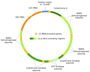
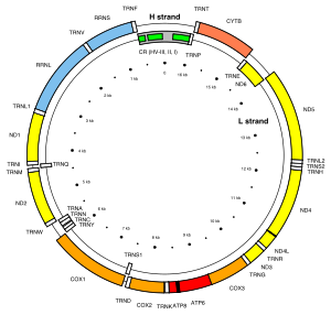
Human mitochondrial DNA with the 37 genes on their respective H- and L-strands.
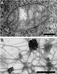
Electron microscopy reveals mitochondrial DNA in discrete foci. Bars: 200 nm. (A) Cytoplasmic section after immunogold labelling with anti-DNA; gold particles marking mtDNA are found near the mitochondrial membrane (black dots in upper right). (B) Whole mount view of cytoplasm after extraction with CSK buffer and immunogold labelling with anti-DNA; mtDNA (marked by gold particles) resists extraction. From Iborra et al., 2004.

Mitochondrial DNA (mtDNA or mDNA) is the DNA located in mitochondria, cellular organelles within eukaryotic cells that convert chemical energy from food into a form that cells can use, adenosine triphosphate (ATP). Mitochondrial DNA is only a small portion of the DNA in a eukaryotic cell; most of the DNA can be found in the cell nucleus and, in plants and algae, also in plastids such as chloroplasts.

In humans, the 16,569 base pairs of mitochondrial DNA encode for only 37 genes. Human mitochondrial DNA was the first significant part of the human genome to be sequenced. In most species, including humans, mtDNA is inherited solely from the mother.

Since animal mtDNA evolves faster than nuclear genetic markers, it represents a mainstay of phylogenetics and evolutionary biology. It also permits an examination of the relatedness of populations, and so has become important in anthropology and biogeography.

Origin

Nuclear and mitochondrial DNA are thought to be of separate evolutionary origin, with the mtDNA being derived from the circular genomes of the bacteria that were engulfed by the early ancestors of today's eukaryotic cells. This theory is called the endosymbiotic theory. Each mitochondrion is estimated to contain 2–10 mtDNA copies. In the cells of extant organisms, the vast majority of the proteins present in the mitochondria (numbering approximately 1500 different types in mammals) are coded for by nuclear DNA, but the genes for some, if not most, of them are thought to have originally been of bacterial origin, having since been transferred to the eukaryotic nucleus during evolution.

The reasons why mitochondria have retained some genes are debated. The existence in some species of mitochondrion-derived organelles lacking a genome suggests that complete gene loss is possible, and transferring mitochondrial genes to the nucleus has several advantages. The difficulty of targeting remotely-produced hydrophobic protein products to the mitochondrion is one hypothesis for why some genes are retained in mtDNA; colocalisation for redox regulation is another, citing the desirability of localised control over mitochondrial machinery. Recent analysis of a wide range of mtDNA genomes suggests that both these features may dictate mitochondrial gene retention.

Mitochondrial inheritance

In most multicellular organisms, mtDNA is inherited from the mother (maternally inherited). Mechanisms for this include simple dilution (an egg contains on average 200,000 mtDNA molecules, whereas a healthy human sperm was reported to contain on average 5 molecules), degradation of sperm mtDNA in the male genital tract, in the fertilized egg, and, at least in a few organisms, failure of sperm mtDNA to enter the egg. Whatever the mechanism, this single parent (uniparental inheritance) pattern of mtDNA inheritance is found in most animals, most plants and in fungi as well.

Female inheritance

In sexual reproduction, mitochondria are normally inherited exclusively from the mother; the mitochondria in mammalian sperm are usually destroyed by the egg cell after fertilization. Also, most mitochondria are present at the base of the sperm's tail, which is used for propelling the sperm cells; sometimes the tail is lost during fertilization. In 1999 it was reported that paternal sperm mitochondria (containing mtDNA) are marked with ubiquitin to select them for later destruction inside the embryo. Some in vitro fertilization techniques, particularly injecting a sperm into an oocyte, may interfere with this.

The fact that mitochondrial DNA is maternally inherited enables genealogical researchers to trace maternal lineage far back in time. (Y-chromosomal DNA, paternally inherited, is used in an analogous way to determine the patrilineal history.) This is usually accomplished on human mitochondrial DNA by sequencing the hypervariable control regions (HVR1 or HVR2), and sometimes the complete molecule of the mitochondrial DNA, as a genealogical DNA test. HVR1, for example, consists of about 440 base pairs. These 440 base pairs are then compared to the control regions of other individuals (either specific people or subjects in a database) to determine maternal lineage. Most often, the comparison is made to the revised Cambridge Reference Sequence. Vilà et al. have published studies tracing the matrilineal descent of domestic dogs to wolves. The concept of the Mitochondrial Eve is based on the same type of analysis, attempting to discover the origin of humanity by tracking the lineage back in time.

mtDNA is highly conserved, and its relatively slow mutation rates (compared to other DNA regions such as microsatellites) make it useful for studying the evolutionary relationships—phylogeny—of organisms. Biologists can determine and then compare mtDNA sequences among different species and use the comparisons to build an evolutionary tree for the species examined. However, due to the slow mutation rates it experiences, it is often hard to distinguish between closely related species to any large degree, so other methods of analysis must be used.

The mitochondrial bottleneck

Entities undergoing uniparental inheritance and with little to no recombination may be expected to be subject to Muller's ratchet, the accumulation of deleterious mutations until functionality is lost. Animal populations of mitochondria avoid this buildup through a developmental process known as the mtDNA bottleneck. The bottleneck exploits stochastic processes in the cell to increase in the cell-to-cell variability in mutant load as an organism develops: a single egg cell with some proportion of mutant mtDNA thus produces an embryo where different cells have different mutant loads. Cell-level selection may then act to remove those cells with more mutant mtDNA, leading to a stabilisation or reduction in mutant load between generations. The mechanism underlying the bottleneck is debated, with a recent mathematical and experimental metastudy providing evidence for a combination of random partitioning of mtDNAs at cell divisions and random turnover of mtDNA molecules within the cell.

Male inheritance

Doubly uniparental inheritance of mtDNA is observed in bivalve mollusks. In those species, females have only one type of mtDNA (F), whereas males have F type mtDNA in their somatic cells, but M type of mtDNA (which can be as much as 30% divergent) in germline cells. Paternally inherited mitochondria have additionally been reported in some insects such as fruit flies, honeybees, and periodical cicadas.

Male mitochondrial inheritance was recently discovered in Plymouth Rock chickens. Evidence supports rare instances of male mitochondrial inheritance in some mammals as well. Specifically, documented occurrences exist for mice, where the male-inherited mitochondria were subsequently rejected. It has also been found in sheep, and in cloned cattle. It has been found in a single case in a human male.

Although many of these cases involve cloned embryos or subsequent rejection of the paternal mitochondria, others document in vivo inheritance and persistence under lab conditions.

Mitochondrial donation

An IVF technique known as mitochondrial donation or mitochondrial replacement therapy (MRT) results in offspring containing mtDNA from a donor female, and nuclear DNA from the mother and father. In the spindle transfer procedure, the nucleus of an egg is inserted into the cytoplasm of an egg from a donor female which has had its nucleus removed, but still contains the donor female's mtDNA. The composite egg is then fertilized with the male's sperm. The procedure is used when a woman with genetically defective mitochondria wishes to procreate and produce offspring with healthy mitochondria. The first known child to be born as a result of mitochondrial donation was a boy born to a Jordanian couple in Mexico on 6 April 2016.

Structure

Circular versus linear

In most multicellular organisms, the mtDNA – or mitogenome – is organized as a circular, covalently closed, double-stranded DNA. But in many unicellular (e.g. the ciliate Tetrahymena or the green alga Chlamydomonas reinhardtii) and in rare cases also in multicellular organisms (e.g. in some species of Cnidaria) the mtDNA is found as linearly organized DNA. Most of these linear mtDNAs possess telomerase-independent telomeres (i.e. the ends of the linear DNA) with different modes of replication, which have made them interesting objects of research, as many of these unicellular organisms with linear mtDNA are known pathogens.

In mammals

For human mitochondrial DNA (and probably for that of metazoans in general), 100–10,000 separate copies of mtDNA are usually present per somatic cell (egg and sperm cells are exceptions). In mammals, each double-stranded circular mtDNA molecule consists of 15,000–17,000 base pairs. The two strands of mtDNA are differentiated by their nucleotide content, with a guanine-rich strand referred to as the heavy strand (or H-strand) and a cytosine-rich strand referred to as the light strand (or L-strand). However, confusion of labeling of these strands is widespread, and appears to originate with a identification of the majority coding strand as the heavy in one influential article in 1999. The light strand encodes 28 genes, and the heavy strand encodes 9 genes for a total of 37 genes. Of the 37 genes, 13 are for proteins (polypeptides), 22 are for transfer RNA (tRNA) and two are for the small and large subunits of ribosomal RNA (rRNA). The human mitogenome contains overlapping genes (ATP8 and ATP6 as well as ND4L and ND4: see the human mitochondrial genome map), a feature that is rare in animal genomes. The 37-gene pattern is also seen among most metazoans, although in some cases one or more of these genes is absent and the mtDNA size range is greater.

The 37 genes of the Cambridge Reference Sequence for human mitochondrial DNA and their locations
Gene Type Product Positions
in the mitogenome
Strand
MT-ATP8 protein coding ATP synthase, Fo subunit 8 (complex V) 08,366–08,572 (overlap with MT-ATP6) L
MT-ATP6 protein coding ATP synthase, Fo subunit 6 (complex V) 08,527–09,207 (overlap with MT-ATP8) L
MT-CO1 protein coding Cytochrome c oxidase, subunit 1 (complex IV) 05,904–07,445 L
MT-CO2 protein coding Cytochrome c oxidase, subunit 2 (complex IV) 07,586–08,269 L
MT-CO3 protein coding Cytochrome c oxidase, subunit 3 (complex IV) 09,207–09,990 L
MT-CYB protein coding Cytochrome b (complex III) 14,747–15,887 L
MT-ND1 protein coding NADH dehydrogenase, subunit 1 (complex I) 03,307–04,262 L
MT-ND2 protein coding NADH dehydrogenase, subunit 2 (complex I) 04,470–05,511 L
MT-ND3 protein coding NADH dehydrogenase, subunit 3 (complex I) 10,059–10,404 L
MT-ND4L protein coding NADH dehydrogenase, subunit 4L (complex I) 10,470–10,766 (overlap with MT-ND4) L
MT-ND4 protein coding NADH dehydrogenase, subunit 4 (complex I) 10,760–12,137 (overlap with MT-ND4L) L
MT-ND5 protein coding NADH dehydrogenase, subunit 5 (complex I) 12,337–14,148 L
MT-ND6 protein coding NADH dehydrogenase, subunit 6 (complex I) 14,149–14,673 H
MT-RNR2 protein coding Humanin
MT-TA transfer RNA tRNA-Alanine (Ala or A) 05,587–05,655 H
MT-TR transfer RNA tRNA-Arginine (Arg or R) 10,405–10,469 L
MT-TN transfer RNA tRNA-Asparagine (Asn or N) 05,657–05,729 H
MT-TD transfer RNA tRNA-Aspartic acid (Asp or D) 07,518–07,585 L
MT-TC transfer RNA tRNA-Cysteine (Cys or C) 05,761–05,826 H
MT-TE transfer RNA tRNA-Glutamic acid (Glu or E) 14,674–14,742 H
MT-TQ transfer RNA tRNA-Glutamine (Gln or Q) 04,329–04,400 H
MT-TG transfer RNA tRNA-Glycine (Gly or G) 09,991–10,058 L
MT-TH transfer RNA tRNA-Histidine (His or H) 12,138–12,206 L
MT-TI transfer RNA tRNA-Isoleucine (Ile or I) 04,263–04,331 L
MT-TL1 transfer RNA tRNA-Leucine (Leu-UUR or L) 03,230–03,304 L
MT-TL2 transfer RNA tRNA-Leucine (Leu-CUN or L) 12,266–12,336 L
MT-TK transfer RNA tRNA-Lysine (Lys or K) 08,295–08,364 L
MT-TM transfer RNA tRNA-Methionine (Met or M) 04,402–04,469 L
MT-TF transfer RNA tRNA-Phenylalanine (Phe or F) 00,577–00,647 L
MT-TP transfer RNA tRNA-Proline (Pro or P) 15,956–16,023 H
MT-TS1 transfer RNA tRNA-Serine (Ser-UCN or S) 07,446–07,514 H
MT-TS2 transfer RNA tRNA-Serine (Ser-AGY or S) 12,207–12,265 L
MT-TT transfer RNA tRNA-Threonine (Thr or T) 15,888–15,953 L
MT-TW transfer RNA tRNA-Tryptophan (Trp or W) 05,512–05,579 L
MT-TY transfer RNA tRNA-Tyrosine (Tyr or Y) 05,826–05,891 H
MT-TV transfer RNA tRNA-Valine (Val or V) 01,602–01,670 L
MT-RNR1 ribosomal RNA Small subunit : SSU (12S) 00,648–01,601 L
MT-RNR2 ribosomal RNA Large subunit : LSU (16S) 01,671–03,229 L

In plants

Great variation in mtDNA gene content and size exists among fungi and plants, although there appears to be a core subset of genes that are present in all eukaryotes (except for the few that have no mitochondria at all). Some plant species have enormous mitochondrial genomes, with Silene conica mtDNA containing as many as 11,300,000 base pairs. Surprisingly, even those huge mtDNAs contain the same number and kinds of genes as related plants with much smaller mtDNAs. The genome of the mitochondrion of the cucumber (Cucumis sativus) consists of three circular chromosomes (lengths 1556, 84 and 45 kilobases), which are entirely or largely autonomous with regard to their replication.

In protists

The smallest mitochondrial genome sequenced to date is the 5,967 bp mtDNA of the parasite Plasmodium falciparum.

Genome diversity

There are six main genome types found in mitochondrial genomes, classified by their structure (e.g. circular versus linear), size, presence of introns or plasmid like structures, and whether the genetic material is a singular molecule or collection of homogeneous or heterogeneous molecules.

Animals

There is only one mitochondrial genome type found in animal cells. This genome usually contains one circular molecule with between 11–28kbp of genetic material (type 1).

Plants and fungi

There are three different genome types found in plants and fungi. The first type is a circular genome that has introns (type 2) and may range from 19 to 1000kbp in length. The second genome type is a circular genome (about 20–1000kbp) that also has a plasmid-like structure (1kb) (type 3). The final genome type that can be found in plant and fungi is a linear genome made up of homogeneous DNA molecules (type 5).

Protists

Protists contain the most diverse mitochondrial genomes, with five different types found in this kingdom. Type 2, type 3 and type 5 mentioned in the plant and fungal genomes also exists in some protist, as well as two unique genome types. The first of these is a heterogeneous collection of circular DNA molecules (type 4) and the final genome type found in protists is a heterogeneous collection of linear molecules (type 6). Genome types 4 and 6 both range from 1–200kbp in size.
Endosymbiotic gene transfer, the process of genes that were coded in the mitochondrial genome being transferred to the cell's main genome likely explains why more complex organisms, such as humans, have smaller mitochondrial genomes than simpler organisms, such as protists.

Genome Type Kingdom Introns Size Shape Description
1 Animal No 11–28kbp Circular Single molecule
2 Fungi, Plant, Protista Yes 19–1000kbp Circular Single molecule
3 Fungi, Plant, Protista No 20–1000kbp Circular Large molecule and small plasmid like structures
4 Protista No 1–200kbp Circular Heterogeneous group of molecules
5 Fungi, Plant, Protista No 1–200kbp Linear Homogeneous group of molecules
6 Protista No 1–200kbp Linear Heterogeneous group of molecules

Replication

Mitochondrial DNA is replicated by the DNA polymerase gamma complex which is composed of a 140 kDa catalytic DNA polymerase encoded by the POLG gene and two 55 kDa accessory subunits encoded by the POLG2 gene. The replisome machinery is formed by DNA polymerase, TWINKLE and mitochondrial SSB proteins. TWINKLE is a helicase, which unwinds short stretches of dsDNA in the 5′ to 3′ direction. All these polypeptides are encoded in the nuclear genome.

During embryogenesis, replication of mtDNA is strictly down-regulated from the fertilized oocyte through the preimplantation embryo. The resulting reduction in per-cell copy number of mtDNA plays a role in the mitochondrial bottleneck, exploiting cell-to-cell variability to ameliorate the inheritance of damaging mutations. At the blastocyst stage, the onset of mtDNA replication is specific to the cells of the trophectoderm. In contrast, the cells of the inner cell mass restrict mtDNA replication until they receive the signals to differentiate to specific cell types.

Transcription

In animal mitochondria, each DNA strand is transcribed continuously and produces a polycistronic RNA molecule. Between most (but not all) protein-coding regions, tRNAs are present (see the human mitochondrial genome map). During transcription, the tRNAs acquire their characteristic L-shape that gets recognized and cleaved by specific enzymes. With the mitochondrial RNA processing, individual mRNA, rRNA, and tRNA sequences are released from the primary transcript. Folded tRNAs therefore act as secondary structure punctuations.

Mutations and disease

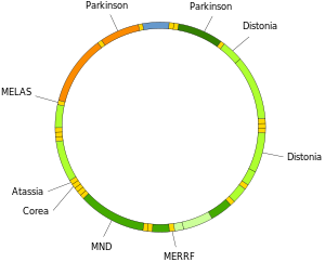
Human mitochondrial DNA with groups of protein-, rRNA- and tRNA-encoding genes.
 
The involvement of mitochondrial DNA in several human diseases.

Susceptibility

The concept that mtDNA is particularly susceptible to reactive oxygen species generated by the respiratory chain due to its proximity remains controversial. mtDNA does not accumulate any more oxidative base damage than nuclear DNA. It has been reported that at least some types of oxidative DNA damage are repaired more efficiently in mitochondria than they are in the nucleus. mtDNA is packaged with proteins which appear to be as protective as proteins of the nuclear chromatin. Moreover, mitochondria evolved a unique mechanism which maintains mtDNA integrity through degradation of excessively damaged genomes followed by replication of intact/repaired mtDNA. This mechanism is not present in the nucleus and is enabled by multiple copies of mtDNA present in mitochondria  The outcome of mutation in mtDNA may be an alteration in the coding instructions for some proteins, which may have an effect on organism metabolism and/or fitness.

Genetic illness

Mutations of mitochondrial DNA can lead to a number of illnesses including exercise intolerance and Kearns–Sayre syndrome (KSS), which causes a person to lose full function of heart, eye, and muscle movements. Some evidence suggests that they might be major contributors to the aging process and age-associated pathologies. Particularly in the context of disease, the proportion of mutant mtDNA molecules in a cell is termed heteroplasmy. The within-cell and between-cell distributions of heteroplasmy dictate the onset and severity of disease and are influenced by complicated stochastic processes within the cell and during development.

Mutations in mitochondrial tRNAs can be responsible for severe diseases like the MELAS and MERRF syndromes.

Mutations in nuclear genes that encode proteins that mitochondria use can also contribute to mitochondrial diseases. These diseases do not follow mitochondrial inheritance patterns, but instead follow Mendelian inheritance patterns.

Use in disease diagnosis

Recently a mutation in mtDNA has been used to help diagnose prostate cancer in patients with negative prostate biopsy.

Relationship with aging

Though the idea is controversial, some evidence suggests a link between aging and mitochondrial genome dysfunction. In essence, mutations in mtDNA upset a careful balance of reactive oxygen species (ROS) production and enzymatic ROS scavenging (by enzymes like superoxide dismutase, catalase, glutathione peroxidase and others). However, some mutations that increase ROS production (e.g., by reducing antioxidant defenses) in worms increase, rather than decrease, their longevity. Also, naked mole rats, rodents about the size of mice, live about eight times longer than mice despite having reduced, compared to mice, antioxidant defenses and increased oxidative damage to biomolecules. Once, there was thought to be a positive feedback loop at work (a 'Vicious Cycle'); as mitochondrial DNA accumulates genetic damage caused by free radicals, the mitochondria lose function and leak free radicals into the cytosol. A decrease in mitochondrial function reduces overall metabolic efficiency. However, this concept was conclusively disproved when it was demonstrated that mice, which were genetically altered to accumulate mtDNA mutations at accelerated rate do age prematurely, but their tissues do not produce more ROS as predicted by the 'Vicious Cycle' hypothesis. Supporting a link between longevity and mitochondrial DNA, some studies have found correlations between biochemical properties of the mitochondrial DNA and the longevity of species. Extensive research is being conducted to further investigate this link and methods to combat aging. Presently, gene therapy and nutraceutical supplementation are popular areas of ongoing research. Bjelakovic et al. analyzed the results of 78 studies between 1977 and 2012, involving a total of 296,707 participants, and concluded that antioxidant supplements do not reduce all-cause mortality nor extend lifespan, while some of them, such as beta carotene, vitamin E, and higher doses of vitamin A, may actually increase mortality.

Neurodegenerative diseases

Increased mtDNA damage is a feature of several neurodegenerative diseases.

The brains of individuals with Alzheimer’s disease have elevated levels of oxidative DNA damage in both nuclear DNA and mtDNA, but the mtDNA has approximately 10-fold higher levels than nuclear DNA. It has been proposed that aged mitochondria is the critical factor in the origin of neurodegeneration in Alzheimer’s disease.

In Huntington’s disease, mutant huntingtin protein causes mitochondria dysfunction involving inhibition of mitochondrial electron transport, higher levels of reactive oxygen species and increased oxidative stress. Mutant huntingtin protein promotes oxidative damage to mtDNA, as well as nuclear DNA, that may contribute to Huntington’s disease pathology.

The DNA oxidation product 8-oxoguanine (8-oxoG) is a well-established marker of oxidative DNA damage. In persons with amyotrophic lateral sclerosis (ALS), the enzymes that normally repair 8-oxoG DNA damages in the mtDNA of spinal motor neurons are impaired. Thus oxidative damage to mtDNA of motor neurons may be a significant factor in the etiology of ALS.

Correlation of the mtDNA base composition with animals lifespan

Animal species mtDNA base composition was retrieved from the MitoAge database and compared to their maximum life span from AnAge database.

Over the past decade, an Israeli research group led by Professor Vadim Fraifeld has shown that extraordinarily strong and significant correlations exist between the mtDNA base composition and animal species-specific maximum life spans. As demonstrated in their work, higher mtDNA guanine + cytosine content (GC%) strongly associates with longer maximum life spans across animal species. An additional astonishing observation is that the mtDNA GC% correlation with the maximum life spans is independent of the well-known correlation between animal species metabolic rate and maximum life spans. The mtDNA GC% and resting metabolic rate explain the differences in animal species maximum life spans in a multiplicative manner (i.e., species maximum life span = their mtDNA GC% * metabolic rate). To support the scientific community in carrying out comparative analyses between mtDNA features and longevity across animals, a dedicated database was built named MitoAge.

Relationship with non-B (non-canonical) DNA structures

Deletion breakpoints frequently occur within or near regions showing non-canonical (non-B) conformations, namely hairpins, cruciforms and cloverleaf-like elements. Moreover, there is data supporting the involvement of helix-distorting intrinsically curved regions and long G-tetrads in eliciting instability events. In addition, higher breakpoint densities were consistently observed within GC-skewed regions and in the close vicinity of the degenerate sequence motif YMMYMNNMMHM. Recently (2017) was found that all mitochodrial genomes sequenced so far contain many of inverted repeats necessary for cruciform DNA formation and these loci are particularly enriched in replication origin sites, D-loops and stem loops.

Use in identification

Unlike nuclear DNA, which is inherited from both parents and in which genes are rearranged in the process of recombination, there is usually no change in mtDNA from parent to offspring. Although mtDNA also recombines, it does so with copies of itself within the same mitochondrion. Because of this and because the mutation rate of animal mtDNA is higher than that of nuclear DNA, mtDNA is a powerful tool for tracking ancestry through females (matrilineage) and has been used in this role to track the ancestry of many species back hundreds of generations.

The rapid mutation rate (in animals) makes mtDNA useful for assessing genetic relationships of individuals or groups within a species and also for identifying and quantifying the phylogeny (evolutionary relationships; see phylogenetics) among different species. To do this, biologists determine and then compare the mtDNA sequences from different individuals or species. Data from the comparisons is used to construct a network of relationships among the sequences, which provides an estimate of the relationships among the individuals or species from which the mtDNAs were taken. mtDNA can be used to estimate the relationship between both closely related and distantly related species. Due to the high mutation rate of mtDNA in animals, the 3rd positions of the codons change relatively rapidly, and thus provide information about the genetic distances among closely related individuals or species. On the other hand, the substitution rate of mt-proteins is very low, thus amino acid changes accumulate slowly (with corresponding slow changes at 1st and 2nd codon positions) and thus they provide information about the genetic distances of distantly related species. Statistical models that treat substitution rates among codon positions separately, can thus be used to simultaneously estimate phylogenies that contain both closely and distantly related species
Mitochondrial DNA was admitted into evidence for the first time ever in a United States courtroom in 1996 during State of Tennessee v. Paul Ware.

In the 1998 United States court case of Commonwealth of Pennsylvania v. Patricia Lynne Rorrer, mitochondrial DNA was admitted into evidence in the State of Pennsylvania for the first time. The case was featured in episode 55 of season 5 of the true crime drama series Forensic Files (season 5).

Mitochondrial DNA was first admitted into evidence in California, United States, in the successful prosecution of David Westerfield for the 2002 kidnapping and murder of 7-year-old Danielle van Dam in San Diego: it was used for both human and dog identification. This was the first trial in the U.S. to admit canine DNA.

The remains of King Richard III were identified by comparing his mtDNA with that of two matrilineal descendants of his sister.

History

Mitochondrial DNA was discovered in the 1960s by Margit M. K. Nass and Sylvan Nass by electron microscopy as DNase-sensitive threads inside mitochondria, and by Ellen Haslbrunner, Hans Tuppy and Gottfried Schatz by biochemical assays on highly purified mitochondrial fractions.

Mitochondrial sequence databases

Several specialized databases have been founded to collect mitochondrial genome sequences and other information. Although most of them focus on sequence data, some of them include phylogenetic or functional information.
  • MitoSatPlant: Mitochondrial microsatellites database of viridiplantae.
  • MitoBreak: the mitochondrial DNA breakpoints database.
  • MitoFish and MitoAnnotator: a mitochondrial genome database of fish.
  • MitoZoa 2.0: a database for comparative and evolutionary analyses of mitochondrial genomes in Metazoa.
  • InterMitoBase: an annotated database and analysis platform of protein-protein interactions for human mitochondria.
  • Mitome: a database for comparative mitochondrial genomics in metazoan animals
  • MitoRes: a resource of nuclear-encoded mitochondrial genes and their products in metazoa

Mitochondrial mutation databases

Several specialized databases exist that report polymorphisms and mutations in the human mitochondrial DNA, together with the assessment of their pathogenicity.
  • MITOMAP: A compendium of polymorphisms and mutations in human mitochondrial DNA.
  • MitImpact: A collection of pre-computed pathogenicity predictions for all nucleotide changes that cause non-synonymous substitutions in human mitochondrial protein coding genes.

Color blindness

From Wikipedia, the free encyclopedia

Color blindness
Synonyms Colour blindness, color deficiency, impaired color vision
Ishihara 9.png
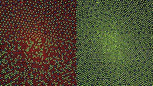
Example of an Ishihara color test plate. With properly configured computer displays, people with normal vision should see the number "74". Many people who are color blind see it as "21", and those with total color blindness may not see any numbers.
Specialty Ophthalmology
Symptoms Decreased ability to see colors
Duration Long term
Causes Genetic (inherited usually X-linked)
Diagnostic method Ishihara color test
Treatment Adjustments to teaching methods, mobile apps
Frequency Red-green: 8% males, 0.5% females (Northern European descent)

Color blindness, also known as color vision deficiency, is the decreased ability to see color or differences in color. Simple tasks such as selecting ripe fruit, choosing clothing, and reading traffic lights can be more challenging. Color blindness may also make some educational activities more difficult. However, problems are generally minor, and most people find that they can adapt. People with total color blindness (achromatopsia) may also have decreased visual acuity and be uncomfortable in bright environments.

The most common cause of color blindness is an inherited problem in the development of one or more of the three sets of color sensing cones in the eye. Males are more likely to be color blind than females, as the genes responsible for the most common forms of color blindness are on the X chromosome. As females have two X chromosomes, a defect in one is typically compensated for by the other, while males only have one X chromosome. Color blindness can also result from physical or chemical damage to the eye, optic nerve or parts of the brain. Diagnosis is typically with the Ishihara color test; however, a number of other testing methods also exist.

There is no cure for color blindness. Diagnosis may allow a person's teacher to change their method of teaching to accommodate the decreased ability to recognize colors. Special lenses may help people with red-green color blindness when under bright conditions. There are also mobile apps that can help people identify colors.

Red-green color blindness is the most common form, followed by blue-yellow color blindness and total color blindness. Red-green color blindness affects up to 8% of males and 0.5% of females of Northern European descent. The ability to see color also decreases in old age. Being color blind may make people ineligible for certain jobs in certain countries. This may include being a pilot, train driver and working in the armed forces. The effect of color blindness on artistic ability, however, is controversial. The ability to draw appears to be unchanged, and a number of famous artists are believed to have been color blind.

Signs and symptoms

Simulation of the normal (above) and dichromatic (below) perception of red and green apples
 
Horizontal traffic light in Halifax, Nova Scotia, Canada

In almost all cases, color blind people retain blue-yellow discrimination, and most color-blind individuals are anomalous trichromats rather than complete dichromats. In practice, this means that they often retain a limited discrimination along the red-green axis of color space, although their ability to separate colors in this dimension is reduced. Color blindness very rarely refers to complete monochromatism.

Dichromats often confuse red and green items. For example, they may find it difficult to distinguish a Braeburn apple from a Granny Smith or red from green of traffic lights without other clues—for example, shape or position. Dichromats tend to learn to use texture and shape clues and so may be able to penetrate camouflage that has been designed to deceive individuals with normal color vision.

Colors of traffic lights are confusing to some dichromats as there is insufficient apparent difference between the red/amber traffic lights and sodium street lamps; also, the green can be confused with a grubby white lamp. This is a risk on high-speed undulating roads where angular cues cannot be used. British Rail color lamp signals use more easily identifiable colors: The red is blood red, the amber is yellow and the green is a bluish color. Most British road traffic lights are mounted vertically on a black rectangle with a white border (forming a "sighting board") and so dichromats can more easily look for the position of the light within the rectangle—top, middle or bottom. In the eastern provinces of Canada horizontally mounted traffic lights are generally differentiated by shape to facilitate identification for those with color blindness. In the United States, this is not done by shape but by position, as the red light is always on the left if the light is horizontal, or on top if the light is vertical. However, a lone flashing light (e.g. red for stop, yellow for caution) is still problematic.

Types

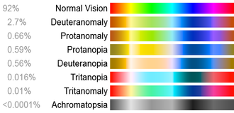
These color charts show how different colorblind people see compared to a person with normal color vision.

Types of color blindness and the terms used


Cone
system
Red Green Blue

N=normal
A=anomalous
N A N A N A
1 Normal vision . . . . . . Trichromat Normal
2 Protanomaly . . . . . . Anomalous Trichromat Partially color blind Red-green
3 Protanopia . . . . . . Dichromat Partially color blind Red-green
4 Deuteranomaly . . . . . . Anomalous Trichromat Partially color blind Red-green
5 Deuteranopia . . . . . . Dichromat Partially color blind Red-green
6 Tritanomaly . . . . . . Anomalous Trichromat Partially color blind Blue-yellow
7 Tritanopia . . . . . . Dichromat Partially color blind Blue-yellow
8 Achromatopsia . . . . . . Monochromat Totally color blind
9 Tetrachromat . . . . . .
10 . . . . . .

Based on clinical appearance, color blindness may be described as total or partial. Total color blindness is much less common than partial color blindness. There are two major types of color blindness: difficulty distinguishing between red and green, and difficulty distinguishing between blue and yellow.

Immunofluorescent imaging is a way to determine red-green color coding. Conventional color coding is difficult for individuals with red–green color blindness (protanopia or deuteranopia) to discriminate. Replacing red with magenta or green with turquoise improves visibility for such individuals.

The different kinds of inherited color blindness result from partial or complete loss of function of one or more of the three different cone systems. When one cone system is compromised, dichromacy results. The most frequent forms of human color blindness result from problems with either the middle (green) or long (red) wavelength sensitive cone systems, and make it hard to discriminate reds, yellows, and greens from one another. They are collectively referred to as "red-green color blindness", though the term is an over-simplification and is somewhat misleading. Other forms of color blindness are much more rare. They include problems in discriminating blues from greens and yellows from reds/pinks, and the rarest form of all, complete color blindness or monochromacy, where one cannot distinguish any color from grey, as in a black-and-white movie or photograph.
Protanopes, deuteranopes, and tritanopes are dichromats; that is, they can match any color they see with some mixture of just two primary colors (in contrast to those with normal sight (trichromats) who can distinguish three primary colors). Dichromats usually know they have a color vision problem, and it can affect their daily lives. Out of the male population, 2% have severe difficulties distinguishing between red, orange, yellow, and green. (Orange and yellow are different combinations of red and green light.) Colors in this range, which appear very different to a normal viewer, appear to a dichromat to be the same or a similar color. The terms protanopia, deuteranopia, and tritanopia come from Greek, and respectively mean "inability to see (anopia) with the first (prot-), second (deuter-), or third (trit-) [cone]".

Anomalous trichromacy is the least serious type of color deficiency. People with protanomaly, deuteranomaly, or tritanomaly are trichromats, but the color matches they make differ from the normal. They are called anomalous trichromats. In order to match a given spectral yellow light, protanomalous observers need more red light in a red/green mixture than a normal observer, and deuteranomalous observers need more green. From a practical standpoint though, many protanomalous and deuteranomalous people have very little difficulty carrying out tasks that require normal color vision. Some may not even be aware that their color perception is in any way different from normal.

Protanomaly and deuteranomaly can be diagnosed using an instrument called an anomaloscope, which mixes spectral red and green lights in variable proportions, for comparison with a fixed spectral yellow. If this is done in front of a large audience of males, as the proportion of red is increased from a low value, first a small proportion of the audience will declare a match, while most will see the mixed light as greenish; these are the deuteranomalous observers. Next, as more red is added the majority will say that a match has been achieved. Finally, as yet more red is added, the remaining, protanomalous, observers will declare a match at a point where normal observers will see the mixed light as definitely reddish.

Red-green color blindness

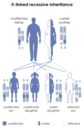
Protanopia, deuteranopia, protanomaly, and deuteranomaly are commonly inherited forms of red-green color blindness which affect a substantial portion of the human population. Those affected have difficulty with discriminating red and green hues due to the absence or mutation of the red or green retinal photoreceptors. It is sex-linked: genetic red-green color blindness affects males much more often than females, because the genes for the red and green color receptors are located on the X chromosome, of which males have only one and females have two. Females (XX) are red-green color blind only if both their X chromosomes are defective with a similar deficiency, whereas males (XY) are color blind if their single X chromosome is defective.

The gene for red-green color blindness is transmitted from a color blind male to all his daughters, who are usually heterozygote carriers and are thus unaffected. In turn, a carrier woman has a 50% chance of passing on a mutated X chromosome region to each of her male offspring. The sons of an affected male will not inherit the trait from him, since they receive his Y chromosome and not his (defective) X chromosome. Should an affected male have children with a carrier or colorblind woman, their daughters may be colorblind by inheriting an affected X chromosome from each parent.

Because one X chromosome is inactivated at random in each cell during a woman's development, deuteranomalous heterozygotes (i.e. female carriers of deuteranomaly) may be tetrachromats, because they will have the normal long wave (red) receptors, the normal medium wave (green) receptors, the abnormal medium wave (deuteranomalous) receptors and the normal autosomal short wave (blue) receptors in their retinas. The same applies to the carriers of protanomaly (who have two types of long wave receptors, normal medium wave receptors, and normal autosomal short wave receptors in their retinas). If, by rare chance, a woman is heterozygous for both protanomaly and deuteranomaly, she could be pentachromatic. This situation could arise if, for instance, she inherited the X chromosome with the abnormal long wave gene (but normal medium wave gene) from her mother who is a carrier of protanomaly, and her other X chromosome from a deuteranomalous father. Such a woman would have a normal and an abnormal long wave receptor, a normal and abnormal medium wave receptor, and a normal autosomal short wave receptor—5 different types of color receptors in all. The degree to which women who are carriers of either protanomaly or deuteranomaly are demonstrably tetrachromatic and require a mixture of four spectral lights to match an arbitrary light is very variable. In many cases it is almost unnoticeable, but in a minority the tetrachromacy is very pronounced. However, Jameson et al. have shown that with appropriate and sufficiently sensitive equipment it can be demonstrated that any female carrier of red-green color blindness (i.e. heterozygous protanomaly, or heterozygous deuteranomaly) is a tetrachromat to a greater or lesser extent.

Since deuteranomaly is by far the most common form of red-green blindness among men of northwestern European descent (with an incidence of 8%) it follows that the proporrtion of carriers (and of potential deuteranomalous tetrachromats) among the females of that genetic stock is 14.7% (i.e. 92% × 8% × 2), based on the Hardy–Weinberg principle.

Illustration of the distribution of cone cells in the fovea of an individual with normal color vision (left), and a color blind (protanopic) retina. The center of the fovea holds very few blue-sensitive cones.
  • Protanopia (1% of males): Lacking the red cones for long-wavelength sensitive retinal cones, those with this condition are unable to distinguish between colors in the green-yellow-red section of the spectrum. They have a neutral point at a cyan-like wavelength around 492 nm (see spectral color for comparison)—that is, they cannot discriminate light of this wavelength from white. For a protanope, the brightness of red, orange, and yellow are much reduced compared to normal. This dimming can be so pronounced that reds may be confused with black or dark gray, and red traffic lights may appear to be extinguished. They may learn to distinguish reds from yellows primarily on the basis of their apparent brightness or lightness, not on any perceptible hue difference. Violet, lavender, and purple are indistinguishable from various shades of blue because their reddish components are so dimmed as to be invisible. For example, pink flowers, reflecting both red light and blue light, may appear just blue to the protanope. A very few people have been found who have one normal eye and one protanopic eye. These unilateral dichromats report that with only their protanopic eye open, they see wavelengths shorter than neutral point as blue and those longer than it as yellow. This is a rare form of color blindness.
  • Deuteranopia (1% of males): Lacking the green cones for medium-wavelength cones, those affected are again unable to distinguish between colors in the green-yellow-red section of the spectrum. Their neutral point is at a slightly longer wavelength, 498 nm, a more greenish hue of cyan. A deuteranope suffers the same hue discrimination problems as protanopes, but without the abnormal dimming. Purple colors are not perceived as something opposite to spectral colors; all these appear similarly. This form of colorblindness is also known as Daltonism after John Dalton (his diagnosis was confirmed as deuteranopia in 1995, some 150 years after his death, by DNA analysis of his preserved eyeball). Equivalent terms for Daltonism in Romanic languages such as daltonismo (Spanish, Portuguese and Italian), daltonisme (French), daltonism (Romanian) are still used to describe color blindess in a broad sense or deuteranopia in a more restricted sense. Deuteranopic unilateral dichromats report that with only their deuteranopic eye open, they see wavelengths shorter than neutral point as blue and longer than it as yellow.
  • Protanomaly (1% of males, 0.01% of females): Having a mutated form of the long-wavelength (red) pigment, whose peak sensitivity is at a shorter wavelength than in the normal retina, protanomalous individuals are less sensitive to red light than normal. This means that they are less able to discriminate colors, and they do not see mixed lights as having the same colors as normal observers. They also suffer from a darkening of the red end of the spectrum. This causes reds to reduce in intensity to the point where they can be mistaken for black. Protanomaly is a fairly rare form of color blindness, making up about 1% of the male population. Both protanomaly and deuteranomaly are carried on the X chromosome.
  • Deuteranomaly (most common—6% of males, 0.4% of females): These individuals have a mutated form of the medium-wavelength (green) pigment. The medium-wavelength pigment is shifted towards the red end of the spectrum resulting in a reduction in sensitivity to the green area of the spectrum. Unlike in protanomaly, the intensity of colors is unchanged. The deuteranomalous person is considered "green weak". For example, in the evening, dark green cars appear to be black to deuteranomalous people. As with protanomates, deuteranomates are poor at discriminating small differences in hues in the red, orange, yellow, green region of the spectrum. They make errors in the naming of hues in this region because the hues appear somewhat shifted towards green. However, unlike protanomates, deuteranomalous people do not have the loss of "brightness" problem.

Blue-yellow color blindness

Those with tritanopia and tritanomaly have difficulty discriminating between bluish and greenish hues, as well as yellowish and reddish hues.

Color blindness involving the inactivation of the short-wavelength sensitive cone system (whose absorption spectrum peaks in the bluish-violet) is called tritanopia or, loosely, blue-yellow color blindness. The tritanope's neutral point occurs near a yellowish 570 nm; green is perceived at shorter wavelengths and red at longer wavelengths. Mutation of the short-wavelength sensitive cones is called tritanomaly. Tritanopia is equally distributed among males and females. Jeremy H. Nathans (with the Howard Hughes Medical Institute) demonstrated that the gene coding for the blue receptor lies on chromosome 7, which is shared equally by males and females. Therefore, it is not sex-linked. This gene does not have any neighbor whose DNA sequence is similar. Blue color blindness is caused by a simple mutation in this gene.
  • Tritanopia (less than 1% of males and females): Lacking the short-wavelength cones, those affected see short-wavelength colors (blue, indigo and a spectral violet) greenish and drastically dimmed, some of these colors even as black. Yellow is indistinguishable from pink, and purple colors are perceived as various shades of red. This form of color blindness is not sex-linked.
  • Tritanomaly (equally rare for males and females [0.01% for both]): Having a mutated form of the short-wavelength (blue) pigment. The short-wavelength pigment is shifted towards the green area of the spectrum. This is the rarest form of anomalous trichromacy color blindness. Unlike the other anomalous trichromacy color deficiencies, the mutation for this color blindness is carried on chromosome 7. Therefore, it is equally prevalent in both male and female populations. The OMIM gene code for this mutation is 304000 "Colorblindness, Partial Tritanomaly".

Total color blindness

Total color blindness is defined as the inability to see color. Although the term may refer to acquired disorders such as cerebral achromatopsia also known as color agnosia, it typically refers to congenital color vision disorders (i.e. more frequently rod monochromacy and less frequently cone monochromacy).

In cerebral achromatopsia, a person cannot perceive colors even though the eyes are capable of distinguishing them. Some sources do not consider these to be true color blindness, because the failure is of perception, not of vision. They are forms of visual agnosia.

Monochromacy is the condition of possessing only a single channel for conveying information about color. Monochromats possess a complete inability to distinguish any colors and perceive only variations in brightness. It occurs in two primary forms:
  1. Rod monochromacy, frequently called achromatopsia, where the retina contains no cone cells, so that in addition to the absence of color discrimination, vision in lights of normal intensity is difficult. While normally rare, achromatopsia is very common on the island of Pingelap, a part of the Pohnpei state, Federated States of Micronesia, where it is called maskun: about 10% of the population there has it, and 30% are unaffected carriers. The island was devastated by a storm in the 18th century (an example of a genetic bottleneck) and one of the few male survivors carried a gene for achromatopsia. The population grew to several thousand before foreign troops introduced diseases to the island in the 1940s.
  2. Cone monochromacy is the condition of having both rods and cones, but only a single kind of cone. A cone monochromat can have good pattern vision at normal daylight levels, but will not be able to distinguish hues. Blue cone monochromacy (X chromosome) is caused by lack of functionality of L and M cones (red and green). It is encoded at the same place as red-green color blindness on the X chromosome. Peak spectral sensitivities are in the blue region of the visible spectrum (near 440 nm). People with this condition generally show nystagmus ("jiggling eyes"), photophobia (light sensitivity), reduced visual acuity, and myopia (nearsightedness). Visual acuity usually falls to the 20/50 to 20/400 range.

Causes

Color vision deficiencies can be classified as acquired or inherited.
  • Acquired: Diseases, drugs (e.g., Plaquenil), and chemicals may cause color blindness.
  • Inherited: There are three types of inherited or congenital color vision deficiencies: monochromacy, dichromacy, and anomalous trichromacy.
    • Monochromacy, also known as "total color blindness", is the lack of ability to distinguish colors (and thus the person views everything as if it were on a black and white television); caused by cone defect or absence. Monochromacy occurs when two or all three of the cone pigments are missing and color and lightness vision is reduced to one dimension.
      • Rod monochromacy (achromatopsia) is an exceedingly rare, nonprogressive inability to distinguish any colors as a result of absent or nonfunctioning retinal cones. It is associated with light sensitivity (photophobia), involuntary eye oscillations (nystagmus), and poor vision.
      • Cone monochromacy is a rare total color blindness that is accompanied by relatively normal vision, electroretinogram, and electrooculogram. Cone monochromacy can also be a result of having more than one type of dichromatic color blindness. People who have, for instance, both protanopia and tritanopia are considered to have cone monochromacy. Since cone monochromacy is the lack of/damage of more than one cone in retinal environment, having two types of dichromacy would be an equivalent.
    • Dichromacy is hereditary. Protanopia and deuteranopia are hereditary and sex-linked, affecting predominantly males.
      • Protanopia is caused by the complete absence of red retinal photoreceptors. Protans have difficulties distinguishing between blue and green colors and also between red and green colors. It is a form of dichromatism in which the subject can only perceive light wavelengths from 400 nm to 650 nm, instead of the usual 700 nm. Pure reds cannot be seen, instead appearing black; purple colors cannot be distinguished from blues; more orange-tinted reds may appear as dim yellows, and all orange-yellow-green shades of too long a wavelength to stimulate the blue receptors appear as a similar yellow hue. It is present in 1% of males.
      • Deuteranopia affects hue discrimination in a similar way to protanopia, but without the dimming effect. Again, it is found in about 1% of the male population.
      • Tritanopia is a very rare color vision disturbance in which only the red and the green cone pigments are present, with a total absence of blue retinal receptors. Blues appear greenish, yellows and oranges appear pinkish, and purple colors appear deep red. It is related to chromosome 7; thus unlike protanopia and deuteranopia, tritanopia and tritanomaly are not sex-linked traits and can be acquired rather than inherited and can be reversed in some cases.
    • Anomalous trichromacy is a common type of inherited color vision deficiency, occurring when one of the three cone pigments is altered in its spectral sensitivity.
      • Protanomaly is a mild color vision defect in which an altered spectral sensitivity of red retinal receptors (closer to green receptor response) results in poor red-green hue discrimination. It is hereditary, sex-linked, and present in 1% of males. In contrast to other defects, in this case the L-cone is present but malfunctioning, whereas in protanopia the L-cone is completely missing.
      • Deuteranomaly, caused by a similar shift in the green retinal receptors, is by far the most common type of color vision deficiency, mildly affecting red-green hue discrimination in 5% of European males. It is hereditary and sex-linked. In contrast to deuteranopia, the green-sensitive cones are not missing but malfunctioning.
      • Tritanomaly is a rare, hereditary color vision deficiency affecting blue-green and yellow-red/pink hue discrimination. It is related to chromosome 7. In contrast to tritanopia, the S-cone is malfunctioning but not missing.

Genetics

X-linked recessive inheritance

Color blindness is typically an inherited genetic disorder. It is most commonly inherited from mutations on the X chromosome, but the mapping of the human genome has shown there are many causative mutations—mutations capable of causing color blindness originate from at least 19 different chromosomes and 56 different genes (as shown online at the Online Mendelian Inheritance in Man (OMIM)).

Two of the most common inherited forms of color blindness are protanomaly (and, more rarely, protanopia–the two together often known as "protans") and deuteranomaly (or, more rarely, deuteranopia—the two together often referred to as "deutans"). Both "protans" and "deutans" (of which the deutans are by far the most common) are known as "red-green color-blind". They comprise about 8% of human males and 0.6% of females of Northern European ancestry.

Some of the inherited diseases known to cause color blindness are:
Inherited color blindness can be congenital (from birth), or it can commence in childhood or adulthood. Depending on the mutation, it can be stationary, that is, remain the same throughout a person's lifetime, or progressive. As progressive phenotypes involve deterioration of the retina and other parts of the eye, certain forms of color blindness can progress to legal blindness, i.e. an acuity of 6/60 (20/200) or worse, and often leave a person with complete blindness.

Color blindness always pertains to the cone photoreceptors in retinas, as it is the cones that detect the color frequencies of light.

About 8% of males, and 0.6% of females, are red-green color blind in some way or another, whether it is one color, a color combination, or another mutation. Males are at a greater risk of inheriting an X-linked mutation because males only have one X chromosome (XY, with the Y chromosome carrying altogether different genes from the X chromosome), and females have two (XX); if a woman inherits a normal X chromosome in addition to the one that carries the mutation, she will not display the mutation. Men do not have a second X chromosome to override the chromosome that carries the mutation. If 8% of variants of a given gene are defective, the probability of a single copy being defective is 8%, but the probability that two copies are both defective is 0.082, i.e. 0.64%.

Other causes

Other causes of color blindness include brain or retinal damage caused by shaken baby syndrome, accidents and other traumas which produce swelling of the brain in the occipital lobe, and damage to the retina caused by exposure to ultraviolet light (wavelengths 10 to 300 nm). Damage often presents itself later in life.

Color blindness may also present itself in the range of degenerative diseases of the eye, such as age-related macular degeneration, and as part of the retinal damage caused by diabetes. Vitamin A deficiency may also cause color blindness.

Some subtle forms of color blindness may be associated with chronic solvent-induced encephalopathy (CSE), caused by long-time exposure to solvent vapors.

Red–green color blindness can be caused by ethambutol, a drug used in the treatment of tuberculosis.

Mechanism

The typical human retina contains two kinds of light cells: the rod cells (active in low light) and the cone cells (active in normal daylight). Normally, there are three kinds of cone cells, each containing a different pigment, which are activated when the pigments absorb light. The spectral sensitivities of the cones differ; one is most sensitive to short wavelengths, one to medium wavelengths, and the third to medium-to-long wavelengths within the visible spectrum, with their peak sensitivities in the blue, green, and yellow-green regions of the spectrum, respectively. The absorption spectra of the three systems overlap, and combine to cover the visible spectrum. These receptors are known as short (S), medium (M), and long (L) wavelength cones, but are also often referred to as blue, green, and red cones, although this terminology is inaccurate.

The receptors are each responsive to a wide range of wavelengths. For example, the long wavelength "red" receptor has its peak sensitivity in the yellow-green, some way from the red end (longest wavelength) of the visible spectrum. The sensitivity of normal color vision actually depends on the overlap between the absorption ranges of the three systems: different colors are recognized when the different types of cone are stimulated to different degrees. Red light, for example, stimulates the long wavelength cones much more than either of the others, and reducing the wavelength causes the other two cone systems to be increasingly stimulated, causing a gradual change in hue.

Many of the genes involved in color vision are on the X chromosome, making color blindness much more common in males than in females because males only have one X chromosome, while females have two. Because this is an X-linked trait, an estimated 2–3% of women have a 4th color cone and can be considered tetrachromats. One such woman has been reported to be a true or functional tetrachromat, as she can discriminate colors most other people can't.

Diagnosis

An Ishihara test image as seen by subjects with normal color vision and by those with a variety of color deficiencies

The Ishihara color test, which consists of a series of pictures of colored spots, is the test most often used to diagnose red-green color deficiencies. A figure (usually one or more Arabic digits) is embedded in the picture as a number of spots in a slightly different color, and can be seen with normal color vision, but not with a particular color defect. The full set of tests has a variety of figure/background color combinations, and enable diagnosis of which particular visual defect is present. The anomaloscope, described above, is also used in diagnosing anomalous trichromacy.
Position yourself about 75cm from your monitor so that the colour test image you are looking at is at eye level, read the description of the image and see what you can see!! It is not necessary in all cases to use the entire set of images. In a large scale examination the test can be simplified to six tests; test, one of tests 2 or 3, one of tests 4, 5, 6, or 7, one of tests 8 or 9, one of tests 10, 11, 12, or 13 and one of tests 14 or 15.
Because the Ishihara color test contains only numerals, it may not be useful in diagnosing young children, who have not yet learned to use numbers. In the interest of identifying these problems early on in life, alternative color vision tests were developed using only symbols (square, circle, car).

Besides the Ishihara color test, the US Navy and US Army also allow testing with the Farnsworth Lantern Test. This test allows 30% of color deficient individuals, whose deficiency is not too severe, to pass.

Another test used by clinicians to measure chromatic discrimination is the Farnsworth-Munsell 100 hue test. The patient is asked to arrange a set of colored caps or chips to form a gradual transition of color between two anchor caps.

The HRR color test (developed by Hardy, Rand, and Rittler) is a red-green color test that, unlike the Ishihara, also has plates for the detection of the tritan defects.

Most clinical tests are designed to be fast, simple, and effective at identifying broad categories of color blindness. In academic studies of color blindness, on the other hand, there is more interest in developing flexible tests to collect thorough datasets, identify copunctal points, and measure just noticeable differences.

Management

There is generally no treatment to cure color deficiencies. ″The American Optometric Association reports a contact lens on one eye can increase the ability to differentiate between colors, though nothing can make you truly see the deficient color.″

Lenses

Optometrists can supply colored spectacle lenses or a single red-tint contact lens to wear on the non-dominant eye, but although this may improve discrimination of some colors, it can make other colors more difficult to distinguish. A 1981 review of various studies to evaluate the effect of the X-chrom contact lens concluded that, while the lens may allow the wearer to achieve a better score on certain color vision tests, it did not correct color vision in the natural environment. A case history using the X-Chrom lens for a rod monochromat is reported and an X-Chrom manual is online.

Lenses that filter certain wavelengths of light can allow people with a cone anomaly, but not dichromacy, to see better separation of colors, especially those with classic "red/green" color blindness. They work by notching out wavelengths that strongly stimulate both red and green cones in a deuter- or protanomalous person, improving the distinction between the two cones' signals. As of 2013, sunglasses that notch out color wavelengths are available commercially.

Apps

Many mobile applications have been developed to help colorblind people to view colors in a better way. Many applications launch a simulation of colorblindness to allow people with normal vision to understand how people with color blindness see the world.

The GNOME desktop environment provides colorblind accessibility using the gnome-mag and the libcolorblind software. Using a gnome applet, the user may switch a color filter on and off, choosing from a set of possible color transformations that will displace the colors in order to disambiguate them. The software enables, for instance, a colorblind person to see the numbers in the Ishihara test.

Epidemiology

Rates of color blindness

Males Females
Dichromacy 2.4% 0.03%
Protanopia (red deficient: L cone absent) 1.3% 0.02%
Deuteranopia (green deficient: M cone absent) 1.2% 0.01%
Tritanopia (blue deficient: S cone absent) 0.001% 0.03%
Anomalous trichromacy 6.3% 0.37%
Protanomaly (red deficient: L cone defect) 1.3% 0.02%
Deuteranomaly (green deficient: M cone defect) 5.0% 0.35%
Tritanomaly (blue deficient: S cone defect) 0.0001% 0.0001%


Color blindness affects a large number of individuals, with protanopia and deuteranopia being the most common types. In individuals with Northern European ancestry, as many as 8 percent of men and 0.4 percent of women experience congenital color deficiency.

The number affected varies among groups. Isolated communities with a restricted gene pool sometimes produce high proportions of color blindness, including the less usual types. Examples include rural Finland, Hungary, and some of the Scottish islands. In the United States, about 7 percent of the male population—or about 10.5 million men—and 0.4 percent of the female population either cannot distinguish red from green, or see red and green differently from how others do (Howard Hughes Medical Institute, 2006). More than 95 percent of all variations in human color vision involve the red and green receptors in male eyes. It is very rare for males or females to be "blind" to the blue end of the spectrum.

Prevalence of red–green color blindness among males
Population Number
studied
%
Arabs (Druzes) 337 10.0
Aboriginal Australians 4,455 1.9
Belgians 9,540 7.4
Bosnians 4,836 6.2
Britons 16,180 6.6
Chinese 1,164 6.9
DR Congolese 929 1.7
Dutch 3,168 8.0
Eskimo 297 2.5
Fijians 608 0.8
French 1,243 8.6
Germans 7,861 7.7
Hutu 1,000 2.9
Indians (Andhra Pradesh) 292 7.5
Iranians 16,180 6.6
Japanese 259,000 4.0
Mexicans 571 2.3
Navajo 571 2.3
Norwegians 9,047 9.0
Russians 1,343 9.2
Scots 463 7.8
Swiss 2,000 8.0
Tibetans 241 5.0
Tswana 407 2.0
Tutsi 1,000 2.5
Serbs 4,750 7.4

History

An 1895 illustration of normal vision and various kinds of color blindness

The first scientific paper on the subject of color blindness, Extraordinary facts relating to the vision of colours, was published by the English chemist John Dalton in 1798 after the realization of his own color blindness. Because of Dalton's work, the general condition has been called daltonism, although in English this term is now used only for deuteranopia.

Society and culture

Design implications

snippet of colored cells in a table (foreground), surrounded in background showing how the image appears in color-blindness simulations.
Testing the colors of a web chart, (center), to ensure that no information is lost to the various forms of color blindness.

Color codes present particular problems for those with color deficiencies as they are often difficult or impossible for them to perceive.

Good graphic design avoids using color coding or using color contrasts alone to express information; this not only helps color blind people, but also aids understanding by normally sighted people by providing them with multiple reinforcing cues.

Designers need to take into account that color-blindness is highly sensitive to differences in material. For example, a red-green colorblind person who is incapable of distinguishing colors on a map printed on paper may have no such difficulty when viewing the map on a computer screen or television. In addition, some color blind people find it easier to distinguish problem colors on artificial materials, such as plastic or in acrylic paints, than on natural materials, such as paper or wood. Third, for some color blind people, color can only be distinguished if there is a sufficient "mass" of color: thin lines might appear black, while a thicker line of the same color can be perceived as having color.

Designers should also note that red-blue and yellow-blue color combinations are generally safe. So instead of the ever-popular "red means bad and green means good" system, using these combinations can lead to a much higher ability to use color coding effectively. This will still cause problems for those with monochromatic color blindness, but it is still something worth considering.

When the need to process visual information as rapidly as possible arises, for example in an emergency situation, the visual system may operate only in shades of gray, with the extra information load in adding color being dropped. This is an important possibility to consider when designing, for example, emergency brake handles or emergency phones.

Occupations

Color blindness may make it difficult or impossible for a person to engage in certain occupations. Persons with color blindness may be legally or practically barred from occupations in which color perception is an essential part of the job (e.g., mixing paint colors), or in which color perception is important for safety (e.g., operating vehicles in response to color-coded signals). This occupational safety principle originates from the Lagerlunda train crash of 1875 in Sweden. Following the crash, Professor Alarik Frithiof Holmgren, a physiologist, investigated and concluded that the color blindness of the engineer (who had died) had caused the crash. Professor Holmgren then created the first test using different-colored skeins to exclude people from jobs in the transportation industry on the basis of color blindness. However, there is a claim that there is no firm evidence that color deficiency did cause the collision, or that it might have not been the sole cause.

Color vision is important for occupations using telephone or computer networking cabling, as the individual wires inside the cables are color-coded using green, orange, brown, blue and white colors. Electronic wiring, transformers, resistors, and capacitors are color-coded as well, using black, brown, red, orange, yellow, green, blue, violet, gray, white, silver, gold.

Driving

Some countries have refused to grant driving licenses to individuals with color blindness. In Romania, there is an ongoing campaign to remove the legal restrictions that prohibit colorblind citizens from getting drivers' licenses.

The usual justification for such restrictions is that drivers of motor vehicles must be able to recognize color-coded signals, such as traffic lights or warning lights.

Piloting aircraft

While many aspects of aviation depend on color coding, only a few of them are critical enough to be interfered with by some milder types of color blindness. Some examples include color-gun signaling of aircraft that have lost radio communication, color-coded glide-path indications on runways, and the like. Some jurisdictions restrict the issuance of pilot credentials to persons who suffer from color blindness for this reason. Restrictions may be partial, allowing color-blind persons to obtain certification but with restrictions, or total, in which case color-blind persons are not permitted to obtain piloting credentials at all.

In the United States, the Federal Aviation Administration requires that pilots be tested for normal color vision as part of their medical clearance in order to obtain the required medical certificate, a prerequisite to obtaining a pilot's certification. If testing reveals color blindness, the applicant may be issued a license with restrictions, such as no night flying and no flying by color signals—such a restriction effectively prevents a pilot from holding certain flying occupations, such as that of an airline pilot, although commercial pilot certification is still possible, and there are a few flying occupations that do not require night flight and thus are still available to those with restrictions due to color blindness (e.g., agricultural aviation). The government allows several types of tests, including medical standard tests (e.g., the Ishihara, Dvorine, and others) and specialized tests oriented specifically to the needs of aviation. If an applicant fails the standard tests, they will receive a restriction on their medical certificate that states: "Not valid for night flying or by color signal control". They may apply to the FAA to take a specialized test, administered by the FAA. Typically, this test is the "color vision light gun test". For this test an FAA inspector will meet the pilot at an airport with an operating control tower. The color signal light gun will be shone at the pilot from the tower, and they must identify the color. If they pass they may be issued a waiver, which states that the color vision test is no longer required during medical examinations. They will then receive a new medical certificate with the restriction removed. This was once a Statement of Demonstrated Ability (SODA), but the SODA was dropped, and converted to a simple waiver (letter) early in the 2000s.

Research published in 2009 carried out by the City University of London's Applied Vision Research Centre, sponsored by the UK's Civil Aviation Authority and the U.S. Federal Aviation Administration, has established a more accurate assessment of color deficiencies in pilot applicants' red/green and yellow-blue color range which could lead to a 35% reduction in the number of prospective pilots who fail to meet the minimum medical threshold.

Art

Inability to distinguish color does not necessarily preclude the ability to become a celebrated artist. The 20th century expressionist painter Clifton Pugh, three-time winner of Australia's Archibald Prize, on biographical, gene inheritance and other grounds has been identified as a protanope. 19th century French artist Charles Méryon became successful by concentrating on etching rather than painting after he was diagnosed as having a red-green deficiency.

Rights of the color blind

Brazil

A Brazilian court ruled that people with color blindness are protected by the Inter-American Convention on the Elimination of All Forms of Discrimination against Person with Disabilities.

At trial, it was decided that the carriers of color blindness have a right of access to wider knowledge, or the full enjoyment of their human condition.

United States

In the United States, under federal anti-discrimination laws such as the Americans with Disabilities Act, color vision deficiencies have not been found to constitute a disability that triggers protection from workplace discrimination.

A famous traffic light on Tipperary Hill in Syracuse, New York, is upside-down due to the sentiments of its Irish American community, but has been criticized due to the potential hazard it poses for color-blind persons.

Research

Some tentative evidence finds that color blind people are better at penetrating certain color camouflages. Such findings may give an evolutionary reason for the high rate of red–green color blindness. There is also a study suggesting that people with some types of color blindness can distinguish colors that people with normal color vision are not able to distinguish. In World War II, color blind observers were used to penetrate camouflage.

In September 2009, the journal Nature reported that researchers at the University of Washington and University of Florida were able to give trichromatic vision to squirrel monkeys, which normally have only dichromatic vision, using gene therapy.

In 2003, a cybernetic device called eyeborg was developed to allow the wearer to hear sounds representing different colors. Achromatopsic artist Neil Harbisson was the first to use such a device in early 2004; the eyeborg allowed him to start painting in color by memorizing the sound corresponding to each color. In 2012, at a TED Conference, Harbisson explained how he could now perceive colors outside the ability of human vision.

Distance education

From Wikipedia, the free encyclopedia https://en.wikipedia.org/wiki/Distance_...