Search This Blog

Thursday, March 7, 2019

Sensory nervous system

From Wikipedia, the free encyclopedia

Sensory nervous system
Gray722-svg.svg
Typical sensory system: the visual system, illustrated by the classic Gray's FIG. 722– This scheme shows the flow of information from the eyes to the central connections of the optic nerves and optic tracts, to the visual cortex. Area V1 is the region of the brain which is engaged in vision.
Details
Identifiers
Latinorgana sensuum
MeSHD012679
TAA15.0.00.000
FMA75259
Anatomical terminology

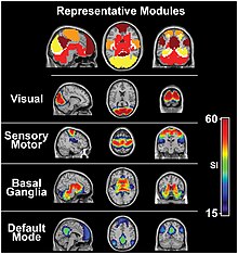
The visual system and the somatosensory system are active even during resting state fMRI
 
Activation and response in the sensory nervous system
 
The sensory nervous system is a part of the nervous system responsible for processing sensory information. A sensory system consists of sensory neurons (including the sensory receptor cells), neural pathways, and parts of the brain involved in sensory perception. Commonly recognized sensory systems are those for vision, hearing, touch, taste, smell, and balance. In short, senses are transducers from the physical world to the realm of the mind where we interpret the information, creating our perception of the world around us.
Organisms need information to solve at least three kinds of problems: (a) to maintain an appropriate environment, i.e., homeostasis; (b) to time activities (e.g., seasonal changes in behavior) or synchronize activities with those of conspecifics; and (c) to locate and respond to resources or threats (e.g., by moving towards resources or evading or attacking threats). Organisms also need to transmit information in order to influence another's behavior: to identify themselves, warn conspecifics of danger, coordinate activities, or deceive.
The receptive field is the area of the body or environment to which a receptor organ and receptor cells respond. For instance, the part of the world an eye can see, is its receptive field; the light that each rod or cone can see, is its receptive field. Receptive fields have been identified for the visual system, auditory system and somatosensory system.

Stimulus

Sensory systems code for four aspects of a stimulus; type (modality), intensity, location, and duration. Arrival time of a sound pulse and phase differences of continuous sound are used for sound localization. Certain receptors are sensitive to certain types of stimuli (for example, different mechanoreceptors respond best to different kinds of touch stimuli, like sharp or blunt objects). Receptors send impulses in certain patterns to send information about the intensity of a stimulus (for example, how loud a sound is). The location of the receptor that is stimulated gives the brain information about the location of the stimulus (for example, stimulating a mechanoreceptor in a finger will send information to the brain about that finger). The duration of the stimulus (how long it lasts) is conveyed by firing patterns of receptors. These impulses are transmitted to the brain through afferent neurons.

Senses and receptors

While debate exists among neurologists as to the specific number of senses due to differing definitions of what constitutes a sense, Gautama Buddha and Aristotle classified five ‘traditional’ human senses which have become universally accepted: touch, taste, smell, sight, and hearing. Other senses that have been well-accepted in most mammals, including humans, include nociception, equilibrioception, kinaesthesia, and thermoception. Furthermore, some nonhuman animals have been shown to possess alternate senses, including magnetoception and electroreception.

Receptors

The initialization of sensation stems from the response of a specific receptor to a physical stimulus. The receptors which react to the stimulus and initiate the process of sensation are commonly characterized in four distinct categories: chemoreceptors, photoreceptors, mechanoreceptors, and thermoreceptors. All receptors receive distinct physical stimuli and transduce the signal into an electrical action potential. This action potential then travels along afferent neurons to specific brain regions where it is processed and interpreted.

Chemoreceptors

Chemoreceptors, or chemosensors, detect certain chemical stimuli and transduce that signal into an electrical action potential. The two primary types of chemoreceptors are:

Photoreceptors

Photoreceptors are capable of phototransduction, a process which converts light (electromagnetic radiation) into, among other types of energy, a membrane potential. The three primary types of photoreceptors are: Cones are photoreceptors which respond significantly to color. In humans the three different types of cones correspond with a primary response to short wavelength (blue), medium wavelength (green), and long wavelength (yellow/red). Rods are photoreceptors which are very sensitive to the intensity of light, allowing for vision in dim lighting. The concentrations and ratio of rods to cones is strongly correlated with whether an animal is diurnal or nocturnal. In humans rods outnumber cones by approximately 20:1, while in nocturnal animals, such as the tawny owl, the ratio is closer to 1000:1. Ganglion Cells reside in the adrenal medulla and retina where they are involved in the sympathetic response. Of the ~1.3 million ganglion cells present in the retina, 1-2% are believed to be photosensitive ganglia. These photosensitive ganglia play a role in conscious vision for some animals, and are believed to do the same in humans.

Mechanoreceptors

Mechanoreceptors are sensory receptors which respond to mechanical forces, such as pressure or distortion. While mechanoreceptors are present in hair cells and play an integral role in the vestibular and auditory systems, the majority of mechanoreceptors are cutaneous and are grouped into four categories:
  • Slowly adapting type 1 receptors have small receptive fields and respond to static stimulation. These receptors are primarily used in the sensations of form and roughness.
  • Slowly adapting type 2 receptors have large receptive fields and respond to stretch. Similarly to type 1, they produce sustained responses to a continued stimuli.
  • Rapidly adapting receptors have small receptive fields and underlie the perception of slip.
  • Pacinian receptors have large receptive fields and are the predominant receptors for high-frequency vibration.

Thermoreceptors

Thermoreceptors are sensory receptors which respond to varying temperatures. While the mechanisms through which these receptors operate is unclear, recent discoveries have shown that mammals have at least two distinct types of thermoreceptors:

Nociceptors

Nociceptors respond to potentially damaging stimuli by sending signals to the spinal cord and brain. This process, called nociception, usually causes the perception of pain. They are found in internal organs, as well as on the surface of the body. Nociceptors detect different kinds of damaging stimuli or actual damage. Those that only respond when tissues are damaged are known as "sleeping" or "silent" nociceptors.
  • Thermal nociceptors are activated by noxious heat or cold at various temperatures.
  • Mechanical nociceptors respond to excess pressure or mechanical deformation.
  • Chemical nociceptors respond to a wide variety of chemicals, some of which are signs of tissue damage. They are involved in the detection of some spices in food.

Sensory cortex

All stimuli received by the receptors listed above are transduced to an action potential, which is carried along one or more afferent neurons towards a specific area of the brain. While the term sensory cortex is often used informally to refer to the somatosensory cortex, the term more accurately refers to the multiple areas of the brain at which senses are received to be processed. For the five traditional senses in humans, this includes the primary and secondary cortices of the different senses: the somatosensory cortex, the visual cortex, the auditory cortex, the primary olfactory cortex, and the gustatory cortex. Other modalities have corresponding sensory cortex areas as well, including the vestibular cortex for the sense of balance.

Somatosensory cortex

Located in the parietal lobe, the primary somatosensory cortex is the primary receptive area for the sense of touch and proprioception in the somatosensory system. This cortex is further divided into Brodmann areas 1, 2, and 3. Brodmann area 3 is considered the primary processing center of the somatosensory cortex as it receives significantly more input from the thalamus, has neurons highly responsive to somatosensory stimuli, and can evoke somatic sensations through electrical stimulation. Areas 1 and 2 receive most of their input from area 3. There are also pathways for proprioception (via the cerebellum), and motor control (via Brodmann area 4).

The human eye is the first element of a sensory system: in this case, vision, for the visual system.

Visual cortex

The visual cortex refers to the primary visual cortex, labeled V1 or Brodmann area 17, as well as the extrastriate visual cortical areas V2-V5. Located in the occipital lobe, V1 acts as the primary relay station for visual input, transmitting information to two primary pathways labeled the dorsal and ventral streams. The dorsal stream includes areas V2 and V5, and is used in interpreting visual ‘where’ and ‘how.’ The ventral stream includes areas V2 and V4, and is used in interpreting ‘what.’ Increases in Task-negative activity are observed in the ventral attention network, after abrupt changes in sensory stimuli, at the onset and offset of task blocks, and at the end of a completed trial.

Auditory cortex

Located in the temporal lobe, the auditory cortex is the primary receptive area for sound information. The auditory cortex is composed of Brodmann areas 41 and 42, also known as the anterior transverse temporal area 41 and the posterior transverse temporal area 42, respectively. Both areas act similarly and are integral in receiving and processing the signals transmitted from auditory receptors

Primary olfactory cortex

Located in the temporal lobe, the primary olfactory cortex is the primary receptive area for olfaction, or smell. Unique to the olfactory and gustatory systems, at least in mammals, is the implementation of both peripheral and central mechanisms of action. The peripheral mechanisms involve olfactory receptor neurons which transduce a chemical signal along the olfactory nerve, which terminates in the olfactory bulb. The chemo-receptors involved in olfactory nervous cascade involve using G-protein receptors to send their chemical signals down said cascade. The central mechanisms include the convergence of olfactory nerve axons into glomeruli in the olfactory bulb, where the signal is then transmitted to the anterior olfactory nucleus, the piriform cortex, the medial amygdala, and the entorhinal cortex, all of which make up the primary olfactory cortex.

In contrast to vision and hearing, the olfactory bulbs are not cross-hemispheric; the right bulb connects to the right hemisphere and the left bulb connects to the left hemisphere. 

Gustatory cortex

The gustatory cortex is the primary receptive area for taste. The word taste is used in a technical sense to refer specifically to sensations coming from taste buds on the tongue. The five qualities of taste detected by the tongue include sourness, bitterness, sweetness, saltiness, and the protein taste quality, called umami. In contrast, the term flavor refers to the experience generated through integration of taste with smell and tactile information. The gustatory cortex consists of two primary structures: the anterior insula, located on the insular lobe, and the frontal operculum, located on the frontal lobe. Similarly to the olfactory cortex, the gustatory pathway operates through both peripheral and central mechanisms. Peripheral taste receptors, located on the tongue, soft palate, pharynx, and esophagus, transmit the received signal to primary sensory axons, where the signal is projected to the nucleus of the solitary tract in the medulla, or the gustatory nucleus of the solitary tract complex. The signal is then transmitted to the thalamus, which in turn projects the signal to several regions of the neocortex, including the gustatory cortex.

The neural processing of taste is affected at nearly every stage of processing by concurrent somatosensory information from the tongue, that is, mouthfeel. Scent, in contrast, is not combined with taste to create flavor until higher cortical processing regions, such as the insula and orbitofrontal cortex.

Human sensory system

The human sensory system consists of the following subsystems:

Vestibular system

From Wikipedia, the free encyclopedia

The vestibular system, in most mammals, is the sensory system that provides the leading contribution to the sense of balance and spatial orientation for the purpose of coordinating movement with balance. Together with the cochlea, a part of the auditory system, it constitutes the labyrinth of the inner ear in most mammals. As movements consist of rotations and translations, the vestibular system comprises two components: the semicircular canals which indicate rotational movements; and the otoliths which indicate linear accelerations. The vestibular system sends signals primarily to the neural structures that control eye movements, and to the muscles that keep an animal upright and in general control posture. The projections to the former provide the anatomical basis of the vestibulo-ocular reflex, which is required for clear vision; while the projections to the latter provide the anatomical means required to enable an animal to maintain its desired position in space.

The brain uses information from the vestibular system in the head and from proprioception throughout the body to enable the animal to understand its body's dynamics and kinematics (including its position and acceleration) from moment to moment. How these two perceptive sources are integrated to provide the underlying structure of the sensorium is unknown.

Semicircular canal system

Cochlea and vestibular system
 
The semicircular canal system detects rotational movements. The semicircular canals are its main tools to achieve this detection.

Structure

Since the world is three-dimensional, the vestibular system contains three semicircular canals in each labyrinth. They are approximately orthogonal (at right angles) to each other, and are the horizontal (or lateral), the anterior semicircular canal (or superior), and the posterior (or inferior) semicircular canal. Anterior and posterior canals may collectively be called vertical semicircular canals.
  • Movement of fluid within the horizontal semicircular canal corresponds to rotation of the head around a vertical axis (i.e. the neck), as when doing a pirouette.
  • The anterior and posterior semicircular canals detect rotations of the head in the sagittal plane (as when nodding), and in the frontal plane, as when cartwheeling. Both anterior and posterior canals are orientated at approximately 45° between frontal and sagittal planes.
The movement of fluid pushes on a structure called the cupula which contains hair cells that transduce the mechanical movement to electrical signals.

Push-pull systems

Push-pull system of the semicircular canals, for a horizontal head movement to the right.
 
The canals are arranged in such a way that each canal on the left side has an almost parallel counterpart on the right side. Each of these three pairs works in a push-pull fashion: when one canal is stimulated, its corresponding partner on the other side is inhibited, and vice versa. 

This push-pull system makes it possible to sense all directions of rotation: while the right horizontal canal gets stimulated during head rotations to the right (Fig 2), the left horizontal canal gets stimulated (and thus predominantly signals) by head rotations to the left.

Vertical canals are coupled in a crossed fashion, i.e. stimulations that are excitatory for an anterior canal are also inhibitory for the contralateral posterior, and vice versa.

Vestibulo-ocular reflex (VOR)

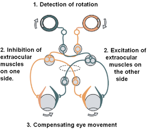
The vestibulo-ocular reflex (VOR) is a reflex eye movement that stabilizes images on the retina during head movement by producing an eye movement in the direction opposite to head movement, thus preserving the image on the center of the visual field. For example, when the head moves to the right, the eyes move to the left, and vice versa. Since slight head movements are present all the time, the VOR is very important for stabilizing vision: patients whose VOR is impaired find it difficult to read, because they cannot stabilize the eyes during small head tremors. The VOR reflex does not depend on visual input and works even in total darkness or when the eyes are closed. 

The vestibulo-ocular reflex. A rotation of the head is detected, which triggers an inhibitory signal to the extraocular muscles on one side and an excitatory signal to the muscles on the other side. The result is a compensatory movement of the eyes.
 
This reflex, combined with the push-pull principle described above, forms the physiological basis of the Rapid head impulse test or Halmagyi-Curthoys-test, in which the head is rapidly and forcefully moved to the side while observing whether the eyes keep looking in the same direction.

Mechanics

The mechanics of the semicircular canals can be described by a damped oscillator. If we designate the deflection of the cupula with , and the head velocity with , the cupula deflection is approximately
α is a proportionality factor, and s corresponds to the frequency. For humans, the time constants T1 and T2 are approximately 3 ms and 5 s, respectively. As a result, for typical head movements, which cover the frequency range of 0.1 Hz and 10 Hz, the deflection of the cupula is approximately proportional to the head-velocity. This is very useful since the velocity of the eyes must be opposite to the velocity of the head in order to maintain clear vision.

Central processing

Signals from the vestibular system also project to the cerebellum (where they are used to keep the VOR effective, a task usually referred to as learning or adaptation) and to different areas in the cortex. The projections to the cortex are spread out over different areas, and their implications are currently not clearly understood.

Projection pathways

The vestibular nuclei on either side of the brain stem exchange signals regarding movement and body position. These signals are sent down the following projection pathways.
  • To the cerebellum. Signals sent to the cerebellum are relayed back as muscle movements of the head, eyes, and posture.
  • To nuclei of cranial nerves III, IV, and VI. Signals sent to these nerves cause the vestibulo-ocular reflex. They allow for the eyes to fix on a moving object while staying in focus.
  • To the reticular formation. Signals sent to the reticular formation signal the new posture the body has taken on, and how to adjust circulation and breathing due to body position.
  • To the spinal cord. Signals sent to the spinal cord allow quick reflex reactions to both the limbs and trunk to regain balance.
  • To the thalamus. Signals sent to the thalamus allow for head and body motor control as well as being conscious of body position.
  • Via the Ventral Pathway, which contributes to vertical orientation and perception of the direction of gravity.

Otolithic organs

While the semicircular canals respond to rotations, the otolithic organs sense linear accelerations. Humans have two otolithic organs on each side, one called the utricle, the other called the saccule. The utricle contains a patch of hair cells and supporting cells called a macula. Similarly, the saccule contains a patch of hair cells and a macula. Each hair cell of a macula has 40-70 stereocilia and one true cilium called a kinocilium. The tips of these cilia are embedded in an otolithic membrane. This membrane is weighted down with protein-calcium carbonate granules called otoliths. These otoliths add to the weight and inertia of the membrane and enhance the sense of gravity and motion. With the head erect, the otolithic membrane bears directly down on the hair cells and stimulation is minimal. When the head is tilted, however, the otolithic membrane sags and bends the stereocilia, stimulating the hair cells. Any orientation of the head causes a combination of stimulation to the utricles and saccules of the two ears. The brain interprets head orientation by comparing these inputs to each other and to other input from the eyes and stretch receptors in the neck, thereby detecting whether the head is tilted or the entire body is tipping. Essentially, these otolithic organs sense how quickly you are accelerating forward or backward, left or right, or up or down. Most of the utricular signals elicit eye movements, while the majority of the saccular signals projects to muscles that control our posture.

While the interpretation of the rotation signals from the semicircular canals is straightforward, the interpretation of otolith signals is more difficult: since gravity is equivalent to a constant linear acceleration, one somehow has to distinguish otolith signals that are caused by linear movements from those caused by gravity. Humans can do that quite well, but the neural mechanisms underlying this separation are not yet fully understood. Humans can sense head tilting and linear acceleration even in dark environments because of the orientation of two groups of hair cell bundles on either side of the striola. Hair cells on opposite sides move with mirror symmetry, so when one side is moved, the other is inhibited. The opposing effects caused by a tilt of the head cause differential sensory inputs from the hair cell bundles allow humans to tell which way the head is tilting, Sensory information is then sent to the brain, which can respond with appropriate corrective actions to the nervous and muscular systems to ensure that balance and awareness are maintained.

Experience from the vestibular system

Experience from the vestibular system is called equilibrioception. It is mainly used for the sense of balance and for spatial orientation. When the vestibular system is stimulated without any other inputs, one experiences a sense of self-motion. For example, a person in complete darkness and sitting in a chair will feel that he or she has turned to the left if the chair is turned to the left. A person in an elevator, with essentially constant visual input, will feel she is descending as the elevator starts to descend. There are a variety of direct and indirect vestibular stimuli which can make people feel they are moving when they are not, not moving when they are, tilted when they are not, or not tilted when they are. Although the vestibular system is a very fast sense used to generate reflexes, including the righting reflex, to maintain perceptual and postural stability, compared to the other senses of vision, touch and audition, vestibular input is perceived with delay.

Pathologies

Diseases of the vestibular system can take different forms, and usually induce vertigo and instability or loss of balance, often accompanied by nausea. The most common vestibular diseases in humans are vestibular neuritis, a related condition called labyrinthitis, Ménière's disease, and BPPV. In addition, the function of the vestibular system can be affected by tumors on the vestibulocochlear nerve, an infarct in the brain stem or in cortical regions related to the processing of vestibular signals, and cerebellar atrophy. 

When the vestibular system and the visual system deliver incongruous results, nausea often occurs. When the vestibular system reports no movement but the visual system reports movement, the motion disorientation is often called motion sickness (or seasickness, car sickness, simulation sickness, or airsickness). In the opposite case, such as when a person is in a zero-gravity environment or during a virtual reality session, the disoriented sensation is often called space sickness or space adaptation syndrome. Either of these "sicknesses" usually ceases once the congruity between the two systems is restored. 

Alcohol can also cause alterations in the vestibular system for short periods and will result in vertigo and possibly nystagmus due to the variable viscosity of the blood and the endolymph during the consumption of alcohol. The common term for this type of sensation is the bed spins.
  • PAN I - The alcohol concentration is higher in the blood than in the vestibular system, hence the endolymph is relatively dense.
  • PAN II - The alcohol concentration is lower in the blood than in the vestibular system, hence the endolymph is relatively dilute.
PAN I will result in subjective vertigo in one direction and typically occurs shortly after ingestion of alcohol when blood alcohol levels are highest. PAN II will eventually cause subjective vertigo in the opposite direction. This occurs several hours after ingestion and after a relative reduction in blood alcohol levels.

Benign paroxysmal positional vertigo (BPPV) is a condition resulting in acute symptoms of vertigo. It is probably caused when pieces that have broken off otoliths have slipped into one of the semicircular canals. In most cases, it is the posterior canal that is affected. In certain head positions, these particles shift and create a fluid wave which displaces the cupula of the canal affected, which leads to dizziness, vertigo and nystagmus. 

A similar condition to BPPV may occur in dogs and other mammals, but the term vertigo cannot be applied because it refers to subjective perception. Terminology is not standardized for this condition.
A common vestibular pathology of dogs and cats is colloquially known as "old dog vestibular disease", or more formally idiopathic peripheral vestibular disease, which causes sudden episode of loss of balance, circling, head tilt, and other signs. This condition is very rare in young dogs but fairly common in geriatric animals, and may affect cats of any age.

Vestibular dysfunction has also been found to correlate with cognitive and emotional disorders, including depersonalization and derealization.

Handedness

From Wikipedia, the free encyclopedia

In human biology, handedness is a better, faster, or more precise performance or individual preference for use of a hand, known as the dominant hand; the less capable or less preferred hand is called the non-dominant hand. Men are somewhat more likely to express a strongly dominant left hand than women. It is estimated that between 70 and 95 percent of the world's population is right-handed.

Types

  • Right-handedness is the most common type. Right-handed people are more skillful with their right hands when performing tasks. Studies suggest 70–95% of the world's population is right-handed.
  • Left-handedness is far less common than right-handedness. Left-handed people are more skillful with their left hands when performing tasks. Studies suggest that approximately 10% of the world population is left-handed.
  • Cross-dominance or mixed-handedness is the change of hand preference between tasks. This is very uncommon in the population with about a 1% prevalence.
  • Ambidexterity, equal ability to use both hands, is exceptionally rare, although it can be learned. A completely ambidextrous person is able to do any task equally well with either hand. Those who learn it still tend to favor their originally dominant hand.

Causes

There are several theories of how handedness develops in individual humans. Occurrences during prenatal development may be important; researchers studied fetuses in utero and determined that handedness in the womb was a very accurate predictor of handedness after birth. In a 2013 study, 39% of infants (6 to 14 months) and 97% of toddlers (18 to 24 months) demonstrated a hand preference.

Division of labor

One common theory as to how handedness affects the hemispheres is the brain hemisphere division of labor. Since speaking and handiwork require fine motor skills, its presumption is that it would be more efficient to have one brain hemisphere do both, rather than having it divided up. Since in most people, the left side of the brain controls speaking, right-handedness predominates. This theory also predicts that left-handed people have a reversed brain division of labor.

Verbal processing in right-handed individuals takes place mostly in the left hemisphere, whereas visuospatial processing is mostly done in the opposite hemisphere. Left-handed individuals have a heterogeneous brain organization in which their brain hemispheres are either organized in the same way as right-handers (but with the hemispheres reversed) or even such that both hemispheres are used for verbal processing. When the average is taken across all types of left-handedness, it shows that left-handers are less lateralized.

Genetic factors

Handedness displays a complex inheritance pattern. For example, if both parents of a child are left-handed, there is a 26% chance of that child being left-handed. A large study of twins from 25,732 families by Medland et al. (2006) has indicated that the heritability of handedness is roughly 24%.

To date, two theoretical single gene models have been proposed to explain the patterns of inheritance of handedness, the first by Marian Annett of the University of Leicester and the second by Chris McManus of UCL

However, the growing weight of evidence from linkage and genome-wide association studies suggests that genetic variance in handedness cannot be explained by a single genetic locus. From these studies McManus et al. now conclude that handedness is polygenic and estimate that at least 40 loci contribute to determining this trait.

Brandler et al. performed a genome-wide association study for a measure of relative hand skill and found that genes involved in the determination of left/right asymmetry in the body play a key role in determining handedness. These results suggest the same mechanisms that determine left/right asymmetry in the body (e.g. Nodal signaling and ciliogenesis) also play a role in the development of brain asymmetry (handedness is an outward reflection of brain asymmetry for motor function).

Epigenetic factors

Twin studies indicate that genetic factors explain 25% of the variance in handedness, while environmental factors explain the remaining 75%. While the molecular basis of handedness epigenetics is largely unclear, Ocklenburg et al. 2017 found that asymmetric methylation of CpG sites plays a key role for gene expression asymmetries that have been related to handedness.

Prenatal hormone exposure

Four studies have indicated that individuals who have had in-utero exposure to diethylstilbestrol (a synthetic estrogen based medication used between 1940 and 1971) were more likely to be left-handed over the clinical control group. Diethylstilbestrol animal studies "suggest that estrogen affects the developing brain, including the part that governs sexual behavior and right and left dominance".

Prenatal vestibular asymmetry

Previc, after reviewing a large number of studies, found evidence that the position of the fetus in the final trimester and a baby's subsequent birth position can affect handedness. About two-thirds of fetuses present with their left occiput (back of the head) at birth. This partly explains why prematurity results in a decrease in right-handedness. Previc argues that asymmetric prenatal positioning creates asymmetric stimulation of the vestibular system, which is involved in the development of handedness. In fact, every major disorder in which patients show reduced right-handedness is associated with either vestibular abnormalities or delay, and asymmetry of the vestibular cortex is strongly correlated with the direction of handedness.

Ultrasound

Another theory is that ultrasound may affect the brains of unborn children, causing higher rates of left-handedness in children whose mothers received ultrasounds during pregnancy. Research on this topic suggests there may exist a weak association between ultrasound screening (sonography used to check on the healthy development of the fetus and mother during pregnancy) and left-handedness.

Handedness developmental timeline

Infants have been known to fluctuate heavily when choosing a hand to lead in grasping and object manipulation tasks. This is especially shown when observing hand dominance in one versus two-handed grasping tasks. Between 36 and 48 months, variability between handedness in one handed grasping begins to decline significantly. This difference can be seen earlier in bi-manual manipulation tasks. Children aged 18 to 36 months showed more hand preference when performing bi-manipulation tasks than simple grasping. The decrease in handedness variability for 36-to-48-month-old children could likely be attributed to preschool or kindergarten attendance. The increase in required single hand grasping activities such as writing or coloring can force children to develop a hand preference.

Correlation with other factors

Intelligence

In his book Right-Hand, Left-Hand, Chris McManus of University College London argues that the proportion of left-handers is increasing and left-handed people as a group have historically produced an above-average quota of high achievers. He says that left-handers' brains are structured differently (in a way that increases their range of abilities) and the genes that determine left-handedness also govern development of the language centers of the brain.

Writing in Scientific American, McManus states that,
Studies in the U.K., U.S. and Australia have revealed that left-handed people differ from right-handers by only one IQ point, which is not noteworthy ... Left-handers' brains are structured differently from right-handers' in ways that can allow them to process language, spatial relations and emotions in more diverse and potentially creative ways. Also, a slightly larger number of left-handers than right-handers are especially gifted in music and math. A study of musicians in professional orchestras found a significantly greater proportion of talented left-handers, even among those who played instruments that seem designed for right-handers, such as violins. Similarly, studies of adolescents who took tests to assess mathematical giftedness found many more left-handers in the population.
Conversely, Joshua Goodman found evidence that left-handers were overrepresented amongst high end of the cognitive spectrum was weak due to methodological and sampling issues in conducted studies. Goodman also found that left-handers were overrepresented at the low end of the cognitive spectrum, with the mentally disabled being twice as likely to be left-handed compared to the general population, as well as generally lower cognitive and non-cognitive abilities amongst left-handed children. Moreover, Ntolka and Papadatou-Pastou in a systematic review and meta-analysis found that it is right-handers who have higher IQ scores, but this difference is negligible (about 1.5 points).

Early childhood intelligence

Nelson, Campbell, and Michel studied infants and whether developing handedness during infancy correlated with language abilities in toddlers. In the article they assessed 38 infants and followed them through to 12 months and then again once they became toddlers from 18–24 months. What they discovered was that when a child developed a consistent use of its right or left hand during infancy (such as using the right hand to put the pacifier back in, or grasping random objects with the left hand), it was more likely to have superior language skills as a toddler. Children who became lateral later than infancy (i.e., when they were toddlers) showed normal development of language and had typical language scores. The researchers used Bayley scales of infant and toddler development to assess all the subjects.

Health

Lower-birth-weight and complications at birth are positively correlated with left-handness.

A variety of neuropsychiatric and developmental disorders like autism spectrum disorders, depression, bipolar disorder, anxiety disorders, schizophrenia, and alcoholism has been associated with left- and mixed-handedness.

A 2012 study showed that nearly 40% of children with cerebral palsy were left-handed, while another study demonstrated that Left-handedness was associated with a 62 percent increased risk of Parkinson's disease in women, but not in men. Another study suggests that the risk of developing multiple sclerosis increases for left-handed women, but the effect is unknown for men at this point.

Left-handed women have a higher risk of breast cancer than right-handed women and the effect is greater post-menopausal.

At least one study maintains that left-handers are more likely to suffer from heart disease, and in a cardiovascular context, are more likely to have reduced longevity.

Left-handers are more likely to suffer bone fractures.

One systematic review concluded: "Left-handers showed no systematic tendency to suffer from disorders of the immune system".

If handedness is entirely genetic, these health problems mean left-handness could be eliminated through natural selection. However, left-handers enjoy an advantage in fighting and sports increasing their likelihood of reproduction.

Income

In a 2006 U.S. study, researchers from Lafayette College and Johns Hopkins University concluded that there was no statistically significant correlation between handedness and earnings for the general population, but among college-educated people, left-handers earned 10 to 15% more than their right-handed counterparts.

More recently, in a 2014 study published by the National Bureau of Economic Research, Harvard economist Joshua Goodman finds that left-handed people earn 10 to 12 percent less over the course of their lives than right-handed people. Goodman attributes this disparity to higher rates of emotional and behavioral problems in left-handed people.

Left-handedness and sports

Interactive sports such as table tennis, badminton and cricket have an overrepresentation of left-handedness, while non-interactive sports such as swimming show no overrepresentation. Smaller physical distance between participants increases the overrepresentation. In fencing, about half the participants are left-handed.

Other, sports-specific factors may increase or decrease the advantage left-handers usually hold in one-on-one situations:
  • In cricket, the overall advantage of a bowler's left-handedness exceeds that resulting from experience alone: even disregarding the experience factor (i.e., even for a batsman whose experience against left-handed bowlers equals his experience against right-handed bowlers), a left-handed bowler challenges the average (i.e., right-handed) batsman more than a right-handed bowler does, because the angle of a bowler's delivery to an opposite-handed batsman is much more penetrating than that of a bowler to a same-handed batsman (see Wasim Akram).
  • In baseball, a right-handed pitcher's curve ball will break away from a right-handed batter and towards a left-handed batter. Historical batting averages show that left-handed batters have a slight advantage over right-handed batters when facing right-handed pitchers. Because there are fewer left-handed pitchers than right-handed pitchers, left-handed batters have more opportunities to face right-handed pitchers than their right-handed counterparts have against left-handed pitchers. Fourteen of the top twenty career batting averages in Major League Baseball history have been posted by left-handed batters. Left-handed batters have a slightly shorter run from the batter's box to first base than right-handers. This gives left-handers a slight advantage in beating throws to first base on infield ground balls.
  • Because a left-handed pitcher faces first base when he is in position to throw to the batter, whereas a right-handed pitcher has his back to first base, a left-handed pitcher has an advantage when attempting to pickoff baserunners at first base.
  • Defensively in baseball, left-handedness is considered an advantage for first basemen because they are better suited to fielding balls hit in the gap between first and second base, and because they do not have to pivot their body around before throwing the ball to another infielder. For the same reason, the other infielder's positions are seen as being advantageous to right-handed throwers. Historically, there have been few left-handed catchers because of the perceived disadvantage a left-handed catcher would have in making the throw to third base, especially with a right-handed hitter at the plate. A left-handed catcher would have a potentially more dangerous time tagging out a baserunner trying to score. With the ball in the glove on the right hand, a left-handed catcher would have to turn his body to the left to tag a runner. In doing so, he can lose the opportunity to brace himself for an impending collision. On the other hand, the Encyclopedia of Baseball Catchers states:
One advantage is a left-handed catcher's ability to frame a right-handed pitcher's breaking balls. A right-handed catcher catches a right-hander's breaking ball across his body, with his glove moving out of the strike zone. A left-handed catcher would be able to catch the pitch moving into the strike zone and create a better target for the umpire.
  • In four wall handball, typical strategy is to play along the left wall forcing the opponent to use their left hand to counter the attack and playing into the strength of a left-handed competitor.
  • In water polo the centre forward position has an advantage in turning to shoot on net when rotating the reverse direction as expected by the centre of the opposition defence and gain an improved position to score.
  • Ice hockey typically uses a strategy in which a defence pairing includes one left-handed and one right-handed defender. A disproportionately large number of ice hockey players of all positions, 62 percent, shoot left, though this does not necessarily indicate left-handedness.
  • In American football, the handedness of a quarterback affects blocking patterns on the offensive line. Tight ends, when only one is used, typically line up on the same side as the throwing hand of the quarterback, while the offensive tackle on the opposite hand, which protects the quarterback's "blind side," is typically the most valued member of the offensive line. While uncommon, there have been several notable left-handed quarterbacks.

Gender

According to a meta-analysis of 144 studies, totaling 1,787,629 participants, the best estimate for the male to female odds ratio was 1.23, indicating 23% more men are left-handed. 11% of men and 9% of women would be approximately 10% overall, at a 1.22 male to female odds ratio.

Sexuality and gender identity

A number of studies examining the relationship between handedness and sexual orientation have reported that a disproportionate minority of homosexual people exhibit left-handedness, though findings are mixed.

A 2001 study also found that children who are genetically male but have different gender identities were more than twice as likely to be left-handed than a clinical control group (19.5% vs. 8.3%, respectively).

Paraphilias (atypical sexual interests) have also been linked to higher rates of left-handedness. A 2008 study analyzing the sexual fantasies of 200 males found "elevated paraphilic interests were correlated with elevated non-right handedness". Greater rates of left-handedness has also been documented among pedophiles.

A 2014 study attempting to analyze the biological markers of asexuality asserts that non-sexual men and women were 2.4 and 2.5 times, respectively, more likely to be left-handed than their heterosexual counterparts.

In culture

Many tools and procedures are designed to facilitate use by right-handed people, often without realizing the difficulties incurred by the left-handed. John W. Santrock has written, "For centuries, left-handers have suffered unfair discrimination in a world designed for right-handers."

McManus noted that, beginning at the time of the Industrial Revolution, workers needed to operate complex machines that were almost certainly designed with right-handers in mind. This would have made left-handers more visible and at the same time appear less capable and more clumsy. During this era, children were taught to write with a dip pen. While a right-hander could smoothly drag the pen across paper from left to right, a dip pen could not easily be pushed across by the left hand without digging into the paper and making blots and stains.

Negative appeal

Moreover, apart from inconvenience, left-handed people have historically been considered unlucky or even malicious for their difference by the right-handed majority. In many European languages, including English, the word for the direction "right" also means "correct" or "proper". Throughout history, being left-handed was considered negative. The Latin adjective sinister means "left" as well as "unlucky", and this double meaning survives in European derivatives of Latin, including the English word "sinister" (only when referring to the bearer's left of a coat of arms).

There are many negative connotations associated with the phrase "left-handed": clumsy, awkward, unlucky, insincere, sinister, malicious, and so on. A "left-handed compliment" is considered one that is unflattering or dismissive in meaning. In French, gauche means both "left" and "awkward" or "clumsy", while droit(e) (cognate to English direct and related to "adroit") means both "right" and "straight", as well as "law" and the legal sense of "right". The name "Dexter" derives from the Latin for "right", as does the word "dexterity" meaning manual skill. As these are all very old words, they would tend to support theories indicating that the predominance of right-handedness is an extremely old phenomenon. 

Black magic is sometimes referred to as the "left-hand path". 

Until very recently in Taiwan (and still in Mainland China, Japan and both North and South Korea), left-handed people were strongly encouraged to switch to being right-handed, or at least switch to writing with the right hand. Due to the importance of stroke order, developed for the comfortable use of right-handed people, it is considered more difficult to write legible Chinese characters with the left hand than it is to write Latin letters, though difficulty is subjective and depends on the writer. Because writing when moving one's hand away from its side towards the other side of the body can cause smudging if the outward side of the hand is allowed to drag across the writing, writing in the Latin alphabet might possibly be less feasible with the left hand than the right under certain circumstances. Conversely, right-to-left alphabets, such as the Arabic and Hebrew, are generally considered easier to write with the left hand in general. Depending on the position and inclination of the writing paper, and the writing method, the left-handed writer can write as neatly and efficiently or as messily and slowly as right-handed writers. Usually the left-handed child needs to be taught how to write correctly with the left hand, since discovering a comfortable left-handed writing method on one's own may not be straightforward.

International Left-Handers Day

International Left-Handers Day is held annually every August 13. It was founded by the Left-Handers Club in 1992, with the club itself having been founded in 1990. International Left-Handers Day is, according to the club, "an annual event when left-handers everywhere can celebrate their sinistrality (left-handedness) and increase public awareness of the advantages and disadvantages of being left-handed." It celebrates their uniqueness and differences, who are from seven to ten percent of the world's population. Thousands of left-handed people in today's society have to adapt to use right-handed tools and objects. Again according to the club, "in the U.K. alone there were over 20 regional events to mark the day in 2001- including left-v-right sports matches, a left-handed tea party, pubs using left-handed corkscrews where patrons drank and played pub games with the left hand only, and nationwide 'Lefty Zones' where left-handers' creativity, adaptability and sporting prowess were celebrated, whilst right-handers were encouraged to try out everyday left-handed objects to see just how awkward it can feel using the wrong equipment!"

In other animals

Kangaroos and other macropod marsupials have a left-hand preference for everyday tasks in the wild. 'True' handedness is unexpected in marsupials because, unlike placental mammals, they lack a corpus callosum. Left-handedness was particularly apparent in the red kangaroo (Macropus rufus) and the eastern gray kangaroo (Macropus giganteus). Red-necked (Bennett's) wallabies (Macropus rufogriseus) preferentially use their left hand for behaviours that involve fine manipulation, but the right for behaviours that require more physical strength. There was less evidence for handedness in arboreal species. Studies of dogs, horses, and domestic cats have shown that females of those species tend to be right-handed, while males are left-handed.

Operator (computer programming)

From Wikipedia, the free encyclopedia https://en.wikipedia.org/wiki/Operator_(computer_programmin...