Search This Blog

Monday, December 9, 2019

Lanthanide probes

From Wikipedia, the free encyclopedia
https://en.wikipedia.org/wiki/Lanthanide_probes

Lanthanide probes are a non-invasive analytical tool commonly used for biological and chemical applications. Lanthanides are metal ions which have their 4f energy level filled and generally refer to elements cerium to lutetium in the periodic table. The fluorescence of lanthanide salts is weak because the energy absorption of the metallic ion is low; hence chelated complexes of lanthanides are most commonly used. The term chelate derives from the Greek word for “claw,” and is applied to name ligands, which attach to a metal ion with two or more donor atoms through dative bonds. The fluorescence is most intense when the metal ion has the oxidation state of 3+. Not all lanthanide metals can be used and the most common are: Sm(III), Eu(III), Tb(III), and Dy(III).

History

EuFOD, an example of a europium complex
 
It has been known since the early 1930s that the salts of certain lanthanides are fluorescent. The reaction of lanthanide salts with nucleic acids was discussed in a number of publications during the 1930s and the 1940s where lanthanum-containing reagents were employed for the fixation of nucleic acid structures. In 1942 complexes of europium, terbium, and samarium were discovered to exhibit unusual luminescence properties when excited by UV light. However, the first staining of biological cells with lanthanides occurred twenty years later when bacterial smears of E. coli were treated with aqueous solutions of a europium complex, which under mercury lamp illumination appeared as bright red spots. Attention to lanthanide probes increased greatly in the mid-1970s when Finnish researchers proposed Eu(III), Sm(III), Tb(III), and Dy(III) polyaminocarboxylates as luminescent sensors in time-resolved luminescent (TRL) immunoassays. Optimization of analytical methods from the 1970s onward for lanthanide chelates and time-resolved luminescence microscopy (TRLM) resulted in the use of lanthanide probes in many scientific, medical and commercial fields.

Techniques

There are two main assaying techniques: heterogeneous and homogeneous. If two lanthanide chelates are used in the analysis one after the other—it is called heterogeneous assaying. The first analyte is linked to a specific binding agent on a solid support such as a polymer and then another reaction couples the first poorly luminescent lanthanide complex with a new better one. This tedious method is used because the second more luminescent compound would not bind without the first analyte already present. Subsequent time resolved detection of the metal-centered luminescent probe yields the desired signal. Antigens, steroids and hormones are routinely assayed with heterogeneous techniques. Homogeneous assays rely on direct coupling of the lanthanide label with an organic acceptor.

The relaxation of excited molecules states often occurs by the emission of light which is called fluorescence. There are two ways of measuring this emitted radiation: as a function of frequency (inverse to wavelength) or time. Conventionally the fluorescence spectrum shows the intensity of fluorescence at different wavelengths, but since lanthanides have relatively long fluorescence decay times (ranging from one microsecond to one millisecond), it is possible to record the fluorescence emission at different decay times from the given excitation energy at time zero. This is called time resolved fluorescence spectroscopy.

Mechanism

Lanthanides can be used because their small size (ionic radius) gives them the ability to replace metal ions inside protein complex such as calcium or nickel. The optical properties of lanthanide ions such as Ln(III) originate in the special features of their electronic [Xe]4fn configurations. These configurations generate many electronic levels, the number of which is given by [14!/n!(14- n)!], translating into 3003 energy levels for Eu(III) and Tb(III).

The energies of these levels are well defined due to the shielding of the 4f orbitals by the filled 5s and 5p sub-shells, and are not very sensitive to the chemical environments in which the lanthanide ions are inserted. Inner-shell 4f-4f transitions span both the visible and near-infrared ranges. They are sharp and easily recognizable. Since these transitions are parity forbidden, the lifetimes of the excited states are long, which allows the use of time resolved spectroscopy, a definitive asset for bioassays and microscopy. The only drawback of f-f transitions are their faint oscillator strengths which may in fact be turned into an advantage.

The energy absorbed by the organic receptor (ligand) is transferred onto Ln(III) excited states, and sharp emission bands originating from the metal ion are detected after rapid internal conversion to the emitting level. The phenomenon is termed sensitization of the metal centered complex (also referred to as antenna effect) and is quite complex. The energy migration path though goes through the long-lived triplet state of the ligand. Ln(III) ions are good quenchers of triplet states so that photobleaching is substantially reduced. The three types of transitions seen for lanthanide probes are: LMCT, 4f-5d, and intraconfigurational 4f-4f. The former two usually occur at energies too high to be relevant for bio-applications.

Applications

Cancer research

Screening tools for the development of new cancer therapies are in high demand worldwide and often require the determination of enzyme kinetics. The high sensitivity of lanthanide luminescence, particularly of time-resolved luminescence has revealed to be an ideal candidate for this purpose. There are several ways of conducting this analysis by the use of fluorogenic enzyme substrates, substrates bearing donor/acceptor groups allowing fluorescence resonance energy transfer (FRET) and immunoassays. For example, guanine nucleotide binding proteins consist of several subunits, one of which comprises those of the Ras subfamily. Ras GTPases act as binary switches by converting guadenosine triphosphate (GTP) into guadenosine diphosphate (GDP). Luminescence of the Tb(III) complex with norfloxacin is sensitive to determine the concentration of phosphate released by the GTP to GDP transformation.

pH probes

Protonation of basic sites in systems comprising a chromophore and a luminescent metal center leads the way for pH sensors. Some initially proposed systems were based on pyridine derivatives but these were not stable in water. More robust sensors have been proposed in which the core is a substituted macrocycle usually bearing phosphinate, carboxylate or four amide coordinating groups. It has been observed that lanthanide luminescent probe emission increases about six-fold when decreasing the pH of the solution from six to two.

Hydrogen peroxide sensor

Hydrogen peroxide can be detected with high sensitivity by the luminescence of lanthanide probes—however only at relatively high pH values. A lanthanide-based analytical procedure was proposed in 2002 based on the finding that the europium complex with various tetracyclines binds hydrogen peroxide forming a luminescent complex.

Estimating molecule size and atom distances

FRET in lanthanide probes is a widely used technique to measure the distance between two points separated by approximately 15–100 Angstrom. Measurements can be done under physiological conditions in vitro with genetically encoded dyes, and often in vivo as well. The technique relies on a distant- dependent transfer of energy from a donor fluorophore to an acceptor dye. Lanthanide probes has been used to study DNA-protein interactions (using a terbium chelate complex) to measure distances in DNA complexes bent by the CAP protein.

Protein conformation

Lanthanide probes have been used to detect conformational changes in proteins. Recently the Shaker potassium ion channel, a voltage-gated channel involved in nerve impulses was measured using this technique. Some scientist also have used lanthanide based luminescence resonance energy transfer (LRET) which is very similar to FRET to study conformational changes in RNA polymerase upon binding to DNA and transcription initiation in prokaryotes. LRET was also used to study the interaction of the proteins dystrophin and actin in muscle cells. Dystrophin is present in the inner muscle cell membrane and is believed to stabilize muscle fibers by binding to actin filaments. Specifically labelled dystrophin with Tb labelled monoclonal antibodies labeled were used.

Virology

Traditional virus diagnostic procedures are being replaced by sensitive immunoassays with lanthanides. The time resolved fluorescence based technique is generally applicable and its performance has also been tested in the assay of viral antigens in clinical specimens.

Medical imaging

Several systems have been proposed which combine MRI capability with lanthanides probes in dual assays. The luminescent probe may for instance serve to localize the MRI contrast agent. This has helped to visualize the delivery of nucleic acids into cultured cells. It should be noted in this case that lanthanides are not used for their fluorescence but their magnetic qualities.

Biology - Receptor-ligand interactions

Lanthanide probes displays unique fluorescence properties, including long lifetime of fluorescence, large Stokes shift and narrow emission peak. These properties is highly advantageous to develop analytical probes for receptor-ligand interactions. Many lanthanide-based fluorescence studies have been developed for GPCRs, including CXCR1, insulin-like family peptide receptor 2, protease-activated receptor 2, β2-adrenergic receptor, and C3a receptor.

Instrumentation

The emitted photons from excited lanthanides are detected by highly sensitive devices and techniques such as single-photon detection. If the lifetime of the excited emitting level is long enough, then time-resolved detection (TRD) can be used to enhance the signal-to-noise ratio. The instrumentation used to perform LRET is relatively simple, although slightly more complex than conventional fluorimeters. The general requirements are a pulsed UV excitation source and time-resolved detection.
Lanthanide probe 1.jpg
Light sources which emit short duration pulses can be divided into the following categories:
The most important factors in the selection of the pulsed light source for are the duration and intensity of the light. Pulsed lasers for the 300 to 500 nm range have now replaced spark caps in fluorescence spectroscopy. There are four general types of pulsing lasers used: lasers with pulsed excitation, lasers with G-switching, mode locked lasers and cavity dumped lasers. Pulsed nitrogen lasers (337 nm) have often been used as an excitation source in time resolved fluorometry.

In time resolved fluorometry the fast photomultiplier tube is the only practical single photon detector. Good single photon resolution is also an advantage in counting photons from long decay fluorescent probes, such as lanthanide chelates.

These commercial instruments are available in the market today: Perkin-Elmer Micro Filter Fluorometer LS-2, Perkin-Elmer Luminescence Spectrometer Model LS 5, and LKB-Wallac Time-Resolved Fluorometer Model 1230.

Ligands

Lanthanide probes ligands must meet several chemical requirements for the probes to work properly. These qualities are: water solubility, large thermodynamic stability at physiological pHs, kinetic inertness and absorption above 330 nm to minimize destruction of live biological materials.

The chelates which have been studied and utilized to date can be classified into the following groups:
  1. Tris chelates (three ligands)
  2. Tetrakis chelates (four ligands)
  3. Mixed ligand complexes
  4. Complexes with neutral donors
  5. Others such as: phthalate, picrate, and salicylate complexes.
The efficiency of the energy transfer from the ligand to the ion is determined ligand-metal bond. The energy transfer is more efficient when bonded covalently than through ionic bonding. Substituents in the ligand which are of electron-donating such as hydroxy, methoxy and methyl groups increase the fluorescence. The opposite effect is seen when an electron-withdrawing group (such as nitro) is attached. Furthermore, the fluorescence intensity is increased by fluorine substitution to the ligand. The energy transfer to the metal ion increases as the electronegativity of the fluorinated group makes the europium-oxygen bond of a more covalent nature. Increased conjugation by aromatic substituents by replacing phenyl by naphtyl groups is shown to enhance fluorescence.

Actinides in the environment

From Wikipedia, the free encyclopedia
https://en.wikipedia.org/wiki/Actinides_in_the_environment
 
Actinides in the environment refer to the sources, environmental behaviour and effects of actinides in Earth's environment. Environmental radioactivity is not limited solely to actinides; non-actinides such as radon and radium are of note. While all actinides are radioactive, there are a lot of actinides or actinide-relating minerals in the Earth's crust such as uranium and thorium. These minerals are helpful in many ways, such as carbon-dating, most detectors, X-rays, and more.

Inhalation versus ingestion

Generally, ingested insoluble actinide compounds, such as high-fired uranium dioxide and mixed oxide (MOX) fuel, will pass through the digestive system with little effect since they cannot dissolve and be absorbed by the body. Inhaled actinide compounds, however, will be more damaging as they remain in the lungs and irradiate the lung tissue. 

Ingested low-fired oxides and soluble salts such as nitrate can be absorbed into the blood stream. If they are inhaled then it is possible for the solid to dissolve and leave the lungs. Hence, the dose to the lungs will be lower for the soluble form. 

Actinium in the environment

Actinium can be naturally found in traces in uranium ores as 227-Ac, an α and β emitter with a half-life of 21.773 years. One ton of uranium ore contains about a tenth of a gram of actinium. It's more commonly made in milligram amounts by the neutron irradiation of 226-Ra in a nuclear reactor. Naturally occurring actinium is composed of 1 radioactive isotope; with 227-Ac being the most abundant (100% natural abundance). 

Thorium in the environment

Monazite, a rare-earth-and-thorium-phosphate mineral, is the primary source of the world's thorium
 
In India, a large amount of thorium ore can be found in the form of monazite in placer deposits of the Western and Eastern coastal dune sands, particularly in the Tamil Nadu coastal areas. The residents of this area are exposed to a naturally occurring radiation dose ten times higher than the worldwide average.

Occurrence

Thorium is found at low levels in most rocks and soils, where it is about three times more abundant than uranium, and is about as common as lead. Soil commonly contains an average of around 6 parts per million (ppm) of thorium. Thorium occurs in several minerals, the most common being the rare earth-thorium-phosphate mineral, monazite, which contains up to about 12% thorium oxide. There are substantial deposits in several countries. 232Th decays very slowly (its half-life is about three times the age of the earth). Other isotopes of thorium occur in the thorium and uranium decay chains. Most of these are short-lived and hence much more radioactive than 232Th, though on a mass basis they are negligible.

Effects in humans

Thorium has been linked to liver cancer. In the past thoria (thorium dioxide) was used as a contrast agent for medical X-ray radiography but its use has been discontinued. It was sold under the name Thorotrast.

Protactinium in the environment

Protactinium-231 occurs naturally in uranium ores such as pitchblende, to the extent of 3 ppm in some ores. Protactinium is naturally present in soil, rock, surface water, groundwater, plants and animals in very low concentrations (on the order of 1 ppt or 0.1 picocouries (pCi)/g).

Uranium in the environment

Uranium is a natural metal which is widely found. It is present in almost all soils and it is more plentiful than antimony, beryllium, cadmium, gold, mercury, silver, or tungsten, and is about as abundant as arsenic or molybdenum. Significant concentrations of uranium occur in some substances such as phosphate rock deposits, and minerals such as lignite, and monazite sands in uranium-rich ores (it is recovered commercially from these sources). 

Seawater contains about 3.3 parts per billion of uranium by weight as uranium (VI) forms soluble carbonate complexes. The extraction of uranium from seawater has been considered as a means of obtaining the element. Because of the very low specific activity of uranium the chemical effects of it upon living things can often outweigh the effects of its radioactivity. Additional uranium has been added to the environment in some locations as a result of the nuclear fuel cycle and the use of depleted uranium in munitions.

Neptunium in the environment

Like plutonium, neptunium has a high affinity for soil. However, it is relatively mobile over the long term, and diffusion of neptunium-237 in groundwater is a major issue in designing a deep geological repository for permanent storage of spent nuclear fuel. 237Np has a half-life of 2.144 million years and is therefore a long-term problem; but its half-life is still much shorter than those of uranium-238, uranium-235, or uranium-236, and 237Np therefore has higher specific activity than those nuclides. It is used only to make Pu-237 when bombarded with neutrons in a lab.

Plutonium in the environment

Sources

Plutonium in the environment has several sources. These include:
  • Atomic batteries
    • In space
    • In pacemakers
  • Bomb detonations
  • Bomb safety trials
  • Nuclear crime
  • Nuclear fuel cycle
  • Nuclear power plants

Environmental chemistry

Plutonium, like other actinides, readily forms a plutonium dioxide (plutonyl) core (PuO2). In the environment, this plutonyl core readily complexes with carbonate as well as other oxygen moieties (OH, NO2, NO3, and SO42−) to form charged complexes which can be readily mobile with low affinities to soil.
  • PuO2CO32−
  • PuO2(CO3)24−
  • PuO2(CO3)36−
PuO2 formed from neutralizing highly acidic nitric acid solutions tends to form polymeric PuO2 which is resistant to complexation. Plutonium also readily shifts valences between the +3, +4, +5 and +6 states. It is common for some fraction of plutonium in solution to exist in all of these states in equilibrium. 

Plutonium is known to bind to soil particles very strongly, see above for an X-ray spectroscopic study of plutonium in soil and concrete. While caesium has very different chemistry to the actinides, it is well known that both caesium and many of the actinides bind strongly to the minerals in soil. Hence it has been possible to use 134Cs labeled soil to study the migration of Pu and Cs is soils. It has been shown that colloidal transport processes control the migration of Cs (and will control the migration of Pu) in the soil at the Waste Isolation Pilot Plant.

Americium in the environment

Americium often enters landfills from discarded smoke detectors. The rules associated with the disposal of smoke detectors are very relaxed in most municipalities. For instance, in the UK it is permissible to dispose of an americium containing smoke detector by placing it in the dustbin with normal household rubbish, but each dustbin worth of rubbish is limited to only containing one smoke detector. The manufacture of products containing americium (such as smoke detectors) as well as nuclear reactors and explosions may also release the americium into the environment.

Picture illustrating David "Radioactive Boyscout" Hahn.
 
In 1999, a truck transporting 900 smoke detectors in France had been reported to have caught fire; it is claimed that this led to a release of americium into the environment. In the U.S., the "Radioactive Boy Scout" David Hahn was able to buy thousands of smoke detectors at remainder prices and concentrate the americium from them.

There have been cases of humans being exposed to americium. The worst case was that of Harold McCluskey, who was exposed to an extremely high dose of americium-241 after an accident involving a glove box. He was subsequently treated with chelation therapy. It is likely that the medical care which he was given saved his life: despite similar biodistribution and toxicity to plutonium, the two radioactive elements have different solution-state chemistries. Americium is stable in the +3 oxidation state, while the +4 oxidation state of plutonium can form in the human body.

The most common isotope americium-241 decays (half-life of 432 years) to neptunium-237 which has a much longer half-life, so in the long term, the issues discussed above for neptunium apply.

Americium released into the environment tends to remain in soil and water at relatively shallow depths and may be taken up by animals and plants during growth; shellfish such as shrimp take up americium-241 in their shells, and parts of grain plants can become contaminated with exposure.

Curium in the environment

Curium can be found in minute amounts in some uranium mineral deposits. It's really only used in spacecraft to power the electrical equipment in rovers. Curium can be dangerous when exposed to because of the radiation its emitting, which is even greater than Americium.

Background radiation

From Wikipedia, the free encyclopedia
https://en.wikipedia.org/wiki/Background_radiation
 
Background radiation is a measure of the level of ionizing radiation present in the environment at a particular location which is not due to deliberate introduction of radiation sources.

Background radiation originates from a variety of sources, both natural and artificial. These include both cosmic radiation and environmental radioactivity from naturally occurring radioactive materials (such as radon and radium), as well as man-made medical X-rays, fallout from nuclear weapons testing and nuclear accidents.

Definition

Background radiation is defined by the International Atomic Energy Agency as "Dose or dose rate (or an observed measure related to the dose or dose rate) attributable to all sources other than the one(s) specified. So a distinction is made between dose which is already in a location, which is defined here as being "background", and the dose due to a deliberately introduced and specified source. This is important where radiation measurements are taken of a specified radiation source, where the existing background may affect this measurement. An example would be measurement of radioactive contamination in a gamma radiation background, which could increase the total reading above that expected from the contamination alone.

However, if no radiation source is specified as being of concern, then the total radiation dose measurement at a location is generally called the background radiation, and this is usually the case where an ambient dose rate is measured for environmental purposes. 

Background dose rate examples

Background radiation varies with location and time, and the following table gives examples:

Average annual human exposure to ionizing radiation in millisieverts (mSv) per year
Radiation source World US Japan Remark
Inhalation of air 1.26 2.28 0.40 mainly from radon, depends on indoor accumulation
Ingestion of food & water 0.29 0.28 0.40 (K-40, C-14, etc.)
Terrestrial radiation from ground 0.48 0.21 0.40 depends on soil and building material
Cosmic radiation from space 0.39 0.33 0.30 depends on altitude
sub total (natural) 2.40 3.10 1.50 sizeable population groups receive 10–20 mSv
Medical 0.60 3.00 2.30 worldwide figure excludes radiotherapy;
US figure is mostly CT scans and nuclear medicine.
Consumer items 0.13
cigarettes, air travel, building materials, etc.
Atmospheric nuclear testing 0.005 0.01 peak of 0.11 mSv in 1963 and declining since; higher near sites
Occupational exposure 0.005 0.005 0.01 worldwide average to workers only is 0.7 mSv, mostly due to radon in mines;
US is mostly due to medical and aviation workers.
Chernobyl accident 0.002 0.01 peak of 0.04 mSv in 1986 and declining since; higher near site
Nuclear fuel cycle 0.0002
0.001 up to 0.02 mSv near sites; excludes occupational exposure
Other 0.003
Industrial, security, medical, educational, and research
sub total (artificial) 0.61 3.14 2.33
Total 3.01 6.24 3.83 millisieverts per year

Natural background radiation

The weather station outside of the Atomic Testing Museum on a hot summer day. Displayed background gamma radiation level is 9.8 μR/h (0.82 mSv/a) This is very close to the world average background radiation of 0.87 mSv/a from cosmic and terrestrial sources.
 
Cloud chambers used by early researchers first detected cosmic rays and other background radiation. They can be used to visualize the background radiation
 
Radioactive material is found throughout nature. Detectable amounts occur naturally in soil, rocks, water, air, and vegetation, from which it is inhaled and ingested into the body. In addition to this internal exposure, humans also receive external exposure from radioactive materials that remain outside the body and from cosmic radiation from space. The worldwide average natural dose to humans is about 2.4 mSv (240 mrem) per year. This is four times the worldwide average artificial radiation exposure, which in 2008 amounted to about 0.6 millisieverts (60 mrem) per year. In some rich countries, like the US and Japan, artificial exposure is, on average, greater than the natural exposure, due to greater access to medical imaging. In Europe, average natural background exposure by country ranges from under 2 mSv (200 mrem) annually in the United Kingdom to more than 7 mSv (700 mrem) annually for some groups of people in Finland.

"Exposure to radiation from natural sources is an inescapable feature of everyday life in both working and public environments. This exposure is in most cases of little or no concern to society, but in certain situations the introduction of health protection measures needs to be considered, for example when working with uranium and thorium ores and other Naturally Occurring Radioactive Material (NORM). These situations have become the focus of greater attention by the Agency in recent years."

Terrestrial sources

Terrestrial radiation, for the purpose of the table above, only includes sources that remain external to the body. The major radionuclides of concern are potassium, uranium and thorium and their decay products, some of which, like radium and radon are intensely radioactive but occur in low concentrations. Most of these sources have been decreasing, due to radioactive decay since the formation of the Earth, because there is no significant amount currently transported to the Earth. Thus, the present activity on earth from uranium-238 is only half as much as it originally was because of its 4.5 billion year half-life, and potassium-40 (half-life 1.25 billion years) is only at about 8% of original activity. But during the time that humans have existed the amount of radiation has decreased very little.

Many shorter half-life (and thus more intensely radioactive) isotopes have not decayed out of the terrestrial environment because of their on-going natural production. Examples of these are radium-226 (decay product of thorium-230 in decay chain of uranium-238) and radon-222 (a decay product of radium-226 in said chain).

Thorium and uranium (and their daughters) primarily undergo alpha and beta decay, and aren't easily detectable. However, many of their daughter products are strong gamma emitters. Thorium-232 is detectable via a 239 keV peak from lead-212, 511, 583 and 2614 keV from thallium-208, and 911 and 969 keV from actinium-228. Uranium-238 manifests as 609, 1120, and 1764 keV peaks of bismuth-214 (cf. the same peak for atmospheric radon). Potassium-40 is detectable directly via its 1461 keV gamma peak.

The level over the sea and other large bodies of water tends to be about a tenth of the terrestrial background. Conversely, coastal areas (and areas by the side of fresh water) may have an additional contribution from dispersed sediment.

Airborne sources

The biggest source of natural background radiation is airborne radon, a radioactive gas that emanates from the ground. Radon and its isotopes, parent radionuclides, and decay products all contribute to an average inhaled dose of 1.26 mSv/a (millisievert per year). Radon is unevenly distributed and varies with weather, such that much higher doses apply to many areas of the world, where it represents a significant health hazard. Concentrations over 500 times the world average have been found inside buildings in Scandinavia, the United States, Iran, and the Czech Republic. Radon is a decay product of uranium, which is relatively common in the Earth's crust, but more concentrated in ore-bearing rocks scattered around the world. Radon seeps out of these ores into the atmosphere or into ground water or infiltrates into buildings. It can be inhaled into the lungs, along with its decay products, where they will reside for a period of time after exposure.

Although radon is naturally occurring, exposure can be enhanced or diminished by human activity, notably house construction. A poorly sealed basement in an otherwise well insulated house can result in the accumulation of radon within the dwelling, exposing its residents to high concentrations. The widespread construction of well insulated and sealed homes in the northern industrialized world has led to radon becoming the primary source of background radiation in some localities in northern North America and Europe. Basement sealing and suction ventilation reduce exposure. Some building materials, for example lightweight concrete with alum shale, phosphogypsum and Italian tuff, may emanate radon if they contain radium and are porous to gas.

Radiation exposure from radon is indirect. Radon has a short half-life (4 days) and decays into other solid particulate radium-series radioactive nuclides. These radioactive particles are inhaled and remain lodged in the lungs, causing continued exposure. Radon is thus assumed to be the second leading cause of lung cancer after smoking, and accounts for 15,000 to 22,000 cancer deaths per year in the US alone. However, the discussion about the opposite experimental results is still going on.

About 100,000 Bq/m3 of radon was found in Stanley Watras's basement in 1984. He and his neighbours in Boyertown, Pennsylvania, United States may hold the record for the most radioactive dwellings in the world. International radiation protection organizations estimate that a committed dose may be calculated by multiplying the equilibrium equivalent concentration (EEC) of radon by a factor of 8 to 9 nSv·m3/Bq·h and the EEC of thoron by a factor of 40 nSv·m3/Bq·h.

Most of the atmospheric background is caused by radon and its decay products. The gamma spectrum shows prominent peaks at 609, 1120, and 1764 keV, belonging to bismuth-214, a radon decay product. The atmospheric background varies greatly with wind direction and meteorological conditions. Radon also can be released from the ground in bursts and then form "radon clouds" capable of traveling tens of kilometers.

Cosmic radiation

Estimate of the maximum dose of radiation received at an altitude of 12 km 20 January 2005, following a violent solar flare. The doses are expressed in microsieverts per hour.
 
The Earth and all living things on it are constantly bombarded by radiation from outer space. This radiation primarily consists of positively charged ions from protons to iron and larger nuclei derived from outside the Solar System. This radiation interacts with atoms in the atmosphere to create an air shower of secondary radiation, including X-rays, muons, protons, alpha particles, pions, electrons, and neutrons. The immediate dose from cosmic radiation is largely from muons, neutrons, and electrons, and this dose varies in different parts of the world based largely on the geomagnetic field and altitude. For example, the city of Denver in the United States (at 1650 meters elevation) receives a cosmic ray dose roughly twice that of a location at sea level. This radiation is much more intense in the upper troposphere, around 10 km altitude, and is thus of particular concern for airline crews and frequent passengers, who spend many hours per year in this environment. During their flights airline crews typically get an additional occupational dose between 2.2 mSv (220 mrem) per year  and 2.19 mSv/year, according to various studies. 

Similarly, cosmic rays cause higher background exposure in astronauts than in humans on the surface of Earth. Astronauts in low orbits, such as in the International Space Station or the Space Shuttle, are partially shielded by the magnetic field of the Earth, but also suffer from the Van Allen radiation belt which accumulates cosmic rays and results from the Earth's magnetic field. Outside low Earth orbit, as experienced by the Apollo astronauts who traveled to the Moon, this background radiation is much more intense, and represents a considerable obstacle to potential future long term human exploration of the moon or Mars.

Cosmic rays also cause elemental transmutation in the atmosphere, in which secondary radiation generated by the cosmic rays combines with atomic nuclei in the atmosphere to generate different nuclides. Many so-called cosmogenic nuclides can be produced, but probably the most notable is carbon-14, which is produced by interactions with nitrogen atoms. These cosmogenic nuclides eventually reach the Earth's surface and can be incorporated into living organisms. The production of these nuclides varies slightly with short-term variations in solar cosmic ray flux, but is considered practically constant over long scales of thousands to millions of years. The constant production, incorporation into organisms and relatively short half-life of carbon-14 are the principles used in radiocarbon dating of ancient biological materials, such as wooden artifacts or human remains.

The cosmic radiation at sea level usually manifests as 511 keV gamma rays from annihilation of positrons created by nuclear reactions of high energy particles and gamma rays. At higher altitudes there is also the contribution of continuous bremsstrahlung spectrum.

Food and water

Two of the essential elements that make up the human body, namely potassium and carbon, have radioactive isotopes that add significantly to our background radiation dose. An average human contains about 17 milligrams of potassium-40 (40K) and about 24 nanograms (10−9 g) of carbon-14 (14C), (half-life 5,730 years). Excluding internal contamination by external radioactive material, these two are largest components of internal radiation exposure from biologically functional components of the human body. About 4,000 nuclei of 40K decay per second, and a similar number of 14C. The energy of beta particles produced by 40K is about 10 times that from the beta particles from 14C decay.

14C is present in the human body at a level of about 3700 Bq (0.1 μCi) with a biological half-life of 40 days. This means there are about 3700 beta particles per second produced by the decay of 14C. However, a 14C atom is in the genetic information of about half the cells, while potassium is not a component of DNA. The decay of a 14C atom inside DNA in one person happens about 50 times per second, changing a carbon atom to one of nitrogen.

The global average internal dose from radionuclides other than radon and its decay products is 0.29 mSv/a, of which 0.17 mSv/a comes from 40K, 0.12 mSv/a comes from the uranium and thorium series, and 12 μSv/a comes from 14C.

Areas with high natural background radiation

Some areas have greater dosage than the country-wide averages. In the world in general, exceptionally high natural background locales include Ramsar in Iran, Guarapari in Brazil, Karunagappalli in India, Arkaroola in Australia, and Yangjiang in China.

The highest level of purely natural radiation ever recorded on the Earth's surface was 90 µGy/h on a Brazilian black beach (areia preta in Portuguese) composed of monazite. This rate would convert to 0.8 Gy/a for year-round continuous exposure, but in fact the levels vary seasonally and are much lower in the nearest residences. The record measurement has not been duplicated and is omitted from UNSCEAR's latest reports. Nearby tourist beaches in Guarapari and Cumuruxatiba were later evaluated at 14 and 15 µGy/h. Note that the values quoted here are in Grays. To convert to Sieverts (Sv) a radiation weighting factor is required; these weighting factors vary from 1 (beta & gamma) to 20 (alpha particles).

The highest background radiation in an inhabited area is found in Ramsar, primarily due to the use of local naturally radioactive limestone as a building material. The 1000 most exposed residents receive an average external effective radiation dose of 6 mSv (600 mrem) per year, six times the ICRP recommended limit for exposure to the public from artificial sources. They additionally receive a substantial internal dose from radon. Record radiation levels were found in a house where the effective dose due to ambient radiation fields was 131 mSv (13.1 rem) per year, and the internal committed dose from radon was 72 mSv (7.2 rem) per year. This unique case is over 80 times higher than the world average natural human exposure to radiation.

Epidemiological studies are underway to identify health effects associated with the high radiation levels in Ramsar. It is much too early to draw unambiguous statistically significant conclusions. While so far support for beneficial effects of chronic radiation (like longer lifespan) has been observed in few places only, a protective and adaptive effect is suggested by at least one study whose authors nonetheless caution that data from Ramsar are not yet sufficiently strong to relax existing regulatory dose limits. However, the recent statistical analyses discussed that there is no correlation between the risk of negative health effects and elevated level of natural background radiation.

Photoelectric

Background radiation doses in the immediate vicinity of particles of high atomic number materials, within the human body, have a small enhancement due to the photoelectric effect.

Neutron background

Most of the natural neutron background is a product of cosmic rays interacting with the atmosphere. The neutron energy peaks at around 1 MeV and rapidly drops above. At sea level, the production of neutrons is about 20 neutrons per second per kilogram of material interacting with the cosmic rays (or, about 100–300 neutrons per square meter per second). The flux is dependent on geomagnetic latitude, with a maximum near the magnetic poles. At solar minimums, due to lower solar magnetic field shielding, the flux is about twice as high vs the solar maximum. It also dramatically increases during solar flares. In the vicinity of larger heavier objects, e.g. buildings or ships, the neutron flux measures higher; this is known as "cosmic ray induced neutron signature", or "ship effect" as it was first detected with ships at sea.

Artificial background radiation

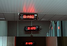
Displays showing ambient radiation fields of 0.120–0.130 μSv/h (1.05–1.14 mSv/a) in a nuclear power plant. This reading includes natural background from cosmic and terrestrial sources.
 

Atmospheric nuclear testing

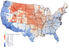
Per capita thyroid doses in the continental United States resulting from all exposure routes from all atmospheric nuclear tests conducted at the Nevada Test Site from 1951–1962.
 
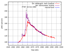
Atmospheric 14C, New Zealand and Austria. The New Zealand curve is representative for the Southern Hemisphere, the Austrian curve is representative for the Northern Hemisphere. Atmospheric nuclear weapon tests almost doubled the concentration of 14C in the Northern Hemisphere.
 
Frequent above-ground nuclear explosions between the 1940s and 1960s scattered a substantial amount of radioactive contamination. Some of this contamination is local, rendering the immediate surroundings highly radioactive, while some of it is carried longer distances as nuclear fallout; some of this material is dispersed worldwide. The increase in background radiation due to these tests peaked in 1963 at about 0.15 mSv per year worldwide, or about 7% of average background dose from all sources. The Limited Test Ban Treaty of 1963 prohibited above-ground tests, thus by the year 2000 the worldwide dose from these tests has decreased to only 0.005 mSv per year.

Occupational exposure

The International Commission on Radiological Protection recommends limiting occupational radiation exposure to 50 mSv (5 rem) per year, and 100 mSv (10 rem) in 5 years.

However, background radiation for occupational doses includes radiation that is not measured by radiation dose instruments in potential occupational exposure conditions. This includes both offsite "natural background radiation" and any medical radiation doses. This value is not typically measured or known from surveys, such that variations in the total dose to individual workers is not known. This can be a significant confounding factor in assessing radiation exposure effects in a population of workers who may have significantly different natural background and medical radiation doses. This is most significant when the occupational doses are very low.

At an IAEA conference in 2002, it was recommended that occupational doses below 1–2 mSv per year do not warrant regulatory scrutiny.

Nuclear accidents

Under normal circumstances, nuclear reactors release small amounts of radioactive gases, which cause small radiation exposures to the public. Events classified on the International Nuclear Event Scale as incidents typically do not release any additional radioactive substances into the environment. Large releases of radioactivity from nuclear reactors are extremely rare. To the present day, there were two major civilian accidents – the Chernobyl accident and the Fukushima I nuclear accidents – which caused substantial contamination [DJS -- What is "substantial"?]. The Chernobyl accident was the only one to cause immediate deaths.

Total doses from the Chernobyl accident ranged from 10 to 50 mSv over 20 years for the inhabitants of the affected areas, with most of the dose received in the first years after the disaster, and over 100 mSv for liquidators. There were 28 deaths from acute radiation syndrome.

Total doses from the Fukushima I accidents were between 1 and 15 mSv for the inhabitants of the affected areas. Thyroid doses for children were below 50 mSv. 167 cleanup workers received doses above 100 mSv, with 6 of them receiving more than 250 mSv (the Japanese exposure limit for emergency response workers).

The average dose from the Three Mile Island accident was 0.01 mSv.

Non-civilian: In addition to the civilian accidents described above, several accidents at early nuclear weapons facilities – such as the Windscale fire, the contamination of the Techa River by the nuclear waste from the Mayak compound, and the Kyshtym disaster at the same compound – released substantial radioactivity into the environment. The Windscale fire resulted in thyroid doses of 5–20 mSv for adults and 10–60 mSv for children. The doses from the accidents at Mayak are unknown.

Nuclear fuel cycle

The Nuclear Regulatory Commission, the United States Environmental Protection Agency, and other U.S. and international agencies, require that licensees limit radiation exposure to individual members of the public to 1 mSv (100 mrem) per year.

Other

Coal plants emit radiation in the form of radioactive fly ash which is inhaled and ingested by neighbours, and incorporated into crops. A 1978 paper from Oak Ridge National Laboratory estimated that coal-fired power plants of that time may contribute a whole-body committed dose of 19 µSv/a to their immediate neighbours in a radius of 500 m. The United Nations Scientific Committee on the Effects of Atomic Radiation's 1988 report estimated the committed dose 1 km away to be 20 µSv/a for older plants or 1 µSv/a for newer plants with improved fly ash capture, but was unable to confirm these numbers by test. When coal is burned, uranium, thorium and all the uranium daughters accumulated by disintegration — radium, radon, polonium — are released. Radioactive materials previously buried underground in coal deposits are released as fly ash or, if fly ash is captured, may be incorporated into concrete manufactured with fly ash.

Other sources of dose uptake

Medical

The global average human exposure to artificial radiation is 0.6 mSv/a, primarily from medical imaging. This medical component can range much higher, with an average of 3 mSv per year across the USA population. Other human contributors include smoking, air travel, radioactive building materials, historical nuclear weapons testing, nuclear power accidents and nuclear industry operation.
A typical chest x-ray delivers 20 µSv (2 mrem) of effective dose. A dental x-ray delivers a dose of 5 to 10 µSv. A CT scan delivers an effective dose to the whole body ranging from 1 to 20 mSv (100 to 2000 mrem). The average American receives about 3 mSv of diagnostic medical dose per year; countries with the lowest levels of health care receive almost none. Radiation treatment for various diseases also accounts for some dose, both in individuals and in those around them.

Consumer items

Cigarettes contain polonium-210, originating from the decay products of radon, which stick to tobacco leaves. Heavy smoking results in a radiation dose of 160 mSv/year to localized spots at the bifurcations of segmental bronchi in the lungs from the decay of polonium-210. This dose is not readily comparable to the radiation protection limits, since the latter deal with whole body doses, while the dose from smoking is delivered to a very small portion of the body.

Radiation metrology

In a radiation metrology laboratory, background radiation refers to the measured value from any incidental sources that affect an instrument when a specific radiation source sample is being measured. This background contribution, which is established as a stable value by multiple measurements, usually before and after sample measurement, is subtracted from the rate measured when the sample is being measured.

This is in accordance with the International Atomic Energy Agency definition of background as being "Dose or dose rate (or an observed measure related to the dose or dose rate) attributable to all sources other than the one(s) specified.

The same issue occurs with radiation protection instruments, where a reading from an instrument may be affected by the background radiation. An example of this is a scintillation detector used for surface contamination monitoring. In an elevated gamma background the scintillator material will be affected by the background gamma, which will add to the reading obtained from any contamination which is being monitored. In extreme cases it will make the instrument unusable as the background swamps the lower level of radiation from the contamination. In such instruments the background can be continually monitored in the "Ready" state, and subtracted from any reading obtained when being used in "Measuring" mode.

Regular Radiation measurement is carried out at multiple levels. Government agencies compile radiation readings as part of environmental monitoring mandates, often making the readings available to the public and sometimes in near-real-time. Collaborative groups and private individuals may also make real-time readings available to the public. Instruments used for radiation measurement include the Geiger–Müller tube and the Scintillation detector. The former is usually more compact and affordable and reacts to several radiation types, while the latter is more complex and can detect specific radiation energies and types. Readings indicate radiation levels from all sources including background, and real-time readings are in general unvalidated, but correlation between independent detectors increases confidence in measured levels. 

List of near-real-time government radiation measurement sites, employing multiple instrument types:
List of international near-real-time collaborative/private measurement sites, employing primarily Geiger-Muller detectors:

Extraterrestrial liquid water

From Wikipedia, the free encyclopedia https://en.wikipedia.org/wiki/Extraterrestrial_liquid_water ...