Search This Blog

Thursday, February 13, 2020

Fusiform face area

From Wikipedia, the free encyclopedia
 
Fusiform face area
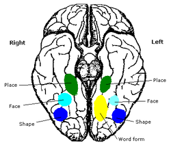
Constudproc - inferior view.png
Human brain, bottom view. Fusiform face area shown in bright blue.
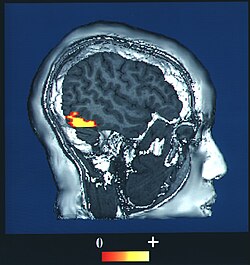
Fusiform face area face recognition.jpg
Computer-enhanced fMRI scan of a person who has been asked to look at faces. The image shows increased blood flow in cerebral cortex that recognizes faces (FFA).

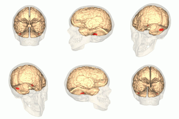
The fusiform face area - FFA (meaning: spindular/spindle-shaped face area) is a part of the human visual system that is specialized for facial recognition. It is located in the Inferior temporal cortex (IT), in the fusiform gyrus (Brodmann area 37).

Structure

The FFA is located in the ventral stream on the ventral surface of the temporal lobe on the lateral side of the fusiform gyrus. It is lateral to the parahippocampal place area. It displays some lateralization, usually being larger in the right hemisphere

The FFA was discovered and continues to be investigated in humans using positron emission tomography (PET) and functional magnetic resonance imaging (fMRI) studies. Usually, a participant views images of faces, objects, places, bodies, scrambled faces, scrambled objects, scrambled places, and scrambled bodies. This is called a functional localizer. Comparing the neural response between faces and scrambled faces will reveal areas that are face-responsive, while comparing cortical activation between faces and objects will reveal areas that are face-selective. 

Function

The human FFA was first described by Justine Sergent in 1992 and later named by Nancy Kanwisher in 1997 who proposed that the existence of the FFA is evidence for domain specificity in the visual system. Studies have recently shown that the FFA is composed of functional clusters that are at a finer spatial scale than prior investigations have measured. Electrical stimulation of these functional clusters selectively distorts face perception, which is causal support for the role of these functional clusters in perceiving the facial image. While it is generally agreed that the FFA responds more to faces than to most other categories, there is debate about whether the FFA is uniquely dedicated to face processing, as proposed by Nancy Kanwisher and others, or whether it participates in the processing of other objects. The expertise hypothesis, as championed by Isabel Gauthier and others, offers an explanation for how the FFA becomes selective for faces in most people. The expertise hypothesis suggests that the FFA is a critical part of a network that is important for individuating objects that are visually similar because they share a common configuration of parts. Gauthier et al., in an adversarial collaboration with Kanwisher, tested both car and bird experts, and found some activation in the FFA when car experts were identifying cars and when bird experts were identifying birds. This finding has been replicated, and expertise effects in the FFA have been found for other categories such as chess displays and x-rays. Recently, it was found that the thickness of the cortex in the FFA predicts the ability to recognize faces as well as vehicles.

A 2009 magnetoencephalography study found that objects incidentally perceived as faces, an example of pareidolia, evoke an early (165-millisecond) activation in the FFA, at a time and location similar to that evoked by faces, whereas other common objects do not evoke such activation. This activation is similar to a face-specific ERP component N170. The authors suggest that face perception evoked by face-like objects is a relatively early process, and not a late cognitive reinterpretation phenomenon.

One case study of agnosia provided evidence that faces are processed in a special way. A patient known as C. K., who suffered brain damage as a result of a car accident, later developed object agnosia. He experienced great difficulty with basic-level object recognition, also extending to body parts, but performed very well at recognizing faces. A later study showed that C. K. was unable to recognize faces that were inverted or otherwise distorted, even in cases where they could easily be identified by normal subjects. This is taken as evidence that the fusiform face area is specialized for processing faces in a normal orientation.

Studies using functional magnetic resonance imaging and electrocorticography have demonstrated that activity in the FFA codes for individual faces and the FFA is tuned for behaviorally relevant facial features. An electrocorticography study found that the FFA is involved in multiple stages of face processing, continuously from when people see a face until they respond to it, demonstrating the dynamic and important role the FFA plays as part of the face perception network.

Another study found that there is stronger activity in the FFA when a person sees a familiar face as opposed to an unfamiliar one. Participants were shown different pictures of faces that either had the same identity, familiar, or faces with separate identities, or unfamiliar. It found that participants were more accurate at matching familiar faces than unfamiliar ones. Using an fMRI, they also found that the participants that were more accurate in identifying familiar faces had more activity in their right fusiform face area and participants that were poor at matching had less activity in their right fusiform area.

History


Function and controversy

The fusiform face area (FFA) is a part of the brain located in the fusiform gyrus with a debated purpose. Some researchers believe that the FFA is evolutionary purposed for face perception. Others believe that the FFA discriminates between any familiar stimuli. 

Psychologists debate whether the FFA is activated by faces for an evolutionary or expertise reason. The conflicting hypotheses stem from the ambiguity in FFA activation, as the FFA is activated by both familiar objects and faces. A study regarding novel objects called greebles determined this phenomenon. When first exposed to greebles, a person's FFA was activated more strongly by faces than by greebles. After familiarising themselves with individual greebles or becoming a greeble expert, a person's FFA was activated equally by faces and greebles. Likewise, children with autism have been shown to develop object recognition at a similarly impaired pace as face recognition. Studies of late patients of autism have discovered that autistic people have lower neuron densities in the FFA This raises an interesting question, however: Is the poor face perception due to a reduced number of cells or is there a reduced number of cells because autistic people seldom perceive faces? Asked simply: Are faces simply objects with which every person has expertise? 

Chinese characters similar to those used in Fu et al., which elicit a response in the FFA

There is evidence supporting the FFA's evolutionary face-perception. Case studies into other dedicated areas of the brain may suggest that the FFA is intrinsically designed to recognize faces. Other studies have recognized areas of the brain essential to recognizing environments and bodies. Without these dedicated areas, people are incapable of recognizing places and bodies. Similar research regarding prosopagnosia has determined that the FFA is essential to the recognition of unique faces. However, these patients are capable of recognizing the same people normally by other means, such as voice. Studies involving language characters have also been conducted in order to ascertain the role of the FFA in face recognition. These studies have found that objects, such as Chinese characters, elicit a high response in different areas of the FFA than those areas that elicit a high response from faces. This data implies that certain areas of the FFA have evolutionary face-perception purposes.

Evidence from infants

The FFA is underdeveloped in children and does not fully develop until adolescence. This calls into question the evolutionary purpose of the FFA, as children show the ability to differentiate faces. Two-year-old babies have been shown to prefer the face of their mother. Although the FFA is underdeveloped in two-year-old babies, they have the ability to recognize their mother. Babies as early as three months old have shown the ability to distinguish between faces. During this time, babies exhibit the ability to differentiate between genders, showing a clear preference for female faces. It is theorized that, in terms of evolution, babies focus on women for food, although the preference could simply reflect a bias for the caregivers they experience. Infants do not appear to use this area for the perception of faces. Recent fMRI work has found no face selective area in the brain of infants 4 to 6 months old. However, given that the adult human brain has been studied far more extensively than the infant brain, and that infants are still undergoing major neurodevelopmental processes, it may simply be that the FFA is not located in anatomically familiar area. It may also be that activation for many different percepts and cognitive tasks in infants is diffuse in terms of neural circuitry, as infants are still undergoing periods of neurogenesis and neural pruning; this may make it more difficult to distinguish the signal, or what we would imagine as visual and complex familiar objects (like faces), from the noise, including static firing rates of neurons, and activity that is dedicated to a different task entirely than the activity of face processing. Infant vision involves only light and dark recognition, recognizing only major features of the face, activating the amygdala. These findings question the evolutionary purpose of the FFA. 

Evidence from emotions

Studies into what else may trigger the FFA validates arguments about its evolutionary purpose. There are countless facial expressions humans use that disturb the structure of the face. These disruptions and emotions are first processed in the amygdala and later transmitted to the FFA for facial recognition. This data is then used by the FFA to determine more static information about the face. The fact that the FFA is so far downstream in the processing of emotion suggests that it has little to do with emotion perception and instead deals in face perception.

Recent evidence, however, shows that the FFA has other functions regarding emotion. The FFA is differentially activated by faces exhibiting different emotions. A study has determined that the FFA is activated more strongly by fearful faces than neutral faces. This implies that the FFA has functions in processing emotion despite its downstream processing and questions its evolutionary purpose to identify faces. 

Additional images

Functional specialization (brain)

 
Functional specialization suggests that different areas in the brain are specialized for different functions.


Historical origins

1848 edition of American Phrenological Journal published by Fowlers & Wells, New York City
 
Phrenology, created by Franz Joseph Gall (1758–1828) and Johann Gaspar Spurzheim (1776–1832) and best known for the idea that one's personality could be determined by the variation of bumps on their skull, proposed that different regions in one's brain have different functions and may very well be associated with different behaviours. Gall and Spurzheim were the first to observe the crossing of pyramidal tracts, thus explaining why lesions in one hemisphere are manifested in the opposite side of the body. However, Gall and Spurzheim did not attempt to justify phrenology on anatomical grounds. It has been argued that phrenology was fundamentally a science of race. Gall considered the most compelling argument in favor of phrenology the differences in skull shape found in sub-Saharan Africans and the anecdotal evidence (due to scientific travelers and colonists) of their intellectual inferiority and emotional volatility. In Italy, Luigi Rolando carried out lesion experiments and performed electrical stimulation of the brain, including the Rolandic area

A
Phineas Gage's accident

Phineas Gage became one of the first lesion case studies in 1848 when an explosion drove a large iron rod completely through his head, destroying his left frontal lobe. He recovered with no apparent sensory, motor, or gross cognitive deficits, but with behaviour so altered that friends described him as "no longer being Gage," suggesting that the damaged areas are involved in "higher functions" such as personality. However, Gage's mental changes are usually grossly exaggerated in modern presentations. 

Subsequent cases (such as Broca's patient Tan) gave further support to the doctrine of specialization. 

Major theories of the brain

Currently, there are two major theories of the brain's cognitive function. The first is the theory of modularity. Stemming from phrenology, this theory supports functional specialization, suggesting the brain has different modules that are domain specific in function. The second theory, distributive processing, proposes that the brain is more interactive and its regions are functionally interconnected rather than specialized. Each orientation plays a role within certain aims and tend to complement each other (see below section `Collaboration´).

Modularity

The theory of modularity suggests that there are functionally specialized regions in the brain that are domain specific for different cognitive processes. Jerry Fodor expanded the initial notion of phrenology by creating his Modularity of the Mind theory. The Modularity of the Mind theory indicates that distinct neurological regions called modules are defined by their functional roles in cognition. He also rooted many of his concepts on modularity back to philosophers like Descartes, who wrote about the mind being composed of "organs" or "psychological faculties". An example of Fodor's concept of modules is seen in cognitive processes such as vision, which have many separate mechanisms for colour, shape and spatial perception.

One of the fundamental beliefs of domain specificity and the theory of modularity suggests that it is a consequence of natural selection and is a feature of our cognitive architecture. Researchers Hirschfeld and Gelman propose that because the human mind has evolved by natural selection, it implies that enhanced functionality would develop if it produced an increase in "fit" behaviour. Research on this evolutionary perspective suggests that domain specificity is involved in the development of cognition because it allows one to pinpoint adaptive problems.

An issue for the modular theory of cognitive neuroscience is that there are cortical anatomical differences from person to person. Although many studies of modularity are undertaken from very specific lesion case studies, the idea is to create a neurological function map that applies to people in general. To extrapolate from lesion studies and other case studies this requires adherence to the universality assumption, that there is no difference, in a qualitative sense, between subjects who are intact neurologically. For example, two subjects would fundamentally be the same neurologically before their lesions, and after have distinctly different cognitive deficits. Subject 1 with a lesion in the "A" region of the brain may show impaired functioning in cognitive ability "X" but not "Y", while subject 2 with a lesion in area "B" demonstrates reduced "Y" ability but "X" is unaffected; results like these allow inferences to be made about brain specialization and localization, also known as using a double dissociation.

The difficulty with this theory is that in typical non-lesioned subjects, locations within the brain anatomy are similar but not completely identical. There is a strong defense for this inherent deficit in our ability to generalize when using functional localizing techniques (fMRI, PET etc.). To account for this problem, the coordinate-based Talairach and Tournoux stereotaxic system is widely used to compare subjects' results to a standard brain using an algorithm. Another solution using coordinates involves comparing brains using sulcal reference points. A slightly newer technique is to use functional landmarks, which combines sulcal and gyral landmarks (the groves and folds of the cortex) and then finding an area well known for its modularity such as the fusiform face area. This landmark area then serves to orient the researcher to the neighboring cortex.

Future developments for modular theories of neuropsychology may lie in "modular psychiatry". The concept is that a modular understanding of the brain and advanced neuro-imaging techniques will allow for a more empirical diagnosis of mental and emotional disorders. There has been some work done towards this extension of the modularity theory with regards to the physical neurological differences in subjects with depression and schizophrenia, for example. Zielasek and Gaeble have set out a list of requirements in the field of neuropsychology in order to move towards neuropsychiatry:
  1. To assemble a complete overview of putative modules of the human mind
  2. To establish module-specific diagnostic tests (specificity, sensitivity, reliability)
  3. To assess how far individual modules, sets of modules or their connections are affected in certain psychopathological situations
  4. To probe novel module-specific therapies like the facial affect recognition training or to retrain access to context information in the case of delusions and hallucinations, in which "hyper-modularity" may play a role [8]
Research in the study of brain function can also be applied to cognitive behaviour therapy. As therapy becomes increasingly refined, it is important to differentiate cognitive processes in order to discover their relevance towards different patient treatments. An example comes specifically from studies on lateral specialization between the left and right cerebral hemispheres of the brain. The functional specialization of these hemispheres are offering insight on different forms of cognitive behaviour therapy methods, one focusing on verbal cognition (the main function of the left hemisphere) and the other emphasizing imagery or spatial cognition (the main function of the right hemisphere). Examples of therapies that involve imagery, requiring right hemisphere activity in the brain, include systematic desensitization and anxiety management training. Both of these therapy techniques rely on the patient's ability to use visual imagery to cope with or replace patients symptoms, such as anxiety. Examples of cognitive behaviour therapies that involve verbal cognition, requiring left hemisphere activity in the brain, include self-instructional training and stress inoculation. Both of these therapy techniques focus on patients' internal self-statements, requiring them to use vocal cognition. When deciding which cognitive therapy to employ, it is important to consider the primary cognitive style of the patient. Many individuals have a tendency to prefer visual imagery over verbalization and vice versa. One way of figuring out which hemisphere a patient favours is by observing their lateral eye movements. Studies suggest that eye gaze reflects the activation of cerebral hemisphere contralateral to the direction. Thus, when asking questions that require spatial thinking, individuals tend to move their eyes to the left, whereas when asked questions that require verbal thinking, individuals usually move their eyes to the right. In conclusion, this information allows one to choose the optimal cognitive behaviour therapeutic technique, thereby enhancing the treatment of many patients.

Areas representing modularity in the brain

 
Fusiform face area
One of the most well known examples of functional specialization is the fusiform face area (FFA). Justine Sergent was one of the first researchers that brought forth evidence towards the functional neuroanatomy of face processing. Using positron emission tomography (PET), Sergent found that there were different patterns of activation in response to the two different required tasks, face processing verses object processing. These results can be linked with her studies of brain-damaged patients with lesions in the occipital and temporal lobes. Patients revealed that there was an impairment of face processing but no difficulty recognizing everyday objects, a disorder also known as prosopagnosia. Later research by Nancy Kanwisher using functional magnetic resonance imaging (fMRI), found specifically that the region of the inferior temporal cortex, known as the fusiform gyrus, was significantly more active when subjects viewed, recognized and categorized faces in comparison to other regions of the brain. Lesion studies also supported this finding where patients were able to recognize objects but unable to recognize faces. This provided evidence towards domain specificity in the visual system, as Kanwisher acknowledges the Fusiform Face Area as a module in the brain, specifically the extrastriate cortex, that is specialized for face perception.

Visual area V4 and V5
While looking at the regional cerebral blood flow (rCBF), using PET, researcher Semir Zeki directly demonstrated functional specialization within the visual cortex known as visual modularity. He localized regions involved specifically in the perception of colour and vision motion. For colour, visual area V4 was located when subjects were shown two identical displays, one being multicoloured and the other shades of grey. This was further supported from lesion studies where individuals were unable to see colours after damage, a disorder known as achromatopsia. Combining PET and magnetic resonance imaging (MRI), subjects viewing a moving checker board pattern verses a stationary checker board pattern located visual area V5, which is now considered to be specialized for vision motion. (Watson et al., 1993) This area of functional specialization was also supported by lesion study patients who's damage caused cerebral motion blindness.

Frontal lobes
Studies have found the frontal lobes to be involved in the executive functions of the brain, which are higher level cognitive processes. This control process is involved in the coordination, planning and organizing of actions towards an individual's goals. It contributes to such things as one's behaviour, language and reasoning. More specifically, it was found to be the function of the prefrontal cortex, and evidence suggest that these executive functions control processes such as planning and decision making, error correction and assisting overcoming habitual responses. Miller and Cummings used PET and functional magnetic imaging (fMRI) to further support functional specialization of the frontal cortex. They found lateralization of verbal working memory in the left frontal cortex and visuospatial working memory in the right frontal cortex. Lesion studies support these findings where left frontal lobe patients exhibited problems in controlling executive functions such as creating strategies. The dorsolateral, ventrolateral and anterior cingulate regions within the prefrontal cortex are proposed to work together in different cognitive tasks, which is related to interaction theories. However, there has also been evidence suggesting strong individual specializations within this network. For instance, Miller and Cummings found that the dorsolateral prefrontal cortex is specifically involved in the manipulation and monitoring of sensorimotor information within working memory.

Right and left hemispheres
During the 1960s, Roger Sperry conducted a natural experiment on epileptic patients who had previously had their corpora callosa cut. The corpus callosum is the area of the brain dedicated to linking both the right and left hemisphere together. Sperry et al.'s experiment was based on flashing images in the right and left visual fields of his participants. Because the participant's corpus callosum was cut, the information processed by each visual field could not be transmitted to the other hemisphere. In one experiment, Sperry flashed images in the right visual field (RVF), which would subsequently be transmitted to the left hemisphere (LH) of the brain. When asked to repeat what they had previously seen, participants were fully capable of remembering the image flashed. However, when the participants were then asked to draw what they had seen, they were unable to. When Sperry et al. flashed images in the left visual field (LVF), the information processed would be sent to the right hemisphere (RH) of the brain. When asked to repeat what they had previously seen, participants were unable to recall the image flashed, but were very successful in drawing the image. Therefore, Sperry concluded that the left hemisphere of the brain was dedicated to language as the participants could clearly speak the image flashed. On the other hand, Sperry concluded that the right hemisphere of the brain was involved in more creative activities such as drawing.

Parahippocampal place area
Located in the parahippocampal gyrus, the parahippocampal place area (PPA) was coined by Nancy Kanwisher and Russell Epstein after an fMRI study showed that the PPA responds optimally to scenes presented containing a spatial layout, minimally to single objects and not at all to faces. It was also noted in this experiment that activity remains the same in the PPA when viewing a scene with an empty room or a room filled with meaningful objects. Kanwisher and Epstein proposed "that the PPA represents places by encoding the geometry of the local environment". In addition, Soojin Park and Marvin Chun posited that activation in the PPA is viewpoint specific, and so responds to changes in the angle of the scene. In contrast, another special mapping area, the retrosplenial cortex (RSC), is viewpoint invariant or does not change response levels when views change. This perhaps indicates a complementary arrangement of functionally and anatomically separate visual processing brain areas.

Extrastriate body area
Located in the lateral occipitotemporal cortex, fMRI studies have shown the extrastriate body area (EBA) to have selective responding when subjects see human bodies or body parts, implying that it has functional specialization. The EBA does not optimally respond to objects or parts of objects but to human bodies and body parts, a hand for example. In fMRI experiments conducted by Downing et al. participants were asked to look at a series of pictures. These stimuli includes objects, parts of objects (for example just the head of a hammer), figures of the human body in all sorts of positions and types of detail (including line drawings or stick men), and body parts (hands or feet) without any body attached. There was significantly more blood flow (and thus activation) to human bodies, no matter how detailed, and body parts than to objects or object parts.

Distributive processing

The cognitive theory of distributed processing suggests that brain areas are highly interconnected and process information in a distributed manner. 

A remarkable precedent of this orientation is the research of Justo Gonzalo on brain dynamics  where several phenomena that he observed could not be explained by the traditional theory of localizations. From the gradation he observed between different syndromes in patients with different cortical lesions, this author proposed in 1952 a functional gradients model, which permits an ordering and an interpretation of multiple phenomena and syndromes. The functional gradients are continuous functions through the cortex describing a distributed specificity, so that, for a given sensory system, the specific gradient, of contralateral character, is maximum in the corresponding projection area and decreases in gradation towards more "central" zone and beyond so that the final decline reaches other primary areas. As a consequence of the crossing and overlapping of the specific gradients, in the central zone where the overlap is greater, there would be an action of mutual integration, rather nonspecific (or multisensory) with bilateral character due to the corpus callosum. This action would be maximum in the central zone and minimal towards the projection areas. As the author stated (p. 20 of the English translation) "a functional continuity with regional variation is then offered, each point of the cortex acquiring different properties but with certain unity with the rest of the cortex. It is a dynamic conception of quantitative localizations". A very similar gradients scheme was proposed by Elkhonon Goldberg in 1989.

Other researchers who provide evidence to support the theory of distributive processing include Anthony McIntosh and William Uttal, who question and debate localization and modality specialization within the brain. McIntosh's research suggests that human cognition involves interactions between the brain regions responsible for processes sensory information, such as vision, audition, and other mediating areas like the prefrontal cortex. McIntosh explains that modularity is mainly observed in sensory and motor systems, however, beyond these very receptors, modularity becomes "fuzzier" and you see the cross connections between systems increase. He also illustrates that there is an overlapping of functional characteristics between the sensory and motor systems, where these regions are close to one another. These different neural interactions influence each other, where activity changes in one area influence other connected areas. With this, McIntosh suggest that if you only focus on activity in one area, you may miss the changes in other integrative areas. Neural interactions can be measured using analysis of covariance in neuroimaging. McIntosh used this analysis to convey a clear example of the interaction theory of distributive processing. In this study, subjects learned that an auditory stimulus signalled a visual event. McIntosh found activation (an increase blood flow), in an area of the occipital cortex, a region of the brain involved in visual processing, when the auditory stimulus was presented alone. Correlations between the occipital cortex and different areas of the brain such as the prefrontal cortex, premotor cortex and superior temporal cortex showed a pattern of co-variation and functional connectivity.

Uttal focusses on the limits of localizing cognitive processes in the brain. One of his main arguments is that since the late 90's, research in cognitive neuroscience has forgotten about conventional psychophysical studies based on behavioural observation. He believes that current research focusses on the technological advances of brain imaging techniques such as MRI and PET scans. Thus, he further suggest that this research is dependent on the assumptions of localization and hypothetical cognitive modules that use such imaging techniques to pursuit these assumptions. Uttal's major concern incorporates many controversies with the validly, over-assumptions and strong inferences some of these images are trying to illustrate. For instance, there is concern over the proper utilization of control images in an experiment. Most of the cerebrum is active during cognitive activity, therefore the amount of increased activity in a region must be greater when compared to a controlled area. In general, this may produce false or exaggerated findings and may increase potential tendency to ignore regions of diminished activity which may be crucial to the particular cognitive process being studied. Moreover, Uttal believes that localization researchers tend to ignore the complexity of the nervous system. Many regions in the brain are physically interconnected in a nonlinear system, hence, Uttal believes that behaviour is produced by a variety of system organizations.

Collaboration

The two theories, modularity and distributive processing, can also be combined. By operating simultaneously, these principles may interact with each other in a collaborative effort to characterize the functioning of the brain. Fodor himself, one of the major contributors to the modularity theory, appears to have this sentiment. He noted that modularity is a matter of degrees, and that the brain is modular to the extent that it warrants studying it in regards to its functional specialization. Although there are areas in the brain that are more specialized for cognitive processes than others, the nervous system also integrates and connects the information produced in these regions. In fact, the proposed distributive scheme of the functional cortical gradientes by J. Gonzalo already tries to join both concepts modular and distributive: regional heterogeneity should be a definitive acquisition (maximum specificity in the projection paths and primary areas), but the rigid separation between projection and association areas would be erased through the continuous functions of gradient.

The collaboration between the two theories not only would provide a more unified perception and understanding of the world but also make available the ability to learn from it.

Lake Tanganyika

From Wikipedia, the free encyclopedia
 
Lake Tanganyika
STS51G-034-0012 Lake Tanganyika June1985.jpg
Lake Tanganyika from space, June 1985
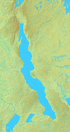
Lake Tanganyika map.png
Lake Tanganyika map
Coordinates6°30′S 29°50′ECoordinates: 6°30′S 29°50′E
Lake typeAncient lake, Rift Valley Lake
Primary inflowsRuzizi River
Malagarasi River
Kalambo River
Primary outflowsLukuga River
Catchment area231,000 km2 (89,000 sq mi)
Basin countriesBurundi
East Congo (DRC)
Tanzania
Zambia

Max. length673 km (418 mi)
Max. width72 km (45 mi)
Surface area32,900 km2 (12,700 sq mi)
Average depth570 m (1,870 ft)
Max. depth1,470 m (4,820 ft)
Water volume18,900 km3 (4,500 cu mi)
Residence time5500 years
Shore length11,828 km (1,136 mi)
Surface elevation773 m (2,536 ft)

SettlementsKigoma, Tanzania
Kalemie, DRC
Bujumbura, Burundi

Official nameTanganyika
Designated2 February 2007
Reference no.1671
1 Shore length is not a well-defined measure.

Lake Tanganyika is an African Great Lake. It is the second-oldest freshwater lake in the world, the second-largest by volume, and the second-deepest, in all cases after Lake Baikal in Siberia. It is the world's longest freshwater lake. The lake is shared between four countries – Tanzania, the Democratic Republic of the Congo (DRC), Burundi, and Zambia, with Tanzania (46%) and DRC (40%) possessing the majority of the lake. It drains into the Congo River system and ultimately into the Atlantic Ocean.

Etymology

The name "Tanganyika" apparently refers to "the great lake spreading out like a plain", or "plain-like lake".

Geography and geological history

Lake Tanganyika is situated within the Albertine Rift, the western branch of the East African Rift, and is confined by the mountainous walls of the valley. It is the largest rift lake in Africa and the second-largest lake by volume in the world. It is the deepest lake in Africa and holds the greatest volume of fresh water, accounting for 16% of the world's available fresh water. It extends for 676 km (420 mi) in a general north-south direction and averages 50 km (31 mi) in width. The lake covers 32,900 km2 (12,700 sq mi), with a shoreline of 1,828 km (1,136 mi), a mean depth of 570 m (1,870 ft) and a maximum depth of 1,471 m (4,826 ft) (in the northern basin). It holds an estimated 18,900 cubic kilometres (4,500 cu mi).

The catchment area of the lake is 231,000 km2 (89,000 sq mi). Two main rivers flow into the lake, as well as numerous smaller rivers and streams (whose lengths are limited by the steep mountains around the lake). The one major outflow is the Lukuga River, which empties into the Congo River drainage. Precipation and evaporation play a greater role than the rivers. At least 90% of the water influx is from rain falling on the lake's surface and at least 90% of the water loss is from direct evaporation.

The major river flowing into the lake is the Ruzizi River, formed about 10,000 years ago, which enters the north of the lake from Lake Kivu. The Malagarasi River, which is Tanzania's second largest river, enters the east side of Lake Tanganyika. The Malagarasi is older than Lake Tanganyika, and before the lake was formed, it probably was a headwater of the Lualaba River, the main Congo River headstream.

The lake has a complex history of changing flow patterns, due to its high altitude, great depth, slow rate of refill, and mountainous location in a turbulently volcanic area that has undergone climate changes. Apparently, it has rarely in the past had an outflow to the sea. It has been described as "practically endorheic" for this reason. The lake's connection to the sea is dependent on a high water level allowing water to overflow out of the lake through the Lukuga River into the Congo. When not overflowing, the lake's exit into the Lukuga River typically is blocked by sand bars and masses of weed, and instead this river depends on its own tributaries, especially the Niemba River, to maintain a flow.

Due to the lake's tropical location, it has a high rate of evaporation. Thus, it depends on a high inflow through the Ruzizi out of Lake Kivu to keep the lake high enough to overflow. This outflow is apparently not more than 12,000 years old, and resulted from lava flows blocking and diverting the Kivu basin's previous outflow into Lake Edward and then the Nile system, and diverting it to Lake Tanganyika. Signs of ancient shorelines indicate that at times, Tanganyika may have been up to 300 m (980 ft) lower than its present surface level, with no outlet to the sea. Even its current outlet is intermittent, thus may not have been operating when first visited by Western explorers in 1858.

The lake may also have at times had different inflows and outflows; inward flows from a higher Lake Rukwa, access to Lake Malawi and an exit route to the Nile have all been proposed to have existed at some point in the lake's history.

Lake Tanganyika is an ancient lake. Its three basins, which in periods with much lower water levels were separate lakes, are of different ages. The central began to form 9–12 million years ago (Mya), the northern 7–8 Mya and the southern 2–4 Mya.

Islands

Of the several islands in Lake Tanganyika, the most important are:
  • Kavala Island (DRC)
  • Mamba-Kayenda Islands (DRC)
  • Milima Island (DRC)
  • Kibishie Island (DRC)
  • Mutondwe Island (Zambia)
  • Kumbula Island (Zambia)

Water characteristics

The lake's water is alkaline with a pH around 9 at depths of 0–100 m (0–330 ft). Below this, it is around 8.7, gradually decreasing to 8.3–8.5 in the deepest parts of Tanganyika. A similar pattern can be seen in the electric conductivity, ranging from about 670 μS/cm in the upper part to 690 μS/cm in the deepest.

Surface temperatures generally range from about 24 °C (75 °F) in the southern part of the lake in early August to 28–29 °C (82–84 °F) in the late rainy season in March—April. At depths greater than 400 m (1,300 ft), the temperature is very stable at 23.1–23.4 °C (73.6–74.1 °F). The water has gradually warmed since the 1800s and this has accelerated with global warming since the 1950s.

The lake is stratified and seasonal mixing generally does not extend beyond depths of 150 m (490 ft). The mixing mainly occurs as upwellings in the south and is wind-driven, but to a lesser extent, up- and downwellings also occur elsewhere in the lake. As a consequence of the stratification, the deep sections contain "fossil water". This also means it has no oxygen (it is anoxic) in the deeper parts, essentially limiting fish and other aerobic organisms to the upper part. Some geographical variations are seen in this limit, but it is typically at depths around 100 m (330 ft) in the northern part of the lake and 240–250 m (790–820 ft) in the south. The oxygen-devoid deepest sections contain high levels of toxic hydrogen sulphide and are essentially lifeless, except for bacteria.

Biology


Reptiles

Lake Tanganyika and associated wetlands are home to Nile crocodiles (including famous giant Gustave), Zambian hinged terrapins, serrated hinged terrapins, and pan hinged terrapins (last species not in the lake itself, but in adjacent lagoons). Storm's water cobra, a threatened subspecies of banded water cobra that feeds mainly on fish, is only found in Lake Tanganyika, where it prefers rocky shores.

Cichlid fish

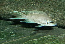
One of the many Tanganyika cichlids is Neolamprologus brichardi. The complex behaviors of this species and its close relative N. pulcher have been studied in detail
 
The lake holds at least 250 species of cichlid fish and undescribed species remain. Almost all (98%) of the Tanganyika cichlids are endemic to the lake and it is thus an important biological resource for the study of speciation in evolution. Some of the endemics do occur slightly into the upper Lukuga River, Lake Tanganyika's outflow, but further spread into the Congo River basin is prevented by physics (Lukuga has fast-flowing sections with many rapids and waterfalls) and chemistry (Tanganyika's water is alkaline, while the Congo's generally is acidic). The cichlids of the African Great Lakes, including Tanganyika, represent the most diverse extent of adaptive radiation in vertebrates.

Although Tanganyika has far fewer cichlid species than Lakes Malawi and Victoria which both have experienced relatively recent explosive species radiations (resulting in many closely related species), its cichlids are the most morphologically and genetically diverse. This is linked to the high age of Tanganyika, as it is far older than the other lakes. Tanganyika has the largest number of endemic cichlid genera of all African lakes. All Tanganyika cichlids are in the subfamily Pseudocrenilabrinae. Of the 10 tribes in this subfamily, half are largely or entirely restricted to the lake (Cyprichromini, Ectodini, Lamprologini, Limnochromini and Tropheini) and another three have species in the lake (Haplochromini, Tilapiini and Tylochromini). Others have proposed splitting the Tanganyika cichlids into as many as 12–16 tribes (in addition to previous mentioned, Bathybatini, Benthochromini, Boulengerochromini, Cyphotilapiini, Eretmodini, Greenwoodochromini, Perissodini and Trematocarini).

Most Tanganyika cichlids live along the shoreline down to a depth of 100 m (330 ft), but some deep-water species regularly descend to 200 m (660 ft). Trematocara species have exceptionally been found at more than 300 m (980 ft), which is deeper than any other cichlid in the world. Some of the deep-water cichlids (e.g., Bathybates, Gnathochromis, Hemibates and Xenochromis) have been caught in places virtually devoid of oxygen, but how they are able to survive there is unclear. Tanganyika cichlids are generally benthic (found at or near the bottom) and/or coastal. No Tanganyika cichlids are truly pelagic and offshore, except for some of the piscivorous Bathybates. Two of these, B. fasciatus and B. leo, mainly feed on Tanganyika sardines. Tanganyika cichlids differ extensively in ecology and include species that are herbivores, detritivores, planktivores, insectivores, molluscivores, scavengers, scale-eaters and piscivores. Their breeding behavior fall into two main groups, the substrate spawners (often in caves or rock crevices) and the mouthbrooders. Among the endemic species are two of the world's smallest cichlids, Neolamprologus multifasciatus and N. similis (both shell dwellers) at up to 4–5 cm (1.6–2.0 in), and one of the largest, the giant cichlid (Boulengerochromis microlepis) at up to 90 cm (3.0 ft).

Many cichlids from Lake Tanganyika, such as species from the genera Altolamprologus, Cyprichromis, Eretmodus, Julidochromis, Lamprologus, Neolamprologus, Tropheus and Xenotilapia, are popular aquarium fish due to their bright colors and patterns, and interesting behaviors. Recreating a Lake Tanganyika biotope to host those cichlids in a habitat similar to their natural environment is also popular in the aquarium hobby.

Other fish

The Tanganyika killifish (Lamprichthys tanganicanus) is the only member of its genus

Lake Tanganyika is home to more than 80 species of non-cichlid fish and about 60% of these are endemic. 

The open waters of the pelagic zone are dominated by four non-cichlid species: Two species of "Tanganyika sardine" (Limnothrissa miodon and Stolothrissa tanganicae) form the largest biomass of fish in this zone, and they are important prey for the forktail lates (Lates microlepis) and sleek lates (L. stappersii). Two additional lates are found in the lake, the Tanganyika lates (L. angustifrons) and bigeye lates (L. mariae), but both these are primarily benthic hunters, although they also may move into open waters. The four lates, all endemic to Tanganyika, have been overfished and larger individuals are rare today.

Among the more unusual fish in the lake are the endemic, facultatively brood parasitic "cuckoo catfish", including at least Synodontis grandiops and S. multipunctatus. A number of others are very similar (e.g., S. lucipinnis and S. petricola) and have often been confused; it is unclear if they have a similar behavior. The facultative brood parasites often lay their eggs synchronously with mouthbroding cichlids. The cichlid pick up the eggs in their mouth as if they were their own. Once the catfish eggs hatch the young eat the cichlid eggs. Six catfish genera are entirely restricted to the lake basin: Bathybagrus, Dinotopterus, Lophiobagrus, Phyllonemus, Pseudotanganikallabes and Tanganikallabes. Although not endemic on a genus level, six species of Chrysichthys catfish are only found in the Tanganyika basin where they live both in shallow and relatively deep waters; in the latter habitat they are the primary predators and scavengers. A unique evolutionary radiation in the lake is the 15 species of Mastacembelus spiny eels, all but one endemic to its basin. Although other African Great Lakes have Synodontis catfish, endemic catfish genera and Mastacembelus spiny eels, the relatively high diversity is unique to Tanganyika, which likely is related to its old age.

Among the non-endemic fish, some are widespread African species but several are only shared with the Malagarasi and Congo River basins, such as the Congo bichir (Polypterus congicus), goliath tigerfish (Hydrocynus goliath), Citharinus citharus, six-banded distichodus (Distichodus sexfasciatus) and mbu puffer (Tetraodon mbu).[50]

Molluscs and crustaceans

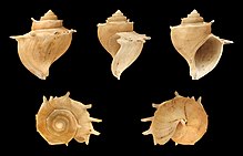
The shell of the endemic thalassoid freshwater snail Tiphobia horei with its elaborate shape and spines.
 
A total of 83 freshwater snail species (65 endemic) and 11 bivalve species (8 endemic) are known from the lake. Among the endemic bivalves are three monotypic genera: Grandidieria burtoni, Pseudospatha tanganyicensis and Brazzaea anceyi. Many of the snails are unusual for species living in freshwater in having noticeably thickened shells and/or distinct sculpture, features more commonly seen in marine snails. They are referred to as thalassoids, which can be translated to "marine-like". All the Tanganyika thalassoids, which are part of Prosobranchia, are endemic to the lake. Initially they were believed to be related to similar marine snails, but they are now known to be unrelated. Their appearance is now believed to be the result of the highly diverse habitats in Lake Tanganyika and evolutionary pressure from snail-eating fish and, in particular, Platythelphusa crabs. A total of 17 freshwater snail genera are endemic to the lake, such as Hirthia, Lavigeria, Paramelania, Reymondia, Spekia, Stanleya, Tanganyicia and Tiphobia. There are about 30 species of non-thalassoid snails in the lake, but only five of these are endemic, including Ferrissia tanganyicensis and Neothauma tanganyicense. The latter is the largest Tanganyika snail and its shell is often used by small shell-dwelling cichlids.

Crustaceans are also highly diverse in Tanganyika with more than 200 species, of which more than half are endemic. They include 10 species of freshwater crabs (9 Platythelphusa and Potamonautes platynotus; all endemic), at least 11 species of small atyid shrimp (Atyella, Caridella and Limnocaridina), an endemic palaemonid shrimp (Macrobrachium moorei), about 100 ostracods, including many endemics, and several copepods. Among these, Limnocaridina iridinae lives inside the mantle cavity of the unionid mussel Pleiodon spekei, making it one of only two known commensal species of freshwater shrimp (the other is the sponge-living Caridina spongicola from Lake Towuti, Indonesia).

Among Rift Valley lakes, Lake Tanganyika far surpasses all others in terms of crustacean and freshwater snail richness (both in total number of species and number of endemics). For example, the only other Rift Valley lake with endemic freshwater crabs are Lake Kivu and Lake Victoria with two species each.

Other invertebrates

The diversity of other invertebrate groups in Lake Tanganyika is often not well-known, but there are at least 20 described species of leeches (12 endemics), 9 sponges (7 endemic), 6 bryozoa (2 endemic), 11 flatworms (7 endemic), 20 nematodes (7 endemic), 28 annelids (17 endemic) and the small hydrozoan jellyfish Limnocnida tanganyicae.

Fishing

Fishermen on Lake Tanganyika

Lake Tanganyika supports a major fishery, which, depending on source, provides 25–40% or c. 60% of the animal protein in the diet of the people living in the region. Currently, there are around 100,000 people directly involved in the fisheries operating from almost 800 sites. The lake is also vital to the estimated 10 million people living in the greater basin.

Lake Tanganyika fish can be found exported throughout East Africa. Major commercial fishing began in the mid-1950s and has, together with global warming (limiting the habitat of temperature sensitive species), had a heavy impact on the fish populations, causing significant declines. In 2016, it was estimated that the total catch was up to 200,000 tonnes. Former industrial fisheries, which boomed in the 1980s, have subsequently collapsed.

Transport

Two ferries carry passengers and cargo along the eastern shore of the lake: MV Liemba between Kigoma and Mpulungu and MV Mwongozo between Kigoma and Bujumbura.
On Dec. 12, 2014, the ferry MV Mutambala capsized on Lake Tanganyika, and more than 120 lives were lost.

History

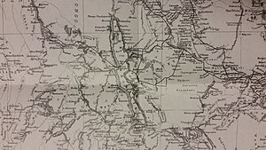
Lake Tanganyika. The black line indicates Henry Morton Stanley's route.

It is thought that early Homo sapiens were making an impact on the region during the stone age. The time period of the Middle Stone Age to Late Stone Age is described as an age of advanced hunter-gatherers. It is believed they would have caused megafaunal extinctions.

There are many methods in which the native people of the area were fishing. Most of them included using a lantern as a lure for fish that are attracted to light. There were three basic forms. One called Lusenga which is a wide net used by one person from a canoe. The second one is using a lift net. This was done by dropping a net deep below the boat using two parallel canoes and then simultaneously pulling it up. The third is called Chiromila which consisted of three canoes. One canoe was stationary with a lantern while another canoe holds one end of the net and the other circles the stationary one to meet up with the net.

The first known Westerners to find the lake were the British explorers Richard Burton and John Speke, in 1858. They located it while searching for the source of the Nile River. Speke continued and found the actual source, Lake Victoria. Later David Livingstone passed by the lake. He noted the name "Liemba" for its southern part, a word probably from the Fipa language, and in 1927 this was chosen as the new name for the conquered German First World War ship Graf von Götzen which is still serving the lake up to the present time.

World War I

The lake was the scene of two celebrated battles during World War I.

With the aid of the Graf Goetzen (named after Count Gustav Adolf Graf von Götzen, the former governor of German East Africa), the Germans had complete control of the lake in the early stages of the war. The ship was used both to ferry cargo and personnel across the lake, and as a base from which to launch surprise attacks on Allied troops.

It therefore became essential for the Allied forces to gain control of the lake themselves. Under the command of Lieutenant Commander Geoffrey Spicer-Simson the British Royal Navy achieved the monumental task of bringing two armed motor boats HMS Mimi and HMS Toutou from England to the lake by rail, road and river to Albertville (since renamed Kalemie in 1971) on the western shore of Lake Tanganyika. The two boats waited until December 1915, and mounted a surprise attack on the Germans, with the capture of the gunboat Kingani. Another German vessel, the Hedwig, was sunk in February 1916, leaving the Götzen as the only German vessel remaining to control the lake.

As a result of their strengthened position on the lake, the Allies started advancing towards Kigoma by land, and the Belgians established an airbase on the western shore at Albertville. It was from there, in June 1916, that they launched a bombing raid on German positions in and around Kigoma. It is unclear whether or not the Götzen was hit (the Belgians claimed to have hit it but the Germans denied this), but German morale suffered and the ship was subsequently stripped of its gun since it was needed elsewhere.

The war on the lake had reached a stalemate by this stage, with both sides refusing to mount attacks. However, the war on land was progressing, largely to the advantage of the Allies, who cut off the railway link in July 1916 and threatened to isolate Kigoma completely. This led the German commander, Gustav Zimmer, to abandon the town and head south. In order to avoid his prize ship falling into Allied hands, Zimmer scuttled the vessel on July 26, 1916. The vessel was later raised in 1924 and renamed MV Liemba (see transport).

Che Guevara

In 1965 Argentinian revolutionary Che Guevara used the western shores of Lake Tanganyika as a training camp for guerrilla forces in the Congo. From his camp, Che and his forces attempted to overthrow the government, but ended up pulling out in less than a year since the National Security Agency (NSA) had been monitoring him the entire time and aided government forces in ambushing his guerrillas.

Recent history

In 1992 Lake Tanganyika featured in the British TV documentary series Pole to Pole. The BBC documentarian Michael Palin stayed on board the MV Liemba and travelled across the lake.

Since 2004 the lake has been the focus of a massive Water and Nature Initiative by the IUCN. The project is scheduled to take five years at a total cost of US$27 million. The initiative is attempting to monitor the resources and state of the lake, set common criteria for acceptable level of sediments, pollution, and water quality in general, and design and establish a lake basin management authority.

Effects of global warming

Because of increasing global temperature there is a direct correlation to lower productivity in Lake Tanganyika. Southern winds create upwells of deep nutrient-rich water on the southern end of the lake. This happens during the cooler months (May to September). These nutrients that are in deep water are vital in maintaining the aquatic food web. The southerly winds are slowing down which limits the ability for the mixing of nutrients. This is correlating with less productivity in the lake.

Alleged Fijian connection

According to a legend of the indigenous people from some parts of the Fiji islands in the South Pacific Ocean, the Fijians originated from Tanganyika. This myth is thought to have originated in relatively recent decades. However, this hypothesis is not tenable and is contradicted by archaeological, linguistic and genetic evidence.

Great Flood of 1862

From Wikipedia, the free encyclopedia https://en.wikipedia.org/wiki/Great_Flood_of_1862   ...