Search This Blog

Thursday, February 13, 2020

Vascular dementia

From Wikipedia, the free encyclopedia
 
Vascular dementia
Other namesArteriosclerotic dementia (in the ICD-9)
Multi-infarct dementia (in the ICD-10)
Vascular cognitive impairment
SpecialtyPsychiatry, neurology Edit this on Wikidata

Vascular dementia (VaD) is dementia caused by problems in the supply of blood to the brain, typically a series of minor strokes, leading to worsening cognitive decline that occurs step by step. The term refers to a syndrome consisting of a complex interaction of cerebrovascular disease and risk factors that lead to changes in the brain structures due to strokes and lesions, and resulting changes in cognition. The temporal relationship between a stroke and cognitive deficits is needed to make the diagnosis.

Signs and symptoms

Differentiating dementia syndromes can be challenging, due to the frequently overlapping clinical features and related underlying pathology. In particular, Alzheimer's dementia often co-occurs with vascular dementia.

People with vascular dementia present with progressive cognitive impairment, acutely or subacutely as in mild cognitive impairment, frequently step-wise, after multiple cerebrovascular events (strokes). Some people may appear to improve between events and decline after further silent strokes. A rapidly deteriorating condition may lead to death from a stroke, heart disease, or infection.

Signs and symptoms are cognitive, motor, behavioral, and for a significant proportion of patients also affective. These changes typically occur over a period of 5–10 years. Signs are typically the same as in other dementias, but mainly include cognitive decline and memory impairment of sufficient severity as to interfere with activities of daily living, sometimes with presence of focal neurologic signs, and evidence of features consistent with cerebrovascular disease on brain imaging (CT or MRI). The neurologic signs localizing to certain areas of the brain that can be observed are hemiparesis, bradykinesia, hyperreflexia, extensor plantar reflexes, ataxia, pseudobulbar palsy, as well as gait problems and swallowing difficulties. People have patchy deficits in terms of cognitive testing. They tend to have better free recall and fewer recall intrusions when compared with patients with Alzheimer's disease. In the more severely affected patients, or patients affected by infarcts in Wernicke's or Broca's areas, specific problems with speaking called dysarthrias and aphasias may be present.

In small vessel disease, the frontal lobes are often affected. Consequently, patients with vascular dementia tend to perform worse than their Alzheimer's disease counterparts in frontal lobe tasks, such as verbal fluency, and may present with frontal lobe problems: apathy, abulia (lack of will or initiative), problems with attention, orientation, and urinary incontinence. They tend to exhibit more perseverative behavior. VaD patients may also present with general slowing of processing ability, difficulty shifting sets, and impairment in abstract thinking. Apathy early in the disease is more suggestive of vascular dementia.

Rare genetic disorders that cause vascular lesions in the brain have other presentation patterns. As a rule, they tend to occur earlier in life and have a more aggressive course. In addition, infectious disorders, such as syphilis, can cause arterial damage, strokes, and bacterial inflammation of the brain. 

Causes

Vascular dementia can be caused by ischemic or hemorrhagic infarcts affecting multiple brain areas, including the anterior cerebral artery territory, the parietal lobes, or the cingulate gyrus. On rare occasion, infarcts in the hippocampus or thalamus are the cause of dementia. A history of stroke increases the risk of developing dementia by around 70%, and recent stroke increases the risk by around 120%. Brain vascular lesions can also be the result of diffuse cerebrovascular disease, such as small vessel disease.

Risk factors for vascular dementia include age, hypertension, smoking, hypercholesterolemia, diabetes mellitus, cardiovascular disease, and cerebrovascular disease. Other risk factors include geographic origin, genetic predisposition, and prior strokes.

Vascular dementia can sometimes be triggered by cerebral amyloid angiopathy, which involves accumulation of beta amyloid plaques in the walls of the cerebral arteries, leading to breakdown and rupture of the vessels. Since amyloid plaques are a characteristic feature of Alzheimer's disease, vascular dementia may occur as a consequence. Cerebral amyloid angiopathy can, however, appear in people with no prior dementia condition. Amyloid beta accumulation is often present in cognitively normal elderly people.

Two reviews of 2018 and 2019 found potentially an association between celiac disease and vascular dementia.

Diagnosis

Several specific diagnostic criteria can be used to diagnose vascular dementia, including the Diagnostic and Statistical Manual of Mental Disorders, Fourth Edition (DSM-IV) criteria, the International Classification of Diseases, Tenth Edition (ICD-10) criteria, the National Institute of Neurological Disorders and Stroke criteria, Association Internationale pour la Recherche et l'Enseignement en Neurosciences (NINDS-AIREN) criteria, the Alzheimer's Disease Diagnostic and Treatment Center criteria, and the Hachinski Ischemic Score (after Vladimir Hachinski).

The recommended investigations for cognitive impairment include: blood tests (for anemia, vitamin deficiency, thyrotoxicosis, infection, etc.), chest X-Ray, ECG, and neuroimaging, preferably a scan with a functional or metabolic sensitivity beyond a simple CT or MRI. When available as a diagnostic tool, single photon emission computed tomography (SPECT) and positron emission tomography (PET) neuroimaging may be used to confirm a diagnosis of multi-infarct dementia in conjunction with evaluations involving mental status examination. In a person already having dementia, SPECT appears to be superior in differentiating multi-infarct dementia from Alzheimer's disease, compared to the usual mental testing and medical history analysis. Advances have led to the proposal of new diagnostic criteria.

The screening blood tests typically include full blood count, liver function tests, thyroid function tests, lipid profile, erythrocyte sedimentation rate, C reactive protein, syphilis serology, calcium serum level, fasting glucose, urea, electrolytes, vitamin B-12, and folate. In selected patients, HIV serology and certain autoantibody testing may be done.

Mixed dementia is diagnosed when people have evidence of Alzheimer's disease and cerebrovascular disease, either clinically or based on neuro-imaging evidence of ischemic lesions.

Pathology

Gross examination of the brain may reveal noticeable lesions and damage to blood vessels. Accumulation of various substances such as lipid deposits and clotted blood appear on microscopic views. The white matter is most affected, with noticeable atrophy (tissue loss), in addition to calcification of the arteries. Microinfarcts may also be present in the gray matter (cerebral cortex), sometimes in large numbers. Although atheroma of the major cerebral arteries is typical in vascular dementia, smaller vessels and arterioles are mainly affected.

Prevention

Early detection and accurate diagnosis are important, as vascular dementia is at least partially preventable. Ischemic changes in the brain are irreversible, but the patient with vascular dementia can demonstrate periods of stability or even mild improvement. Since stroke is an essential part of vascular dementia, the goal is to prevent new strokes. This is attempted through reduction of stroke risk factors, such as high blood pressure, high blood lipid levels, atrial fibrillation, or diabetes mellitus. Meta-analyses have found that medications for high blood pressure are effective at prevention of pre-stroke dementia, which means that high blood pressure treatment should be started early. These medications include angiotensin converting enzyme inhibitors, diuretics, calcium channel blockers, sympathetic nerve inhibitors, angiotensin II receptor antagonists or adrenergic antagonists. Elevated lipid levels, including HDL, were found to increase risk of vascular dementia. However, six large recent reviews showed that therapy with statin drugs was ineffective in treatment or prevention of this dementia. Aspirin is a medication that is commonly prescribed for prevention of strokes and heart attacks; it is also frequently given to patients with dementia. However, its efficacy in slowing progression of dementia or improving cognition has not been supported by studies. Smoking cessation and Mediterranean diet have not been found to help patients with cognitive impairment; physical activity was consistently the most effective method of preventing cognitive decline.

Treatment

Currently, there are no medications that have been approved specifically for prevention or treatment of vascular dementia. The use of medications for treatment of Alzheimer's dementia, such as cholinesterase inhibitors and memantine, has shown small improvement of cognition in vascular dementia. This is most likely due to the drugs' actions on co-existing AD-related pathology. Multiple studies found a small benefit in VaD treatment with: memantine, a non-competitive N-methyl-D-aspartate (NMDA) receptor antagonist; cholinesterase inhibitors galantamine, donepezil, rivastigmine; and ginkgo biloba extract.

In those with celiac disease or non-celiac gluten sensitivity, a strict gluten-free diet may relieve symptoms of mild cognitive impairment. It should be started as soon as possible. There is no evidence that a gluten free diet is useful against advanced dementia. People with no digestive symptoms are less likely to receive early diagnosis and treatment.

General management of dementia includes referral to community services, aid with judgment and decision-making regarding legal and ethical issues (e.g., driving, capacity, advance directives), and consideration of caregiver stress. Behavioral and affective symptoms deserve special consideration in this patient group. These problems tend to resist conventional psychopharmacological treatment, and often lead to hospital admission and placement in permanent care. 

Prognosis

Many studies have been conducted to determine average survival of patients with dementia. The studies were frequently small and limited, which caused contradictory results in the connection of mortality to the type of dementia and the patient's gender. A very large study conducted in Netherlands in 2015 found that the one-year mortality was three to four times higher in patients after their first referral to a day clinic for dementia, when compared to the general population. If the patient was hospitalized for dementia, the mortality was even higher than in patients hospitalized for cardiovascular disease. Vascular dementia was found to have either comparable or worse survival rates when compared to Alzheimer's Disease; another very large 2014 Swedish study found that the prognosis for VaD patients was worse for male and older patients.

Unlike Alzheimer's Disease, which weakens the patient, causing them to succumb to bacterial infections like pneumonia, vascular dementia can be a direct cause of death due to the possibility of a fatal interruption in the brain's blood supply. 

Epidemiology

Vascular dementia is the second-most-common form of dementia after Alzheimer's disease (AD) in older adults. The prevalence of the illness is 1.5% in Western countries and approximately 2.2% in Japan. It accounts for 50% of all dementias in Japan, 20% to 40% in Europe and 15% in Latin America. 25% of stroke patients develop new-onset dementia within one year of their stroke. One study found that in the United States, the prevalence of vascular dementia in all people over the age of 71 is 2.43%, and another found that the prevalence of the dementias doubles with every 5.1 years of age. The incidence peaks between the fourth and the seventh decades of life and 80% of patients have a history of hypertension

A recent meta-analysis identified 36 studies of prevalent stroke (1.9 million participants) and 12 studies of incident stroke (1.3 million participants). For prevalent stroke, the pooled hazard ratio for all-cause dementia was 1.69 (95% confidence interval: 1.49–1.92; P < .00001; I2 = 87%). For incident stroke, the pooled risk ratio was 2.18 (95% confidence interval: 1.90–2.50; P < .00001; I2 = 88%). Study characteristics did not modify these associations, with the exception of sex, which explained 50.2% of between-study heterogeneity for prevalent stroke. These results confirm that stroke is a strong, independent, and potentially modifiable risk factor for all-cause dementia.
White matter

From Wikipedia, the free encyclopedia
 
White matter
Grey matter and white matter - very high mag.jpg
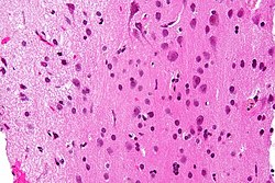
Micrograph showing white matter with its characteristic fine meshwork-like appearance (left of image - lighter shade of pink) and grey matter, with the characteristic neuronal cell bodies (right of image - dark shade of pink). HPS stain.
Human brain right dissected lateral view description.JPG
Human brain right dissected lateral view, showing grey matter (the darker outer parts), and white matter (the inner and prominently whiter parts).
Details
LocationCentral nervous system
Identifiers
Latinsubstantia alba
MeSHD066127
TAA14.1.00.009
FMA83929

White matter structure of human brain (taken by MRI).

White matter refers to areas of the central nervous system (CNS) that are mainly made up of myelinated axons, also called tracts. Long thought to be passive tissue, white matter affects learning and brain functions, modulating the distribution of action potentials, acting as a relay and coordinating communication between different brain regions.

White matter is named for its relatively light appearance resulting from the lipid content of myelin. However, the tissue of the freshly cut brain appears pinkish-white to the naked eye because myelin is composed largely of lipid tissue veined with capillaries. Its white color in prepared specimens is due to its usual preservation in formaldehyde

Structure


White matter

White matter is composed of bundles, which connect various gray matter areas (the locations of nerve cell bodies) of the brain to each other, and carry nerve impulses between neurons. Myelin acts as an insulator, which allows electrical signals to jump, rather than coursing through the axon, increasing the speed of transmission of all nerve signals.

The total number of long range fibers within a cerebral hemisphere is 2% of the total number of cortico-cortical fibers (across cortical areas) and is roughly the same number as those that communicate between the two hemispheres in the brain's largest white tissue structure, the corpus callosum. Schüz and Braitenberg note "As a rough rule, the number of fibres of a certain range of lengths is inversely proportional to their length."

White matter in nonelderly adults is 1.7–3.6% blood.

Grey matter

The other main component of the brain is grey matter (actually pinkish tan due to blood capillaries), which is composed of neurons. The substantia nigra is a third colored component found in the brain that appears darker due to higher levels of melanin in dopaminergic neurons than its nearby areas. Note that white matter can sometimes appear darker than grey matter on a microscope slide because of the type of stain used. Cerebral- and spinal white matter do not contain dendrites, neural cell bodies, or shorter axons, which can only be found in grey matter. 

Location

White matter forms the bulk of the deep parts of the brain and the superficial parts of the spinal cord. Aggregates of grey matter such as the basal ganglia (caudate nucleus, putamen, globus pallidus, substantia nigra, subthalamic nucleus, nucleus accumbens) and brainstem nuclei (red nucleus, cranial nerve nuclei) are spread within the cerebral white matter.

The cerebellum is structured in a similar manner as the cerebrum, with a superficial mantle of cerebellar cortex, deep cerebellar white matter (called the "arbor vitae") and aggregates of grey matter surrounded by deep cerebellar white matter (dentate nucleus, globose nucleus, emboliform nucleus, and fastigial nucleus). The fluid-filled cerebral ventricles (lateral ventricles, third ventricle, cerebral aqueduct, fourth ventricle) are also located deep within the cerebral white matter.

Myelinated axon length

Men have more white matter than women both in volume and in length of myelinated axons. At the age of 20, the total length of myelinated fibers in men is 176,000 km while that of a woman is 149,000 km. There is a decline in total length with age of about 10% each decade such that a man at 80 years of age has 97,200 km and a female 82,000 km. Most of this reduction is due to the loss of thinner fibers.

Function

White matter is the tissue through which messages pass between different areas of gray matter within the central nervous system. The white matter is white because of the fatty substance (myelin) that surrounds the nerve fibers (axons). This myelin is found in almost all long nerve fibers, and acts as an electrical insulation. This is important because it allows the messages to pass quickly from place to place. 

Unlike gray matter, which peaks in development in a person's twenties, the white matter continues to develop, and peaks in middle age.

Research

Multiple sclerosis (MS) is the most common of the inflammatory demyelinating diseases of the central nervous system which affect white matter. In MS lesions, the myelin sheath around the axons is deteriorated by inflammation. Alcohol use disorders are associated with a decrease in white matter volume.

Amyloid plaques in white matter may be associated with Alzheimer's disease and other neurodegenerative diseases. Other changes that commonly occur with age include the development of leukoaraiosis, which is a rarefaction of the white matter that can be correlated with a variety of conditions, including loss of myelin pallor, axonal loss, and diminished restrictive function of the blood–brain barrier.

White matter lesions on magnetic resonance imaging are linked to several adverse outcomes, such as cognitive impairment and depression. White matter hyperintensity are more than often present with vascular dementia, particularly among small vessel/subcortical subtypes of vascular dementia.

Volume

Smaller volumes (in terms of group averages) of white matter might be associated with larger deficits in attention, declarative memory, executive functions, intelligence, and academic achievement. However, volume change is continuous throughout one's lifetime due to neuroplasticity, and is a contributing factor rather than determinant factor of certain functional deficits due to compensating effects in other brain regions. The integrity of white matter declines due to aging. Nonetheless, regular aerobic exercise appears to either postpone the aging effect or in turn enhance the white matter integrity in the long run. Changes in white matter volume due to inflammation or injury may be a factor in the severity of obstructive sleep apnea.

Imaging

The study of white matter has been advanced with the neuroimaging technique called diffusion tensor imaging where magnetic resonance imaging (MRI) brain scanners are used. As of 2007, more than 700 publications have been published on the subject.

A 2009 paper by Jan Scholz and colleagues used diffusion tensor imaging (DTI) to demonstrate changes in white matter volume as a result of learning a new motor task (e.g. juggling). The study is important as the first paper to correlate motor learning with white matter changes. Previously, many researchers had considered this type of learning to be exclusively mediated by dendrites, which are not present in white matter. The authors suggest that electrical activity in axons may regulate myelination in axons. Or, gross changes in the diameter or packing density of the axon might cause the change. A more recent DTI study by Sampaio-Baptista and colleagues reported changes in white matter with motor learning along with increases in myelination.

Language center

From Wikipedia, the free encyclopedia
https://en.wikipedia.org/wiki/Language_center

The term language center (or more accurately centers, e.g. Broca's area and Wernicke's area) refers to the areas of the brain which serve a particular function for speech processing and production. Language is a core system, which gives humans the capacity to solve difficult problems and provides them with a unique type of social interaction. Language allows individuals to attribute symbols (e.g. words or signs) to specific concepts and display them through sentences and phrases that follow proper grammatical rules. Moreover, speech is the mechanism in which language is orally expressed.

Language Areas of the brain. The Angular Gyrus is represented in orange, Supramarginal Gyrus is represented in yellow, Broca's area is represented in blue, Wernicke's area is represented in green and the Primary Auditory Cortex is represented in pink.

Information is exchanged in a larger system including language-related regions. These regions are connected by white matter fiber tracts that make possible the transmission of information between regions. The white matter fibers bunches were recognized to be important for language production after suggesting that it is possible to make a connection between multiple language centers. The three classical language areas that are involved in language production and processing are Broca’s and Wernicke’s areas, and angular gyrus.

Broca’s area

Broca's Area was first suggested to play a role in speech function by the French neurologist and anthropologist Paul Broca in 1861. The basis for this discovery was the analysis of speech problems resulting from injuries to this region of the brain, located in the inferior frontal gyrus. Paul Broca had a patient called Leborgne who could only pronounce the word “tan” when speaking. Paul Broca, after working with another patient with similar impairment, concluded that damage in the inferior frontal gyrus affected articulate language.

Broca’s area is well-known for being the syntactic processing  “center”. It has been known since Paul Broca associated speech production with an area in the posterior inferior frontal gyrus, which he called “Broca’s area”. Although this area is in charge of speech production, its particular role in the language system is unknown. However, it is involved in phonological, semantic, and syntactic processing and working memory. The anterior region of Broca’s area is involved in semantic processing, while the posterior region in the phonological processing (Bohsali, 2015). Moreover, the whole of Broca’s area has been shown to have a higher activation while doing reading tasks than other types of tasks.

In a simple explanation of speech production, this area approaches phonological word representation chronologically divided into segments of syllables which then is sent to different motor areas where they are converted into a phonetic code. The study of how this area produces speech has been made with paradigms using both single and complex words.

Broca’s area is correlated with phonological segmentation, unification, and syntactic processing, which are all connected to linguistic information. This area, although it synchronizes the transformation of information within cortical systems involved in spoken word production, does not contribute to the production of single words. The inferior frontal lobe is the one in charge of word production.

Broca's and Wernicke's area
 
Language Areas of the brain. The Angular Gyrus is represented in orange, Supramarginal Gyrus is represented in yellow, Broca's area is represented in blue, Wernicke's area is represented in green and the Primary Auditory Cortexis represented in pink.
 
Furthermore, Broca’s area is structurally related to the thalamus and both are engaged in language processing. The connectivity between both areas is two thalamic nuclei, the pulvinar, and the ventral nucleus, which are involved in language processing and linguistic functions similar to BA 44 and 45 in Broca’s area. Pulvinar is connected to many frontal regions of the frontal cortex and ventral nucleus is involved in speech production. The frontal speech regions of the brain have been shown to participate in speech sound perception.

Broca's Area is today still considered an important language center, playing a central role in processing syntax, grammar, and sentence structure. 

Wernicke’s area

Wernicke’s area was named for German doctor Carl Wernicke, who discovered it in 1874 in the course of his research into aphasias (loss of ability to speak).This area of the brain is involved in language comprehension. Therefore, Wernicke’s area is for understanding oral language. Besides Wernicke’s area, the left posterior superior temporal gyrus (pSTG), middle temporal gyrus (MTG), inferior temporal gyrus (ITG), supramarginal gyrus (SMG), and angular gyrus (AG) participate in language comprehension. Therefore, language comprehension is not located in a specific area. Contrarily, it involves large regions of the inferior parietal lobe and left temporal.

While the finale of speech production is a sequence of muscle movements, the activation of knowledge about the sequence of phonemes (consonants and vowel speech sounds) that creates a word is a phonological retrieval. Wernicke’s area contributes to phonological retrieval. All speech production tasks (e.g. word retrieval, repetition, and reading aloud) require phonological retrieval. The phonological retrieval system involved in speech repetition is the auditory phoneme perception system and the visual letter perception system is the one that serves for reading aloud. The communicative speech production entails a phase preceding phonological retrieval. The speech comprehension implicates representing sequences of phonemes onto word meaning.

Angular gyrus

The angular gyrus is an important element in processing concrete and abstract concepts. It also has a role in verbal working memory during retrieval for verbal information and in visual memory for when turning written language into spoken language. The left AG is activated in semantic processing requiring concept retrieval and conceptual integration. Moreover, the left AG is activated during problems of multiplication and addition requiring retrieval of arithmetic factors in verbal memory. Therefore, it is involved in verbal coding of numbers.

Insular cortex

The insula is implicated in speech and language, partaking of functional and structural connections with motor, linguistic, sensory, and limbic brain areas. The knowledge about the function of the insula in speech production comes from different studies with patients who suffered from apraxia of speech. These studies have led researchers to know about the involvement of different parts of the insula. These parts are: the left anterior insula, which is related to speech production; and the bilateral anterior insula, involved in misleading speech comprehension.

Speech and language disorders

Many different sources state that the study of the brain and therefore, language disorders, originated in the 19th century and linguistic analysis of those disorders began throughout the 20th century. Studying language impairments in the brain after injuries aids to comprehend how the brain works and how it changes after an injury. When this happens, the brain suffers an impairment that is referred to as “aphasia”. Lesions to Broca's Area resulted primarily in disruptions to speech production; damage to Wernicke's Area, which is located in the lower part of the temporal lobe, lead mainly to disruptions in speech reception.

There are numerous distinctive ways in which language can be affected. Phonemic paraphasia, an attribute of conduction aphasia and Wernicke aphasia, is not the speech comprehension impairment. Instead, it is the speech production damage, where the desire phonemes are selected erroneously or in an incorrect sequence. Therefore, although Wernicke’s aphasia, a combination of phonological retrieval and semantic systems impairment, affects speech comprehension, it also involves speech production damage. Phonemic paraphasia and anomia (impaired word retrieval) are the results of phonological retrieval impairment.

Another lesion that involves impairment in language production and processing is the “apraxia of speech”, a difficulty synchronizing articulators essential for speech production. This lesion is located in the superior pre-central gyrus of the insula and is more likely to occur to patients with Broca’s aphasia. Dominant ventral anterior (VA) nucleus, another type of lesion, is the result of word-finding and semantic paraphasia’s difficulties engaging in language processing. Moreover, individuals with thalamic lesions experience difficulties linking semantic concepts with correct phonological representations in word production.

Dyslexia is a language processing disorder. It involves learning difficulties such as reading, writing, word recognition, phonological recording, numeracy, and spelling. Although having access to appropriate intervention during childhood, these difficulties continue throughout the lifespan. Moreover, children are diagnosed with dyslexia when more than one factor affecting learning, such as reading, appears visible. Children diagnosed with dyslexia that have difficulties in concrete cognitive functioning is called an assumption of specificity, and it helps to diagnose dyslexia.

Some characteristics that distinguish dyslexics are incompetent phonological processing abilities causing misread of unfamiliar words and affecting comprehension; inadequacy of working memory affecting speaking, reading, and writing; errors in oral reading; oral skills difficulties as expressing oneself; and writing skills problems like expressing and spelling errors. Dyslexics not only experience learning difficulties but also other secondary characteristics as having difficulties organizing, planning, social interactions, motor skills, visual perception, and short-term memory. These characteristics affect personal and academic life.

Dysarthria is a motor speech disorder caused by damage in the central and/or peripheral nervous system and it is related to degenerative neurological diseases, such as Parkinson’s disease, cerebrovascular accident (CVA) and traumatic brain injury (TBI). Dysarthria is caused by a mechanical difficulty in the vocal cords or neurological disease-producing abnormal articulation of phonemes, such as instead of “b” a “p”. A type of dyspraxia based on distortions of words is called apraxic dysarthria This type is related to facial apraxia and motor aphasia if Broca’s area is involved.

Current scientific consensus

Improvements in computer technology, in the late 20th century, has allowed a better understanding of the correlation between brain and language, and the disorder that this entails. This improvement has permitted a better visualization of the brain structure in high resolution three-dimensional images. It has also allowed to observe brain activity through the blood flow (Dronkers, Ivanova, & Baldo, 2017).

New medical imaging techniques such as PET and fMRI have allowed researchers to generate pictures showing which areas of a living brain are active at a given time. Functional magnetic resonance imaging (fMRI) is a technique used for locating, in the brain, particular functions to different activity related. This technique shows the location and magnitude of neural activity variations, influenced by external stimulation and fluctuation at rest. MRI is a technique that was developed in the 20th century to observe brain activity in healthy and abnormal brains. Diffusion-weighted magnetic resonance imaging or diffusion tensor imaging (DTI) is a technique use for track white matter bundles in vivo and gives information of the internal fibrous structure by the measure of water diffusion. This diffusion tensor is used for infer white matter connectivity.

In the past, research was primarily based on observations of loss of ability resulting from damage to the cerebral cortex. Indeed, medical imaging has represented a radical step forward for research on speech processing. Since then, a whole series of relatively large areas of the brain are involved in speech processing. In more recent research, subcortical regions (those lying below the cerebral cortex such as the putamen and the caudate nucleus), as well as the pre-motor areas (BA 6), have received increased attention. It is now generally assumed that the following structures of the cerebral cortex near the primary and secondary auditory cortices play a fundamental role in speech processing:

The left hemisphere is usually dominant in right-handed people, although bilateral activations are not uncommon in the area of syntactic processing. It is now accepted that the right hemisphere plays an important role in the processing of suprasegmental acoustic features like prosody; which is “the rhythmic and melodic variations in speech”. There are two types of prosodic information: emotional prosody (right hemisphere), which is the emotional that the speaker gives to the speech, and linguistic prosody (left hemisphere), the syntactic and thematic structure of the speech.

Most areas of speech processing develop in the second year of life in the dominant half (hemisphere) of the brain, which often (though not necessarily) corresponds to the opposite of the dominant hand. 98% of right-handed people are left-hemisphere dominant, and the majority of left-handed people are as well.

Computerized tomographic (CT) scans is another technique of the 1970s, which produce low spatial resolution but provides the location of the injury in vivo. Moreover, Voxel-based Lesion Symptom Mapping (VLSM) and Voxel-Based Morphometry (VBM) techniques contributed to the understanding that specific brain regions have different roles when supporting speech processing.[2] VLSM has been used to observe complex language functions sustained by different regions. Furthermore, VBM is a helpful technique to analysis language impairments related to neurodegenerative disease.

Older models

The differentiation of speech production into only two large sections of the brain (i.e. Broca's and Wernicke's areas), that was accepted long before the advent of medical imaging techniques, is now considered outdated. Broca's Area was first suggested to play a role in speech function by the French neurologist and anthropologist Paul Broca in 1861. The basis for this discovery was the analysis of speech problems resulting from injuries to this region of the brain, located in the inferior frontal gyrus. Lesions to Broca's Area resulted primarily in disruptions to speech production. Damage to Wernicke's Area, which is located in the lower part of the temporal lobe, lead mainly to disruptions in speech reception. This area was named for German doctor Carl Wernicke, who discovered it in 1874 in the course of his research into aphasias (loss of ability to speak).

Broca's Area is today still considered an important language center, playing a central role in processing syntax, grammar, and sentence structure.

In summary, these early research efforts demonstrated that semantic and structural speech production takes place in different areas of the brain.

Visual modularity

From Wikipedia, the free encyclopedia
https://en.wikipedia.org/wiki/Visual_modularity

In cognitive neuroscience, visual modularity is an organizational concept concerning how vision works. The way in which the primate visual system operates is currently under intense scientific scrutiny. One dominant thesis is that different properties of the visual world (color, motion, form and so forth) require different computational solutions which are implemented in anatomically/functionally distinct regions that operate independently – that is, in a modular fashion.


Motion processing

Akinetopsia is an intriguing condition brought about by damage to the Extrastriate cortex MT+ that renders humans and monkeys unable to perceive motion, seeing the world in a series of static "frames" instead and indicates that there might be a "motion centre" in the brain. Of course, such data can only indicate that this area is at least necessary to motion perception, not that it is sufficient; however, other evidence has shown the importance of this area to primate motion perception. Specifically, physiological, neuroimaging, perceptual, electrical- and transcranial magnetic stimulation evidence (Table 1) all come together on the area V5/hMT+. Converging evidence of this type is supportive of a module for motion processing. However, this view is likely to be incomplete: other areas are involved with motion perception, including V1, V2 and V3a  and areas surrounding V5/hMT+ (Table 2). A recent fMRI study put the number of motion areas at twenty-one. Clearly, this constitutes a stream of diverse anatomical areas. The extent to which this is ‘pure’ is in question: with Akinetopsia come severe difficulties in obtaining structure from motion. V5/hMT+ has since been implicated in this function as well as determining depth. Thus the current evidence suggests that motion processing occurs in a modular stream, although with a role in form and depth perception at higher levels.

Evidence for a "motion centre" in the primate brain
Methodology Finding
Physiology (single cell recording) Cells directionally and speed selective in MT/V5
Neuroimaging Greater activation for motion information than static information in V5/MT
Electrical-stimulation & perceptual Following electrical stimulation of V5/MT cells perceptual decisions are biased towards the stimulated neuron’s direction preference
Magnetic-stimulation Motion perception is also briefly impaired in humans by a strong magnetic pulse over the corresponding scalp region to hMT+
Psychophysics Perceptual asynchrony among motion, color and orientation.

Evidence for a motion processing area surrounding V5
Methodology Finding
Physiology (single cell recording) Complex motion involving contraction/expansion and rotation found to activate neurons in medial superior temporal area (MST)
Neuroimaging Biological motion activated superior temporal sulcus
Neuroimaging Tool use activated middle temporal gyrus and inferior temporal sulcus
Neuropsychology Damage to visual area V5 results in akinetopsia

Color processing

Similar converging evidence suggests modularity for color. Beginning with Gowers’ finding that damage to the fusiform/lingual gyri in occipitotemporal cortex correlates with a loss in color perception (achromatopsia), the notion of a "color centre" in the primate brain has had growing support. Again, such clinical evidence only implies that this region is critical to color perception, and nothing more. Other evidence, however, including neuroimaging and physiology converges on V4 as necessary to color perception. A recent meta-analysis has also shown a specific lesion common to achromats corresponding to V4. From another direction altogether it has been found that when synaesthetes experience color by a non-visual stimulus, V4 is active. On the basis of this evidence it would seem that color processing is modular. However, as with motion processing it is likely that this conclusion is inaccurate. Other evidence shown in Table 3 implies different areas’ involvement with color. It may thus be more instructive to consider a multistage color processing stream from the retina through to cortical areas including at least V1, V2, V4, PITd and TEO. Consonant with motion perception, there appears to be a constellation of areas drawn upon for color perception. In addition, V4 may have a special, but not exclusive, role. For example, single cell recording has shown that only V4 cells respond to the color of a stimuli rather than its waveband, whereas other areas involved with color do not.

Evidence against a "color center" in the primate brain
Other areas involved with color/Other functions of V4
Wavelength sensitive cells in V1 and V2
anterior parts of the inferior temporal cortex
posterior parts of the superior temporal sulcus (PITd)
Area in or near TEO
Shape detection
Link between vision, attention and cognition

Form processing

Another clinical case that would a priori suggest a module for modularity in visual processing is visual agnosia. The well studied patient DF is unable to recognize or discriminate objects owing to damage in areas of the lateral occipital cortex although she can see scenes without problem – she can literally see the forest but not the trees. Neuroimaging of intact individuals reveals strong occipito-temporal activation during object presentation and greater activation still for object recognition. Of course, such activation could be due to other processes, such as visual attention. However, other evidence that shows a tight coupling of perceptual and physiological changes suggests activation in this area does underpin object recognition. Within these regions are more specialized areas for face or fine grained analysis, place perception and human body perception. Perhaps some of the strongest evidence for the modular nature of these processing systems is the double dissociation between object- and face (prosop-) agnosia. However, as with color and motion, early areas are implicated too, lending support to the idea of a multistage stream terminating in the inferotemporal cortex rather than an isolated module. 

Functional modularity

One of the first uses of the term "module" or "modularity" occurs in the influential book "Modularity of Mind" by philosopher Jerry Fodor. A detailed application of this idea to the case of vision was published by Pylyshyn (1999), who argued that there is a significant part of vision that is not responsive to beliefs and is "cognitively impenetrable".

Much of the confusion concerning modularity exists in neuroscience because there is evidence for specific areas (e.g. V4 or V5/hMT+) and the concomitant behavioral deficits following brain insult (thus taken as evidence for modularity). In addition, evidence shows other areas are involved and that these areas subserve processing of multiple properties (e.g. V1) (thus taken as evidence against modularity). That these streams have the same implementation in early visual areas, like V1, is not inconsistent with a modular viewpoint: to adopt the canonical analogy in cognition, it is possible for different software to run on the same hardware. A consideration of psychophysics and neuropsychological data would suggest support for this. For example, psychophysics has shown that percepts for different properties are realized asynchronously. In addition, although achromats experience other cognitive defects they do not have motion deficits when their lesion is restricted to V4, or total loss of form perception. Relatedly, Zihl and colleagues' akinetopsia patient shows no deficit to color or object perception (although deriving depth and structure from motion is problematic, see above) and object agnostics do not have damaged motion or color perception, making the three disorders triply dissociable. Taken together this evidence suggests that even though distinct properties may employ the same early visual areas they are functionally independent. Furthermore, that the intensity of subjective perceptual experience (e.g. color) correlates with activity in these specific areas (e.g. V4), the recent evidence that synaesthetes show V4 activation during the perceptual experience of color, as well as the fact that damage to these areas results in concomitant behavioral deficits (the processing may be occurring but perceivers do not have access to the information) are all evidence for visual modularity.

Modularity of mind

From Wikipedia, the free encyclopedia
https://en.wikipedia.org/wiki/Modularity_of_mind
 
Modularity of mind is the notion that a mind may, at least in part, be composed of innate neural structures or mental modules which have distinct, established, and evolutionarily developed functions. However, different definitions of "module" have been proposed by different authors.

Early investigations

Historically, questions regarding the functional architecture of the mind have been divided into two different theories of the nature of the faculties. The first can be characterized as a horizontal view because it refers to mental processes as if they are interactions between faculties such as memory, imagination, judgement, and perception, which are not domain specific (e.g., a judgement remains a judgement whether it refers to a perceptual experience or to the conceptualization/comprehension process). The second can be characterized as a vertical view because it claims that the mental faculties are differentiated on the basis of domain specificity, are genetically determined, are associated with distinct neurological structures, and are computationally autonomous.

The vertical vision goes back to the 19th century movement called phrenology and its founder Franz Joseph Gall, who claimed that the individual mental faculties could be associated precisely, in a sort of one-to-one correspondence, with specific physical areas of the brain. Hence, someone's level of intelligence, for example, could be literally "read off" from the size of a particular bump on his posterior parietal lobe. This simplistic view of modularity has been disproven over the course of the last century.

Fodor's Modularity of Mind

In the 1980s, however, Jerry Fodor revived the idea of the modularity of mind, although without the notion of precise physical localizability. Drawing from Noam Chomsky's idea of the language acquisition device and other work in linguistics as well as from the philosophy of mind and the implications of optical illusions, he became a major proponent of the idea with the 1983 publication of Modularity of Mind.

According to Fodor, a module falls somewhere between the behaviorist and cognitivist views of lower-level processes.

Behaviorists tried to replace the mind with reflexes which Fodor describes as encapsulated (cognitively impenetrable or unaffected by other cognitive domains) and non-inferential (straight pathways with no information added). Low level processes are unlike reflexes in that they are inferential. This can be demonstrated by poverty of the stimulus arguments in which the proximate stimulus, that which is initially received by the brain (such as the 2D image received by the retina), cannot account for the resulting output (for example, our 3D perception of the world), thus necessitating some form of computation.

In contrast, cognitivists saw lower level processes as continuous with higher level processes, being inferential and cognitively penetrable (influenced by other cognitive domains, such as beliefs). The latter has been shown to be untrue in some cases, such as with many visual illusions (ex. Müller-Lyer illusion), which can persist despite a person's awareness of their existence. This is taken to indicate that other domains, including one's beliefs, cannot influence such processes.

Fodor arrives at the conclusion that such processes are inferential like higher order processes and encapsulated in the same sense as reflexes.

Although he argued for the modularity of "lower level" cognitive processes in Modularity of Mind he also argued that higher level cognitive processes are not modular since they have dissimilar properties. The Mind Doesn't Work That Way, a reaction to Steven Pinker's How the Mind Works, is devoted to this subject. 

Fodor (1983) states that modular systems must—at least to "some interesting extent"—fulfill certain properties:
  1. Domain specificity: modules only operate on certain kinds of inputs—they are specialised
  2. Informational encapsulation: modules need not refer to other psychological systems in order to operate
  3. Obligatory firing: modules process in a mandatory manner
  4. Fast speed: probably due to the fact that they are encapsulated (thereby needing only to consult a restricted database) and mandatory (time need not be wasted in determining whether or not to process incoming input)
  5. Shallow outputs: the output of modules is very simple
  6. Limited accessibility
  7. Characteristic ontogeny: there is a regularity of development
  8. Fixed neural architecture.
Pylyshyn (1999) has argued that while these properties tend to occur with modules, one—information encapsulation—stands out as being the real signature of a module; that is the encapsulation of the processes inside the module from both cognitive influence and from cognitive access. One example is that conscious awareness of the Müller-Lyer illusion being an illusion does not correct visual processing.

Evolutionary psychology and massive modularity

Other perspectives on modularity come from evolutionary psychology, particularly from the work of Leda Cosmides and John Tooby. This perspective suggests that modules are units of mental processing that evolved in response to selection pressures. On this view, much modern human psychological activity is rooted in adaptations that occurred earlier in human evolution, when natural selection was forming the modern human species.

Evolutionary psychologists propose that the mind is made up of genetically influenced and domain-specific mental algorithms or computational modules, designed to solve specific evolutionary problems of the past. Cosmides and Tooby also state in a brief "primer" on their website, that "…the brain is a physical system. It functions like a computer," "…the brain’s function is to process information," "different neural circuits are specialized for solving different adaptive problems," and "our modern skulls house a stone age mind."

The definition of module has caused confusion and dispute. J. A. Fodor initially defined module as "functionally specialized cognitive systems" that have nine features but not necessarily all at the same time. In his views modules can be found in peripheral processing such as low-level visual processing but not in central processing. Later he narrowed the two essential features to domain-specificity and information encapsulation. Frankenhuis and Ploeger write that domain-specificity means that "a given cognitive mechanism accepts, or is specialized to operate on, only a specific class of information". Information encapsulation means that information processing in the module cannot be affected by information in the rest of the brain. One example is that being aware that a certain optical illusion, caused by low level processing, is false does not prevent the illusion from persisting.

Evolutionary psychologists instead usually define modules as functionally specialized cognitive systems that are domain-specific and may also contain innate knowledge about the class of information processed. Modules can be found also for central processing. This theory is sometimes referred to as massive modularity. 

A 2010 review by evolutionary psychologists Confer et al. suggested that domain general theories, such as for "rationality," has several problems: 1. Evolutionary theories using the idea of numerous domain-specific adaptions have produced testable predictions that have been empirically confirmed; the theory of domain-general rational thought has produced no such predictions or confirmations. 2. The rapidity of responses such as jealousy due to infidelity indicates a domain-specific dedicated module rather than a general, deliberate, rational calculation of consequences. 3. Reactions may occur instinctively (consistent with innate knowledge) even if a person has not learned such knowledge. One example being that in the ancestral environment it is unlikely that males during development learn that infidelity (usually secret) may cause paternal uncertainty (from observing the phenotypes of children born many months later and making a statistical conclusion from the phenotype dissimilarity to the cuckolded fathers). With respect to general purpose problem solvers, Barkow, Cosmides, and Tooby (1992) have suggested in The Adapted Mind: Evolutionary Psychology and The Generation of Culture that a purely general problem solving mechanism is impossible to build due to the frame problem. Clune et al. (2013) have argued that computer simulations of the evolution of neural nets suggest that modularity evolves because, compared to non-modular networks, connection costs are lower.

Several groups of critics, including psychologists working within evolutionary frameworks, argue that the massively modular theory of mind does little to explain adaptive psychological traits. Proponents of other models of the mind argue that the computational theory of mind is no better at explaining human behavior than a theory with mind entirely a product of the environment. Even within evolutionary psychology there is discussion about the degree of modularity, either as a few generalist modules or as many highly specific modules. Other critics suggest that there is little empirical support in favor of the domain-specific theory beyond performance on the Wason selection task, a task critics state is too limited in scope to test all relevant aspects of reasoning. Moreover, critics argue that Cosmides and Tooby's conclusions contain several inferential errors and that the authors use untested evolutionary assumptions to eliminate rival reasoning theories.

Wallace (2010) observes that the evolutionary psychologists' definition of "mind" has been heavily influenced by cognitivism and/or information processing definitions of the mind. Critics point out that these assumptions underlying evolutionary psychologists' hypotheses are controversial and have been contested by some psychologists, philosophers, and neuroscientists. For example, Jaak Panksepp, an affective neuroscientist, point to the "remarkable degree of neocortical plasticity within the human brain, especially during development" and states that "the developmental interactions among ancient special-purpose circuits and more recent general-purpose brain mechanisms can generate many of the "modularized" human abilities that evolutionary psychology has entertained."

Philosopher David Buller agrees with the general argument that the human mind has evolved over time but disagrees with the specific claims evolutionary psychologists make. He has argued that the contention that the mind consists of thousands of modules, including sexually dimorphic jealousy and parental investment modules, are unsupported by the available empirical evidence. He has suggested that the "modules" result from the brain's developmental plasticity and that they are adaptive responses to local conditions, not past evolutionary environments. However, Buller has also stated that even if massive modularity is false this does not necessarily have broad implications for evolutionary psychology. Evolution may create innate motives even without innate knowledge.

In contrast to modular mental structure, some theories posit domain-general processing, in which mental activity is distributed across the brain and cannot be decomposed, even abstractly, into independent units. A staunch defender of this view is William Uttal, who argues in The New Phrenology (2003) that there are serious philosophical, theoretical, and methodological problems with the entire enterprise of trying to localise cognitive processes in the brain. Part of this argument is that a successful taxonomy of mental processes has yet to be developed.

Merlin Donald argues that over evolutionary time the mind has gained adaptive advantage from being a general problem solver. The mind, as described by Donald, includes module-like "central" mechanisms, in addition to more recently evolved "domain-general" mechanisms.

Grandmother cell

From Wikipedia, the free encyclopedia
 
The sketch of the idea of the grandmother cell: a neuron that reacts selectively on a pattern: Jennifer Aniston cell, Dodecahedron cell, and 'Grandmother cell' cell, which reacts on the copy of this cartoon.
 
The grandmother cell, sometimes called the "Jennifer Aniston neuron", is a hypothetical neuron that represents a complex but specific concept or object. It activates when a person "sees, hears, or otherwise sensibly discriminates" a specific entity, such as his or her grandmother. The term was in use at least as early as 1966 amongst staff and students in the Department of Experimental Psychology, University of Cambridge, England. A similar concept, that of the gnostic neuron, was proposed two years later by Jerzy Konorski.

Support


Face selective cells

Visual neurons in the inferior temporal cortex of the monkey fire selectively to hands and faces. These cells are selective in that they do not fire for other visual objects important for monkeys such as fruit and genitalia. Research finds that some of these cells can be trained to show high specificity for arbitrary visual objects, and these would seem to fit the requirements of gnostic/grandmother cells. In addition, evidence exists for cells in the human hippocampus that have highly selective responses to gnostic categories including highly selective responses to individual human faces.

However most of the reported face-selective cells are not grandmother/gnostic cells since they do not represent a specific percept, that is, they are not cells narrowly selective in their activations for one face and only one face irrespective of transformations of size, orientation, and color. Even the most selective face cells usually also discharge, if more weakly, to a variety of individual faces. Furthermore, face-selective cells often vary in their responsiveness to different aspects of faces. This suggests that cell responsiveness arises from the need of a monkey to differentiate among different individual faces rather than among other categories of stimuli such as bananas with their discrimination properties linked to the fact that different individual faces are much more similar to each other in their overall organization and fine detail than other kinds of stimuli. Moreover, it has been suggested that these cells might in fact be responding as specialized feature detector neurons that only function in the holistic context of a face construct.

One idea has been that such cells form ensembles for the coarse or distributed coding of faces rather than detectors for specific faces. Thus, a specific grandmother may be represented by a specialized ensemble of grandmother or near grandmother cells.

Individual specific recognition cells

In 2005, a UCLA and Caltech study found evidence of different cells that fire in response to particular people, such as Bill Clinton or Jennifer Aniston. A neuron for Halle Berry, for example, might respond "to the concept, the abstract entity, of Halle Berry", and would fire not only for images of Halle Berry, but also to the actual name "Halle Berry". However, there is no suggestion in that study that only the cell being monitored responded to that concept, nor was it suggested that no other actress would cause that cell to respond (although several other presented images of actresses did not cause it to respond). The researchers believe that they have found evidence for sparseness, rather than for grandmother cells.

Further evidence for the theory that a small neural network provides facial recognition was found from analysis of cell recording studies of macaque monkeys. By formatting faces as points in a high-dimensional linear space, the scientists discovered that each face cell’s firing rate is proportional to the projection of an incoming face stimulus onto a single axis in this space, allowing a face cell ensemble of about 200 cells to encode the location of any face in the space.

Sparseness vs distributed representations

The grandmother cell hypothesis, is an extreme version of the idea of sparseness, and is not without critics. The opposite of the grandmother cell theory is the distributed representation theory, that states that a specific stimulus is coded by its unique pattern of activity over a large group of neurons widely distributed in the brain.

The arguments against the sparseness include:
  1. According to some theories, one would need thousands of cells for each face, as any given face must be recognised from many different angles – profile, 3/4 view, full frontal, from above, etc.
  2. Rather than becoming more and more specific as visual processing proceeds from retina through the different visual centres of the brain, the image is partially dissected into basic features such as vertical lines, colour, speed, etc., distributed in various modules separated by relatively large distances. How all these disparate features are re-integrated to form a seamless whole is known as the binding problem.

Pontifical cells

William James in 1890 proposed a related idea of a pontifical cell. The pontifical cell is defined as a putative, and implausible cell which had all our experiences. It is in this different from a concept specific cell in that it is the site of experience of sense data. James's 1890 pontifical cell was instead a cell "to which the rest of the brain provided a representation" of a grandmother. The experience of grandmother occurred in this cell.

Great Flood of 1862

From Wikipedia, the free encyclopedia https://en.wikipedia.org/wiki/Great_Flood_of_1862   ...