Search This Blog

Tuesday, November 17, 2020

Chemical imaging

From Wikipedia, the free encyclopedia

 
Chemical imaging (as quantitative – chemical mapping) is the analytical capability to create a visual image of components distribution from simultaneous measurement of spectra and spatial, time information. Hyperspectral imaging measures contiguous spectral bands, as opposed to multispectral imaging which measures spaced spectral bands.

The main idea - for chemical imaging, the analyst may choose to take as many data spectrum measured at a particular chemical component in spatial location at time; this is useful for chemical identification and quantification. Alternatively, selecting an image plane at a particular data spectrum (PCA - multivariable data of wavelength, spatial location at time) can map the spatial distribution of sample components, provided that their spectral signatures are different at the selected data spectrum.

Software for chemical imaging is most specific and distinguished from chemical methods such as chemometrics.

Imaging instrumentation has three components: a radiation source to illuminate the sample, a spectrally selective element, and usually a detector array (the camera) to collect the images. The data format is called a hypercube. The data set may be visualized as a data cube, a three-dimensional block of data spanning two spatial dimensions (x and y), with a series of wavelengths (lambda) making up the third (spectral) axis. The hypercube can be visually and mathematically treated as a series of spectrally resolved images (each image plane corresponding to the image at one wavelength) or a series of spatially resolved spectra.

History

Commercially available laboratory-based chemical imaging systems emerged in the early 1990s (ref. 1-5). In addition to economic factors, such as the need for sophisticated electronics and extremely high-end computers, a significant barrier to commercialization of infrared imaging was that the focal plane array (FPA) needed to read IR images were not readily available as commercial items. As high-speed electronics and sophisticated computers became more commonplace, and infrared cameras became readily commercially available, laboratory chemical imaging systems were introduced.

Initially used for novel research in specialized laboratories, chemical imaging became a more commonplace analytical technique used for general R&D, quality assurance (QA) and quality control (QC) in less than a decade. The rapid acceptance of the technology in a variety of industries (pharmaceutical, polymers, semiconductors, security, forensics and agriculture) rests in the wealth of information characterizing both chemical composition and morphology. The parallel nature of chemical imaging data makes it possible to analyze multiple samples simultaneously for applications that require high throughput analysis in addition to characterizing a single sample.

Applications

Hyperspectral imaging is most often applied to either solid or gel samples, and has applications in chemistry, biology, medicine, pharmacy (see also for example: food science, biotechnology, agriculture and industry. NIR, IR and Raman chemical imaging is also referred to as hyperspectral, spectroscopic, spectral or multispectral imaging (also see microspectroscopy). However, other ultra-sensitive and selective imaging techniques are also in use that involve either UV-visible or fluorescence microspectroscopy. Many imaging techniques can be used to analyze samples of all sizes, from the single molecule to the cellular level in biology and medicine, and to images of planetary systems in astronomy, but different instrumentation is employed for making observations on such widely different systems.

Any material that depends on chemical gradients for functionality may be amenable to study by an analytical technique that couples spatial and chemical characterization. To efficiently and effectively design and manufacture such materials, the ‘what’ and the ‘where’ must both be measured. The demand for this type of analysis is increasing as manufactured materials become more complex. Chemical imaging techniques are critical to understanding modern manufactured products and in some cases is a non-destructive technique so that samples are preserved for further testing.

Many materials, both manufactured and naturally occurring, derive their functionality from the spatial distribution of sample components. For example, extended release pharmaceutical formulations can be achieved by using a coating that acts as a barrier layer. The release of active ingredient is controlled by the presence of this barrier, and imperfections in the coating, such as discontinuities, may result in altered performance. In the semi-conductor industry, irregularities or contaminants in silicon wafers or printed micro-circuits can lead to failure of these components. The functionality of biological systems is also dependent upon chemical gradients – a single cell, tissue, and even whole organs function because of the very specific arrangement of components. It has been shown that even small changes in chemical composition and distribution may be an early indicator of disease.

Principles

Chemical imaging shares the fundamentals of vibrational spectroscopic techniques, but provides additional information by way of the simultaneous acquisition of spatially resolved spectra. It combines the advantages of digital imaging with the attributes of spectroscopic measurements. Briefly, vibrational spectroscopy measures the interaction of light with matter. Photons that interact with a sample are either absorbed or scattered; photons of specific energy are absorbed, and the pattern of absorption provides information, or a fingerprint, on the molecules that are present in the sample.

On the other hand, in terms of the observation setup, chemical imaging can be carried out in one of the following modes: (optical) absorption, emission (fluorescence), (optical) transmission or scattering (Raman). A consensus currently exists that the fluorescence (emission) and Raman scattering modes are the most sensitive and powerful, but also the most expensive.

In a transmission measurement, the radiation goes through a sample and is measured by a detector placed on the far side of the sample. The energy transferred from the incoming radiation to the molecule(s) can be calculated as the difference between the quantity of photons that were emitted by the source and the quantity that is measured by the detector. In a diffuse reflectance measurement, the same energy difference measurement is made, but the source and detector are located on the same side of the sample, and the photons that are measured have re-emerged from the illuminated side of the sample rather than passed through it. The energy may be measured at one or multiple wavelengths; when a series of measurements are made, the response curve is called a spectrum.

A key element in acquiring spectra is that the radiation must somehow be energy selected – either before or after interacting with the sample. Wavelength selection can be accomplished with a fixed filter, tunable filter, spectrograph, an interferometer, or other devices. For a fixed filter approach, it is not efficient to collect a significant number of wavelengths, and multispectral data are usually collected. Interferometer-based chemical imaging requires that entire spectral ranges be collected, and therefore results in hyperspectral data. Tunable filters have the flexibility to provide either multi- or hyperspectral data, depending on analytical requirements.

Spectra are typically measured with an imaging spectrometer, based on a Focal Plane Array.

Terminology

Some words common in spectroscopy, optical microscopy and photography have been adapted or their scope modified for their use in chemical imaging. They include: resolution, field of view and magnification. There are two types of resolution in chemical imaging. The spectral resolution refers to the ability to resolve small energy differences; it applies to the spectral axis. The spatial resolution is the minimum distance between two objects that is required for them to be detected as distinct objects. The spatial resolution is influenced by the field of view, a physical measure of the size of the area probed by the analysis. In imaging, the field of view is a product of the magnification and the number of pixels in the detector array. The magnification is a ratio of the physical area of the detector array divided by the area of the sample field of view. Higher magnifications for the same detector image a smaller area of the sample.

Types of vibrational chemical imaging instruments

Chemical imaging has been implemented for mid-infrared, near-infrared spectroscopy and Raman spectroscopy. As with their bulk spectroscopy counterparts, each imaging technique has particular strengths and weaknesses, and are best suited to fulfill different needs.

Mid-infrared chemical imaging

A set of stones scanned with a Specim LWIR-C hyperspectral imager in the thermal infrared range from 7.7 μm to 12.4 μm. Minerals such as quartz and feldspar spectra are clearly recognizable.

Mid-infrared (MIR) spectroscopy probes fundamental molecular vibrations, which arise in the spectral range 2,500-25,000 nm. Commercial imaging implementations in the MIR region employ hyperspectral imagers or Fourier Transform Infrared (FT-IR) interferometers, depending on the application. The MIR absorption bands tend to be relatively narrow and well-resolved; direct spectral interpretation is often possible by an experienced spectroscopist. MIR spectroscopy can distinguish subtle changes in chemistry and structure, and is often used for the identification of unknown materials. The absorptions in this spectral range are relatively strong; for this reason, sample presentation is important to limit the amount of material interacting with the incoming radiation in the MIR region. Data can be collected in reflectance, transmission, or emission mode. Water is a very strong absorber of MIR radiation and wet samples often require advanced sampling procedures (such as attenuated total reflectance). Commercial instruments include point and line mapping, and imaging. Mid-infrared chemical imaging can also be performed with nanometer level spatial resolution using atomic force microscope based infrared spectroscopy (AFM-IR).

Remote chemical imaging of a simultaneous release of SF6 and NH3 at 1.5km using the Telops Hyper-Cam imaging spectrometer

Atmospheric windows in the infrared spectrum are also employed to perform chemical imaging remotely. In these spectral regions the atmospheric gases (mainly water and CO2) present low absorption and allow infrared viewing over kilometer distances. Target molecules can then be viewed using the selective absorption/emission processes described above. An example of the chemical imaging of a simultaneous release of SF6 and NH3 is shown in the image.

Near-infrared chemical imaging

The analytical near infrared (NIR) region spans the range from 780 nm to 2,500 nm. The absorption bands seen in this spectral range arise from overtones and combination bands of O-H, N-H, C-H and S-H stretching and bending vibrations. Absorption is one to two orders of magnitude smaller in the NIR compared to the MIR; this phenomenon eliminates the need for extensive sample preparation. Thick and thin samples can be analyzed without any sample preparation, it is possible to acquire NIR chemical images through some packaging materials, and the technique can be used to examine hydrated samples, within limits. Intact samples can be imaged in transmittance or diffuse reflectance.

The lineshapes for overtone and combination bands tend to be much broader and more overlapped than for the fundamental bands seen in the MIR. Often, multivariate methods are used to separate spectral signatures of sample components. NIR chemical imaging is particularly useful for performing rapid, reproducible and non-destructive analyses of known materials. NIR imaging instruments are typically based on a hyperspectral camera, a tunable filter or an FT-IR interferometer. External light source is always needed, such as sun (outdoor scans, remote sensing) or a halogen lamp (laboratory, industrial measurements).

Raman chemical imaging

The Raman shift chemical imaging spectral range spans from approximately 50 to 4,000 cm−1; the actual spectral range over which a particular Raman measurement is made is a function of the laser excitation frequency. The basic principle behind Raman spectroscopy differs from the MIR and NIR in that the x-axis of the Raman spectrum is measured as a function of energy shift (in cm−1) relative to the frequency of the laser used as the source of radiation. Briefly, the Raman spectrum arises from inelastic scattering of incident photons, which requires a change in polarizability with vibration, as opposed to infrared absorption, which requires a change in dipole moment with vibration. The end result is spectral information that is similar and in many cases complementary to the MIR. The Raman effect is weak - only about one in 107 photons incident to the sample undergoes Raman scattering. Both organic and inorganic materials possess a Raman spectrum; they generally produce sharp bands that are chemically specific. Fluorescence is a competing phenomenon and, depending on the sample, can overwhelm the Raman signal, for both bulk spectroscopy and imaging implementations.

Raman chemical imaging requires little or no sample preparation. However, physical sample sectioning may be used to expose the surface of interest, with care taken to obtain a surface that is as flat as possible. The conditions required for a particular measurement dictate the level of invasiveness of the technique, and samples that are sensitive to high power laser radiation may be damaged during analysis. It is relatively insensitive to the presence of water in the sample and is therefore useful for imaging samples that contain water such as biological material.

Fluorescence Imaging (Ultraviolet, visible and near infrared regions)

Emission microspectroscopy is a sensitive technique with excitation and emission ranging from the ultraviolet, visible and NIR regions. As such, it has numerous biomedical, biotechnological and agricultural applications. There are several powerful, highly specific and sensitive fluorescence techniques that are currently in use, or still being developed; among the former are FLIM, FRAP, FRET and FLIM-FRET; among the latter are NIR fluorescence and probe-sensitivity enhanced NIR fluorescence microspectroscopy and nanospectroscopy techniques (see "Further reading" section). Fluorescence emission microspectroscopy and imaging are also commonly used to locate protein crystals in solution, for the characterization of metamaterials and biotechnology devices.

Sampling and samples

The value of imaging lies in the ability to resolve spatial heterogeneities in solid-state or gel/gel-like samples. Imaging a liquid or even a suspension has limited use as constant sample motion serves to average spatial information, unless ultra-fast recording techniques are employed as in fluorescence correlation microspectroscopy or FLIM observations where a single molecule may be monitored at extremely high (photon) detection speed. High-throughput experiments (such as imaging multi-well plates) of liquid samples can however provide valuable information. In this case, the parallel acquisition of thousands of spectra can be used to compare differences between samples, rather than the more common implementation of exploring spatial heterogeneity within a single sample.

Similarly, there is no benefit in imaging a truly homogeneous sample, as a single point spectrometer will generate the same spectral information. Of course the definition of homogeneity is dependent on the spatial resolution of the imaging system employed. For MIR imaging, where wavelengths span from 3-10 micrometres, objects on the order of 5 micrometres may theoretically be resolved. The sampled areas are limited by current experimental implementations because illumination is provided by the interferometer. Raman imaging may be able to resolve particles less than 1 micrometre in size, but the sample area that can be illuminated is severely limited. With Raman imaging, it is considered impractical to image large areas and, consequently, large samples. FT-NIR chemical/hyperspectral imaging usually resolves only larger objects (>10 micrometres), and is better suited for large samples because illumination sources are readily available. However, FT-NIR microspectroscopy was recently reported to be capable of about 1.2 micron (micrometer) resolution in biological samples Furthermore, two-photon excitation FCS experiments were reported to have attained 15 nanometer resolution on biomembrane thin films with a special coincidence photon-counting setup.

Detection limit

The concept of the detection limit for chemical imaging is quite different from for bulk spectroscopy, as it depends on the sample itself. Because a bulk spectrum represents an average of the materials present, the spectral signatures of trace components are simply overwhelmed by dilution. In imaging however, each pixel has a corresponding spectrum. If the physical size of the trace contaminant is on the order of the pixel size imaged on the sample, its spectral signature will likely be detectable. If however, the trace component is dispersed homogeneously (relative to pixel image size) throughout a sample, it will not be detectable. Therefore, detection limits of chemical imaging techniques are strongly influenced by particle size, the chemical and spatial heterogeneity of the sample, and the spatial resolution of the image.

Data analysis

Data analysis methods for chemical imaging data sets typically employ mathematical algorithms common to single point spectroscopy or to image analysis. The reasoning is that the spectrum acquired by each detector is equivalent to a single point spectrum; therefore pre-processing, chemometrics and pattern recognition techniques are utilized with the similar goal to separate chemical and physical effects and perform a qualitative or quantitative characterization of individual sample components. In the spatial dimension, each chemical image is equivalent to a digital image and standard image analysis and robust statistical analysis can be used for feature extraction.

Software

Multispectral image

From Wikipedia, the free encyclopedia
 
 
Video by SDO simultaneously showing sections of the Sun at various wavelengths

A multispectral image is one that captures image data within specific wavelength ranges across the electromagnetic spectrum. The wavelengths may be separated by filters or detected via the use of instruments that are sensitive to particular wavelengths, including light from frequencies beyond the visible light range, i.e. infrared and ultra-violet. Spectral imaging can allow extraction of additional information the human eye fails to capture with its visible receptors for red, green and blue. It was originally developed for military target identification and reconnaissance. Early space-based imaging platforms incorporated multispectral imaging technology to map details of the Earth related to coastal boundaries, vegetation, and landforms. Multispectral imaging has also found use in document and painting analysis.

Multispectral imaging measures light in a small number (typically 3 to 15) of spectral bands.

 Hyperspectral imaging is a special case of spectral imaging where often hundreds of contiguous spectral bands are available.

Applications

Military target tracking

Multispectral imaging measures light emission and is often used in detecting or tracking military targets. In 2003, researchers at the United States Army Research Laboratory and the Federal Laboratory Collaborative Technology Alliance reported a dual band multispectral imaging focal plane array (FPA). This FPA allowed researchers to look at two infrared (IR) planes at the same time. Because mid-wave infrared (MWIR) and long wave infrared (LWIR) technologies measure radiation inherent to the object and require no external light source, they also are referred to as thermal imaging methods.

The brightness of the image produced by a thermal imager depends on the objects emissivity and temperature.  Every material has an infrared signature that aids in the identification of the object. These signatures are less pronounced in hyperspectral systems (which image in many more bands than multispectral systems) and when exposed to wind and, more dramatically, to rain. Sometimes the surface of the target may reflect infrared energy. This reflection may misconstrue the true reading of the objects’ inherent radiation. Imaging systems that use MWIR technology function better with solar reflections on the target's surface and produce more definitive images of hot objects, such as engines, compared to LWIR technology. However, LWIR operates better in hazy environments like smoke or fog because less scattering occurs in the longer wavelengths. Researchers claim that dual-band technologies combine these advantages to provide more information from an image, particularly in the realm of target tracking.

For nighttime target detection, thermal imaging outperformed single-band multispectral imaging. Citation. Dual band MWIR and LWIR technology resulted in better visualization during the nighttime than MWIR alone. Citation Citation. The US Army reports that its dual band LWIR/MWIR FPA demonstrated better visualizing of tactical vehicles than MWIR alone after tracking them through both day and night.  

Land mine detection

By analyzing the emissivity of ground surfaces, multispectral imaging can detect the presence of underground missiles. Surface and sub-surface soil possess different physical and chemical properties that appear in spectral analysis. Disturbed soil has increased emissivity in the wavelength range of 8.5 to 9.5 micrometers while demonstrating no change in wavelengths greater than 10 micrometers. The US Army Research Laboratory's dual MWIR/LWIR FPA used "red" and "blue" detectors to search for areas with enhanced emissivity. The red detector acts as a backdrop, verifying realms of undisturbed soil areas, as it is sensitive to the 10.4 micrometer wavelength. The blue detector is sensitive to wavelengths of 9.3 micrometers. If the intensity of the blue image changes when scanning, that region is likely disturbed. The scientists reported that fusing these two images increased detection capabilities.

Ballistic missile detection

Intercepting an intercontinental ballistic missile (ICBM) in its boost phase requires imaging of the hard body as well as the rocket plumes. MWIR presents a strong signal from highly heated objects including rocket plumes, while LWIR produces emissions from the missile's body material. The US Army Research Laboratory reported that with their dual-band MWIR/LWIR technology, tracking of the Atlas 5 Evolved Expendable Launch Vehicles, similar in design to ICBMs, picked up both the missile body and plumage.

Space-based imaging

Most radiometers for remote sensing (RS) acquire multispectral images. Dividing the spectrum into many bands, multispectral is the opposite of panchromatic, which records only the total intensity of radiation falling on each pixel. Usually, Earth observation satellites have three or more radiometers. Each acquires one digital image (in remote sensing, called a 'scene') in a small spectral band. The bands are grouped into wavelength regions based on the origin of the light and the interests of the researchers.

Weather Forecasting

Modern weather satellites produce imagery in a variety of spectra. 

Multispectral imaging combines two to five spectral imaging bands of relatively large bandwidth into a single optical system. A multispectral system usually provides a combination of visible (0.4 to 0.7 µm), near infrared (NIR; 0.7 to 1 µm), short-wave infrared (SWIR; 1 to 1.7 µm), mid-wave infrared (MWIR; 3.5 to 5 µm) or long-wave infrared (LWIR; 8 to 12 µm) bands into a single system. — Valerie C. Coffey

In the case of Landsat satellites, several different band designations have been used, with as many as 11 bands (Landsat 8) comprising a multispectral image. Spectral imaging with a higher radiometric resolution (involving hundreds or thousands of bands), finer spectral resolution (involving smaller bands), or wider spectral coverage may be called hyperspectral or ultraspectral.

Documents and artworks

The technology has also assisted in the interpretation of ancient papyri, such as those found at Herculaneum, by imaging the fragments in the infrared range (1000 nm). Often, the text on the documents appears to the naked eye as black ink on black paper. At 1000 nm, the difference in how paper and ink reflect infrared light makes the text clearly readable. It has also been used to image the Archimedes palimpsest by imaging the parchment leaves in bandwidths from 365–870 nm, and then using advanced digital image processing techniques to reveal the undertext with Archimedes' work. Multispectral imaging has been used in a Mellon Foundation project at Yale University to compare inks in medieval English manuscripts.

Multispectral imaging can be employed for investigation of paintings and other works of art. The painting is irradiated by ultraviolet, visible and infrared rays and the reflected radiation is recorded in a camera sensitive in this regions of the spectrum. The image can also be registered using the transmitted instead of reflected radiation. In special cases the painting can be irradiated by UV, VIS or IR rays and the fluorescence of pigments or varnishes can be registered.

Multispectral imaging has also been used to examine discolorations and stains on old books and manuscripts. Comparing the "spectral fingerprint" of a stain to the characteristics of known chemical substances can make it possible to identify the stain. This technique has been used to examine medical and alchemical texts, seeking hints about the activities of early chemists and the possible chemical substances they may have used in their experiments. Like a cook spilling flour or vinegar on a cookbook, an early chemist might have left tangible evidence on the pages of the ingredients used to make medicines.

Spectral bands

The wavelengths are approximate; exact values depend on the particular satellite's instruments:

  • Blue, 450–515..520 nm, is used for atmosphere and deep water imaging, and can reach depths up to 150 feet (50 m) in clear water.
  • Green, 515..520–590..600 nm, is used for imaging vegetation and deep water structures, up to 90 feet (30 m) in clear water.
  • Red, 600..630–680..690 nm, is used for imaging man-made objects, in water up to 30 feet (9 m) deep, soil, and vegetation.
  • Near infrared (NIR), 750–900 nm, is used primarily for imaging vegetation.
  • Mid-infrared (MIR), 1550–1750 nm, is used for imaging vegetation, soil moisture content, and some forest fires.
  • Far-infrared (FIR), 2080–2350 nm, is used for imaging soil, moisture, geological features, silicates, clays, and fires.
  • Thermal infrared, 10400-12500 nm, uses emitted instead of reflected radiation to image geological structures, thermal differences in water currents, fires, and for night studies.
  • Radar and related technologies are useful for mapping terrain and for detecting various objects.

Spectral band usage

For different purposes, different combinations of spectral bands can be used. They are usually represented with red, green, and blue channels. Mapping of bands to colors depends on the purpose of the image and the personal preferences of the analysts. Thermal infrared is often omitted from consideration due to poor spatial resolution, except for special purposes.

  • True-color uses only red, green, and blue channels, mapped to their respective colors. As a plain color photograph, it is good for analyzing man-made objects, and is easy to understand for beginner analysts.
  • Green-red-infrared, where the blue channel is replaced with near infrared, is used for vegetation, which is highly reflective in near IR; it then shows as blue. This combination is often used to detect vegetation and camouflage.
  • Blue-NIR-MIR, where the blue channel uses visible blue, green uses NIR (so vegetation stays green), and MIR is shown as red. Such images allow the water depth, vegetation coverage, soil moisture content, and the presence of fires to be seen, all in a single image.

Many other combinations are in use. NIR is often shown as red, causing vegetation-covered areas to appear red.

Classification

Unlike other Aerial photographic and satellite image interpretation work, these multispectral images do not make it easy to identify directly the feature type by visual inspection. Hence the remote sensing data has to be classified first, followed by processing by various data enhancement techniques so as to help the user to understand the features that are present in the image.

Such classification is a complex task which involves rigorous validation of the training samples depending on the classification algorithm used. The techniques can be grouped mainly into two types.

  • Supervised classification techniques
  • Unsupervised classification techniques

Supervised classification makes use of training samples. Training samples are areas on the ground for which there is Ground truth, that is, what is there is known. The spectral signatures of the training areas are used to search for similar signatures in the remaining pixels of the image, and we will classify accordingly. This use of training samples for classification is called supervised classification. Expert knowledge is very important in this method since the selection of the training samples and a biased selection can badly affect the accuracy of classification. Popular techniques include the Maximum likelihood principle and Convolutional neural network. The Maximum likelihood principle calculates the probability of a pixel belonging to a class (i.e. feature) and allots the pixel to its most probable class. Newer Convolutional neural network based methods account for both spatial proximity and entire spectra to determine the most likely class.

In case of unsupervised classification no prior knowledge is required for classifying the features of the image. The natural clustering or grouping of the pixel values, i.e. the gray levels of the pixels, are observed. Then a threshold is defined for adopting the number of classes in the image. The finer the threshold value, the more classes there will be. However, beyond a certain limit the same class will be represented in different classes in the sense that variation in the class is represented. After forming the clusters, ground truth validation is done to identify the class the image pixel belongs to. Thus in this unsupervised classification apriori information about the classes is not required. One of the popular methods in unsupervised classification is k-means clustering.

Multispectral data analysis software

  • MicroMSI is endorsed by the NGA.
  • Opticks is an open-source remote sensing application.
  • Multispec is freeware multispectral analysis software.
  • Gerbil is open source multispectral visualization and analysis software.

Monday, November 16, 2020

Conservation and restoration of paintings

Paintings conservation laboratory, Heritage Conservation Centre, Singapore

The conservation and restoration of paintings is carried out by professional painting conservators. Paintings cover a wide range of various mediums, materials, and their supports (i.e. the painted surface made from fabric, paper, wood panel, fabricated board, or other). Painting types include fine art to decorative and functional objects spanning from acrylics, frescoes, and oil paint on various surfaces, egg tempera on panels and canvas, lacquer painting, water color and more. Knowing the materials of any given painting and its support allows for the proper restoration and conservation practices. All components of a painting will react to its environment differently, and impact the artwork as a whole. These material components along with collections care (also known as preventive conservation) will determine the longevity of a painting. The first steps to conservation and restoration is preventive conservation followed by active restoration with the artist's intent in mind.

Basic care

Typical, traditional oil, acrylic, and many other types of paintings are made up of various different types of materials, from their paint layers to the materials that make up their supports. Each of these materials requires specific care in handling, displaying, storage, added protective measures, and general environmental conditions. Providing the proper care to each of these materials ensures that the overall condition of the painting is protected.

Backing boards

Using good protective measures such as attaching a rigid backing to a painting on canvas provides several protections. It reduces the effects of rapid changes in relative humidity around the painting, provides some protection from pressure or direct contact against the canvas back, and protects from vibrations caused by handling or moving. Backing boards also serve to protect from dust and dirt, cracks and deformations from handling, and insect activity. Some of the most commonly used types of backing boards include foam core, heritage board, matboard, cardboard/millboard, coroplast, corrugated plastic sheets, acrylic sheeting, mylar, and fabric.

Framing

The frames around paintings are not just for aesthetic appearances. Frames are also used to protect the more sensitive parts of a painting when handled by hand, and they reduce the potential for damage if the painting is dropped. There are also specialists that work on the conservation and restoration of painting frames.

Handling and moving

The movement of objects places an object at a much greater risk of damage than when it is on display or in storage. Certain techniques and equipment are used any time an art work needs to be transported. These techniques and equipment include using padding lifts and dollies, moving small, fragile objects on carts instead of carrying by hand; lifting objects from underneath by their sturdiest part; and taking extra time and care when on ladders or stairs. In many cases gloves are worn to protect the art work from any dirt or oil that may be on a conservator or object handlers hands. When handling canvas paintings specifically, never presume that the frame is stable and firmly attached. Do not lift or carry a painting by its stretcher bar, or insert your fingers between the stretcher bar and the canvas.

Display and storage

It is estimated that a lack of proper routine maintenance and care is responsible for 95 percent of conservation treatments; the remaining 5 percent results from mishandling objects When developing display and storage methods for works of art, issues regarding relative humidity, temperature, light, pollutants, and pests need to be considered. Location and the types of storage units must be considered as well. Storage areas should be located in areas away from pipes and heating systems, as well as out of areas that are likely to flood and collect dust and dirt. Storage units should be sturdy, adjustable for collections growth so all collections sizes are safely stored, made of materials that will not cause any damage to the paintings (i.e. metal racks), and be free of any hardware or supports that stick out.

Causes of deterioration

Moisture, heat, light, pollutants, and pests can slowly or suddenly cause damage to a painting. These agents of deterioration impact all of the components that make up a painting in various ways.

Relative humidity and temperature

Too low or too high relative humidity (RH) as well as rapid changes in relative humidity can be damaging to paintings. According to the Canadian Conservation Institute, there are four types of incorrect relative humidity: dampness over 75% RH, RH above or below a critical value for that object, RH above 0%, and RH fluctuations. "Generally accepted temperature and relative humidity standards for most museum objects and artifacts are 65°-70° F (18°-21° C) at 47%-55% RH." The best method of controlling the environment is by using a centralized climate control or HVAC system where incoming air is washed, cleaned, heated, or cooled, adjusted to specific conditions, and then injected into the storage space. An appropriate alternative is a localized climate control system where air conditioners cool the air and absorb some of its moisture while filtering out gross particles. They do not condition the air, nor do they filter air pollutants.

Light

Both visible and ultraviolet light can cause damage to paintings. In particular, organic materials such as paper, fabric, wood, leather, and colored surfaces. "Fugitive dyes and colorants used in paints will eventually discolor under exposure to ultraviolet light. The fading of pigments and dyes in paintings will affect the color balance of the image." Damage from natural and artificial light exposure can be mitigated by displaying paintings out of direct sunlight, use of blinds, shades, curtains, or shudders, filters on nearby windows, installing dimmers and appropriate wattage light bulbs, and displaying paintings a safe distance from a light source to limit heat exposure.

Pollutants

Pollutants can be described as gasses, aerosols, liquids, or solids that have a chemical reaction with any part of a painting. There are three types of pollutants. Airborne pollutants, pollutants transferred by contact, and intrinsic pollutants.

Airborne pollutants which originate from atmospheric sources (ozone, hydrogen sulfide, sulfur dioxide, soot, salts), or emissive products, objects, and people (sulfur-based gases, organic acids, lint, and dander). Their effects can include acidification of papers, corrosion of metals, discoloration of colorants, and efflorescence of calcium-based objects.

Pollutants transferred by contact include plasticizer from PVC, sulfur compounds from natural rubber, staining materials from wood, viscous compounds from old polyurethane foams, fatty acids from people or from greasy objects, and impregnation of residue of cleaning agents. The effects of these pollutants can include discoloration or corrosion of a paintings surface.

Intrinsic pollutants are composite objects that have compounds that are harmful to other parts of an object. The effects of these pollutants includes deterioration of the object, acidification, discoloration or staining on an object, speed up degradation processes caused by oxygen, water vapor, or other pollutants.

Pests

Pests such as rodents and insects have the potential to cause considerable damage to works of art. Preventive measures that may be taken to protect paintings from pests include upgrading building structures to obstruct pest entry, installing better cabinetry with good seals, better control of temperature and humidity in collections and storage areas, keeping food and other organic materials from collection areas, and treatment of outbreaks. Materials that are commonly damaged by pests include: natural fibers, wood, paper, starch adhesives, and egg tempera.

Painting mediums and preventive conservation

Acrylic paintings

Acrylic Paintings were introduced in the 1950s and the material differs from oil paint in chemical and physical properties. There are two types of Acrylic paints used in acrylic paintings. There is solvent-based and water-based. Solvent-based acrylic paints are soluble in mineral spirits, and water-based acrylic paints are water-soluble. Acrylic paint differs from oil paint in both its quick drying time, and how the paint dries. Acrylic paint dries in as little as thirty minutes, and dries by the evaporation of solvent of water.

"Il Mio Unicerso" X0385UNAT

Preventive conservation

Acrylic paintings require attentive preventive care. Due to the soft nature of the paint attracts and hold dirt and debris creating difficulty when cleaning resulting in darkening colors over time. Due to the characteristic of acrylic paint, varnishes will diminish top layers of the paint and effect the colors vibrancy. Storage of acrylic paintings should be clean and free of dust and heat- below room temperature is best as it will reduce further softening of the top layer of the paint. Exposing acrylic paintings to temperatures ranging near sub zero will case damaging cracking. Acrylic paint is highly susceptible to mold growth. This is a growing concern for artists and conservators as removal causes some degree of damage to the original paint.

Conservation and restoration methods

Preventive care seems to be the best method of conservation. However, after more than 10 years of investigation, conservators are now better able to understand the risks of swelling, extraction, and gloss changes that are associated with surface cleaning treatments. Wet cleaning systems are now being developed that help to minimize the risks associated with cleaning acrylic paints.

Blacklight or fluorescent/luminous paintings

Black Light or Luminous Paint is typically made up of fluorescent dyes mixed into paint. These dyes are not a typical dye, but rather a pigment that is suspended in a carrier or resin. This pigment is what gives off a glow when exposed to ultraviolet light. This glow or light is created by the energy that is released from the pigment. While the fluorescent paint layers reflect light, the paint layer darkens over time and decreases in fluorescence.

Preventive conservation

The intensity of fluorescent paints can decline quite rapidly, making it difficult for conservators to care for. This is because the brightening agent that is mixed into the paint is not stable. Some fluorescent paintings can only be displayed in the dark with UV-lights. These requirements can make choosing appropriate lighting and exhibition and storage space for preventive conservation challenging. For fluorescent paintings that are displayed in dark rooms with UV lights, it is recommended to have an automatic lighting system.

Conservation and restoration methods

Conserving . The age of the fluorescent pigments must be determined in order to develop a close matching pigment use for retouching. A painting can lose its effect under UV-light if the retouches and fillings are not closely matched and are too light or too dark.

Egg Tempera

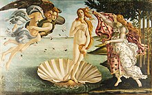
Egg Tempera is made up of egg yolk, water, and pigment. These ingredients are mixed together to create a thick paste that dries quickly, but can take six to twelve months before it completely cures. Egg Tempera's fast drying property makes it difficult to correct or revise.

Sandro Botticelli - La nascita di Venere - Google Art Project - edited

Preventive conservation

Tempera paintings have many of the same problems of condition and conservation as other painting mediums. These include changes in the work due to unstable and fugitive pigments. The aging of a paintings supports and ground will also impact the paint layer. For example, cracks can form in a gesso ground due to the embrittlement and movement of its support, then become visible in the paint film.

Tempera can develop cracks over time that are visible to the naked eye, and flaking caused by air bubbles. Tempera paintings are thought to be more resistant to materials typically used during cleanings. However, they are susceptible to abrasions from routine dusting, washings, and removal of old varnish layers.

Conservation and restoration methods

The paint surfaces of many tempera paintings have become abraded, most likely from routine dusting and cleanings. It is unclear how tempera paintings were originally varnished due to the need for sensitive methods of analysis. Modern tempera paintings are almost always unvarnished and more prone to mold attacks.

Enamel

Enamel paints, not to be confused with vitreous enamel, are nitro-cellulose based paints originally designed for commercial use, but have also been used in artist's paintings such as Jackson Pollock and Pablo Picasso. Enamel paints are oil, latex, alkyd, and water based. This paint dries rapidly and has a glossy finish once dry.

R Bampton Coach Painting & Carriage Lining enamel advert at the Louwman museum

Preventive conservation

Like all painting mediums, enamel is subject to damage from improper handling and environmental stressors. Jackson Pollock's Mural for example was subjected to several moves, likely having been rolled and unrolled each time. These moves may have taken a toll on its condition. The paint flaked, and the original stretcher weakened causing the painting to have a noticeable sag. Another of Jackson Pollock's paintings, Echo, endured yellowing at the top of the canvas due to strong museum display lights.

Conservation and restoration methods

Conservation treatments can take the form of adhering a lining to the canvas with wax-resin to the reverse side, replacing the painting's original stretcher, and varnishing the painting. In Jackson Pollock's Echo, solvents were used to remove a thin layer of the canvas to even out the work's coloring.

Encaustic

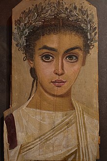
Encaustic is a method of painting that involves dry pigments mixed with hot beeswax, then applied to the surface of a support such as wood or canvas. A completed painting is then finished by taking a source of heat to reheat the surface and fuse it together. Encaustic paintings do not require a varnish, are resistant to moisture, and do not yellow.

Mummy portrait of a girl, AD 120–150, Roman Egypt, wax encaustic painting on sycamore wood, Liebieghaus, Frankfurt am Main (14304151412)

Preventive conservation

Encaustic paintings are considered very durable. However, the waxes used in encaustic paints can soften or melt above certain temperatures. This may cause the upper layers to slide or detach and cause irreversible damage. Controlling the light, temperature, and humidity levels can prevent this type of damage from occurring.

Conservation and restoration methods

Surface cleaning on encaustic paintings can typically be done with distilled water and swabs is sufficient. For more challenging cleanings, solutions made of beeswax and carbon tetrachloride can be used. One of the biggest challenges in treating encaustic paintings is identifying the different waxes used to determine the appropriate treatments. Infra-red photography and gas chromotography can be used to identify the various types of waxes.

Frescos

Frescos are types of mural paintings where the pigment is painted directly into a fresh lime mortar surface. These types of paintings are susceptible to damage from vandalism, time, environmental stressors, and climate changes. Frescos, like most works of cultural heritage, have specific climate parameters for preservation. Humidity and water damage cause mold to develop. The mold aspergillus versicolor can grow on frescos and consume nutrients effectively causing pigment discoloration and wall detachment from rot.

A restorer filling the gaps of the damaged frescos in the crypt of Saint Eustorgio church in Milan, Italy.

Preventive conservation

Ideally, buildings with frescos would be outfitted with central air with humidity adjusting features to keep the paintings in a cool and dry environment with low humidity. Frescos can be found typically in old churches and other ancient structures such as temples and tombs. These types of structures can be limited with additional means like environmental controls. Deterioration of frescos can be caused by environmental pollutants. These pollutants can be physical, chemical, or biological. The many layers can deteriorate from the materials chemical compositions reacting to pollutants or environmental conditions such as humidity, temperature, light, and pH.

Chemical Degradation- Evident with pigment discoloration, stains, and the presence of biofilm.

Physical Degradation- Evident with cracking of layers.

Conservation and restoration methods

Frescos can be repaired by methods of detaching sections of the fresco. Surface repairs for frescos can be less invasive. Conservators can remedy cracks and minor detachments of frescos with injections of epoxy resin containing micronized silica and lime putty.

Lacquer

There are a variety of lacquers that have been, and continue to be used such as Urushi (unprocessed lacquer), Guangqi (processed), Nitrocellulose, lacquers with acrylic resins, and water-based lacquers, but the most well known lacqueris Urushi lacquer. This lacquer paint is made from raw lacquer or sap taken from trees. It is then heated, filtered, and applied in thin layers to supports such as wood or metal. The lacquer is left to cure before it is polished, and another layer is added. The number of layers may vary, and each can be left in its natural transparent state, or colored with pigments to create Lacquer painting.

Lacquer painting over wood, Northern Wei

Preventive conservation

While lacquer is a hard material, it is best to first prevent damage and loss by maintaining proper environmental conditions. Lacquer is susceptible to cracks and loose joins from fluctuating temperatures and relative humidity. Extended exposure to light can also cause lacquer to lose its durability. Over exposure can also cause discoloration and loss of lustre. Avoiding exposure to unfiltered daylight and fluorescent lamps can help to prevent this type of damamge. Temperatures should be kept as low and consistent as possible to avoid changes in relative humidity which can cause condensation. Condensation can cause shrinkage and swelling in the wood that he lacquer is applied to.

Conservation and restoration methods

Treatments to lacquer paintings may include consolidation and repair work before or after cleaning. Consolidation can be used to repair cupping and flaking.

Oil paintings

Oil paint is a medium made up of pigments and a drying oil binding agent. Various other ingredients can be mixed in to condition the paint in several ways and modify its various properties and drying.  Oil paintings are painted on various surface support types. Oil on canvas, oil on board, and oil on metal are only some examples of oil paintings on various surfaces. Oil paintings are susceptible to damage from vandalism, time, improper handling, environmental stressors and temperature changes.

Restored Painting- oil on canvas; by Bartolomeo Better; Italian. 17th-Century.

They are also susceptible to damage in low relative humid conditions, and fluctuations can create stresses in the paint layers.

Preventive conservation

Preventive care for oil paintings is essential for preservation. Excessive light with heat can cause fading to pigments. Proper storage with climate and lighting controls are important especially depending on the support structure. The wooden stretcher behind an oil painting on canvas will expand and contract with moisture causing possible buckling of the canvas and cracking, flaking, or shattering of the paint. Paintings should be stored off the ground in case of flooding. Moisture and water damage can cause mold to develop along with various other issues depending on the materials involved: rot (natural materials), rusting (in metals), warping (of wooden supports), etc.

Conservation and restoration methods

Treatment methods may include rejoining split wooden panels, mending torn fabric, or consolidating lifting paint flakes. Cleaning old and yellowing varnish, and revarnish the painting.

Pastels

There are two types of Pastels. Pastels that are made of pigment particles bound together with a binding agent, and oil pastels that have pigments mixed with wax and non-drying oil. Pastels that are pigment particles bound together take on a more chalky and loose powdery characterization, and are secured to its supports using fixative or diluted resin in solution. Oil pastels never fully dry, and are sensitive to scrapes, dust and dirt.

Double Portrait by Francis Cotes, pastel, Speed Art Museum

Preventive conservation

In general, works of art on paper should be stored in a cool and relatively dry room with minimal exposure to light. Pastel artworks should be matted and framed. Framing should be under ultraviolet filtering acrylic sheeting. Using a glaze over the surface of the oil pastel works can help to protect the oil pastel from damage. Limited exhibition time and low light intensity is recommended to limit light exposure. Excessive light exposure can cause pigment fading and discoloration in the paper.

Conservation and restoration methods

Preventive conservation is key. Some damage to works of art on paper is irreversible, but there are some methods of restoration that can be used to treat damages such as structural tension in the paper created by previous restoration treatments. This may include removal of the secondary and adhering a new support or even an internal cardboard support.

Watercolor and gouache

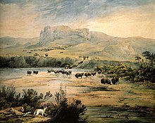
Water color and Gouache paintings are pigments mixed into water-soluble gums that are applied onto paper or rigid board supports. Due to its thin washes and light colors, watercolor paintings are very light sensitive. Also, due to their exposed support they are vulnerable to damage from dirt, dust, and pollutants. Gouache paintings can form layers like acrylic and oil paint, but is still vulnerable to the same agents of deterioration as watercolors.

Landscape with Herd of Buffalo on the Upper Missouri. Watercolor by Karl Bodmer 1833

Preventive conservation

Damage to Watercolor and Gouache paintings can be prevented and mitigated by maintaining temperature and relative humidity within acceptable ranges, and low light conditions. As with pastel works, watercolor and gouache paintings should be mounted and framed. Damage that may occur are disfiguring brown spots from mold growth, paper turning brown and brittle from cardboard supports, yellow stains from adhesive tapes, and cockling and undulations.

Conservation and restoration methods

Some treatments to watercolor works may include a washing treatment to remove discoloration from acidic mounts, tapes, and adhesives. Stains from these products can also be treated with solvents. Tears and losses can be repaired with products such as wheat starch paste or methyl cellulose, and weakened paper can be strengthened by attaching a lining.

Scroll and screen paintings

Scroll Paintings, Hanging and Hand, and screen paintings are made of ink, color, pigment, silk and paper. Scroll paintings often are multiple layers of paper and silk attached to wooden bars called a stave and dowel. Screens are often single panels that are joined together by paper hinges that fold into each other like an accordion.

Japanese Arhat Painting

Preventive conservation

Scrolls and Screens are vulnerable to damage from fluctuations in temperature and humidity. Exposure to light for extended periods of time can cause silk and pigments to fade, and paper to darken. Glazes and films that filter ultraviolet light can help to prevent damage from UV radiation. Creases and abrasions may also form on scroll paintings from repeated rolling and unrolling, squeezing the scroll or tying the cord too tight. Screens can become distorted from uneven tension between the back and front side panels.

Conservation and restoration methods

Conservation treatments require significant research as the variations in technique and materials among Asian scroll and screen paintings is great. In general some types of conservation treatments that may be conducted on scroll paintings and screens include remounting, consolidation of pignments, removing old backings, and in-painting and retouching. In-painting and retouching materials for scrolls and screens is not reversible. In-painting and retouches should only be done on losses or fills. Inpainted areas can darken due to losses that have a heavy concentration of animal gelatin/alum for inpainting.

Painting supports and preventive conservation

The material that makes up the support can have a major impact on the overall deterioration of an artwork, it can also determine the best method for handling, storing, and displaying an object.

Architectural structures (i.e. walls & ceilings)

Architectural Structures such as walls and ceilings are typical supports for fresco and mural style paintings. Preserving the both the paint layer and the support (wall) is crucial. Regular maintenance of the building and structure is necessary to safeguarding wall paintings. Monitoring environmental conditions, limiting visitor access, and temporary closures to public access can be used to help preserve the paintings. Conservation treatments may take the form of reconstruction using traditional materials and techniques, and complete or partial coverings with protective layers.

Canvas

Canvas, typically made from Linen, hemp, jute, burlap, and cotton, is often stretched onto a wooden frame called a stretcher. Canvas can also come in the form of canvas board. As with all parts of a painting, deterioration is inevitable. Re-lining or lining treatments was used to added support to the original canvas until it was determined that it created more damage. Strip-lining is now used instead. Strip-lining involves reinforcing tacking that has weakened. Tears in the canvas are repaired with adhesive or sewing.

Ceramics

Ceramics vary widely in their construction, style, and use. There are three types of ceramic objects. They are earthenware, stoneware, and porcelain. Each of these types of ceramic objects are fired at different degrees and come in different colors.

The materials used in their construction are often a clay body, and some times mixed with sand, shell, chalk, mica, and ground-up fired ceramics. The surface of these ceramic objects are finished with glaze and fired in a number of ways. Decorations with gold, paint or enamel are applied over the glaze. These overglaze decorations are susceptible to abrasions or chemical damage from cleaning and handling. A ceramic object is also vulnerable to weeping and crizzling from fluctuations in relative humidity and temperature.

Cloth or textiles

Cloth or textiles are typically made from natural fibers such as wool, silk, cotton, linen, and hemp. However, some textiles more recently have been created with modified natural fibers such as Rayon. Textiles can make up many different objects from cushions to dresses. All with varying degrees and types of degradation. Light typically causes dyes to fade, and some fibers (silk) are more sensitive to physical damage from light. Low light levels are recommended to prevent damage. Mold and mildew caused by high levels of humidity and improper storage can cause irreversible damage. Damage can be mitigated with proper relative humidity levels and storing of textiles in acid-free tissue or clean cotton sheets. Textiles are especially vulnerable to attacks from pests such as moths and silverfish.

Glass

Paintings on Glass are particularly challenging for conservation because first the fragile nature of glass, and second the smooth surface of glass that makes it difficult for paint to adhere to the surface. Glass objects are not subject to the same vulnerability to environmental conditions as other types of paintings, but are most susceptible to damage from improper handling and inappropriate cleaning methods.

Ivory

Paintings on ivory are typically small, and the most commonly used paints were (watercolor, tempera, and gouache) directly on the surface of the ivory. The ivory was usually thin, translucent in appearance, and typically attached to a secondary support made of paper or card. Miniatures of these designs were frequently sealed in metal lockets or cases. The paint on the surface of ivory is very delicate and can be easily rubbed off, small amounts of water (breath, condensation or residues from cleaning) can affect the image. Ivory is also very sensitive to environmental changes. Ivory supports can be prone susceptible to warping and splitting from fluctuations in relative humidity.

Metal

Metal Various types of metal plates have been used as supports for paintings. These include: "silver, tin leaf, iron with tin on either side, copper, or copper coated with silver, tin, lead, or zinc. Enamel paint has been used on copper, but typically oil paint was used on metal supports. Metal does not respond to changes in relative humidity by expanding and contracting. However, metal can corrode over time staining paint or creating eruptions and flake in the paint. It is also susceptible to physical deterioration such as dents, tears, and scratches.

Outdoor murals

Outdoor Murals are typically painted on cementitious materials. As the paintings are located outside, they are subjected to harsh environmental conditions. Some of the damages that murals are subjected to are graffiti, cracking, changes in color, and fading from UV radiation. Research is being conducted to determine reversibility, and UV barrier coatings.

Paper

Works of art on paper range from watercolor paintings, prints, posters, and drawings using a variety of media from watercolor, charcoal, pastels, and colored inks. Due to the fibers in its construction, paper is vulnerable to various types of damage. Paper is easily torn, creased, or stained during handling. When exposed to light colors fade and the paper itself can discolor too. Works of art like watercolors and Japanese prints are especially vulnerable to fading. High relative humidity can cause paper to swell making it appear wavy or winkled. When exposed to long periods of high humidity, mold can form.

Wood

Wooden supports depending on what kind of object they are used for are made from hard and soft woods. Some types of woods that artists use are poplar, beech, spruce, pine, chestnut, cherry, mahogany, and cypress. Wooden supports are susceptible to several kinds of deterioration. These include insect infestation, fluctuations in humidity/temperature causing warping and cracking, and structural damages. Cradling was previously used to correct warping by thinning the original structural support and then adhering the cradle to the reverse side of the support. However, it has become widely understood that cradling can be harmful to the paint and ground layers. Conservators today instead work to preserve what remains of the wood support rather than making corrections.

Material combination issues

Conservation treatments and processes

After determining an artworks condition, stability, history of previous restoration, and documenting and photographing the examination, future conservation treatments can be determined. The results of conservation treatments often yield a more "stable paint layer and support, more appropriate aesthetic presentation through cleaning, and a more unified paintings through the reintegration of the paint losses. It is not possible to restore a painting to its original form, but with careful preservation, documentation and restoration, conservators can help to extend the life expectancy of a painting." These treatments depend on the materials that make up an artwork. They can include:

Consolidation

Securing areas of loose paint with adhesive.

Cleaning

Removing or reducing "dirt, grime, discolored varnish, and retouching with solvent mixtures or mechanical means."

Facing

Securing the paint layer with tissue and adhesive before corrective structural procedures.

Transferring

Involves removing the original canvas or wood support, leaving the paint and/or ground layer, and re adhering the layers to a new support.

Cradling

Cradling involves applying a wood latticework on the backside of a panel painting to prevent warping. Before cradling, the wood support is typically thinned.

Lining

The lining of paintings involves adhering a new canvas to the reverse side of the original canvases for added support.

Lining Removal

Removing the old lining of a canvas because the adhesive has failed and resulted in delamination between the original canvas and the lining canvas.

Varnishing

Applying "saturating varnish of either a synthetic resin or a stabilized natural resin varnish."

Filling

Adding "putty-like material to areas of paint loss."

Inpainting

Inpainting is applying synthetic or natural resin medium restoration paints to areas of paint loss to restore the "visual unity" of the painting.

Scientific tools used

Conservators need to analyze the inner layers of paintings and their support to identify pigments and unstable layers. They use various tools for types of imaging to disguise materials and damages.

Multispectral imaging

Multispectral imaging is the capture of a single image viewed in different wavelengths. For imaging purposes of conservation, paintings are recorded in the wavelengths: ultraviolet (UV), visible (VIS) and infrared (IR).

VIS- Visible Light- The photo image visible to the naked eye representing the actual colors of the painting.

RAK-Raking Light- Lighting the painting from the sides to show the small cracks and surface texture clearly.

UVF- Ultraviolet Fluorescence- Lighting the painting with ultraviolet radiation to resulting in fluorescent glowing from the varnishes and previous conservation.

IRR-Infrared Reflectography- when the painting is exposed to infrared radiation and its reflection is recorded. The underpainting or drawing of the painting will become visible with IR radiation.

IRFC- Infrared False Color- When the visible image and infrared image are overlapped to view the areas of different material usage and retouches.

IRF-Infrared Fluorescence- The painting is exposed to both visible light and infrared light causing particular pigments to fluoresce revealing the chemicals and types of paint used.

IRTR- Infrared Transmitted- The painting is flooded with infrared radiation and the verso of the painting transmits radiation. The image taken of the verso will show underdrawings and alterations made by the artist.

Instruments

  • Digital Camera (high quality)- can capture visible light and UV and sometimes IR images with filters.
  • Infrared Sensitive Camera- to capture longer wavelengths

X-Ray radiology

X-rays are often taken to detect areas of various heavy metals found in paint such as lead, tin, mercuric sulfide.

X-Ray fluorescence

X-Ray Fluorescence is a technique used to identify the chemical composition of the materials used to make the paint.

Scanning macro-XRF & neutron activation autoradiography (NAAR)

Scanning techniques that images with element specific characters that are able to reveal underlying paint layers

Terahertz imaging

Terahertz imaging is able to show hidden layers and reveal defects like delamination and void, highlight previous restorations like relining and can be used before restoration.

Censorship in the United States

From Wikipedia, the free encyclopedia https://en.wikipedia.org/wiki/Censorshi...