Search This Blog

Saturday, February 13, 2021

Neuroimaging

From Wikipedia, the free encyclopedia

Neuroimaging
Parasagittal MRI of human head in patient with benign familial macrocephaly prior to brain injury (ANIMATED).gif
Para-sagittal MRI of the head in a patient with benign familial macrocephaly.
Purposeindirectly(directly) image structure, function/pharmacology of the nervous system

Neuroimaging or brain imaging is the use of various techniques to either directly or indirectly image the structure, function, or pharmacology of the nervous system. It is a relatively new discipline within medicine, neuroscience, and psychology. Physicians who specialize in the performance and interpretation of neuroimaging in the clinical setting are neuroradiologists. Neuroimaging falls into two broad categories:

Functional imaging enables, for example, the processing of information by centers in the brain to be visualized directly. Such processing causes the involved area of the brain to increase metabolism and "light up" on the scan. One of the more controversial uses of neuroimaging has been researching "thought identification" or mind-reading.

History

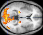
Functional magnetic resonance imaging (fMRI) of a head, from top to base of the skull

The first chapter of the history of neuroimaging traces back to the Italian neuroscientist Angelo Mosso who invented the 'human circulation balance', which could non-invasively measure the redistribution of blood during emotional and intellectual activity.

In 1918, the American neurosurgeon Walter Dandy introduced the technique of ventriculography. X-ray images of the ventricular system within the brain were obtained by injection of filtered air directly into one or both lateral ventricles of the brain. Dandy also observed that air introduced into the subarachnoid space via lumbar spinal puncture could enter the cerebral ventricles and also demonstrate the cerebrospinal fluid compartments around the base of the brain and over its surface. This technique was called pneumoencephalography.

In 1927, Egas Moniz introduced cerebral angiography, whereby both normal and abnormal blood vessels in and around the brain could be visualized with great precision.

In the early 1970s, Allan McLeod Cormack and Godfrey Newbold Hounsfield introduced computerized axial tomography (CAT or CT scanning), and ever more detailed anatomic images of the brain became available for diagnostic and research purposes. Cormack and Hounsfield won the 1979 Nobel Prize for Physiology or Medicine for their work. Soon after the introduction of CAT in the early 1980s, the development of radioligands allowed single photon emission computed tomography (SPECT) and positron emission tomography (PET) of the brain.

More or less concurrently, magnetic resonance imaging (MRI or MR scanning) was developed by researchers including Peter Mansfield and Paul Lauterbur, who were awarded the Nobel Prize for Physiology or Medicine in 2003. In the early 1980s MRI was introduced clinically, and during the 1980s a veritable explosion of technical refinements and diagnostic MR applications took place. Scientists soon learned that the large blood flow changes measured by PET could also be imaged by the correct type of MRI. Functional magnetic resonance imaging (fMRI) was born, and since the 1990s, fMRI has come to dominate the brain mapping field due to its low invasiveness, lack of radiation exposure, and relatively wide availability.

In the early 2000s, the field of neuroimaging reached the stage where limited practical applications of functional brain imaging have become feasible. The main application area is crude forms of brain-computer interface.

Indications

Neuroimaging follows a neurological examination in which a physician has found cause to more deeply investigate a patient who has or may have a neurological disorder.

One of the more common neurological problems which a person may experience is simple syncope. In cases of simple syncope in which the patient's history does not suggest other neurological symptoms, the diagnosis includes a neurological examination but routine neurological imaging is not indicated because the likelihood of finding a cause in the central nervous system is extremely low and the patient is unlikely to benefit from the procedure.

Neuroimaging is not indicated for patients with stable headaches which are diagnosed as migraine. Studies indicate that presence of migraine does not increase a patient's risk for intracranial disease. A diagnosis of migraine which notes the absence of other problems, such as papilledema, would not indicate a need for neuroimaging. In the course of conducting a careful diagnosis, the physician should consider whether the headache has a cause other than the migraine and might require neuroimaging.

Another indication for neuroimaging is CT-, MRI- and PET-guided stereotactic surgery or radiosurgery for treatment of intracranial tumors, arteriovenous malformations and other surgically treatable conditions.

Brain imaging techniques

Computed axial tomography

Computed tomography (CT) or Computed Axial Tomography (CAT) scanning uses a series of x-rays of the head taken from many different directions. Typically used for quickly viewing brain injuries, CT scanning uses a computer program that performs a numerical integral calculation (the inverse Radon transform) on the measured x-ray series to estimate how much of an x-ray beam is absorbed in a small volume of the brain. Typically the information is presented as cross-sections of the brain.

Diffuse optical imaging

Diffuse optical imaging (DOI) or diffuse optical tomography (DOT) is a medical imaging modality which uses near infrared light to generate images of the body. The technique measures the optical absorption of haemoglobin, and relies on the absorption spectrum of haemoglobin varying with its oxygenation status. High-density diffuse optical tomography (HD-DOT) has been compared directly to fMRI using response to visual stimulation in subjects studied with both techniques, with reassuringly similar results. HD-DOT has also been compared to fMRI in terms of language tasks and resting state functional connectivity.

Event-related optical signal

Event-related optical signal (EROS) is a brain-scanning technique which uses infrared light through optical fibers to measure changes in optical properties of active areas of the cerebral cortex. Whereas techniques such as diffuse optical imaging (DOT) and near-infrared spectroscopy (NIRS) measure optical absorption of haemoglobin, and thus are based on blood flow, EROS takes advantage of the scattering properties of the neurons themselves and thus provides a much more direct measure of cellular activity. EROS can pinpoint activity in the brain within millimeters (spatially) and within milliseconds (temporally). Its biggest downside is the inability to detect activity more than a few centimeters deep. EROS is a new, relatively inexpensive technique that is non-invasive to the test subject. It was developed at the University of Illinois at Urbana-Champaign where it is now used in the Cognitive Neuroimaging Laboratory of Dr. Gabriele Gratton and Dr. Monica Fabiani.

Magnetic resonance imaging

Sagittal MRI slice at the midline.

Magnetic resonance imaging (MRI) uses magnetic fields and radio waves to produce high quality two- or three-dimensional images of brain structures without the use of ionizing radiation (X-rays) or radioactive tracers.

the record for the highest spatial resolution of a whole intact brain (postmortem) is 100 microns, from Massachusetts General Hospital. The data was published in NATURE on 30th of October 2019.

Functional magnetic resonance imaging
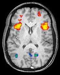
Axial MRI slice at the level of the basal ganglia, showing fMRI BOLD signal changes overlaid in red (increase) and blue (decrease) tones.

Functional magnetic resonance imaging (fMRI) and arterial spin labeling (ASL) relies on the paramagnetic properties of oxygenated and deoxygenated hemoglobin to see images of changing blood flow in the brain associated with neural activity. This allows images to be generated that reflect which brain structures are activated (and how) during the performance of different tasks or at resting state. According to the oxygenation hypothesis, changes in oxygen usage in regional cerebral blood flow during cognitive or behavioral activity can be associated with the regional neurons as being directly related to the cognitive or behavioral tasks being attended.

Most fMRI scanners allow subjects to be presented with different visual images, sounds and touch stimuli, and to make different actions such as pressing a button or moving a joystick. Consequently, fMRI can be used to reveal brain structures and processes associated with perception, thought and action. The resolution of fMRI is about 2-3 millimeters at present, limited by the spatial spread of the hemodynamic response to neural activity. It has largely superseded PET for the study of brain activation patterns. PET, however, retains the significant advantage of being able to identify specific brain receptors (or transporters) associated with particular neurotransmitters through its ability to image radiolabelled receptor "ligands" (receptor ligands are any chemicals that stick to receptors).

As well as research on healthy subjects, fMRI is increasingly used for the medical diagnosis of disease. Because fMRI is exquisitely sensitive to oxygen usage in blood flow, it is extremely sensitive to early changes in the brain resulting from ischemia (abnormally low blood flow), such as the changes which follow stroke. Early diagnosis of certain types of stroke is increasingly important in neurology, since substances which dissolve blood clots may be used in the first few hours after certain types of stroke occur, but are dangerous to use afterward. Brain changes seen on fMRI may help to make the decision to treat with these agents. With between 72% and 90% accuracy where chance would achieve 0.8%, fMRI techniques can decide which of a set of known images the subject is viewing.

Magnetoencephalography

Magnetoencephalography (MEG) is an imaging technique used to measure the magnetic fields produced by electrical activity in the brain via extremely sensitive devices such as superconducting quantum interference devices (SQUIDs) or spin exchange relaxation-free (SERF) magnetometers. MEG offers a very direct measurement of neural electrical activity (compared to fMRI for example) with very high temporal resolution but relatively low spatial resolution. The advantage of measuring the magnetic fields produced by neural activity is that they are likely to be less distorted by surrounding tissue (particularly the skull and scalp) compared to the electric fields measured by electroencephalography (EEG). Specifically, it can be shown that magnetic fields produced by electrical activity are not affected by the surrounding head tissue, when the head is modeled as a set of concentric spherical shells, each being an isotropic homogeneous conductor. Real heads are non-spherical and have largely anisotropic conductivities (particularly white matter and skull). While skull anisotropy has a negligible effect on MEG (unlike EEG), white matter anisotropy strongly affects MEG measurements for radial and deep sources. Note, however, that the skull was assumed to be uniformly anisotropic in this study, which is not true for a real head: the absolute and relative thicknesses of diploƫ and tables layers vary among and within the skull bones. This makes it likely that MEG is also affected by the skull anisotropy, although probably not to the same degree as EEG.

There are many uses for MEG, including assisting surgeons in localizing a pathology, assisting researchers in determining the function of various parts of the brain, neurofeedback, and others.

Positron emission tomography

Positron emission tomography (PET) and brain positron emission tomography, measure emissions from radioactively labeled metabolically active chemicals that have been injected into the bloodstream. The emission data are computer-processed to produce 2- or 3-dimensional images of the distribution of the chemicals throughout the brain. The positron emitting radioisotopes used are produced by a cyclotron, and chemicals are labeled with these radioactive atoms. The labeled compound, called a radiotracer, is injected into the bloodstream and eventually makes its way to the brain. Sensors in the PET scanner detect the radioactivity as the compound accumulates in various regions of the brain. A computer uses the data gathered by the sensors to create multicolored 2- or 3-dimensional images that show where the compound acts in the brain. Especially useful are a wide array of ligands used to map different aspects of neurotransmitter activity, with by far the most commonly used PET tracer being a labeled form of glucose.

The greatest benefit of PET scanning is that different compounds can show blood flow and oxygen and glucose metabolism in the tissues of the working brain. These measurements reflect the amount of brain activity in the various regions of the brain and allow to learn more about how the brain works. PET scans were superior to all other metabolic imaging methods in terms of resolution and speed of completion (as little as 30 seconds) when they first became available. The improved resolution permitted better study to be made as to the area of the brain activated by a particular task. The biggest drawback of PET scanning is that because the radioactivity decays rapidly, it is limited to monitoring short tasks. Before fMRI technology came online, PET scanning was the preferred method of functional (as opposed to structural) brain imaging, and it continues to make large contributions to neuroscience.

PET scanning is also used for diagnosis of brain disease, most notably because brain tumors, strokes, and neuron-damaging diseases which cause dementia (such as Alzheimer's disease) all cause great changes in brain metabolism, which in turn causes easily detectable changes in PET scans. PET is probably most useful in early cases of certain dementias (with classic examples being Alzheimer's disease and Pick's disease) where the early damage is too diffuse and makes too little difference in brain volume and gross structure to change CT and standard MRI images enough to be able to reliably differentiate it from the "normal" range of cortical atrophy which occurs with aging (in many but not all) persons, and which does not cause clinical dementia.

Single-photon emission computed tomography

Single-photon emission computed tomography (SPECT) is similar to PET and uses gamma ray-emitting radioisotopes and a gamma camera to record data that a computer uses to construct two- or three-dimensional images of active brain regions. SPECT relies on an injection of radioactive tracer, or "SPECT agent," which is rapidly taken up by the brain but does not redistribute. Uptake of SPECT agent is nearly 100% complete within 30 to 60 seconds, reflecting cerebral blood flow (CBF) at the time of injection. These properties of SPECT make it particularly well-suited for epilepsy imaging, which is usually made difficult by problems with patient movement and variable seizure types. SPECT provides a "snapshot" of cerebral blood flow since scans can be acquired after seizure termination (so long as the radioactive tracer was injected at the time of the seizure). A significant limitation of SPECT is its poor resolution (about 1 cm) compared to that of MRI. Today, SPECT machines with Dual Detector Heads are commonly used, although Triple Detector Head machines are available in the marketplace. Tomographic reconstruction, (mainly used for functional "snapshots" of the brain) requires multiple projections from Detector Heads which rotate around the human skull, so some researchers have developed 6 and 11 Detector Head SPECT machines to cut imaging time and give higher resolution.

Like PET, SPECT also can be used to differentiate different kinds of disease processes which produce dementia, and it is increasingly used for this purpose. Neuro-PET has a disadvantage of requiring the use of tracers with half-lives of at most 110 minutes, such as FDG. These must be made in a cyclotron, and are expensive or even unavailable if necessary transport times are prolonged more than a few half-lives. SPECT, however, is able to make use of tracers with much longer half-lives, such as technetium-99m, and as a result, is far more widely available.

Cranial ultrasound

Cranial ultrasound is usually only used in babies, whose open fontanelles provide acoustic windows allowing ultrasound imaging of the brain. Advantages include the absence of ionising radiation and the possibility of bedside scanning, but the lack of soft-tissue detail means MRI is preferred for some conditions.

Functional ultrasound imaging

Functional ultrasound imaging (fUS) is a medical ultrasound imaging technique of detecting or measuring changes in neural activities or metabolism, for example, the loci of brain activity, typically through measuring blood flow or hemodynamic changes. Functional ultrasound relies on Ultrasensitive Doppler and ultrafast ultrasound imaging which allows high sensitivity blood flow imaging.

Advantages and concerns of neuroimaging techniques

Functional Magnetic Resonance Imaging (fMRI)

fMRI is commonly classified as a minimally-to-moderate risk due to its non-invasiveness compared to other imaging methods. fMRI uses blood oxygenation level dependent (BOLD)-contrast in order to produce its form of imaging. BOLD-contrast is a naturally occurring process in the body so fMRI is often preferred over imaging methods that require radioactive markers to produce similar imaging. A concern in the use of fMRI is its use in individuals with medical implants or devices and metallic items in the body. The magnetic resonance (MR) emitted from the equipment can cause failure of medical devices and attract metallic objects in the body if not properly screened for. Currently, the FDA classifies medical implants and devices into three categories, depending on MR-compatibility: MR-safe (safe in all MR environments), MR-unsafe (unsafe in any MR environment), and MR-conditional (MR-compatible in certain environments, requiring further information).

Computed Tomography (CT) Scan

The CT scan was introduced in the 1970s and quickly became one of the most widely used methods of imaging. A CT scan can be performed in under a second and produce rapid results for clinicians, with its ease of use leading to an increase in CT scans performed in the United States from 3 million in 1980 to 62 million in 2007. Clinicians oftentimes take multiple scans, with 30% of individuals undergoing at least 3 scans in one study of CT scan usage. CT scans can expose patients to levels of radiation 100-500 times higher than traditional x-rays, with higher radiation doses producing better resolution imaging.

While easy to use, increases in CT scan use, especially in asymptomatic patients, is a topic of concern since patients are exposed to significantly high levels of radiation.

Positron Emission Tomography (PET)

In PET scans, imaging does not rely on intrinsic biological processes, but relies on a foreign substance injected into the bloodstream traveling to the brain. Patients are injected with radioisotopes that are metabolized in the brain and emit positrons to produce a visualization of brain activity. The amount of radiation a patient is exposed to in a PET scan is relatively small, comparable to the amount of environmental radiation an individual is exposed to across a year. PET radioisotopes have limited exposure time in the body as they commonly have very short half-lives (~2 hours) and decay rapidly. Currently, fMRI is a preferred method of imaging brain activity compared to PET, since it does not involve radiation, has a higher temporal resolution than PET, and is more readily available in most medical settings.

Magnetoencephalography (MEG) and Electroencephalography (EEG)

The high temporal resolution of MEG and EEG allow these methods to measure brain activity down to the millisecond. Both MEG and EEG do not require exposure of the patient to radiation to function. EEG electrodes detect electrical signals produced by neurons to measure brain activity and MEG uses oscillations in the magnetic field produced by these electrical currents to measure activity. A barrier in the widespread usage of MEG is due to pricing, as MEG systems can cost millions of dollars. EEG is a much more widely used method to achieve such temporal resolution as EEG systems cost much less than MEG systems. A disadvantage of EEG and MEG is that both methods have poor spatial resolution when compared to fMRI.

Criticism and cautions

Some scientists have criticized the brain image-based claims made in scientific journals and the popular press, like the discovery of "the part of the brain responsible" for functions like talents, specific memories, or generating emotions such as love. Many mapping techniques have a relatively low resolution, including hundreds of thousands of neurons in a single voxel. Many functions also involve multiple parts of the brain, meaning that this type of claim is probably both unverifiable with the equipment used, and generally based on an incorrect assumption about how brain functions are divided. It may be that most brain functions will only be described correctly after being measured with much more fine-grained measurements that look not at large regions but instead at a very large number of tiny individual brain circuits. Many of these studies also have technical problems like small sample size or poor equipment calibration which means they cannot be reproduced - considerations which are sometimes ignored to produce a sensational journal article or news headline. In some cases the brain mapping techniques are used for commercial purposes, lie detection, or medical diagnosis in ways which have not been scientifically validated.


Friday, February 12, 2021

Neurolinguistics

From Wikipedia, the free encyclopedia

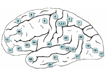
Surface of the human brain, with Brodmann areas numbered
 
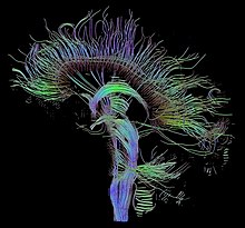
An image of neural pathways in the brain taken using diffusion tensor imaging

Neurolinguistics is the study of the neural mechanisms in the human brain that control the comprehension, production, and acquisition of language. As an interdisciplinary field, neurolinguistics draws methods and theories from fields such as neuroscience, linguistics, cognitive science, communication disorders and neuropsychology. Researchers are drawn to the field from a variety of backgrounds, bringing along a variety of experimental techniques as well as widely varying theoretical perspectives. Much work in neurolinguistics is informed by models in psycholinguistics and theoretical linguistics, and is focused on investigating how the brain can implement the processes that theoretical and psycholinguistics propose are necessary in producing and comprehending language. Neurolinguists study the physiological mechanisms by which the brain processes information related to language, and evaluate linguistic and psycholinguistic theories, using aphasiology, brain imaging, electrophysiology, and computer modeling.

History

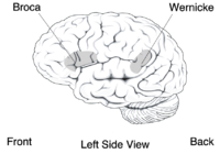
Neurolinguistics is historically rooted in the development in the 19th century of aphasiology, the study of linguistic deficits (aphasias) occurring as the result of brain damage. Aphasiology attempts to correlate structure to function by analyzing the effect of brain injuries on language processing. One of the first people to draw a connection between a particular brain area and language processing was Paul Broca, a French surgeon who conducted autopsies on numerous individuals who had speaking deficiencies, and found that most of them had brain damage (or lesions) on the left frontal lobe, in an area now known as Broca's area. Phrenologists had made the claim in the early 19th century that different brain regions carried out different functions and that language was mostly controlled by the frontal regions of the brain, but Broca's research was possibly the first to offer empirical evidence for such a relationship, and has been described as "epoch-making" and "pivotal" to the fields of neurolinguistics and cognitive science. Later, Carl Wernicke, after whom Wernicke's area is named, proposed that different areas of the brain were specialized for different linguistic tasks, with Broca's area handling the motor production of speech, and Wernicke's area handling auditory speech comprehension.

The work of Broca and Wernicke established the field of aphasiology and the idea that language can be studied through examining physical characteristics of the brain. Early work in aphasiology also benefited from the early twentieth-century work of Korbinian Brodmann, who "mapped" the surface of the brain, dividing it up into numbered areas based on each area's cytoarchitecture (cell structure) and function; these areas, known as Brodmann areas, are still widely used in neuroscience today.

The coining of the term "neurolinguistics" is attributed to Edith Crowell Trager, Henri Hecaen and Alexandr Luria, in the late 1940s and 1950s; Luria's book "Problems in Neurolinguistics" is likely the first book with Neurolinguistics in the title. Harry Whitaker popularized neurolinguistics in the United States in the 1970s, founding the journal "Brain and Language" in 1974.

Although aphasiology is the historical core of neurolinguistics, in recent years the field has broadened considerably, thanks in part to the emergence of new brain imaging technologies (such as PET and fMRI) and time-sensitive electrophysiological techniques (EEG and MEG), which can highlight patterns of brain activation as people engage in various language tasks; electrophysiological techniques, in particular, emerged as a viable method for the study of language in 1980 with the discovery of the N400, a brain response shown to be sensitive to semantic issues in language comprehension. The N400 was the first language-relevant event-related potential to be identified, and since its discovery EEG and MEG have become increasingly widely used for conducting language research.

Interaction with other fields

Neurolinguistics is closely related to the field of psycholinguistics, which seeks to elucidate the cognitive mechanisms of language by employing the traditional techniques of experimental psychology; today, psycholinguistic and neurolinguistic theories often inform one another, and there is much collaboration between the two fields.

Much work in neurolinguistics involves testing and evaluating theories put forth by psycholinguists and theoretical linguists. In general, theoretical linguists propose models to explain the structure of language and how language information is organized, psycholinguists propose models and algorithms to explain how language information is processed in the mind, and neurolinguists analyze brain activity to infer how biological structures (populations and networks of neurons) carry out those psycholinguistic processing algorithms. For example, experiments in sentence processing have used the ELAN, N400, and P600 brain responses to examine how physiological brain responses reflect the different predictions of sentence processing models put forth by psycholinguists, such as Janet Fodor and Lyn Frazier's "serial" model, and Theo Vosse and Gerard Kempen's "unification model". Neurolinguists can also make new predictions about the structure and organization of language based on insights about the physiology of the brain, by "generalizing from the knowledge of neurological structures to language structure".

Neurolinguistics research is carried out in all the major areas of linguistics; the main linguistic subfields, and how neurolinguistics addresses them, are given in the table below.

Subfield Description Research questions in neurolinguistics
Phonetics the study of speech sounds how the brain extracts speech sounds from an acoustic signal, how the brain separates speech sounds from background noise
Phonology the study of how sounds are organized in a language how the phonological system of a particular language is represented in the brain
Morphology and lexicology the study of how words are structured and stored in the mental lexicon how the brain stores and accesses words that a person knows
Syntax the study of how multiple-word utterances are constructed how the brain combines words into constituents and sentences; how structural and semantic information is used in understanding sentences
Semantics the study of how meaning is encoded in language

Topics considered

Neurolinguistics research investigates several topics, including where language information is processed, how language processing unfolds over time, how brain structures are related to language acquisition and learning, and how neurophysiology can contribute to speech and language pathology.

Localizations of language processes

Much work in neurolinguistics has, like Broca's and Wernicke's early studies, investigated the locations of specific language "modules" within the brain. Research questions include what course language information follows through the brain as it is processed, whether or not particular areas specialize in processing particular sorts of information, how different brain regions interact with one another in language processing, and how the locations of brain activation differ when a subject is producing or perceiving a language other than his or her first language.

Time course of language processes

Another area of neurolinguistics literature involves the use of electrophysiological techniques to analyze the rapid processing of language in time. The temporal ordering of specific patterns of brain activity may reflect discrete computational processes that the brain undergoes during language processing; for example, one neurolinguistic theory of sentence parsing proposes that three brain responses (the ELAN, N400, and P600) are products of three different steps in syntactic and semantic processing.

Language acquisition

Another topic is the relationship between brain structures and language acquisition. Research in first language acquisition has already established that infants from all linguistic environments go through similar and predictable stages (such as babbling), and some neurolinguistics research attempts to find correlations between stages of language development and stages of brain development, while other research investigates the physical changes (known as neuroplasticity) that the brain undergoes during second language acquisition, when adults learn a new language. Neuroplasticity is observed when both Second Language acquisition and Language Learning experience are induced, the result of this language exposure concludes that an increase of gray and white matter could be found in children, young adults and the elderly.

Ping Li, Jennifer Legault, Kaitlyn A. Litcofsky, May 2014. Neuroplasticity as a function of second language learning: Anatomical changes in the human brain Cortex: A Journal Devoted to the Study of the Nervous System & Behavior, 410.1016/j.cortex.2014.05.00124996640

Language pathology

Neurolinguistic techniques are also used to study disorders and breakdowns in language, such as aphasia and dyslexia, and how they relate to physical characteristics of the brain.

Technology used

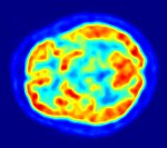
Images of the brain recorded with PET (top) and fMRI (bottom). In the PET image, the red areas are the most active. In the fMRI image, the yellowest areas are the areas that show the greatest difference in activation between two tasks (watching a moving stimulus, versus watching a black screen).

Since one of the focuses of this field is the testing of linguistic and psycholinguistic models, the technology used for experiments is highly relevant to the study of neurolinguistics. Modern brain imaging techniques have contributed greatly to a growing understanding of the anatomical organization of linguistic functions. Brain imaging methods used in neurolinguistics may be classified into hemodynamic methods, electrophysiological methods, and methods that stimulate the cortex directly.

Hemodynamic

Hemodynamic techniques take advantage of the fact that when an area of the brain works at a task, blood is sent to supply that area with oxygen (in what is known as the Blood Oxygen Level-Dependent, or BOLD, response). Such techniques include PET and fMRI. These techniques provide high spatial resolution, allowing researchers to pinpoint the location of activity within the brain; temporal resolution (or information about the timing of brain activity), on the other hand, is poor, since the BOLD response happens much more slowly than language processing. In addition to demonstrating which parts of the brain may subserve specific language tasks or computations, hemodynamic methods have also been used to demonstrate how the structure of the brain's language architecture and the distribution of language-related activation may change over time, as a function of linguistic exposure.

In addition to PET and fMRI, which show which areas of the brain are activated by certain tasks, researchers also use diffusion tensor imaging (DTI), which shows the neural pathways that connect different brain areas, thus providing insight into how different areas interact. Functional near-infrared spectroscopy (fNIRS) is another hemodynamic method used in language tasks.

Electrophysiological

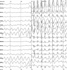
Brain waves recorded using EEG

Electrophysiological techniques take advantage of the fact that when a group of neurons in the brain fire together, they create an electric dipole or current. The technique of EEG measures this electric current using sensors on the scalp, while MEG measures the magnetic fields that are generated by these currents. In addition to these non-invasive methods, electrocorticography has also been used to study language processing. These techniques are able to measure brain activity from one millisecond to the next, providing excellent temporal resolution, which is important in studying processes that take place as quickly as language comprehension and production. On the other hand, the location of brain activity can be difficult to identify in EEG; consequently, this technique is used primarily to how language processes are carried out, rather than where. Research using EEG and MEG generally focuses on event-related potentials (ERPs), which are distinct brain responses (generally realized as negative or positive peaks on a graph of neural activity) elicited in response to a particular stimulus. Studies using ERP may focus on each ERP's latency (how long after the stimulus the ERP begins or peaks), amplitude (how high or low the peak is), or topography (where on the scalp the ERP response is picked up by sensors). Some important and common ERP components include the N400 (a negativity occurring at a latency of about 400 milliseconds), the mismatch negativity, the early left anterior negativity (a negativity occurring at an early latency and a front-left topography), the P600, and the lateralized readiness potential.

Experimental design

Experimental techniques

Neurolinguists employ a variety of experimental techniques in order to use brain imaging to draw conclusions about how language is represented and processed in the brain. These techniques include the subtraction paradigm, mismatch design, violation-based studies, various forms of priming, and direct stimulation of the brain.

Subtraction

Many language studies, particularly in fMRI, use the subtraction paradigm, in which brain activation in a task thought to involve some aspect of language processing is compared against activation in a baseline task thought to involve similar non-linguistic processes but not to involve the linguistic process. For example, activations while participants read words may be compared to baseline activations while participants read strings of random letters (in attempt to isolate activation related to lexical processing—the processing of real words), or activations while participants read syntactically complex sentences may be compared to baseline activations while participants read simpler sentences.

Mismatch paradigm

The mismatch negativity (MMN) is a rigorously documented ERP component frequently used in neurolinguistic experiments. It is an electrophysiological response that occurs in the brain when a subject hears a "deviant" stimulus in a set of perceptually identical "standards" (as in the sequence s s s s s s s d d s s s s s s d s s s s s d). Since the MMN is elicited only in response to a rare "oddball" stimulus in a set of other stimuli that are perceived to be the same, it has been used to test how speakers perceive sounds and organize stimuli categorically. For example, a landmark study by Colin Phillips and colleagues used the mismatch negativity as evidence that subjects, when presented with a series of speech sounds with acoustic parameters, perceived all the sounds as either /t/ or /d/ in spite of the acoustic variability, suggesting that the human brain has representations of abstract phonemes—in other words, the subjects were "hearing" not the specific acoustic features, but only the abstract phonemes. In addition, the mismatch negativity has been used to study syntactic processing and the recognition of word category.

Violation-based

Many studies in neurolinguistics take advantage of anomalies or violations of syntactic or semantic rules in experimental stimuli, and analyzing the brain responses elicited when a subject encounters these violations. For example, sentences beginning with phrases such as *the garden was on the worked, which violates an English phrase structure rule, often elicit a brain response called the early left anterior negativity (ELAN). Violation techniques have been in use since at least 1980, when Kutas and Hillyard first reported ERP evidence that semantic violations elicited an N400 effect. Using similar methods, in 1992, Lee Osterhout first reported the P600 response to syntactic anomalies. Violation designs have also been used for hemodynamic studies (fMRI and PET): Embick and colleagues, for example, used grammatical and spelling violations to investigate the location of syntactic processing in the brain using fMRI. Another common use of violation designs is to combine two kinds of violations in the same sentence and thus make predictions about how different language processes interact with one another; this type of crossing-violation study has been used extensively to investigate how syntactic and semantic processes interact while people read or hear sentences.

Priming

In psycholinguistics and neurolinguistics, priming refers to the phenomenon whereby a subject can recognize a word more quickly if he or she has recently been presented with a word that is similar in meaning or morphological makeup (i.e., composed of similar parts). If a subject is presented with a "prime" word such as doctor and then a "target" word such as nurse, if the subject has a faster-than-usual response time to nurse then the experimenter may assume that word nurse in the brain had already been accessed when the word doctor was accessed. Priming is used to investigate a wide variety of questions about how words are stored and retrieved in the brain and how structurally complex sentences are processed.

Stimulation

Transcranial magnetic stimulation (TMS), a new noninvasive technique for studying brain activity, uses powerful magnetic fields that are applied to the brain from outside the head. It is a method of exciting or interrupting brain activity in a specific and controlled location, and thus is able to imitate aphasic symptoms while giving the researcher more control over exactly which parts of the brain will be examined. As such, it is a less invasive alternative to direct cortical stimulation, which can be used for similar types of research but requires that the subject's scalp be removed, and is thus only used on individuals who are already undergoing a major brain operation (such as individuals undergoing surgery for epilepsy). The logic behind TMS and direct cortical stimulation is similar to the logic behind aphasiology: if a particular language function is impaired when a specific region of the brain is knocked out, then that region must be somehow implicated in that language function. Few neurolinguistic studies to date have used TMS; direct cortical stimulation and cortical recording (recording brain activity using electrodes placed directly on the brain) have been used with macaque monkeys to make predictions about the behavior of human brains.

Subject tasks

In many neurolinguistics experiments, subjects do not simply sit and listen to or watch stimuli, but also are instructed to perform some sort of task in response to the stimuli. Subjects perform these tasks while recordings (electrophysiological or hemodynamic) are being taken, usually in order to ensure that they are paying attention to the stimuli. At least one study has suggested that the task the subject does has an effect on the brain responses and the results of the experiment.

Lexical decision

The lexical decision task involves subjects seeing or hearing an isolated word and answering whether or not it is a real word. It is frequently used in priming studies, since subjects are known to make a lexical decision more quickly if a word has been primed by a related word (as in "doctor" priming "nurse").

Grammaticality judgment, acceptability judgment

Many studies, especially violation-based studies, have subjects make a decision about the "acceptability" (usually grammatical acceptability or semantic acceptability) of stimuli. Such a task is often used to "ensure that subjects [are] reading the sentences attentively and that they [distinguish] acceptable from unacceptable sentences in the way the [experimenter] expect[s] them to do."

Experimental evidence has shown that the instructions given to subjects in an acceptability judgment task can influence the subjects' brain responses to stimuli. One experiment showed that when subjects were instructed to judge the "acceptability" of sentences they did not show an N400 brain response (a response commonly associated with semantic processing), but that they did show that response when instructed to ignore grammatical acceptability and only judge whether or not the sentences "made sense".

Probe verification

Some studies use a "probe verification" task rather than an overt acceptability judgment; in this paradigm, each experimental sentence is followed by a "probe word", and subjects must answer whether or not the probe word had appeared in the sentence. This task, like the acceptability judgment task, ensures that subjects are reading or listening attentively, but may avoid some of the additional processing demands of acceptability judgments, and may be used no matter what type of violation is being presented in the study.

Truth-value judgment

Subjects may be instructed not to judge whether or not the sentence is grammatically acceptable or logical, but whether the proposition expressed by the sentence is true or false. This task is commonly used in psycholinguistic studies of child language.

Active distraction and double-task

Some experiments give subjects a "distractor" task to ensure that subjects are not consciously paying attention to the experimental stimuli; this may be done to test whether a certain computation in the brain is carried out automatically, regardless of whether the subject devotes attentional resources to it. For example, one study had subjects listen to non-linguistic tones (long beeps and buzzes) in one ear and speech in the other ear, and instructed subjects to press a button when they perceived a change in the tone; this supposedly caused subjects not to pay explicit attention to grammatical violations in the speech stimuli. The subjects showed a mismatch response (MMN) anyway, suggesting that the processing of the grammatical errors was happening automatically, regardless of attention—or at least that subjects were unable to consciously separate their attention from the speech stimuli.

Another related form of experiment is the double-task experiment, in which a subject must perform an extra task (such as sequential finger-tapping or articulating nonsense syllables) while responding to linguistic stimuli; this kind of experiment has been used to investigate the use of working memory in language processing.

Thought disorder

From Wikipedia, the free encyclopedia
 
Thought disorder
Other namesFormal thought disorder (FTD), thinking disorder
Cloth embroidered by a schizophrenia sufferer.jpg
An embroidered cloth produced by a person with schizophrenia, showing the nonsensical associations between words and ideas characteristic of thought disorder
SpecialtyPsychiatry

A thought disorder (TD) is any disturbance in cognition that adversely affects language and thought content, and thereby communication. A variety of thought disorders were said to be characteristic of people with schizophrenia. A content-thought disorder is typically characterised by the experience of multiple delusional fragments. The term, thought disorder, is often used to refer to a formal thought disorder.

A formal thought disorder (FTD) is a disruption of the form or structure of thought. Formal thought disorder, also known as disorganised thinking, results in disorganised speech, and is recognised as a major feature of schizophrenia, and other psychoses. FTD is also associated with conditions including mood disorders, dementia, mania, and neurological diseases.

Types of thought disorder include derailment, pressured speech, poverty of speech, tangentiality, repeating things, and thought blocking.

Formal thought disorder is a disorder of the form of thought rather than of content of thought that covers hallucinations and delusions. FTD unlike hallucinations and delusions, is an observable objective sign of psychosis. FTD is a common, and core symptom of a psychotic disorder and may be seen as a marker of its severity, and also as a predictor of prognosis. It reflects a cluster of cognitive, linguistic, and affective disturbances, that has generated research interest from the fields of cognitive neuroscience, neurolinguistics, and psychiatry.

Eugen Bleuler, who named schizophrenia, held that thought disorder was its defining characteristic.

However, disturbances of thinking and speech such as clanging or echolalia may be present in Tourette syndrome, or other symptoms as found in delirium. A clinical difference exists between these two groups. Those with psychoses are less likely to show an awareness or concern about the disordered thinking, while those with other disorders do show awareness and concerns about not being able to think straight.

Content-thought disorder

Content-thought disorder is a thought disturbance in which a person experiences multiple, fragmented delusions, typically a feature of schizophrenia, and some other mental disorders including obsessive–compulsive disorder, and mania. Content-thought disorder is not limited to delusions, other possible abnormalities include preoccupation (centering thought to a particular idea in association with strong affection), obsession (a persistent thought, idea, or image that is intrusive or inappropriate, and is distressing or upsetting), compulsion (the need to perform an act persistently and repetitively—without it necessarily leading to an actual reward or pleasure—to reduce distress), magical thinking (belief that one's thoughts by themselves can bring about effects in the world, or that thinking something corresponds with doing the same thing), overvalued ideas (false/exaggerated belief that is held with conviction but not with delusional intensity), ideas of reference (belief that innocuous or coincident events experienced have strong personal significance) or influence (belief that other people or external agents are covertly exerting powers over oneself), persecutory ideas, phobias (irrational fears of objects or circumstances), suicidal ideas, violent ideas, and homicidal ideas.

The cores of thought content disturbance are abnormal beliefs and convictions, after accounting for the person's culture and backgrounds, and range from overvalued ideas to fixed delusions. Typically, abnormal beliefs and delusions are non-specific diagnostically, even if some delusions are more prevalent in one disorder than another. Also, normal, or neurotypical, thought—consisting of awareness, concerns, beliefs, preoccupations, wishes, fantasies, imagination, and concepts—can be illogical, and can contain beliefs and prejudices/biases that are obviously contradictory. Individuals also have considerable variations, and the same person's thinking also may shift considerably from time to time.

In psychosis, delusions are the most common thought-content abnormalities. A delusion is a firm and fixed belief based on inadequate grounds not amenable to rational argument or evidence to the contrary, and not in sync with regional, cultural and educational background. Common examples in mental status examination include: erotomanic (belief that someone is in love with oneself), grandiose (belief that one is the greatest, strongest, fastest, richest, and/or most intelligent person ever), persecutory (belief that the person, or someone to whom the person is close, is being malevolently treated in some way), reference (belief that insignificant remarks, events, or objects in one's environment have personal meaning or significance), thought broadcasting (belief that others can hear or are aware of one's thoughts), thought insertion (belief that one's thoughts are not one's own, but rather belong to someone else and have been inserted into one's mind), thought withdrawal (belief that thoughts have been 'taken out' of one's mind, and one has no power over this), outside control (belief that outside forces are controlling one's thoughts, feelings, and actions), infidelity (belief that a partner is cheating on oneself), somatic (belief that one has a disease or medical condition), and nihilistic (belief that the mind, body, the world at large, or parts thereof, no longer exist). Delusions are common in people with mania, depression, schizoaffective disorder, delirium, dementia, substance use disorder, schizophrenia, and delusional disorders

Formal thought disorder

Formal thought disorder (FTD), or simply thought disorder, is also known as disorganized speech – evident from disorganized thinking, and is one of the hallmark features of schizophrenia. Formal thought disorder is a disorder of the form of thought rather than of content of thought that covers hallucinations and delusions. FTD, unlike hallucinations and delusions, is an observable objective sign of psychosis. FTD is a common, and core symptom of a psychotic disorder and may be seen as a marker of its severity, and also as a predictor of prognosis. It reflects a cluster of cognitive, linguistic, and affective disturbances, that has generated research interest from the fields of cognitive neuroscience, neurolinguistics, and psychiatry.

FTD is a complex, multidimensional syndrome characterized by deficiencies in the logical organizing of thought needed to achieve goals. FTD can be subdivided into clusters of positive and negative symptoms, as well as objective versus subjective symptoms. Within the scale of positive and negative symptoms they have been grouped into positive formal thought disorder (posFTD) and negative formal thought disorder (negFTD). Positive subtypes were those of pressure of speech, tangentiality, derailment, incoherence, and illogicality. Negative subtypes were those of poverty of speech and poverty of content. The two groups were posited to be at either end of a spectrum of normal speech. However, later studies showed these to be poorly correlated. A comprehensive measure of formal thought disorder is the Thought and Language Disorder (TALD) Scale.

Nancy Andreasen preferred to call the thought disorders collectively as thought-language-communication disorders (TLC disorders). Within the Thought, Language, Communication (TLC) Scale up to seven domains of FTD have been described with most of the variance accounted for by just two or three domains. Some TLC disorders are more suggestive of a severe disorder and given priority by listing them in the first 11 items.

It has been proposed that formal thought disorder relates to neurocognition via semantic memory. Semantic network impairment in people with schizophrenia—measured by the difference between fluency (number of animals' names produced in 60 seconds) and phonological fluency (number of words beginning with "F" produced in 60 seconds)—predicts severity of formal thought disorder, suggesting that verbal information (through semantic priming) is unavailable. Other hypotheses include working memory deficit (being confused about what has already been said in a conversation) and attentional focus.

Signs and symptoms

In the general population there will always be abnormalities in language, and their presence or absence is therefore not diagnostic of any condition. Language abnormalities can occur in schizophrenia and other disorders such as mania or depression, and can also occur in anybody who may simply be tired or stressed. To distinguish thought disorder, patterns of speech, severity of symptoms, their frequency, and resulting functional impairment can be considered.

Symptoms of thought disorder include derailment, pressured speech, poverty of speech, tangentiality, and thought blocking. FTD is a hallmark feature of schizophrenia, but is also associated with other conditions including mood disorders, dementia, mania, and neurological diseases. Impaired attention, poor memory, and difficulty formulating abstract concepts may also reflect thought disorder, and can be observed or assessed with mental status tests such as serial sevens or memory tests.

Types

There are many types of thought disorder. They are also referred to as symptoms of formal thought disorder of which 30 are described including:

  • Alogia (also poverty of speech) – A poverty of speech, either in amount or content. Under negative/positive symptom classification of schizophrenia, it is classified as a negative symptom. When classifying symptoms into more dimensions, poverty of speech content—paucity of meaningful content with normal amount of speech—is a disorganization symptom, whereas poverty of speech—loss of speech production—is a negative symptom. Under SANS, thought blocking is considered a part of alogia, and so is increased latency in response.
  • Blocking or thought blocking, also called deprivation of thought or obstructive thought – An abrupt stop in the middle of a train of thought which may or not be able to be continued.
  • Circumstantial speech (also circumstantial thinking) — An inability to answer a question without giving excessive, unnecessary detail. This differs from tangential thinking, in that the person does eventually return to the original point. For example, the patient answers the question "how have you been sleeping lately?" with "Oh, I go to bed early, so I can get plenty of rest. I like to listen to music or read before bed. Right now I'm reading a good mystery. Maybe I'll write a mystery someday. But it isn't helping, reading I mean. I have been getting only 2 or 3 hours of sleep at night."
  • Clanging – A severe form of flight of ideas whereby ideas are related only by similar or rhyming sounds rather than actual meaning. This may be heard as excessive rhyming and/or alliteration. e.g. "Many moldy mushrooms merge out of the mildewy mud on Mondays." "I heard the bell. Well, hell, then I fell." It is most commonly seen in bipolar disorder (manic phase), although it is often observed in patients with primary psychoses, namely schizophrenia and schizoaffective disorder.
  • Derailment (also loose association and knight's move thinking) – Thought frequently moves from one idea to another which is obliquely related or unrelated, often appearing in speech but also in writing, e.g. "The next day when I'd be going out you know, I took control, like uh, I put bleach on my hair in California."
  • Distractible speech – During mid speech, the subject is changed in response to a nearby stimulus. e.g. "Then I left San Francisco and moved to... Where did you get that tie?"
  • Echolalia – Echoing of another's speech that may only be committed once, or may be continuous in repetition. This may involve repeating only the last few words or last word of the examiner's sentences. This can be a symptom of Tourette's Syndrome, e.g. "What would you like for dinner?", "That's a good question. That's a good question. That's a good question. That's a good question."
  • Evasion - the next logical idea in a sequence is replaced with another idea closely but not accurately or appropriately related to it. Also called paralogia and perverted logic. Example: "I... er ah... you are uh... I think you have... uh-- acceptable erm... uh... hair."
  • Flight of ideas - a form of formal thought disorder marked by abrupt leaps from one topic to another, possibly with discernable links between successive ideas, perhaps governed by similarities between subjects or, in somewhat higher grades, by rhyming, puns, and word plays, or by innocuous environmental stimuli – e.g., the sound of birds chirping. It is most characteristic of the manic phase of bipolar illness.
  • Illogicality – Conclusions are reached that do not follow logically (non-sequiturs or faulty inferences). e.g. "Do you think this will fit in the box?" draws a reply like "Well duh; it's brown, isn't it?"
  • Incoherence (word salad) – Speech that is unintelligible because, though the individual words are real words, the manner in which they are strung together results in incoherent gibberish, e.g. the question "Why do people comb their hair?" elicits a response like "Because it makes a twirl in life, my box is broken help me blue elephant. Isn't lettuce brave? I like electrons, hello please!"
  • Neologisms – forms completely new words or phrases whose origins and meanings are usually unrecognizable. Example is "I got so angry I picked up a dish and threw it at the geshinker." These may also involve elisions of two words that are similar in meaning or in sound. Although neologisms may sometimes refer to words that are formed incorrectly but whose origins are understandable (e.g. "headshoe" for hat), these can be more clearly referred to as word approximations.
  • Overinclusion is failure to eliminate ineffective, inappropriate, irrelevant, extraneous details associated with a particular stimulus.
  • Perseveration – Persistent repetition of words or ideas even when another person attempts to change the topic. e.g. "It's great to be here in Nevada, Nevada, Nevada, Nevada, Nevada." This may also involve repeatedly giving the same answer to different questions. e.g. "Is your name Mary?" "Yes." "Are you in the hospital?" "Yes." "Are you a table?" "Yes." Perseveration can include palilalia and logoclonia, and can be an indication of organic brain disease such as Parkinson's.
  • Phonemic paraphasia – Mispronunciation; syllables out of sequence. e.g. "I slipped on the lice and broke my arm."
  • Pressured speech Rapid speech without pauses, difficult to interrupt.
  • Self reference – Patient repeatedly and inappropriately refers back to self. e.g. "What's the time?", "It's 7 o'clock. That's my problem."
  • Semantic paraphasia – Substitution of inappropriate word. e.g. "I slipped on the coat, on the ice I mean, and broke my book."
  • Stilted speech Sentences may be stilted or vague. Speech characterized by the use of words or phrases that are flowery, excessive, and pompous, e.g. "The attorney comported himself indecorously."
  • Tangential speech – Wandering from the topic and never returning to it or providing the information requested. For example, in answer to the question "Where are you from?", the person answers "My dog is from England. They have good fish and chips there. Fish breathe through gills."
  • Verbigeration – Meaningless and stereotyped repetition of words or phrases replacing understandable speech, as seen in schizophrenia.

Use of term

Some recent (2015, 2017) psychiatric/psychological glossaries defined thought disorder as disturbed thinking or cognition that affects communication, language, or thought content including poverty of ideas, neologisms, paralogia, word salad, and delusions —which are disturbance of both thought content and thought form—and suggested the more specific terms of content thought disorder and formal thought disorder, with content thought disorder defined as a thought disturbance characterized by multiple fragmented delusions, and formal thought disorder defined as disturbance in the form or structure of thinking. For example, DSM-5 (2013) only used the word formal thought disorder, mostly as a synonym of disorganized thinking and disorganized speech. This is in contrast with ICD-10 (1992) which only used the word "thought disorder", always accompanied with "delusion" and "hallucination" separately, and a general medical dictionary (2002) that although generally defined thought disorders similarly to the psychiatric glossaries, but also used the word in other entries as ICD-10 did.

The recent psychiatric text (2017) also mentioned when describing thought disorder as a "disorganization syndrome" within the context of schizophrenia:

"Thought disorder" here refers to disorganization of the form of thought and not content. An older use of the term "thought disorder" included the phenomena of delusions and sometimes hallucinations, but this is confusing and ignores the clear differences in the relationships between symptoms that have become apparent over the past 30 years. Delusions and hallucinations should be identified as psychotic symptoms, and thought disorder should be taken to mean formal thought disorders or a disorder of verbal cognition.

— Phenomenology of Schizophrenia (2017), THE SYMPTOMS OF SCHIZOPHRENIA

The same text also mentioned that some clinicians use the term "formal thought disorder" broadly referring to abnormalities in thought form plus any psychotic cognitive sign or symptom, and that various studies examining cognition and subsymdromes in schizophrenia may refer to formal thought disorder as "conceptual disorganization" or "disorganization factor."

Still, there may be other dissenting opinions, including:

Unfortunately, "thought disorder" is often involved rather loosely to refer to both formal thought disorder and delusional content. For the sake of clarity, the unqualiļ¬ed use of the phrase "thought disorder" should be discarded from psychiatric communication. Even the designation "formal thought disorder" covers too wide a territory. It should always be made clear whether one is referring to derailment or loose associations, ļ¬‚ight of ideas, or circumstantiality.

— The Mental Status Examination, The Medical Basis of Psychiatry (2016)

Course, diagnosis, and prognosis

It was believed that thought disorder occurred only in schizophrenia, but later findings indicate it may occur in other psychiatric conditions including mania, and occurs even in people without mental illness. Also, people with schizophrenia don't all exhibit thought disorder, so not having any thought disorder doesn't mean the person doesn't have schizophrenia, i.e. the condition is not very specific to the disease.

When adopting specific definitions of thought disorder subtypes and classifying them as positive and negative symptoms, Nancy Andreasen found that different subtypes of thought disorder occur at different frequencies among those with manic, depression, and schizophrenia. People with mania have pressured speech as the most prominent symptom, but also have relatively high rates of derailment, tangentiality, and incoherence which are as prominent as in those with schizophrenia. They are likelier to have pressured speech, distractibility, and circumstantiality.

People with schizophrenia have more negative thought disorder including poverty of speech and poverty of content of speech, but also have relatively high rates of certain positive thought disorders.

Derailment, loss of goal, poverty of content of speech, tangentiality and illogicality are particularly characteristic of schizophrenia. People with depression have relatively less thought disorders; the most prominent are poverty of speech, poverty of content of speech, and circumstantiality. She found the diagnostic usefulness of dividing the symptoms into subtypes, such as having negative thought disorders without the full affective symptoms highly suggest schizophrenia.

She also found prognostic values of negative/positive symptom divisions. In manic patients, most thought disorders return to normal levels 6 months after evaluation which suggests that thought disorders in this condition, although as severe as in schizophrenia, tend to be recoverable. In people with schizophrenia, however, negative thought disorders remain after six months, and sometimes worsen. Positive thought disorders get better somewhat. Also, negative thought disorder is a good predictor of some outcomes, e.g. patients with prominent negative thought disorders do worse on social functioning six months later. So, in general, having more prominent negative symptoms suggest a worse outcome. Nevertheless, some people may do well, respond to medication, and have normal brain function. The positive symptoms are similar vice versa.

At illness onset, prominent thought disorder also predicts worse prognosis, including:

  • illness begins earlier
  • increased risk of hospitalization
  • decreased functional outcomes
  • increased disability rates
  • increased inappropriate social behaviors

Thought disorder unresponsive to treatment also predicts worse illness course. In schizophrenia, thought disorders' severity tend to be more stable than hallucinations and delusions. Prominent thought disorders are more unlikely to diminish in middle age compared to positive symptoms. Less severe thought disorder may occur during the prodromal and residual periods of schizophrenia.

DSM-5 include delusions, hallucinations, disorganized thought process (formal thought disorder), and disorganized or abnormal motor behavior (including catatonia) as key symptoms in "psychosis." Although not specific to different diagnoses, some aspects of psychosis are characteristic of some diagnoses. Schizophrenia spectrum disorders (e.g., schizoaffective disorder, schizophreniform disorder) typically consist of prominent hallucinations and/or delusions as well as formal thought disorder—displayed as severe behavioral abnormalities including disorganized, bizarre, and catatonic behavior. Psychotic disorders due to general medical conditions and substance-induced psychotic disorders typically consist of delusions and/or hallucinations. Delusional disorder and shared psychotic disorder, which are more rare, typically consist of persistent delusions. Research found that most formal thought disorders are commonly found in schizophrenia and mood disorders, but poverty of speech content is more common in schizophrenia.

Experienced clinicians may distinguish true psychosis, such as in schizophrenia, and bipolar mania, from malingering, when an individual fakes illness for other gains, by clinical presentations. For example, malingerers feign thought contents with no irregularities in form such as derailment or looseness of associations. Negative symptoms including alogia may not be present. In addition, chronic thought disorder is typically distressing.

Typically, autism spectrum disorders (ASD), whose diagnosis requires onset of symptoms prior to 3 years of age, can be distinguished from early-onset schizophrenia by disease onset occurrence (schizophrenia manifestation under age 10 is extremely rare) and the fact that ASD patients don't display formal thought disorders. However, it has been suggested that individuals with autism spectrum disorders (ASD) display language disturbances like those found in schizophrenia; a 2008 study found that children and adolescents with ASD showed significantly more illogical thinking and loose associations than control subjects. The illogical thinking was related to cognitive functioning and executive control; the loose associations were related to communication symptoms and to parent reports of stress and anxiety.

Criticisms

The concept of thought disorder has been criticized as being based on circular or incoherent definitions. For example, symptoms of thought disorder are inferred from disordered speech, based on the assumption that disordered speech arises because of disordered thought. Incoherence, or word salad, refers to speech that is semantically unconnected and conveys no meaning to the listener.

Furthermore, although thought disorder is typically associated with psychosis, similar phenomena can appear in different disorders, potentially leading to misdiagnosis—for example, in the case of incomplete yet potentially fruitful thought processes.

Another criticism related to the separation of symptoms of schizophrenia into negative/positive symptoms, including thought disorder, is that it oversimplifies the complexity of thought disorder and its relationship with other positive symptoms. Later factor analysis studies found that negative symptoms tend to correlate with one another, while positive symptoms tend to separate into two groups. The three clusters became roughly known as negative symptoms, psychotic symptoms, and disorganization symptoms. Alogia, a thought disorder traditionally classified as a negative symptom, can be separated into two separate groups: poverty of speech content as a disorganization symptom, and poverty of speech, response latency, and thought blocking as negative symptoms. Nevertheless, the efforts that led to the positive/negative symptom diametrics may allow the more accurate characterization of schizophrenia in the later works.

Lie point symmetry

From Wikipedia, the free encyclopedia https://en.wikipedia.org/wiki/Lie_point_symmetry     ...