Search This Blog

Thursday, April 22, 2021

Medication costs

From Wikipedia, the free encyclopedia

Medication costs, also known as drug costs are a common health care cost for many people and health care systems. Prescription costs are the costs to the end consumer. Medication costs are influenced by multiple factors such as patents, stakeholder influence, and marketing expenses. A number of countries including Canada, parts of Europe, and Brasil use external reference pricing as a means to compare drug prices and to determine a base price for a particular medication.

Medication costs can be listed in a number of ways including cost per defined daily dose, cost per specific period of time, cost per prescribed daily dose, and cost proportional to gross national product.

Definition

Medication costs can be the selling price from the manufacturer, that price together with shipping, the wholesale price, the retail price, and the dispensed price.

The dispensed price or prescription cost is defined as a cost which the patient has to pay to get medicines or treatments which are written as directions on prescription by a prescribers. The cost is generally influenced by a financial relationship between pharmaceutical manufacturers, wholesale distributors and pharmacies. In addition to the financial relationship, each nation has different systems to control the cost of prescriptions. In the United States, a pharmacy benefit manager, a third-party organization, such as private insurances or government-run health insurances will implement cost containment programs, such as establishing a formulary, to contain the cost.  In the United Kingdom, the government negotiates an overall cap on drugs bill growth with the pharmaceutical industry. In addition a government agency, the National Institute of Health and Care Excellence (NICE) assesses cost effectiveness of individual prescription drugs pricing. The National Health Service also may negotiate direct with individual pharmaceutical companies for certain specialised medicines, as well as running competitive procurements for generic drugs and for patented medicines where there is more than one drug available for a condition. Prescription costs are a regular health care cost for the sick and may mean economic hardship for the underprivileged. With healthcare insurance, the patient in the U.S. pays a co-pay (the amount the patient must pay for each drug or medical visit), a deductible (the amount the patient has to pay before the insurance starts sharing the cost) and co-insurance (the amount the patient has to pay after deductible) for prescription costs. After reaching the out of pocket maximum, the insurance company will pay 100% of the prescription cost. The amount the patient has to pay depends on the healthcare insurance plan the patient has.

As of 2017, prescription costs range from just more than 15% in high income countries to 25% in lower-middle income countries and low income countries.

Factors

Drug costs in different countries
Drug US Canada UK Spain Netherlands
Etanercept $2,225 $1,646 $1,117 $1,386 $1,509
Celecoxib $225 $51 $112 $164 $112
Glatiramer $3,903 $1,400 $862 $1,191 $1,190
Duloxetine $194 $110 $46 $71 $52
Adalimumab $2,246 $1,950 $1,102 $1,498 $1,498
Esomeprazole $215 $30 $42 $58 $23

Pricing any pharmaceutical drug for sale to the general public is daunting. Per Forbes, setting a high ceiling price for a new drug could be problematic as physicians could shy away from prescribing the drug, because the cost could be too great for the benefit. Setting too low of a price could imply inferiority, that the drug is too "weak" for the market. There are many different pricing strategies and factors that go into the research and evaluation of a future drug’s price with whole departments within US pharmaceutical companies like Pfizer devoted to cost analysis. Regardless of the pricing strategy the common theme within all factors is to maximize profits.

This chart shows discrepancies in drug pricing in different countries, which indicates differences in both market conditions and government regulation. For instance, Canada has federal Patented Medicine Prices Review Board (PMPRB), which does not set prices of drugs, but it reviews to determine if the prices are not excessive.

Marketing expenses

A study has placed the amount spent on drug marketing at 2-19 times that on drug research.

Research and development

Much research, needed to create drugs is done by the public sector. In addition, pharmaceutical companies also do much research prior to producing medications. The table shows research and development statistics for pharmaceutical companies as of 2013 per Astra Zeneca.

Pharmaceutical company Number of drugs approved Average R&D spending per drug (in $ Millions) Total R&D spending from 1997-2011 (in $ Millions)
AstraZeneca 5 $11,790.93 $58,955
GlaxoSmithKline 10 $8,170.81 $81,708
Sanofi 8 $7,909.26 $63,274
Roche Holding 11 $7,803.77 $85,841
Pfizer 14 $7,727.03 $108,178
Johnson & Johnson 15 $5,885.65 $88,285
Eli Lilly & Co. 11 $4,577.04 $50,347
Abbott Laboratories 8 $4,496.21 $35,970
Merck & Co Inc. 16 $4,209.99 $67,360
Bristol-Meyers Squibb Co. 11 $4,152.26 $45,675
Novartis 21 $3,983.13 $83,646
Amgen Inc. 9 $3,692.14 $33,229

Severin Schwan, the CEO of the Swiss company Roche, reported in 2012 that Roche’s research and development costs in 2014 amounted to $8.4 billion, a quarter of the entire National Institutes of Health budget. Given the profit-driven nature of pharmaceutical companies and their research and development expenses, companies use their research and development expenses as a starting point to determine appropriate yet profitable prices.

Pharmaceutical companies spend a large amount on research and development before a drug is released to the market and costs can be further divided into three major fields: the discovery into the drug’s specific medical field, clinical trials, and failed drugs.

Discovery

The process of drug discovery can involve scientists determining the germs, viruses, and bacteria that cause a specific disease or illness. The time frame can range from 3–20 years and costs can range between several million to tens of millions of dollars. Research teams attempt to break down disease components to find abnormal events/processes taking place in the body. Only then do scientists work on developing chemical compounds to treat these abnormalities with the aid of computer models.

After "discovery" and a creation of a chemical compound, pharmaceutical companies move forward with the Investigational New Drug (IND) Application from the FDA. After the investigation into the drug and given approval, pharmaceutical companies can move into pre-clinical trials and clinical trials.

Trials

Drug development and pre-clinical trials focus on non-human subjects and work on animals such as rats.

The Food and Drug Administration requires at least 3 phases of clinical trials that assess the side effects and the effectiveness of the drug. An analysis of trial costs of approved drugs by the FDA from 2015-2016 found that out of 138 clinical trials, 59 new therapeutic agents were approved by the FDA. These trials have a median estimated cost of $19 million US dollars.

  • Phase 1 lasts several months and aims to assess the safety and dosage of the drug. The purpose is to determine how the drug affects the body.
  • Phase 2 lasts several months to two years and aims to assess the efficacy and side effect profile of the drug.
  • Phase 3 lasts 1 to 4 years and aims to continue assessing and monitoring the efficacy and side effects of the drug. Phase 3 aims to determine the risks and benefits of a drug to its intended patient population.
  • Phase 4 trials occur after the drug is approved by the FDA and aims to continue monitoring safety and efficacy of the drug.

Of these phases, the phase 3 is the most costly process of drug development. A single phase 3 trial can cost upwards of $100 million. It accounts for about 90 percent of the cost to pharmaceutical companies to develop a medication.

Failed drugs

The processes of "discovery" and clinical trials amounts to approximately 12 years from research lab to the patient, in which about 10% of all drugs that start pre-clinical trials ever make it to actual human testing. Each pharmaceutical company (who have hundreds of drugs moving in and out of these phases) will never recuperate the costs of "failed drugs". Thus, profits made from one drug need to cover the costs of previous "failed drugs".

Relationship

Overall, research and development expenses relating to a pharmaceutical drug amount to the billions. For example, it was reported that AstraZeneca spent upwards on average of $11 billion per drug for research and developmental purposes. The average of $11 billion only comprises the "discovery" costs, pre-clinical and clinical trial costs, and other expenses. With the addition of "failed drug" costs, the $11 billion easily amounts to over $20 billion in expenses. Therefore, an appropriate figure like $60 billion would be approximate sales figure that a pharmaceutical company like AstraZeneca would aim to generate to cover these costs and make a profit at the same time.

Total research and development costs provide pharmaceutical companies a ballpark estimation of total expenses. This is important in setting projected profit goals for a particular drug and thus, is one of the most necessary steps pharmaceutical companies take in pricing a particular drug.

Stakeholders

Patients and doctors can also have some input in pricing, though indirectly. Customers in the United States have been protesting the high prices for recent "miracle" drugs like Daraprim and Harvoni, both of which attempt to cure or treat major diseases (HIV/AIDS and hepatitis C). Public outcry has worked in many cases to control and even decide the pricing for some drugs. For example, there was severe backlash over Daraprim, a drug that treats toxoplasmosis. Turing Pharmaceuticals under the leadership of Martin Shkreli raised the price of the drug 5,500% from $13.50 to $750 per pill. After denouncement from 2016 presidential candidates Hillary Clinton and Bernie Sanders, Martin Shkreli said he would reduce the price but later decided not to.

With the recent trend of price gouging, legislators have introduced reform to curb these hikes, effectively controlling the pricing of drugs in the United States. Hillary Clinton announced a proposal to help patients with chronic and severe health conditions by placing a nationwide monthly cap of $250 on prescription out-of-pocket drugs.

Research for a drug that is curing something no one has ever cured before will cost much more than research for the medicine of a very common disease that has known treatments. Also, there would be more patients for a more common ailment so that prices would be lower. Soliris only treats two extremely rare diseases, so the number of consumers is low, making it an orphan drug. Soliris still makes money because of its high price of over $400,000 per year per patient. The benefit of this drug is immense because it cures very rare diseases that would cost much more money to treat otherwise, which saves insurance companies and health agencies millions of dollars. Hence, insurance companies and health agencies are willing to pay these prices.

Public policy

Policy makers in some countries have placed controls on the amount pharmaceutical companies can raise the price of drugs. In 2017, Democratic party leaders proposed the creation of a new federal agency to investigate and perhaps fine drug manufacturers who make unjustified price increases. Pharmaceutical companies would be required to submit a justification for a drug with a “significant price increase” within at least 30 days of implementation. Under the terms of the proposal, Mylan’s well-publicized price increase for its EpiPen product would fall below the criteria for a significant price increase, while the 5000% overnight increase of Turing Pharmaceuticals Daraprim (pyrimethamine) would be subject to regulatory action.

Patents and monopoly rights

One of the most important factors that determine the cost of a drug is the availability of competing drugs and treatments. Having two or more manufacturers producing drugs for the same disease tends to reduce costs.

Patent laws give pharmaceutical companies the exclusive right to market a drug for a period of time, allowing them to extract a high monopoly price. For example, U.S. patent law grants a monopoly for 20 years after filing. After that period, the same product from different manufacturers - known as generic drugs - can be sold, usually resulting in a substantial price reduction and possible shift in market share. Two patents that are commonly used are process patents and drug product patents. Process patents only provide developers intellectual claim to the methods in which the product was manufactured, so a competitor can make the same drug by a different method without violating the patent.

In some cases, a new treatment is more effective than an older treatment, or a given drug may work better than competitors for only some patients. The availability of an imperfect substitution erodes prices to a lesser degree than would a perfect substitute.

Some countries grant additional protections from competition for a limited period, such as test data exclusivity or supplementary protection certificates. Additional incentives are available in some jurisdictions for manufacturers of orphan drugs for rare diseases, including extended monopoly protection, tax credits, waived fees, and relaxed approval processes due to the small number of affected patients.

Transparency

In the United States in 2019 there were efforts to improve drug price transparency in television advertising. The pharmaceutical industry, however, successfully challenged the legislation.

Effect on consumers

When the price of medicine goes up the quality of life of consumers who need the medicine decreases. Consumers who have increased costs for medicine are more likely to change their lifestyle to spend less money on groceries, entertainment, and routine family needs. They are more likely to go into debt or postpone paying their existing debts. High drug prices can prevent people from saving for retirement. It is not uncommon for typical people to have challenges paying medical bills. Some people fail to get the medical care they need due to lack of money to pay for it. In low and middle income countries up to 90% of people pay for medications out of pocket.

Consumers respond to higher drug prices by doing what they can to save drug costs. The most commonly recommended course of action for consumers who seek to lower their drug costs is for them to tell their own doctor and pharmacist that they need to save money and then ask for advice. Doctors and pharmacists are professionals who know their fields and are the most likely source of information about options for reducing cost.

Depending on the country and health policies implemented, there are also options to search for the most convenient and affordable health insurance plans without having to consult a healthcare provider or obtain insurance through the employer. However, those who seek to purchase insurance individually through the individual market are most likely to be underinsured and therefore could potentially have a higher prescription cost.

There can be significant variation of prices for drugs in different pharmacies, even within a single geographical area. Because of this, some people check prices at multiple pharmacies to seek lower prices. Online pharmacies can offer low prices but many consumers using online services have experienced Internet fraud and other problems.

Some consumers lower costs by asking their doctor for generic drugs when available. Because pharmaceutical companies often set prices by pills rather than by dose, consumers can sometimes buy double-dose pills, split the pills themselves with their doctor's permission, and save money in the process.

By region

United States

Prescription drug prices in the United States have been among the highest in the world. The high cost of prescription drugs became a major topic of discussion in the new millennium, leading up to the U.S. health care reform debate of 2009, and received renewed attention in 2015. High prices have been attributed to monopolies given to manufacturers by the government and a lack of ability for organizations to negotiate prices.

Individuals are able to enroll in health insurance plans, which often include prescription medication coverage. However, insurance companies decide which drugs they will cover by creating a formulary. If a medication is not on this list, the insurance company may require people to pay more money out-of-pocket compared to other medications that are on the formulary. There are also often tiers within this approved drug list, as the insurance company may be willing to cover a portion of one drug but prefer and completely cover a cheaper alternative.

Medicare Part D is a branch of Medicare that helps to cover costs of prescription medications for patients aged 65 and up. From 2010 to 2018, the Part D plan "nearly quadrupled" its spending on the catastrophic coverage phase. This increase in spending is attributed to the rising pricing of prescription medications.

United Kingdom

It varies by region in the United Kingdom. In Wales, Scotland and Northern Ireland prescription costs have been completely abolished, however in England the current prescription cost for adults as of November 2020 is £9.15 per item dispensed. There are subsidised costs for those claiming Universal Credit.

Developing world

In developing countries medications make up between 25 and 70% of health care costs. Many medications are beyond the reach of the majority of the population. There have been attempts both by international agreements and by pharmaceutical companies to provide drugs at low cost, either supplied by manufacturers who own the drugs, or manufactured locally as generic versions of drugs which are elsewhere protected by patent. Countries without manufacturing capability may import such generics.

The legal framework regarding generic versions of patented drugs is formalised in the Doha Declaration on Trade-Related Aspects of Intellectual Property Rights and later agreements.

Bioelectricity

From Wikipedia, the free encyclopedia

In biology, developmental bioelectricity refers to the regulation of cell, tissue, and organ-level patterning and behavior as the result of endogenous electrically mediated signaling. Cells and tissues of all types use ion fluxes to communicate electrically. The charge carrier in bioelectricity is the ion (charged atom), and an electric current and field is generated whenever a net ion flux occurs. Endogenous electric currents and fields, ion fluxes, and differences in resting potential across tissues comprise an ancient and highly conserved communicating and signaling system. It functions alongside (in series and in parallel to) biochemical factors, transcriptional networks, and other physical forces to regulate the cell behavior and large-scale patterning during embryogenesis, regeneration, cancer, and many other processes.

Figure 1 - The morphogenetic field of pattern formation and maintenance during the lifespan.

Contextualization of the field

Developmental bioelectricity is a sub-discipline of biology, related to, but distinct from, neurophysiology and bioelectromagnetics. Developmental bioelectricity refers to the endogenous ion fluxes, transmembrane and transepithelial voltage gradients, and electric currents and fields produced and sustained in living cells and tissues. This electrical activity is often used during embryogenesis, regeneration, and cancer - it is one layer of the complex field of signals that impinge upon all cells in vivo and regulate their interactions during pattern formation and maintenance (Figure 1). This is distinct from neural bioelectricity (classically termed electrophysiology), which refers to the rapid and transient spiking in well-recognized excitable cells like neurons and myocytes; and from bioelectromagnetics, which refers to the effects of applied electromagnetic radiation, and endogenous electromagnetics such as biophoton emission and magnetite.

Figure 2 - Membrane potential and transepithelial potential.
 
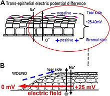
Figure 3 - Electric potential difference across corneal epithelium, and the generation of wound electric fields.
 
Figure 4 - Distribution of bioelectric potential in the flank of a frog embryo stained with voltage-sensitive fluorescent dye.

Overview of the field: terminology and basic definitions

The inside/outside discontinuity at the cell surface enabled by a lipid bilayer membrane (capacitor) is at the core of bioelectricity. The plasma membrane was an indispensable structure for the origin and evolution of life itself. It provided compartmentalization permitting the setting of a differential voltage/potential gradient (battery or voltage source) across the membrane, probably allowing early and rudimentary bioenergetics that fueled cell mechanisms. During evolution, the initially purely passive diffusion of ions (charge carriers), become gradually controlled by the acquisition of ion channels, pumps, exchangers, and transporters. These energetically free (resistors or conductors, passive transport) or expensive (current sources, active transport) translocators set and fine tune voltage gradients – resting potentials – that are ubiquitous and essential to life's physiology, ranging from bioenergetics, motion, sensing, nutrient transport, toxins clearance, and signaling in homeostatic and disease/injury conditions. Upon stimuli or barrier breaking (short-circuit) of the membrane, ions powered by the voltage gradient (electromotive force) diffuse or leak, respectively, through the cytoplasm and interstitial fluids (conductors), generating measurable electric currents – net ion fluxes – and fields. Some ions (such as calcium) and molecules (such as hydrogen peroxide) modulate targeted translocators to produce a current or to enhance, mitigate or even reverse an initial current, being switchers.

Endogenous bioelectric signals are produced in cells by the cumulative action of ion channels, pumps, and transporters. In non-excitable cells, the resting potential across the plasma membrane (Vmem) of individual cells propagate across distances via electrical synapses known as gap junctions (conductors), which allow cells to share their resting potential with neighbors. Aligned and stacked cells (such as in epithelia) generate transepithelial potentials (battery in series) and electric fields (Figures 2 and 3), which likewise propagate across tissues. Tight junctions (resistors) efficiently mitigate the paracellular ion diffusion and leakage, precluding the voltage short circuit. Together, these voltages and electric fields form rich and dynamic and patterns (Figure 5) inside living bodies that demarcate anatomical features, thus acting like blueprints for gene expression and morphogenesis in some instances. More than correlations, these bioelectrical distributions are dynamic, evolving with time and with the microenvironment and even long-distant conditions to serve as instructive influences over cell behavior and large-scale patterning during embryogenesis, regeneration, and cancer suppression. Bioelectric control mechanisms are an important emerging target for advances in regenerative medicine, birth defects, cancer, and synthetic bioengineering.

Brief history of the field: the pioneers in bioelectricity

The modern roots of developmental bioelectricity can be traced back to the entire 18th century. Several seminal works stimulating muscle contractions using Leyden jars culminated with the publication of classical studies by Luigi Galvani in 1791 (De viribus electricitatis in motu musculari) and 1794. In these, Galvani thought to have uncovered intrinsic electric-producing ability in living tissues or “animal electricity”. Alessandro Volta showed that the frog's leg muscle twitching was due to a static electricity generator and from dissimilar metals contact. Galvani showed, in a 1794 study, twitching without metal electricity by touching the leg muscle with a deviating cut sciatic nerve, definitively showing “animal electricity”. Unknowingly, Galvani with this and related experiments discovered the injury current (ion leakage driven by the intact membrane/epithelial potential) and injury potential (potential difference between injured and intact membrane/epithelium). The injury potential was, in fact, the electrical source behind the leg contraction, as realized in the next century. Subsequent work ultimately extended this field broadly beyond nerve and muscle to all cells, from bacteria to non-excitable mammalian cells.

Building on earlier studies, further glimpses of developmental bioelectricity occurred with the discovery of wound-related electric currents and fields in the 1840s, when one of the founding fathers of modern electrophysiologyEmil du Bois-Reymond – reported macroscopic level electrical activities in frog, fish and human bodies. He recorded minute electric currents in live tissues and organisms with a then state-of-the-art galvanometer made of insulated copper wire coils. He unveiled the fast-changing electricity associated with muscle contraction and nerve excitation – the action potentials. At the same time, du Bois-Reymond also reported in detail less fluctuating electricity at wounds – injury current and potential – he made to himself.

Figure 5 - Some sample cell types and their resting potentials, revealing that actively proliferating and plastic cells cluster in the depolarized end of the continuum, while terminally-differentiated mature cell types tend to be strongly polarized.

Bioelectricity work began in earnest at the beginning of the 20th century. Since then, several waves of research produced important functional data showing the role that bioelectricity plays in the control of growth and form. In the 1920s and 1930s, E. J. Lund and H. S. Burr were some of the most prolific authors in this field. Lund measured currents in a large number of living model systems, correlating them to changes in patterning. In contrast, Burr used a voltmeter to measure voltage gradients, examining developing embryonic tissues and tumors, in a range of animals and plants. Applied electric fields were demonstrated to alter the regeneration of planaria by Marsh and Beams in the 1940s and 1950s, inducing the formation of heads or tails at cut sites, reversing the primary body polarity. The introduction and development of the vibrating probe, the first device for quantitative non-invasive characterization of the extracellular minute ion currents, by Lionel Jaffe and Richard Nuccittelli, revitalized the field in the 1970s. They were followed by researchers such as Joseph Vanable, Richard Borgens, Ken Robinson, and Colin McCaig, among many others, who showed roles of endogenous bioelectric signaling in limb development and regeneration, embryogenesis, organ polarity, and wound healing. C.D. Cone studied the role of resting potential in regulating cell differentiation and proliferation and subsequent work has identified specific regions of the resting potential spectrum that correspond to distinct cell states such as quiescent, stem, cancer, and terminally differentiated (Figure 5).

Although this body of work generated a significant amount of high-quality physiological data, this large-scale biophysics approach has historically been in the shadow of the limelight of biochemical gradients and genetic networks in biology education, funding, and overall popularity among biologists. A key factor that contributed to this field lagging behind molecular genetics and biochemistry is that bioelectricity is inherently a living phenomenon – it cannot be studied in fixed specimens. Working with bioelectricity is more complex than traditional approaches to developmental biology, both methodologically and conceptually, as it typically requires a highly interdisciplinary approach.

Methodology for studying bioelectric signaling: electrode-based techniques

The gold standard techniques to quantitatively extract electric dimensions from living specimens, ranging from cell to organism levels, are the glass microelectrode (or micropipette), the vibrating (or self-referencing) voltage probe, and the vibrating ion-selective microelectrode. The former is inherently invasive and the two latter are non-invasive, but all are ultra-sensitive and fast-responsive sensors extensively used in a plethora of physiological conditions in widespread biological models.

The glass microelectrode was developed in the 1940s to study the action potential of excitable cells, deriving from the seminal work by Hodgkin and Huxley in the giant axon squid. It is simply a liquid salt bridge connecting the biological specimen with the electrode, protecting tissues from leachable toxins and redox reactions of the bare electrode. Owing to its low impedance, low junction potential and weak polarization, silver electrodes are standard transducers of the ionic into electric current that occurs through a reversible redox reaction at the electrode surface.

The vibrating probe was introduced in biological studies in the 1970s. The voltage-sensitive probe is electroplated with platinum to form a capacitive black tip ball with large surface area. When vibrating in an artificial or natural DC voltage gradient, the capacitive ball oscillates in a sinusoidal AC output. The amplitude of the wave is proportional to the measuring potential difference at the frequency of the vibration, efficiently filtered by a lock-in amplifier that boosts probe's sensitivity.

The vibrating ion-selective microelectrode was first used in 1990 to measure calcium fluxes in various cells and tissues. The ion-selective microelectrode is an adaptation of the glass microelectrode, where an ion-specific liquid ion exchanger (ionophore) is tip-filled into a previously silanized (to prevent leakage) microelectrode. Also, the microelectrode vibrates at low frequencies to operate in the accurate self-referencing mode. Only the specific ion permeates the ionophore, therefore the voltage readout is proportional to the ion concentration in the measuring condition. Then, flux is calculated using the Fick's first law.

Emerging optic-based techniques, for example, the pH optrode (or optode), which can be integrated into a self-referencing system may become an alternative or additional technique in bioelectricity laboratories. The optrode does not require referencing and is insensitive to electromagnetism simplifying system setting up and making it a suitable option for recordings where electric stimulation is simultaneously applied.

Much work to functionally study bioelectric signaling has made use of applied (exogenous) electric currents and fields via DC and AC voltage-delivering apparatus integrated with agarose salt bridges. These devices can generate countless combinations of voltage magnitude and direction, pulses, and frequencies. Currently, lab-on-a-chip mediated application of electric fields is gaining ground in the field with the possibility to allow high-throughput screening assays of the large combinatory outputs.

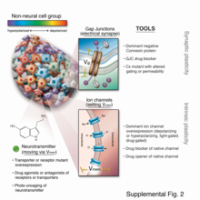
Figure 6 - Tools for manipulating non-neural bioelectricity include pharmacological and genetic reagents to alter cell connectivity (control gap junctions), cell Vmem (control ion channels/pumps), and bioelectrically-guided 2nd messengers (control neurotransmitters and other small molecules).

Methodology for studying bioelectric signaling: molecular-age reagents and approaches

The remarkable progress in molecular biology over the last six decades has produced powerful tools that facilitate the dissection of biochemical and genetic signals; yet, they tend to not be well-suited for bioelectric studies in vivo. Prior work relied extensively on current applied directly by electrodes, reinvigorated by significant recent advances in materials science and extracellular current measurements, facilitated by sophisticated self-referencing electrode systems. While electrode applications for manipulating neutrally-controlled body processes have recently attracted much attention, the nervous system is just the tip of the iceberg when it comes to the opportunities for controlling somatic processes, as most cell types are electrically active and respond to ionic signals from themselves and their neighbors (Figure 6).

In the last 15 years, a number of new molecular techniques have been developed that allowed bioelectric pathways to be investigated with a high degree of mechanistic resolution, and to be linked to canonical molecular cascades. These include (1) pharmacological screens to identify endogenous channels and pumps responsible for specific patterning events; (2) voltage-sensitive fluorescent reporter dyes and genetically-encoded fluorescent voltage indicators for the characterization of the bioelectric state in vivo; (3) panels of well-characterized dominant ion channels that can be misexpressed in cells of interest to alter the bioelectric state in desired ways; and (4) computational platforms that are coming on-line to assist in building predictive models of bioelectric dynamics in tissues.

Compared with the electrode-based techniques, the molecular probes provide a wider spatial resolution and facilitated dynamic analysis over time. Although calibration or titration can be possible, molecular probes are typically semi-quantitative, whereas electrodes provide absolute bioelectric values. Another advantage of fluorescence and other probes is their less-invasive nature and spatial multiplexing, enabling the simultaneous monitoring of large areas of embryonic or other tissues in vivo during normal or pathological pattering processes.

Role in early development

Work in model systems such as Xenopus laevis and zebrafish has revealed a role for bioelectric signaling in the development of heart, face, eye, brain, and other organs. Screens have identified roles for ion channels in size control of structures such as the zebrafish fin, while focused gain-of-function studies have shown for example that bodyparts can be re-specified at the organ level – for example creating entire eyes in gut endoderm. As in the brain, developmental bioelectrics can integrate information across significant distance in the embryo, for example such as the control of brain size by bioelectric states of ventral tissue. and the control of tumorigenesis at the site of oncogene expression by bioelectric state of remote cells.

Human disorders, as well as numerous mouse mutants show that bioelectric signaling is important for human development (Tables 1 and 2). Those effects are pervasively linked to channelopathies, which are human disorders that result from mutations that disrupt ion channels.

Several channelopathies result in morphological abnormalities or congenital birth defects in addition to symptoms that affect muscle and or neurons. For example, mutations that disrupt an inwardly rectifying potassium channel Kir2.1 cause dominantly inherited Andersen-Tawil Syndrome (ATS). ATS patients experience periodic paralysis, cardiac arrhythmias, and multiple morphological abnormalities that can include cleft or high arched palate, cleft or thin upper lip, flattened philtrum, micrognathia, dental oligodontia, enamel hypoplasia, delayed dentition eruption, malocclusion, broad forehead, wide set eyes, low set ears, syndactyly, clinodactyly, brachydactyly, and dysplastic kidneys. Mutations that disrupt another inwardly rectifying K+ channel Girk2 encoded by KCNJ6 cause Keppen-Lubinsky syndrome which includes microcephaly, a narrow nasal bridge, a high arched palate, and severe generalized lipodystrophy (failure to generate adipose tissue). KCNJ6 is in the Down syndrome critical region such that duplications that include this region lead to craniofacial and limb abnormalities and duplications that do not include this region do not lead to morphological symptoms of Down syndrome. Mutations in KCNH1, a voltage gated potassium channel lead to Temple-Baraitser (also known as Zimmermann- Laband) syndrome. Common features of Temple-Baraitser syndrome include absent or hypoplastic of finger and toe nails and phalanges and joint instability. Craniofacial defects associated with mutations in KCNH1 include cleft or high arched palate, hypertelorism, dysmorphic ears, dysmorphic nose, gingival hypertrophy, and abnormal number of teeth.

Mutations in CaV1.2, a voltage gated Ca2+ channel, lead to Timothy syndrome which causes severe cardiac arrhythmia (long-QT) along with syndactyly and similar craniofacial defects to Andersen-Tawil syndrome including cleft or high-arched palate, micrognathia, low set ears, syndactyly and brachydactyly. While these channelopathies are rare, they show that functional ion channels are important for development. Furthermore, in utero exposure to anti-epileptic medications that target some ion channels also cause increased incidence of birth defects such as oral clefting. The effects of both genetic and exogenous disruption of ion channels lend insight into the importance of bioelectric signaling in development.

Role in wound healing and cell guidance

One of the best-understood roles for bioelectric gradients is at the tissue-level endogenous electric fields utilized during wound healing. It is challenging to study wound-associated electric fields, because these fields are weak, less fluctuating, and do not have immediate biological responses when compared to nerve pulses and muscle contraction. The development of the vibrating and glass microelectrodes, demonstrated that wounds indeed produced and, importantly, sustained measurable electric currents and electric fields. These techniques allow further characterization of the wound electric fields/currents at cornea and skin wounds, which show active spatial and temporal features, suggesting active regulation of these electrical phenomena. For example, the wound electric currents are always the strongest at the wound edge, which gradually increased to reach a peak about 1 hour after injury. At wounds in diabetic animals, the wound electric fields are significantly compromised. Understanding the mechanisms of generation and regulation of the wound electric currents/fields is expected to reveal new approaches to manipulate the electrical aspect for better wound healing.

How are the electric fields at a wound produced? Epithelia actively pump and differentially segregate ions. In the cornea epithelium, for example, Na+ and K+ are transported inwards from tear fluid to extracellular fluid, and Cl− is transported out of the extracellular fluid into the tear fluid. The epithelial cells are connected by tight junctions, forming the major electrical resistive barrier, and thus establishing an electrical gradient across the epithelium – the transepithelial potential (TEP). Breaking the epithelial barrier, as occurs in any wounds, creates a hole that breaches the high electrical resistance established by the tight junctions in the epithelial sheet, short-circuiting the epithelium locally. The TEP therefore drops to zero at the wound. However, normal ion transport continues in unwounded epithelial cells beyond the wound edge (typically <1 mm away), driving positive charge flow out of the wound and establishing a steady, laterally-oriented electric field (EF) with the cathode at the wound. Skin also generates a TEP, and when a skin wound is made, similar wound electric currents and fields arise, until the epithelial barrier function recovers to terminate the short-circuit at the wound. When wound electric fields are manipulated with pharmacological agents that either stimulate or inhibit transport of ions, the wound electric fields also increase or decrease, respectively. Wound healing can be speed up or slowed down accordingly in cornea wounds.

How do electric fields affect wound healing? To heal wounds, cells surrounding the wound must migrate and grow directionally into the wound to cover the defect and restore the barrier. Cells important to heal wounds respond remarkably well to applied electric fields of the same strength that are measured at wounds. The whole gamut of cell types and their responses following injury are affected by physiological electric fields. Those include migration and division of epithelial cells, sprouting and extension of nerves, and migration of leukocytes and endothelial cells. The most well studied cellular behavior is directional migration of epithelial cells in electric fields – electrotaxis. The epithelial cells migrate directionally to the negative pole (cathode), which at a wound is the field polarity of the endogenous vectorial electric fields in the epithelium, pointing (positive to negative) to the wound center. Epithelial cells of the cornea, keratinocytes from the skin, and many other types of cells show directional migration at electric field strengths as low as a few mV mm−1. Large sheets of monolayer epithelial cells, and sheets of stratified multilayered epithelial cells also migrate directionally. Such collective movement closely resembles what happens during wound healing in vivo, where cell sheets move collectively into the wound bed to cover the wound and restore the barrier function of the skin or cornea.

How cells sense such minute extracellular electric fields remains largely elusive. Recent research has started to identify some genetic, signaling and structural elements underlying how cells sense and respond to small physiological electric fields. These include ion channels, intracellular signaling pathways, membrane lipid rafts, and electrophoresis of cellular membrane components.

Role in animal regeneration

In the early 20th century, Albert Mathews seminally correlated regeneration of a cnidarian polyp with the potential difference between polyp and stolon surfaces, and affected regeneration by imposing countercurrents. Amedeo Herlitzka, following on the wound electric currents footsteps of his mentor, du Bois-Raymond, theorized about electric currents playing an early role in regeneration, maybe initiating cell proliferation. Using electric fields overriding endogenous ones, Marsh and Beams astoundingly generated double-headed planarians and even reversed the primary body polarity entirely, with tails growing where a head previously existed. After these seed studies, variations of the idea that bioelectricity could sense injury and trigger or at least be a major player in regeneration have spurred over the decades until the present day. A potential explanation lies on resting potentials (primarily Vmem and TEP), which can be, at least in part, dormant sensors (alarms) ready to detect and effectors (triggers) ready to react to local damage.

Following up on the relative success of electric stimulation on non-permissive frog leg regeneration using an implanted bimetallic rod in the late 1960s, the bioelectric extracellular aspect of amphibian limb regeneration was extensively dissected in the next decades. Definitive descriptive and functional physiological data was made possible owing to the development of the ultra-sensitive vibrating probe and improved application devices. Amputation invariably leads to a skin-driven outward current and a consequent lateral electric field setting the cathode at the wound site. Although initially pure ion leakage, an active component eventually takes place and blocking ion translocators typically impairs regeneration. Using biomimetic exogenous electric currents and fields, partial regeneration was achieved, which typically included tissue growth and increased neuronal tissue. Conversely, precluding or reverting endogenous electric current and fields impairs regeneration. These studies in amphibian limb regeneration and related studies in lampreys and mammals  combined with those of bone fracture healing and in vitro studies, led to the general rule that migrating (such as keratinocytes, leucocytes and endothelial cells) and outgrowing (such as axons) cells contributing to regeneration undergo electrotaxis towards the cathode (injury original site). Congruently, an anode is associated with tissue resorption or degeneration, as occurs in impaired regeneration and osteoclastic resorption in bone. Despite these efforts, the promise for a significant epimorphic regeneration in mammals remains a major frontier for future efforts, which includes the use of wearable bioreactors to provide an environment within which pro-regenerative bioelectric states can be driven and continued efforts at electrical stimulation.

Recent molecular work has identified proton and sodium flux as being important for tail regeneration in Xenopus tadpoles, and shown that regeneration of the entire tail (with spinal cord, muscle, etc.) could be triggered in a range of normally non-regenerative conditions by either molecular-genetic, pharmacological, or optogenetc methods. In planaria, work on bioelectric mechanism has revealed control of stem cell behavior, size control during remodeling, anterior-posterior polarity, and head shape. Gap junction-mediated alteration of physiological signaling produces 2-headed worms in Dugesia japonica; remarkably, these animals continue to regenerate as 2-headed in future rounds of regeneration months after the gap junction-blocking reagent has left the tissue. This stable, long-term alteration of the anatomical layout to which animals regenerate, without genomic editing, is an example of epigenetic inheritance of body pattern, and is also the only available “strain” of planarian species exhibiting an inherited anatomical change that is different from the wild-type.

Figure 7 - Voltage changes can be transduced to downstream effector mechanisms via a variety of 2nd messenger processes, including Vmem-dependent movement of small signaling molcules like serotonin through transporters or gap junctions, voltage-sensitive phosphatases, voltage-gated calcium channels (which trigger calcium-signaling cascades), and dimerization of receptors in the cell surface.
 
Figure 8 - Bioelectricity and genetic expression work together in an integrated fashion; nothing is downstream.
 
Figure 9 - Misexpression of specific ion channels in diverse areas of frog embryos can induce the creation of ectopic organs, such as eyes on gut tissue.

Role in cancer

Defection of cells from the normally tight coordination of activity towards an anatomical structure results in cancer; it is thus no surprise that bioelectricity – a key mechanism for coordinating cell growth and patterning – is a target often implicated in cancer and metastasis. Indeed, it has long been known that gap junctions have a key role in carcinogenesis and progression. Channels can behave as oncogenes and are thus suitable as novel drug targets. Recent work in amphibian models has shown that depolarization of resting potential can trigger metastatic behavior in normal cells, while hyperpolarization (induced by ion channel misexpression, drugs, or light) can suppress tumorigenesis induced by expression of human oncogenes. Depolarization of resting potential appears to be a bioelectric signature by which incipient tumor sites can be detected non-invasively. Refinement of the bioelectric signature of cancer in biomedical contexts, as a diagnostic modality, is one of the possible applications of this field. Excitingly, the ambivalence of polarity – depolarization as marker and hyperpolarization as treatment – make it conceptually possible to derive theragnostic (portmanteau of therapeutics with diagnostics) approaches, designed to simultaneously detect and treat early tumors, in this case based on the normalization of the membrane polarization.

Role in pattern regulation

Recent experiments using ion channel opener/blocker drugs, as well as dominant ion channel misexpression, in a range of model species, has shown that bioelectricity, specifically, voltage gradients instruct not only stem cell behavior but also large-scale patterning. Patterning cues are often mediated by spatial gradients of cell resting potentials, or Vmem, which can be transduced into second messenger cascades and transcriptional changes by a handful of known mechanisms (Figure 7). These potentials are set by the function of ion channels and pumps, and shaped by gap junctional connections which establish developmental compartments (isopotential cell fields). Because both gap junctions and ion channels are themselves voltage-sensitive, cell groups implement electric circuits with rich feedback capabilities (Figure 8). The outputs of developmental bioelectric dynamics in vivo represent large-scale patterning decisions such as the number of heads in planaria, the shape of the face in frog development, and the size of tails in zebrafish. Experimental modulation of endogenous bioelectric prepatterns have enabled converting body regions (such as the gut) to a complete eye (Figure 9), inducing regeneration of appendages such as tadpole tails at non-regenerative contexts, and conversion of flatworm head shapes and contents to patterns appropriate to other species of flatworms, despite a normal genome. Recent work has shown the use of physiological modeling environments for identifying predictive interventions to target bioelectric states for repair of embryonic brain defects under a range of genetic and pharmacologically-induced teratologies.

Future of the field

Life is ultimately an electrochemical enterprise; research in this field is progressing along several frontiers. First is the reductive program of understanding how bioelectric signals are produced, how voltage changes in the cell membrane are able to regulate cell behavior, and what are the genetic and epigenetic downstream targets of bioelectric signals. A few mechanisms that transduce bioelectric change into alterations of gene expression are already known, including the bioelectric control of movement of small second-messenger molecules through cells, including serotonin and butyrate, voltage sensitive phosphatases, among others. Also known are numerous gene targets of voltage signaling, such as Notch, BMP, FGF, and HIF-1α. Thus, the proximal mechanisms of bioelectric signaling within single cells are becoming well-understood, and advances in optogenetics and magnetogenetics continue to facilitate this research program. More challenging however is the integrative program of understanding how specific patterns of bioelectric dynamics help control the algorithms that accomplish large-scale pattern regulation (regeneration and development of complex anatomy). The incorporation of bioelectrics with chemical signaling in the emerging field of probing cell sensory perception and decision-making is an important frontier for future work.

Bioelectric modulation has shown control over complex morphogenesis and remodeling, not merely setting individual cell identity. Moreover, a number of the key results in this field have shown that bioelectric circuits are non-local – regions of the body make decisions based on bioelectric events at a considerable distance. Such non-cell-autonomous events suggest distributed network models of bioelectric control; new computational and conceptual paradigms may need to be developed to understand spatial information processing in bioelectrically-active tissues. It has been suggested that results from the fields of primitive cognition and unconventional computation are relevant to the program of cracking the bioelectric code. Finally, efforts in biomedicine and bioengineering are developing applications such as wearable bioreactors for delivering voltage-modifying reagents to wound sites, and ion channel-modifying drugs (a kind of electroceutical) for repair of birth defects and regenerative repair. Synthetic biologists are likewise starting to incorporate bioelectric circuits into hybrid constructs.

Medicalization

From Wikipedia, the free encyclopedia 

Medicalization or medicalisation (see spelling differences) is the process by which human conditions and problems come to be defined and treated as medical conditions, and thus become the subject of medical study, diagnosis, prevention, or treatment. Medicalization can be driven by new evidence or hypotheses about conditions; by changing social attitudes or economic considerations; or by the development of new medications or treatments.

Medicalization is studied from a sociologic perspective in terms of the role and power of professionals, patients, and corporations, and also for its implications for ordinary people whose self-identity and life decisions may depend on the prevailing concepts of health and illness. Once a condition is classified as medical, a medical model of disability tends to be used in place of a social model. Medicalization may also be termed "pathologization" or (pejoratively) "disease mongering". Since medicalization is the social process through which a condition becomes a medical disease in need of treatment, medicalization may be viewed as a benefit to human society. According to this view, the identification of a condition as a disease will lead to the treatment of certain symptoms and conditions, which will improve overall quality of life.

Development of the concept

The concept of medicalization was devised by sociologists to explain how medical knowledge is applied to behaviors which are not self-evidently medical or biological. The term medicalization entered the sociology literature in the 1970s in the works of Irving Zola, Peter Conrad and Thomas Szasz, among others. According to Dr. Cassell's book, The Nature of Suffering and the Goals of Medicine (2004), the expansion of medical social control is being justified as a means of explaining deviance. These sociologists viewed medicalization as a form of social control in which medical authority expanded into domains of everyday existence, and they rejected medicalization in the name of liberation. This critique was embodied in works such as Conrad's "The discovery of hyperkinesis: notes on medicalization of deviance", published in 1973 (hyperkinesis was the term then used to describe what we might now call ADHD). Nevertheless, opium was used to pacify children in ancient Egypt before 2000 BC.

These sociologists did not believe medicalization to be a new phenomenon, arguing that medical authorities had always been concerned with social behavior and traditionally functioned as agents of social control (Foucault, 1965; Szasz,1970; Rosen). However, these authors took the view that increasingly sophisticated technology had extended the potential reach of medicalization as a form of social control, especially in terms of "psychotechnology" (Chorover,1973).

In the 1975 book Limits to medicine: Medical nemesis (1975), Ivan Illich put forth one of the earliest uses of the term "medicalization". Illich, a philosopher, argued that the medical profession harms people through iatrogenesis, a process in which illness and social problems increase due to medical intervention. Illich saw iatrogenesis occurring on three levels: the clinical, involving serious side effects worse than the original condition; the social, whereby the general public is made docile and reliant on the medical profession to cope with life in their society; and the structural, whereby the idea of aging and dying as medical illnesses effectively "medicalized" human life and left individuals and societies less able to deal with these "natural" processes.

The concept of medicalization dovetailed with some aspects of the 1970s feminist movement. Critics such as Ehrenreich and English (1978) argued that women's bodies were being medicalized by the predominantly male medical profession. Menstruation and pregnancy had come to be seen as medical problems requiring interventions such as hysterectomies.

Marxists such as Vicente Navarro (1980) linked medicalization to an oppressive capitalist society. They argued that medicine disguised the underlying causes of disease, such as social inequality and poverty, and instead presented health as an individual issue. Others examined the power and prestige of the medical profession, including the use of terminology to mystify and of professional rules to exclude or subordinate others.

Tiago Correia (2017) offers an alternative perspective on medicalization. He argues that medicalization needs to be detached from biomedicine to overcome much of the criticism it has faced, and to protect its value in contemporary sociological debates. Building on Gadamer's hermeneutical view of medicine, he focuses on medicine's common traits, regardless of empirical differences in both time and space. Medicalization and social control are viewed as distinct analytical dimensions that in practice may or may not overlap. Correia contends that the idea of "making things medical" needs to include all forms of medical knowledge in a global society, not simply those forms linked to the established (bio)medical professions. Looking at "knowledge", beyond the confines of professional boundaries, may help us understand the multiplicity of ways in which medicalization can exist in different times and societies, and allow contemporary societies to avoid such pitfalls as "demedicalization" (through a turn towards complementary and alternative medicine) on the one hand, or the over-rapid and unregulated adoption of biomedical medicine in non-western societies on the other. The challenge is to determine what medical knowledge is present, and how it is being used to medicalize behaviors and symptoms.

Professionals, patients, corporations and society

Several decades on the definition of medicalization is complicated, if for no other reason than because the term is so widely used. Many contemporary critics position pharmaceutical companies in the space once held by doctors as the supposed catalysts of medicalization. Titles such as "The making of a disease" or "Sex, drugs, and marketing" critique the pharmaceutical industry for shunting everyday problems into the domain of professional biomedicine. At the same time, others reject as implausible any suggestion that society rejects drugs or drug companies and highlight that the same drugs that are allegedly used to treat deviances from societal norms also help many people live their lives. Even scholars who critique the societal implications of brand-name drugs generally remain open to these drugs' curative effects — a far cry from earlier calls for a revolution against the biomedical establishment. The emphasis in many quarters has come to be on "overmedicalization" rather than "medicalization" in itself.

Others, however, argue that in practice the process of medicalization tends to strip subjects of their social context, so they come to be understood in terms of the prevailing biomedical ideology, resulting in a disregard for overarching social causes such as unequal distribution of power and resources. A series of publications by Mens Sana Monographs have focused on medicine as a corporate capitalist enterprise.

Conversation between doctor and patient

The physician's role in this present-day notion of medicalization is similarly complex. On the one hand, the doctor remains an authority figure who prescribes pharmaceuticals to patients. However, in some countries, such as the US, ubiquitous direct-to-consumer advertising encourages patients to ask for particular drugs by name, thereby creating a conversation between consumer and drug company that threatens to cut the doctor out of the loop. Additionally, there is a widespread concern regarding the extent of the pharmaceutical marketing direct to doctors and other healthcare professionals. Examples of this direct marketing are visits by salespeople, funding of journals, training courses or conferences, incentives for prescribing, and the routine provision of "information" written by the pharmaceutical company.

The role of patients in this economy has also changed. Once regarded as passive victims of medicalization, patients can now occupy active positions as advocates, consumers, or even agents of change.

The antithesis of medicalization is the process of paramedicalization, where human conditions come under the attention of alternative medicine, traditional medicine or any of numerous non-medical health approaches. Medicalization and paramedicalization can sometimes be contradictory and conflicting, but they also support and strengthen each other since they both ensure that questions of health and illness stay in sharp focus in defining human conditions and problems.

Areas

A 2002 editorial in the British Medical Journal warned of inappropriate medicalization leading to disease mongering, where the boundaries of the definition of illnesses are expanded to include personal problems as medical problems or risks of diseases are emphasized to broaden the market for medications. The authors noted:

Inappropriate medicalisation carries the dangers of unnecessary labelling, poor treatment decisions, iatrogenic illness, and economic waste, as well as the opportunity costs that result when resources are diverted away from treating or preventing more serious disease. At a deeper level it may help to feed unhealthy obsessions with health, obscure or mystify sociological or political explanations for health problems, and focus undue attention on pharmacological, individualised, or privatised solutions.

For many years, marginalized psychiatrists (such as Peter Breggin, Paula Caplan, Thomas Szasz) and outside critics (such as Stuart A. Kirk) have "been accusing psychiatry of engaging in the systematic medicalization of normality". More recently these concerns have come from insiders who have worked for and promoted the American Psychiatric Association (e.g., Robert Spitzer, Allen Frances).

Benjamin Rush, the father of American psychiatry, claimed that Black people had black skin because they were ill with hereditary leprosy. Consequently, he considered vitiligo as a "spontaneous cure".

According to Franco Basaglia and his followers, whose approach pointed out the role of psychiatric institutions in the control and medicalization of deviant behaviors and social problems, psychiatry is used as the provider of scientific support for social control to the existing establishment, and the ensuing standards of deviance and normality brought about repressive views of discrete social groups. As scholars have long argued, governmental and medical institutions code menaces to authority as mental diseases during political disturbances.

The HIV/AIDS pandemic allegedly caused from the 1980s a "profound re-medicalization of sexuality". The diagnosis of premenstrual dysphoric disorder has caused some controversy, and psychologist Peggy Kleinplatz has criticized the diagnosis as the medicalization of normal human behavior, that occurred while fluoxetine (also known as Prozac) was being repackaged as a PMDD therapy under the trade named Sarafem. Although it has received less attention, it is claimed that masculinity has also faced medicalization, being deemed damaging to health and requiring regulation or enhancement through drugs, technologies or therapy.

According to Kittrie, a number of phenomena considered "deviant", such as alcoholism, drug addiction, prostitution, pedophilia, and masturbation ("self-abuse"), were originally considered as moral, then legal, and now medical problems. Innumerable other conditions such as obesity, smoking cigarettes, draft malingering, bachelorhood, divorce, unwanted pregnancy, kleptomania, and grief, have been declared a disease by medical and psychiatric authorities who hold impeccable institutional credentials. Due to these perceptions, peculiar deviants were subjected to moral, then legal, and now medical modes of social control. Similarly, Conrad and Schneider concluded their review of the medicalization of deviance by identifying three major paradigms that have reigned over deviance designations in different historical periods: deviance as sin; deviance as crime; and deviance as sickness.

According to Mike Fitzpatrick, resistance to medicalization was a common theme of the gay liberation, anti-psychiatry, and feminist movements of the 1970s, but now there is actually no resistance to the advance of government intrusion in lifestyle if it is thought to be justified in terms of public health. Moreover, the pressure for medicalization also comes from society itself. Feminists, who once opposed state intervention as oppressive and patriarchal, now demand more coercive and intrusive measures to deal with child abuse and domestic violence.

According to Thomas Szasz, "the therapeutic state swallows up everything human on the seemingly rational ground that nothing falls outside the province of health and medicine, just as the theological state had swallowed up everything human on the perfectly rational ground that nothing falls outside the province of God and religion".

Unnecessary health care

From Wikipedia, the free encyclopedia

Unnecessary health care (overutilization, overuse, or overtreatment) is health care provided with a higher volume or cost than is appropriate. In the United States, where health care costs are the highest as a percentage of GDP, overuse was the predominant factor in its expense, accounting for about a third of its health care spending ($750 billion out of $2.6 trillion) in 2012.

Factors that drive overuse include paying health professionals more to do more (fee-for-service), defensive medicine to protect against litigiousness, and insulation from price sensitivity in instances where the consumer is not the payer—the patient receives goods and services but insurance pays for them (whether public insurance, private, or both). Such factors leave many actors in the system (doctors, patients, pharmaceutical companies, device manufacturers) with inadequate incentive to restrain health care prices or overuse. This drives payers, such as national health insurance systems or the U.S. Centers for Medicare and Medicaid Services, to focus on medical necessity as a condition for payment. However, the threshold between necessity and lack thereof can often be subjective.

Overtreatment, in the strict sense, may refer to unnecessary medical interventions, including treatment of a self-limited condition (overdiagnosis) or to extensive treatment for a condition that requires only limited treatment.

It is economically linked with overmedicalization.

Definition

A forerunner of the term was what Jack Wennberg called unwarranted variation, different rates of treatments based upon where people lived, not clinical rationale. He had discovered that in studies that began in 1967 and were published in the 1970s and the 1980s: "The basic premise – that medicine was driven by science and by physicians capable of making clinical decisions based on well-established fact and theory – was simply incompatible with the data we saw. It was immediately apparent that suppliers were more important in driving demand than had been previously realized."

In 2008, US bioethicist Ezekiel J. Emanuel and health economist Victor R. Fuchs defined unnecessary health care as "overutilization", health care provided with a higher volume or cost than is appropriate. Recently, economists have sought to understand unnecessary health care in terms of misconsumption rather than overconsumption.

In 2009 two US physicians wrote in an editorial, that unnecessary care was "defined as services which show no demonstrable benefit to patients" and might represent 30% of U.S. medical care. They referred to a 2003 study on regional variations in Medicare spending, which found, "Medicare enrollees in higher-spending regions receive more care than those in lower-spending regions, but do not have better health outcomes or satisfaction with care."

In January 2012, the American College of Physicians Ethics, Professionalism, and Human Rights Committee suggested that overtreatment can also be understood in contrast to 'parsimonious care', defined as "care that utilizes the most efficient means to effectively diagnose a condition and treat a patient."

In April 2012, Berwick, from the Institute for Healthcare Improvement, and Andrew Hackbarth from the RAND Corporation defined overtreatment as "subjecting patients to care that, according to sound science and the patients' own preferences, cannot possibly help them—care rooted in outmoded habits, supply-driven behaviors, and ignoring science." They wrote that trying to do something (treatment or testing) for all patients who might need it inevitably entails doing that same thing for some patients who might not need it." In uncertain situations, "some non-beneficial care was the necessary byproduct of optimal clinical decision making."

In October 2015, two pediatricians said that considering "overtreatment as an ethical violation" could help see the conflicting incentives of health care workers for treatment or nontreatment.

Cost

In the US, the country which spends the most on health care per person globally, patients have fewer doctor visits and fewer days in hospitals than people in other countries do, but prices are high, there is more use of some procedures and new drugs than elsewhere, and doctor salaries are double the levels in other countries. The New York Times reported "no one knows for sure" how much unnecessary care exists in the United States. Overuse of medical care is no longer a large fraction of total health care spending, which was $3.3 trillion in 2016.

Researchers in 2014 analyzed many services listed as low value by Choosing Wisely and other sources. They looked at spending in 2008–2009 and found that these services represented 0.6% or 2.7% of Medicare costs and there was no significant pattern of particular types of physicians ordering these low value services. The Institute of Medicine in 2010 gave two estimates of "unnecessary services," using different methodologies: 0.2% or 1% to 5% of health spending, which was US$2.6 trillion. The Institute of Medicine quoted that 2010 report in a 2012 report to support an estimate of 8% ($210 billion) in unnecessary services, without explaining the discrepancy. This IOM 2012 report also said there were $555 billion in other wasted spending, which have an "unknown overlap" with each other and the $210 billion. The United States National Academy of Sciences estimated in 2005, without giving its methods or sources, that "between $.30 and $.40 of every dollar spent on health care is spent on the costs of poor quality," amounting to" slightly more than a half-trillion dollars a year... wasted on overuse, underuse, misuse, duplication, system failures, unnecessary repetition, poor communication, and inefficiency. In 2003 Fisher et al. found that there was "no apparent regional health benefit for Medicare recipients from doing more, whether 'more' is expressed as hospitalizations, surgical procedures, or consultations within the hospital." Up to 30% of Medicare spending could be cut in 2003 without harming patients.

When care is overused, patients are put at risk of complications unnecessarily, with documented harm to patients from overuse of surgeries and other treatments.

Causes

Physicians' decisions are the proximate cause of unnecessary care, though the potential incentives and penalties they face can influence their choices.

Third-party payers and fee-for-service

When public or private insurance cover expenses and doctors are paid under a fee-for-service (FFS) model, neither has an incentive to consider the cost of treatment, a combination that contributes to waste. Fee-for-service is a large incentive for overuse because health care providers (such as doctors and hospitals) receive revenue from the overtreatment.

Atul Gawande investigated Medicare FFS reimbursements in McAllen, Texas, for a 2009 article in the New Yorker. In 2006, the town of McAllen was the second-most expensive Medicare market, behind Miami. Costs per beneficiary were almost twice the national average.

In 1992, however, McAllen had been almost exactly in line with the Medicare spending average. After looking at other potential explanations such as relatively poorer health or medical malpractice, Gawande concluded the town was a chief example of the overuse of medical services. Gawande concluded that a business culture (physicians viewing their practices as a revenue stream) had established itself there, in contrast to a culture of low-cost high-quality medicine at the Mayo Clinic and in the Grand Junction, Colorado, market. Gawande advised:

As America struggles to extend healthcare coverage while curbing health care costs, we face a decision that is more important than whether we have a public-insurance option, more important than whether we will have a single-payer system in the long run or a mixture of public and private insurance, as we do now. The decision is whether we are going to reward the leaders who are trying to build a new generation of Mayos and Grand Junctions. If we don't, McAllen won't be an outlier. It will be our future.

Medical malpractice laws and defensive medicine

To protect themselves from legal prosecution U.S. physicians have an incentive to order clinically unnecessary tests or tests of little potential value. While defensive medicine is a favored explanation for high medical costs by physicians, Gawande estimated in 2010 it only contributed to 2.4% of the total $2.3 trillion of U.S. health care spending in 2008.

Direct-to-consumer advertising

Direct-to-consumer advertising can encourage patients to ask for drugs, devices, diagnostics, or procedures. Sometimes service providers will simply give these treatments or services rather than attempting the potentially more unpleasant task of convincing the patient what they have requested is not needed, or is likely to cause more harm than good.

Physician predispositions

Dartmouth Medical School professor Gilbert Welch argued 2016 that certain predispositions by physicians and the general public may lead to unnecessary health care, including:

  • Attempting to mitigate a risk without considering how small or unlikely the potential benefit is
  • Attempting to fix an underlying problem, instead of using a less-risky monitoring or coping strategy
  • Acting too quickly, when waiting for more information might be wiser
  • Acting without considering the benefits of doing nothing
  • Discounting downsides of diagnostic testing
  • Preferring newer over older treatments without considering the cost of new treatments or the effectiveness of older ones
  • Treating patients with terminal illness to maximize life span over quality of life, without probing a patient's preferences

Examples

Imaging

Overuse of diagnostic imaging, such as X-rays and CT scans, is defined as any application unlikely to improve patient care. Factors that contribute to overuse include "self-referral, patient wishes, inappropriate financially motivated factors, health system factors, industry, media, lack of awareness" and defensive medicine. Respected organizations—such as the American College of Radiology (ACR), Royal College of Radiologists (RCR) and the World Health Organization (WHO)—have developed "appropriateness criteria". The Canadian Association of Radiologists estimated in 2009 that 30% of imaging was unnecessary in the Canadian health care system. 2008 Medicare claims showed overuse with chest CT's. Financial incentives have also been shown to have a significant impact on dental X-ray use with dentists who are paid a separate fee for each X-ray providing more X-rays.

Overuse of imaging can lead to a diagnosis of a condition that would have otherwise remained irrelevant (overdiagnosis).

Physician self-referral

One type of overuse can be physician self-referral. Multiple studies have replicated the finding that when non-radiologists have an ownership interest in the fees generated by radiology equipment—and can self-refer—their use of imaging is unnecessarily higher. The majority of U.S. growth in imaging use (the fastest-growing physician service) comes from self-referring nonradiologists. In 2004, this overuse was estimated to contribute to $16 billion of annual U.S. health care costs.

As of a 2018 review evidence of overtreatment overmedicalization, and overdiagnosis in Pediatrics have been use of commercial rehydration solution, antidepressants, and parenteral nutrition; overmedicalization with planned early deliveries, immobilization of ankle injuries, use of hydrolyzed infant formula; and overdiagnosis of hypoxemia among children recovering from bronchiolitis.

Others

  • Hospitalizations for those with chronic conditions who could be treated as outpatients
  • Surgeries in Medicare patients in their last year of life; regions with high levels had higher death rates
  • Antibiotic use for viral or self-limiting infections (an overmedication that can promote antibiotic resistance)
  • Opiate prescriptions carry the risk of addiction. In some cases, the number of pills prescribed might exceed what is actually needed for pain relief from a given condition, or a different pain management technique or medication would be effective but less risky.
  • Many blood transfusions in the U.S. are given without checking to see if they are needed after a previous transfusion, or are given in cases where monitoring, recovering the patient's own blood, or iron therapy would be effective and reduce the risk of complications
  • An estimated one in eight coronary stents (used in $20,000 procedures) with nonacute indications (U.S.)
  • Heart bypass surgeries at Redding Medical Center which resulted in an FBI raid
  • Screening patients with advanced cancer for other cancers
  • Annual cervical cancer screening in women with medical histories of normal pap smear and HPV test results

Reduction efforts

Utilization management (utilization review) has evolved over decades among both public and private payers in an attempt to reduce overuse. In this effort, insurers employ physicians to review the actions of other physicians and detect overuse. Utilization review has a poor reputation among most clinicians as a corrupted system in which utilization reviewers have their own perverse incentives (i.e., find ways to deny coverage no matter what) and in some cases are not practicing physicians, lacking real-world clinical insight or wisdom. Results of a recent systematic review found that many studies focused more on reductions in utilization than in improving clinically meaningful measures.

The 2010 U.S. health care reform, the Patient Protection and Affordable Care Act, did not contain serious strategies to reduce overuse; "the public has made it clear that it does not want to be told what medical care it can and cannot have." Uwe Reinhardt, a health economist at Princeton, said "the minute you attack overutilization, you will be called a Nazi before the day is out".

Professional societies and other groups have begun to push for policy changes that would encourage clinicians to avoid providing unnecessary care. Most physicians accept that laboratory tests are overused, but "it remains difficult to persuade them to consider the possibility that they, too, might be overutilizing laboratory tests." In November 2011, the American Board of Internal Medicine Foundation began the Choosing Wisely campaign, which aims to raise awareness of overtreatment and change physician behavior by publicizing lists of tests and treatments that are often overused, and which doctors and patients should try to avoid.

In the UK, 2011, online platform AskMyGP was launched to decrease the amount of unnecessary medical appointments. In the app patients are given a questionnaire about their symptoms, which then assesses the patient's need for medical care. The program was a success, and as of January 2018 has managed over 29,000 patient episodes.

In April 2012, the Lown Institute and the New America Foundation Health Policy Program convened the 'Avoiding Avoidable Care' conference. It was the first major medical conference to focus entirely on overuse, and it included presentations from speakers including Bernard Lown, Don Berwick, Christine Cassel, Amitabh Chandra, JudyAnn Bigby, and Julio Frenk. A second meeting was planned for December 2013.

Since the meeting, the Lown Institute has focused its work on deepening the understanding of overuse and generating public discussion of the ethical and cultural drivers of overuse, especially on the role of the hidden curriculum in medical school and residency.

Patient safety committees, which are charged with reviewing the quality of care, can view overutilization as adverse event.

Anti-environmentalism

From Wikipedia, the free encyclopedia https://en.wikipedia.org/wiki/Anti-environmentalism Anti-environmentalism is a set of ideas and actio...