Search This Blog

Saturday, September 18, 2021

Artificial organ

From Wikipedia, the free encyclopedia
 

An artificial organ is a human made organ device or tissue that is implanted or integrated into a human — interfacing with living tissue — to replace a natural organ, to duplicate or augment a specific function or functions so the patient may return to a normal life as soon as possible. The replaced function does not have to be related to life support, but it often is. For example, replacement bones and joints, such as those found in hip replacements, could also be considered artificial organs.

Implied by definition, is that the device must not be continuously tethered to a stationary power supply or other stationary resources such as filters or chemical processing units. (Periodic rapid recharging of batteries, refilling of chemicals, and/or cleaning/replacing of filters would exclude a device from being called an artificial organ.) Thus, a dialysis machine, while a very successful and critically important life support device that almost completely replaces the duties of a kidney, is not an artificial organ.

Purpose

Constructing and installing artificial organs, an extremely research-intensive and expensive process initially, may entail many years of ongoing maintenance services not needed by a natural organ:

The use of any artificial organ by humans is almost always preceded by extensive experiments with animals. Initial testing in humans is frequently limited to those either already facing death or who have exhausted every other treatment possibility.

Examples

Artificial limbs

A prosthetic arm

Artificial arms and legs, or prosthetics, are intended to restore a degree of normal function to amputees. Mechanical devices that allow amputees to walk again or continue to use two hands have probably been in use since ancient times, the most notable one being the simple peg leg. Since then, the development of artificial limbs has progressed rapidly. New plastics and other materials, such as carbon fiber have allowed artificial limbs to become stronger and lighter, limiting the amount of extra energy necessary to operate the limb. Additional materials have allowed artificial limbs to look much more realistic. Prostheses can roughly be categorized as upper- and lower-extremity and can take many shapes and sizes.

New advances in artificial limbs include additional levels of integration with the human body. Electrodes can be placed into nervous tissue, and the body can be trained to control the prosthesis. This technology has been used in both animals and humans. The prosthetic can be controlled by the brain using a direct implant or implant into various muscles.

Bladder

The two main methods for replacing bladder function involve either redirecting urine flow or replacing the bladder in situ. Standard methods for replacing the bladder involve fashioning a bladder-like pouch from intestinal tissue. As of 2017 methods to grow bladders using stem cells had been attempted in clinical research but this procedure was not part of medicine.

Brain

A diagram of a hippocampal prosthesis
 

Neural prostheses are a series of devices that can substitute a motor, sensory or cognitive modality that might have been damaged as a result of an injury or a disease.

Neurostimulators, including deep brain stimulators, send electrical impulses to the brain in order to treat neurological and movement disorders, including Parkinson's disease, epilepsy, treatment resistant depression, and other conditions such as urinary incontinence. Rather than replacing existing neural networks to restore function, these devices often serve by disrupting the output of existing malfunctioning nerve centers to eliminate symptoms.

Scientists in 2013 created a mini brain that developed key neurological components until the early gestational stages of fetal maturation.

Corpora cavernosa

To treat erectile dysfunction, both corpora cavernosa can be irreversibly surgically replaced with manually inflatable penile implants. This is a drastic therapeutic surgery meant only for men who suffer from complete impotence who have resisted all other treatment approaches. An implanted pump in the (groin) or (scrotum) can be manipulated by hand to fill these artificial cylinders, normally sized to be direct replacements for the natural corpora cavernosa, from an implanted reservoir in order to achieve an erection.

Ear

An illustration of a cochlear implant
 

In cases when a person is profoundly deaf or severely hard of hearing in both ears, a cochlear implant may be surgically implanted. Cochlear implants bypass most of the peripheral auditory system to provide a sense of sound via a microphone and some electronics that reside outside the skin, generally behind the ear. The external components transmit a signal to an array of electrodes placed in the cochlea, which in turn stimulates the cochlear nerve.

In the case of an outer ear trauma, a craniofacial prosthesis may be necessary.

Thomas Cervantes and his colleagues, who are from Massachusetts General Hospital, built an artificial ear from sheep cartilage by a 3D printer. With a lot of calculations and models, they managed to build an ear shaped like a typical human one. Modeled by a plastic surgeon, they had to adjust several times so the artificial ear can have curves and lines just like a human ear. The researchers said "The technology is now under development for clinical trials, and thus we have scaled up and redesigned the prominent features of the scaffold to match the size of an adult human ear and to preserve the aesthetic appearance after implantation." Their artificial ears have not been announced as successful, but they are still currently developing the project. Each year, thousands of children were born with a congenital deformity called microtia, where the external ear does not fully develop. This could be a major step forward in medical and surgical microtia treatment.

Eye

A bionic eye

The most successful function-replacing artificial eye so far is actually an external miniature digital camera with a remote unidirectional electronic interface implanted on the retina, optic nerve, or other related locations inside the brain. The present state of the art yields only partial functionality, such as recognizing levels of brightness, swatches of color, and/or basic geometric shapes, proving the concept's potential.

Various researchers have demonstrated that the retina performs strategic image preprocessing for the brain. The problem of creating a completely functional artificial electronic eye is even more complex. Advances towards tackling the complexity of the artificial connection to the retina, optic nerve, or related brain areas, combined with ongoing advances in computer science, are expected to dramatically improve the performance of this technology.

Heart

An artificial heart

Cardiovascular-related artificial organs are implanted in cases where the heart, its valves, or another part of the circulatory system is in disorder. The artificial heart is typically used to bridge the time to heart transplantation, or to permanently replace the heart in case heart transplantation is impossible. Artificial pacemakers represent another cardiovascular device that can be implanted to either intermittently augment (defibrillator mode), continuously augment, or completely bypass the natural living cardiac pacemaker as needed. Ventricular assist devices are another alternative, acting as mechanical circulatory devices that partially or completely replace the function of a failing heart, without the removal of the heart itself.

Besides these, lab-grown hearts and 3D bioprinted hearts are also being researched. Currently, scientists are limited in their ability to grow and print hearts due to difficulties in getting blood vessels and lab-made tissues to function cohesively.

Kidney

It has been reported that scientists at the University of California, San Francisco, are developing an implantable artificial kidney. As of 2018, these scientists have made significant advancements with the technology but are still identifying methods to prevent the blood clotting associated with their machine.

The list of the patients who are waiting on kidneys is long, and kidneys are rare compared to other organs. Many people couldn't wait for their surgeries. Scientists feel the urge of developing an artificial kidney, they have been working hard in order to make a kidney that can function perfectly, and hopefully can replace human kidneys. Thanks to the NIBIB Quantum grantees, artificial kidney development advanced, they computed a simulation of how blood flow, they combined their work with a rare expertise in artificial kidney. “As developers of this technology know all too well, it is especially frustrating to deal with blood clots, which can both plug up the device, making it useless, and cause dangers to other parts of the body where blood flow would be compromised,” said Rosemarie Hunziker, Director of the NIBIB program in Tissue Engineering and Regenerative Medicine.

An artificial kidney would allow blood filtrate continuously, that would help reduce kidney disease illness and increase the quality of life of patients.

Liver

HepaLife is developing a bioartificial liver device intended for the treatment of liver failure using stem cells. The artificial liver is designed to serve as a supportive device, either allowing the liver to regenerate upon failure, or to bridge the patient's liver functions until transplant is available. It is only made possible by the fact that it uses real liver cells (hepatocytes), and even then, it is not a permanent substitute.

Researchers from Japan found that a mixture of human liver precursor cells (differentiated from human induced pluripotent stem cells [iPSCs]) and two other cell types can spontaneously form three-dimensional structures dubbed “liver buds.”

Lungs

An artificial lung by MC3

With some almost fully functional, artificial lungs promise to be a great success in the near future. An Ann Arbor company MC3 is currently working on this type of medical device.

Extracorporeal membrane oxygenation (ECMO) can be used to take significant load off of the native lung tissue and heart. In ECMO, one or more catheters are placed into the patient and a pump is used to flow blood over hollow membrane fibers, which exchange oxygen and carbon dioxide with the blood. Similar to ECMO, Extracorporeal CO2 Removal (ECCO2R) has a similar set-up, but mainly benefits the patient through carbon dioxide removal, rather than oxygenation, with the goal of allowing the lungs to relax and heal.

Ovaries

The ground work for the development of the artificial ovary was laid in the early 1990s.

Reproductive age patients who develop cancer often receive chemotherapy or radiation therapy, which damages oocytes and leads to early menopause. An artificial human ovary has been developed at Brown University with self-assembled microtissues created using novel 3-D petri dish technology. In a study funded and conducted by the NIH in 2017, scientists were successful in printing 3-D ovaries and implanting them in sterile mice. In the future, scientists hope to replicate this in larger animals as well as humans. The artificial ovary will be used for the purpose of in vitro maturation of immature oocytes and the development of a system to study the effect of environmental toxins on folliculogenesis.

Pancreas

An artificial pancreas is used to substitute endocrine functionality of a healthy pancreas for diabetic and other patients who require it. It can be used to improve insulin replacement therapy until glycemic control is practically normal as evident by the avoidance of the complications of hyperglycemia, and it can also ease the burden of therapy for the insulin-dependent. Approaches include using an insulin pump under closed loop control, developing a bio-artificial pancreas consisting of a biocompatible sheet of encapsulated beta cells, or using gene therapy.

Red blood cells

Artificial red blood cells (RBC) have already been in projects for about 60 years, but they started getting interest when the HIV-contaminated-donor blood crisis. Artificial RBCs will be dependent 100% on nanotechnology. A successful artificial RBC should be able to totally replace human RBC, which means it can carry on all the functions that a human RBC does.

The first artificial RBC, made by Chang and Poznanski in 1968, was made to transport Oxygen and Carbon Dioxide, also antioxidant functions.

Scientists are working on a new kind of artificial RBC, which is one-fiftieth the size of a human RBC. They are made from purified human hemoglobin proteins that have been coated with a synthetic polymer. Thanks to the special materials of the artificial RBC, they can capture oxygen when blood pH is high, and release oxygen when blood pH is low. The polymer coating also keeps the hemoglobin from reacting with nitric oxide in the bloodstream, thus preventing dangerous constriction of the blood vessels. Allan Doctor, MD, stated that the artificial RBC can be used by anyone, with any blood type because the coating is immune silent.

Testes

Men whom have sustained testicular abnormalities through birth defects or injury have been able to replace the damaged testicle with a testicular prosthesis. Although the prosthetic does not restore biological reproductive function, the device has been shown to improve mental health for these patients.

Thymus

An implantable machine that performs the function of a thymus does not exist. However, researchers have been able to grow a thymus from reprogrammed fibroblasts. They expressed hope that the approach could one day replace or supplement neonatal thymus transplantation.

As of 2017, researchers at UCLA developed an artificial thymus that, although not yet implantable, is capable of performing all functions of a true thymus.

The artificial thymus would play an important role in the immune system, it would use blood stem cells to produce more T cells, which would help the body fight infections, it would also grant the body the ability to eliminate cancer cells. Since when people become old, their thymus don't work well, an artificial thymus would be a good choice to replace an old, not-functioning-well thymus.

The idea of using T cells to fight against infections has been around for a time, but until recently, the idea of using a T cell source, an artificial thymus is proposed. “We know that the key to creating a consistent and safe supply of cancer-fighting T cells would be to control the process in a way that deactivates all T cell receptors in the transplanted cells, except for the cancer-fighting receptors,” said Dr. Gay Crooks of UCLA. The scientist also found that the T cells produced by the artificial thymus carried a diverse range of T cell receptors and worked similarly to the T cells produced by a normal thymus. Since they can work like human thymus, artificial thymus can supply a consistent amount of T cells to the body for the patients who are in need of treatments.

Trachea

The field of artificial tracheas went through a period of high interest and excitement with the work of Paolo Macchiarini at the Karolinska Institute and elsewhere from 2008 to around 2014, with front-page coverage in newspapers and on television. Concerns were raised about his work in 2014 and by 2016 he had been fired and high level management at Karolinska had been dismissed, including people involved in the Nobel Prize.

As of 2017 engineering a trachea—a hollow tube lined with cells—had proved more challenging then originally thought; challenges include the difficult clinical situation of people who present as clinical candidates, who generally have been through multiple procedures already; creating an implant that can become fully developed and integrate with host while withstanding respiratory forces, as well as the rotational and longitudinal movement the trachea undergoes.

Enhancement

It is also possible to construct and install an artificial organ to give its possessor abilities that are not naturally occurring. Research is proceeding in areas of vision, memory, and information processing. Some current research focuses on restoring short-term memory in accident victims and long-term memory in dementia patients.

One area of success was achieved when Kevin Warwick carried out a series of experiments extending his nervous system over the internet to control a robotic hand and the first direct electronic communication between the nervous systems of two humans.

This might also include the existing practice of implanting subcutaneous chips for identification and location purposes (ex. RFID tags).

Microchips

Organ chips are devices containing hollow microvessels filled with cells simulating tissue and/or organs as a microfluidic system that can provide key chemical and electrical signal information. This is distinct from an alternative use of the term microchip, which refers to small, electronic chips that are commonly used as an identifier and can also contain a transponder.

This information can create various applications such as creating "human in vitro models" for both healthy and diseased organs, drug advancements in toxicity screening as well as replacing animal testing.

Using 3D cell culture techniques enables scientists to recreate the complex extracellular matrix, ECM, found in in vivo to mimic human response to drugs and human diseases. Organs on chips are used to reduce the failure rate in new drug development; microengineering these allows for a microenvironment to be modeled as an organ.

See also


Liver transplantation

From Wikipedia, the free encyclopedia
 
Liver transplantation
Human Hepar.jpg
A healthy human liver removed at autopsy
SpecialtyHepatology, Transplant surgery
ComplicationsPrimary nonfunction of graft, hepatic artery thrombosis, portal vein thrombosis, biliary stenosis, biliary leak, ischemic cholangiopathy

Liver transplantation or hepatic transplantation is the replacement of a diseased liver with the healthy liver from another person (allograft). Liver transplantation is a treatment option for end-stage liver disease and acute liver failure, although availability of donor organs is a major limitation. The most common technique is orthotopic transplantation, in which the native liver is removed and replaced by the donor organ in the same anatomic position as the original liver. The surgical procedure is complex, requiring careful harvest of the donor organ and meticulous implantation into the recipient. Liver transplantation is highly regulated, and only performed at designated transplant medical centers by highly trained transplant physicians and supporting medical team. The duration of the surgery ranges from 4 to 18 hours depending on outcome. Favorable outcomes require careful screening for eligible recipient, as well as a well-calibrated live or cadaveric donor match.

Medical uses

Liver transplantation is a potential treatment for acute or chronic conditions which cause irreversible and severe ("end-stage") liver dysfunction. Since the procedure carries relatively high risks, is resource-intensive, and requires major life modifications after surgery, it is reserved for dire circumstances.

Judging the appropriateness/effectiveness of liver transplant on case-by-case basis is critically important (see Contraindications), as outcomes are highly variable.

Contraindications

Although liver transplantation is the most effective treatment for many forms of end-stage liver disease, the tremendous limitation in allograft availability and widely variable post-surgical outcomes make case selection critically important. Assessment of a person's transplant eligibility is made by a multi-disciplinary team that includes surgeons, medical doctors, and other providers.

The first step in evaluation is to determine whether the patient has irreversible liver-based disease which will be cured by getting a new liver. Thus, those with diseases which are primarily based outside the liver or have spread beyond the liver are generally considered poor candidates. Some examples include:

  • someone with advanced liver cancer, with known/likely spread beyond the liver
  • active alcohol/substance use
  • severe heart/lung disease
  • existing high cholesterol levels in the patient
  • dyslipidemia 

Importantly, many contraindications to liver transplantation are considered reversible; a person initially deemed "transplant-ineligible" may later become a favorable candidate if their situation changes. Some examples include:

  • partial treatment of liver cancer, such that risk of spread beyond liver is decreased (for those with primary liver cancer or secondary spread to the liver, the medical team will likely rely heavily on the opinion of the patient's primary provider, the oncologist, and the radiologist)
  • cessation of substance use (time period of abstinence is variable)
  • improvement in heart function, e.g. by percutaneous coronary intervention or bypass surgery
  • treated HIV infection (see Special populations)
  • for those with high cholesterol or triglyceride levels or other dyslipidemias, using lifestyle changes (diet, portions, exercise) and drugs and counseling to lower one's levels, and to control any hyperglycemia or (pre-)diabetes or obesity

Risks/complications

Graft rejection

After a liver transplantation, immune-mediated rejection (also known as rejection) of the allograft may happen at any time. Rejection may present with lab findings: elevated AST, ALT, GGT; abnormal liver function values such as prothrombin time, ammonia level, bilirubin level, albumin concentration; and abnormal blood glucose. Physical findings may include encephalopathy, jaundice, bruising and bleeding tendency. Other nonspecific presentation may include malaise, anorexia, muscle ache, low fever, slight increase in white blood count and graft-site tenderness.

Three types of graft rejection may occur: hyperacute rejection, acute rejection, and chronic rejection.

  • Hyperacute rejection is caused by preformed anti-donor antibodies. It is characterized by the binding of these antibodies to antigens on vascular endothelial cells. Complement activation is involved and the effect is usually profound. Hyperacute rejection happens within minutes to hours after the transplant procedure.
  • Acute rejection is mediated by T cells (versus B-cell-mediated hyperacute rejection). It involves direct cytotoxicity and cytokine mediated pathways. Acute rejection is the most common and the primary target of immunosuppressive agents. Acute rejection is usually seen within days or weeks of the transplant.
  • Chronic rejection is the presence of any sign and symptom of rejection after one year. The cause of chronic rejection is still unknown, but an acute rejection is a strong predictor of chronic rejections.

Biliary complications

Biliary complications include biliary stenosis, biliary leak, and ischemic cholangiopathy. The risk of ischemic cholangiopathy increases with longer durations of cold ischemia time, which is the time that the organ does not receive blood flow (after death/removal until graft placement).

Vascular complications

Vascular complications include thrombosis, stenosis, pseudoaneurysm, and rupture of the hepatic artery. Venous complications occur less often compared with arterial complications, and include thrombosis or stenosis of the portal vein, hepatic vein, or vena cava.

Technique

Before transplantation, liver-support therapy might be indicated (bridging-to-transplantation). Artificial liver support like liver dialysis or bioartificial liver support concepts are currently under preclinical and clinical evaluation. Virtually all liver transplants are done in an orthotopic fashion; that is, the native liver is removed and the new liver is placed in the same anatomic location. The transplant operation can be conceptualized as consisting of the hepatectomy (liver removal) phase, the anhepatic (no liver) phase, and the postimplantation phase. The operation is done through a large incision in the upper abdomen. The hepatectomy involves division of all ligamentous attachments to the liver, as well as the common bile duct, hepatic artery, hepatic vein and portal vein. Usually, the retrohepatic portion of the inferior vena cava is removed along with the liver, although an alternative technique preserves the recipient's vena cava ("piggyback" technique).

The donor's blood in the liver will be replaced by an ice-cold organ storage solution, such as UW (Viaspan) or HTK until the allograft liver is implanted. Implantation involves anastomoses (connections) of the inferior vena cava, portal vein, and hepatic artery. After blood flow is restored to the new liver, the biliary (bile duct) anastomosis is constructed, either to the recipient's own bile duct or to the small intestine. The surgery usually takes between five and six hours, but may be longer or shorter due to the difficulty of the operation and the experience of the surgeon.

The large majority of liver transplants use the entire liver from a non-living donor for the transplant, particularly for adult recipients. A major advance in pediatric liver transplantation was the development of reduced size liver transplantation, in which a portion of an adult liver is used for an infant or small child. Further developments in this area included split liver transplantation, in which one liver is used for transplants for two recipients, and living donor liver transplantation, in which a portion of a healthy person's liver is removed and used as the allograft. Living donor liver transplantation for pediatric recipients involves removal of approximately 20% of the liver (Couinaud segments 2 and 3).

Further advance in liver transplant involves only resection of the lobe of the liver involved in tumors and the tumor-free lobe remains within the recipient. This speeds up the recovery and the patient stay in the hospital quickly shortens to within 5–7 days.

Radiofrequency ablation of the liver tumor can be used as a bridge while awaiting liver transplantation.

Cooling

Between removal from donor and transplantation into the recipient, the allograft liver is stored in a temperature-cooled preservation solution. The reduced temperature slows down the process of deterioration from normal metabolic processes, and the storage solution itself is designed to counteract the unwanted effects of cold ischemia. Although this "static" cold storage method has long been standard technique, various dynamic preservation methods are under investigation. For example, systems which use a machine to pump blood through the explanted liver (after it is harvested from the body) during a transfer have met some success (see Research section for more).

Living donor transplantation

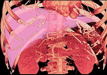
Volume rendering image created with computed tomography, which can be used to evaluate the volume of the liver of a potential donor.

Living donor liver transplantation (LDLT) has emerged in recent decades as a critical surgical option for patients with end stage liver disease, such as cirrhosis and/or hepatocellular carcinoma often attributable to one or more of the following: long-term alcohol use disorder, long-term untreated hepatitis C infection, long-term untreated hepatitis B infection. The concept of LDLT is based on (1) the remarkable regenerative capacities of the human liver and (2) the widespread shortage of cadaveric livers for patients awaiting transplant. In LDLT, a piece of healthy liver is surgically removed from a living person and transplanted into a recipient, immediately after the recipient’s diseased liver has been entirely removed.

Historically, LDLT began with terminal pediatric patients, whose parents were motivated to risk donating a portion of their compatible healthy livers to replace their children's failing ones. The first report of successful LDLT was by Silvano Raia at the University of Sao Paulo Faculty of Medicine in July 1989. It was followed by Christoph Broelsch at the University of Chicago Medical Center in November 1989, when two-year-old Alyssa Smith received a portion of her mother's liver. Surgeons eventually realized that adult-to-adult LDLT was also possible, and now the practice is common in a few reputable medical institutes. It is considered more technically demanding than even standard, cadaveric donor liver transplantation, and also poses the ethical problems underlying the indication of a major surgical operation (hemihepatectomy or related procedure) on a healthy human being. In various case series, the risk of complications in the donor is around 10%, and very occasionally a second operation is needed. Common problems are biliary fistula, gastric stasis and infections; they are more common after removal of the right lobe of the liver. Death after LDLT has been reported at 0% (Japan), 0.3% (USA) and <1% (Europe), with risks likely to decrease further as surgeons gain more experience in this procedure. Since the law was changed to permit altruistic non-directed living organ donations in the UK in 2006, the first altruistic living liver donation took place in Britain in December 2012.

In a typical adult recipient LDLT, 55 to 70% of the liver (the right lobe) is removed from a healthy living donor. The donor's liver will regenerate approaching 100% function within 4–6 weeks, and will almost reach full volumetric size with recapitulation of the normal structure soon thereafter. It may be possible to remove up to 70% of the liver from a healthy living donor without harm in most cases. The transplanted portion will reach full function and the appropriate size in the recipient as well, although it will take longer than for the donor.

Living donors are faced with risks and/or complications after the surgery. Blood clots and biliary problems have the possibility of arising in the donor post-op, but these issues are remedied fairly easily. Although death is a risk that a living donor must be willing to accept prior to the surgery, the mortality rate of living donors in the United States is low. The LDLT donor's immune system does diminish as a result of the liver regenerating, so certain foods which would normally cause an upset stomach could cause serious illness.

Donor requirements

CT scan performed for evaluation of a potential donor. The image shows an unusual variation of hepatic artery. The left hepatic artery supplies not only left lobe but also segment 8. The anatomy makes right lobe donation impossible. Even used as left lobe or lateral segment donation, it would be very technically challenging in anastomosing the small arteries.

Any member of the family, parent, sibling, child, spouse or a volunteer can donate their liver. The criteria for a liver donation include:

  • Being in good health
  • Having a blood type that matches or is compatible with the recipient's, although some centres now perform blood group incompatible transplants with special immunosuppression protocols.
  • Having a charitable desire of donation without financial motivation
  • Being between 20 and 60 years old (18 to 60 years old in some places)
  • Have an important personal relationship with the recipient
  • Being of similar or larger size than the recipient
  • Before one becomes a living donor, the donor must undergo testing to ensure that the individual is physically fit, in excellent health, and not having uncontrolled high blood pressure, liver disease, diabetes or heart disease. Sometimes CT scans or MRIs are done to image the liver. In most cases, the work up is done in 2–3 weeks.

Complications

Living donor surgery is done at a major center. Very few individuals require any blood transfusions during or after surgery. All potential donors should know there is a 0.5 to 1.0 percent chance of death. Other risks of donating a liver include bleeding, infection, painful incision, possibility of blood clots and a prolonged recovery. The vast majority of donors enjoy complete and full recovery within 2–3 months.

Pediatric transplantation

In children, due to their smaller abdominal cavity, there is only space for a partial segment of liver, usually the left lobe of the donor's liver. This is also known as a "split" liver transplant. There are four anastomoses required for a "split" liver transplant: hepaticojejunostomy (biliary drainage connecting to a roux limb of jejunum), portal venous anatomosis, hepatic arterial anastomosis, and inferior vena cava anastomosis.

In children, living liver donor transplantations have become very accepted. The accessibility of adult parents who want to donate a piece of the liver for their children/infants has reduced the number of children who would have otherwise died waiting for a transplant. Having a parent as a donor also has made it a lot easier for children - because both patients are in the same hospital and can help boost each other's morale.

Benefits

There are several advantages of living liver donor transplantation over cadaveric donor transplantation, including:

  • Transplant can be done on an elective basis because the donor is readily available
  • There are fewer possibilities for complications and death than there would be while waiting for a cadaveric organ donor
  • Because of donor shortages, UNOS has placed limits on cadaveric organ allocation to foreigners who seek medical help in the USA. With the availability of living donor transplantation, this will now allow foreigners a new opportunity to seek medical care in the USA.

Screening for donors

Living donor transplantation is a multidisciplinary approach. All living liver donors undergo medical evaluation. Every hospital which performs transplants has dedicated nurses that provide specific information about the procedure and answer questions that families may have. During the evaluation process, confidentiality is assured on the potential donor. Every effort is made to ensure that organ donation is not made by coercion from other family members. The transplant team provides both the donor and family thorough counseling and support which continues until full recovery is made.

All donors are assessed medically to ensure that they can undergo the surgery. Blood type of the donor and recipient must be compatible but not always identical. Other things assessed prior to surgery include the anatomy of the donor liver. However, even with mild variations in blood vessels and bile duct, surgeons today are able to perform transplantation without problems. The most important criterion for a living liver donor is to be in excellent health.

Post-transplant immunosuppression

Like most other allografts, a liver transplant will be rejected by the recipient unless immunosuppressive drugs are used. The immunosuppressive regimens for all solid organ transplants are fairly similar, and a variety of agents are now available. Most liver transplant recipients receive corticosteroids plus a calcineurin inhibitor such as tacrolimus or ciclosporin, (also spelled cyclosporine and cyclosporin) plus a purine antagonist such as mycophenolate mofetil. Clinical outcome is better with tacrolimus than with ciclosporin during the first year of liver transplantation. If the patient has a co-morbidity such as active hepatitis B, high doses of hepatitis B immunoglubins are administrated in liver transplant patients.

Liver transplantation is unique in that the risk of chronic rejection also decreases over time, although the great majority of recipients need to take immunosuppressive medication for the rest of their lives. It is possible to be slowly taken off anti rejection medication but only in certain cases. It is theorized that the liver may play a yet-unknown role in the maturation of certain cells pertaining to the immune system. There is at least one study by Thomas E. Starzl's team at the University of Pittsburgh which consisted of bone marrow biopsies taken from such patients which demonstrate genotypic chimerism in the bone marrow of liver transplant recipients.

Recovery and outcomes

The prognosis following liver transplant is variable, depending on overall health, technical success of the surgery, and the underlying disease process affecting the liver. There is no exact model to predict survival rates; those with transplant have a 58% chance of surviving 15 years. Failure of the new liver (primary nonfunction in liver transplantation or PNF) occurs in 10% to 15% of all cases. These percentages are contributed to by many complications. Early graft failure is probably due to preexisting disease of the donated organ. Others include technical flaws during surgery such as revascularization that may lead to a nonfunctioning graft.

History

As with many experimental models used in early surgical research, the first attempts at liver transplantation were performed on dogs. The earliest published reports of canine liver transplantations were performed in 1955 by Vittorio Staudacher at Opedale Maggiore Policlinico in Milan, Italy. This initial attempt varied significantly from contemporary techniques; for example, Staudacher reported "arterialization" of the donor portal vein via the recipient hepatic artery, and use of cholecystostomy for biliary drainage.

The first attempted human liver transplant was performed in 1963 by Thomas Starzl, although the pediatric patient died intraoperatively due to uncontrolled bleeding. Multiple subsequent attempts by various surgeons remained unsuccessful until 1967, when Starzl transplanted a 19 month old girl with hepatoblastoma who was able to survive for over 1 year before dying of metastatic disease. Despite the development of viable surgical techniques, liver transplantation remained experimental through the 1970s, with one year patient survival in the vicinity of 25%. The introduction of ciclosporin by Sir Roy Calne, Professor of Surgery Cambridge, markedly improved patient outcomes, and the 1980s saw recognition of liver transplantation as a standard clinical treatment for both adult and pediatric patients with appropriate indications. Liver transplantation is now performed at over one hundred centers in the US, as well as numerous centres in Europe and elsewhere.

The limited supply of liver allografts from non-living donors relative to the number of potential recipients spurred the development of living donor liver transplantation. The first altruistic living liver donation in Britain was performed in December 2012 in St James University Hospital Leeds.

Society and culture

Famous liver transplant recipients

See also: Category:Liver transplant recipients and List of organ transplant donors and recipients

Research directions

Cooling

There is increasing interest in improving methods for allograft preservation following organ harvesting. The standard "static cold storage" technique relies on decreased temperature to slow of anaerobic metabolic breakdown. This is currently being investigated at cold (hypothermic), body temperature (normothermic), and under body temperature (subnormothermic). Hypothermic machine perfusion has been used successfully at Columbia University and at the University of Zurich. A 2014 study showed that the liver preservation time could be significantly extended using a supercooling technique, which preserves the liver at subzero temperatures (-6 °C)  More recently, the first randomised controlled clinical trial comparing machine preservation with conventional cold storage showed comparable outcomes, with better early function, fewer discarded organs, and longer preservation times compared with cold stored livers.

Special populations

Alcohol dependence

The high incidence of liver transplants given to those with alcoholic cirrhosis has led to a recurring controversy regarding the eligibility of such patients for liver transplant. The controversy stems from the view of alcoholism as a self-inflicted disease and the perception that those with alcohol-induced damage are depriving other patients who could be considered more deserving. It is an important part of the selection process to differentiate transplant candidates who suffer from alcoholism as opposed to those who were susceptible to non-dependent alcohol use. The latter who gain control of alcohol use have a good prognosis following transplantation. Once a diagnosis of alcoholism has been established, however, it is necessary to assess the likelihood of future sobriety.

HIV

Historically, HIV was considered an absolute contraindication to liver transplantation. This was in part due to concern that the infection would be worsened by the immunosuppressive medication which is required after transplantation.

However, with the advent of highly active antiretroviral therapy (HAART), people with HIV have much improved prognosis. Transplantation may be offered selectively, although consideration of overall health and life circumstances may still be limiting. Uncontrolled HIV disease (AIDS) remains an absolute contraindication.

Molecular machine

From Wikipedia, the free encyclopedia

A molecular machine, nanite, or nanomachine is a molecular component that produces quasi-mechanical movements (output) in response to specific stimuli (input). In cellular biology, macromolecular machines frequently perform tasks essential for life, such as DNA replication and ATP synthesis. The expression is often more generally applied to molecules that simply mimic functions that occur at the macroscopic level. The term is also common in nanotechnology where a number of highly complex molecular machines have been proposed that are aimed at the goal of constructing a molecular assembler.

For the last several decades, chemists and physicists alike have attempted, with varying degrees of success, to miniaturize machines found in the macroscopic world. Molecular machines are at the forefront of cellular biology research. The 2016 Nobel Prize in Chemistry was awarded to Jean-Pierre Sauvage, Sir J. Fraser Stoddart, and Bernard L. Feringa for the design and synthesis of molecular machines.

Types

Molecular machines can be divided into two broad categories; artificial and biological. In general, artificial molecular machines (AMMs) refer to molecules that are artificially designed and synthesized whereas biological molecular machines can commonly be found in nature and have evolved into their forms after abiogenesis on Earth.

Artificial

A wide variety of artificial molecular machines (AMMs) have been synthesized by chemists which are rather simple and small compared to biological molecular machines. The first AMM, a molecular shuttle, was synthesized by Sir J. Fraser Stoddart. A molecular shuttle is a rotaxane molecule where a ring is mechanically interlocked onto an axle with two bulky stoppers. The ring can move between two binding sites with various stimuli such as light, pH, solvents, and ions. As the authors of this 1991 JACS paper noted: "Insofar as it becomes possible to control the movement of one molecular component with respect to the other in a [2]rotaxane, the technology for building molecular machines will emerge", mechanically interlocked molecular architectures spearheaded AMM design and synthesis as they provide directed molecular motion. Today a wide variety of AMMs exists as listed below.

Overcrowded alkane molecular motor.

Molecular motors

Molecular motors are molecules that are capable of directional rotary motion around a single or double bond. Single bond rotary motors are generally activated by chemical reactions whereas double bond rotary motors are generally fueled by light. The rotation speed of the motor can also be tuned by careful molecular design. Carbon nanotube nanomotors have also been produced.

Molecular propeller

A molecular propeller is a molecule that can propel fluids when rotated, due to its special shape that is designed in analogy to macroscopic propellers. It has several molecular-scale blades attached at a certain pitch angle around the circumference of a nanoscale shaft.

Daisy chain [2]rotaxane. These molecules are considered as building blocks for artificial muscle.

Molecular switch

A molecular switch is a molecule that can be reversibly shifted between two or more stable states. The molecules may be shifted between the states in response to changes in pH, light (photoswitch), temperature, an electric current, microenvironment, or the presence of a ligand.

Rotaxane based molecular shuttle.

Molecular shuttle

A molecular shuttle is a molecule capable of shuttling molecules or ions from one location to another. A common molecular shuttle consists of a rotaxane where the macrocycle can move between two sites or stations along the dumbbell backbone.

Nanocar

Nanocars are single molecule vehicles that resemble macroscopic automobiles and are important for understanding how to control molecular diffusion on surfaces. The first nanocars were synthesized by James M. Tour in 2005. They had an H shaped chassis and 4 molecular wheels (fullerenes) attached to the four corners. In 2011, Ben Feringa and co-workers synthesized the first motorized nanocar which had molecular motors attached to the chassis as rotating wheels. The authors were able to demonstrate directional motion of the nanocar on a copper surface by providing energy from a scanning tunneling microscope tip. Later, in 2017, the world's first ever Nanocar Race took place in Toulouse.

Molecular balance

A molecular balance is a molecule that can interconvert between two and more conformational or configurational states in response to the dynamic of multiple intra- and intermolecular driving forces, such as hydrogen bonding, solvophobic/hydrophobic effects, π interactions, and steric and dispersion interactions. Molecular balances can be small molecules or macromolecules such as proteins. Cooperatively folded proteins, for example, have been used as molecular balances to measure interaction energies and conformational propensities.

Molecular tweezers

Molecular tweezers are host molecules capable of holding items between their two arms. The open cavity of the molecular tweezers binds items using non-covalent bonding including hydrogen bonding, metal coordination, hydrophobic forces, van der Waals forces, π interactions, or electrostatic effects. Examples of molecular tweezers have been reported that are constructed from DNA and are considered DNA machines.

Molecular sensor

A molecular sensor is a molecule that interacts with an analyte to produce a detectable change. Molecular sensors combine molecular recognition with some form of reporter, so the presence of the item can be observed.

Molecular logic gate

A molecular logic gate is a molecule that performs a logical operation on one or more logic inputs and produces a single logic output. Unlike a molecular sensor, the molecular logic gate will only output when a particular combination of inputs are present.

Molecular assembler

A molecular assembler is a molecular machine able to guide chemical reactions by positioning reactive molecules with precision.

Molecular hinge

A molecular hinge is a molecule that can be selectively switched from one configuration to another in a reversible fashion. Such configurations must have distinguishable geometries; for instance, azobenzene groups in a linear molecule may undergo cis-trans isomerizations when irradiated with ultraviolet light, triggering a reversible transition to a bent or V-shaped conformation. Molecular hinges typically rotate in a crank-like motion around a rigid axis, such as a double bond or aromatic ring. However, macrocyclic molecular hinges with more clamp-like mechanisms have also been synthesized.

Biological

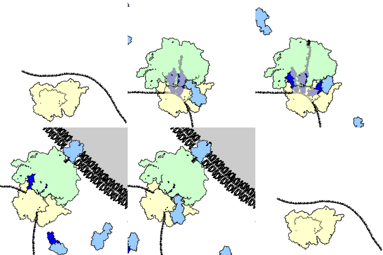
A ribosome performing the elongation and membrane targeting stages of protein translation. The ribosome is green and yellow, the tRNAs are dark blue, and the other proteins involved are light blue. The produced peptide is released into the endoplasmic reticulum.

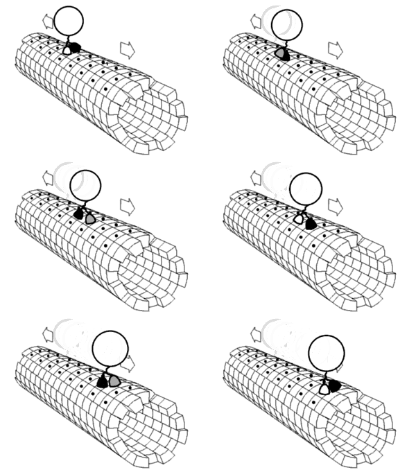
The most complex macromolecular machines are found within cells, often in the form of multi-protein complexes. Important examples of biological machines include motor proteins such as myosin, which is responsible for muscle contraction, kinesin, which moves cargo inside cells away from the nucleus along microtubules, and dynein, which moves cargo inside cells towards the nucleus and produces the axonemal beating of motile cilia and flagella. "[I]n effect, the [motile cilium] is a nanomachine composed of perhaps over 600 proteins in molecular complexes, many of which also function independently as nanomachines ... Flexible linkers allow the mobile protein domains connected by them to recruit their binding partners and induce long-range allostery via protein domain dynamics." Other biological machines are responsible for energy production, for example ATP synthase which harnesses energy from proton gradients across membranes to drive a turbine-like motion used to synthesise ATP, the energy currency of a cell. Still other machines are responsible for gene expression, including DNA polymerases for replicating DNA, RNA polymerases for producing mRNA, the spliceosome for removing introns, and the ribosome for synthesising proteins. These machines and their nanoscale dynamics are far more complex than any molecular machines that have yet been artificially constructed.

Some biological molecular machines

These biological machines might have applications in nanomedicine. For example, they could be used to identify and destroy cancer cells. Molecular nanotechnology is a speculative subfield of nanotechnology regarding the possibility of engineering molecular assemblers, biological machines which could re-order matter at a molecular or atomic scale. Nanomedicine would make use of these nanorobots, introduced into the body, to repair or detect damages and infections. Molecular nanotechnology is highly theoretical, seeking to anticipate what inventions nanotechnology might yield and to propose an agenda for future inquiry. The proposed elements of molecular nanotechnology, such as molecular assemblers and nanorobots are far beyond current capabilities.

Research

The construction of more complex molecular machines is an active area of theoretical and experimental research. A number of molecules, such as molecular propellers, have been designed, although experimental studies of these molecules are inhibited by the lack of methods to construct these molecules. In this context, theoretical modeling can be extremely useful to understand the self-assembly/disassembly processes of rotaxanes, important for the construction of light-powered molecular machines. This molecular-level knowledge may foster the realization of ever more complex, versatile, and effective molecular machines for the areas of nanotechnology, including molecular assemblers.

Although currently not feasible, some potential applications of molecular machines are transport at the molecular level, manipulation of nanostructures and chemical systems, high density solid-state informational processing and molecular prosthetics. Many fundamental challenges need to be overcome before molecular machines can be used practically such as autonomous operation, complexity of machines, stability in the synthesis of the machines and the working conditions.

Inequality (mathematics)

From Wikipedia, the free encyclopedia https://en.wikipedia.org/wiki/Inequality...