Search This Blog

Monday, November 1, 2021

Viral hepatitis

From Wikipedia, the free encyclopedia
Jump to navigation Jump to search
Viral hepatitis
Ground glass hepatocytes high mag cropped 2.jpg
Micrograph showing ground glass hepatocytes, which are seen in chronic hepatitis B infections (a type of viral hepatitis), and represent accumulations of viral antigen in the endoplasmic reticulum. H&E stain.
SpecialtyInfectious disease, gastroenterology 

Viral hepatitis is liver inflammation due to a viral infection. It may present in acute form as a recent infection with relatively rapid onset, or in chronic form.

The most common causes of viral hepatitis are the five unrelated hepatotropic viruses hepatitis A, B, C, D, and E. Other viruses can also cause liver inflammation, including cytomegalovirus, Epstein-Barr virus, and yellow fever. There also have been scores of recorded cases of viral hepatitis caused by herpes simplex virus.

Mode Of Transmission

Viral hepatitis is either transmitted through contaminated food or water (A, E) or via blood and body fluids (B, C). The viruses which get transmitted through water and food are mostly self-limited resulting in acute illness with full resolution. The blood borne viruses (B, C) can cause both acute and chronic liver disease and can be transmitted from mother to child during birth, through contact with body fluids during sex, unsafe injections and through unscreened blood transfusions.

The most common types of hepatitis can be prevented or treated. Hepatitis A and hepatitis B can be prevented by vaccination. Effective treatments for hepatitis C are available but costly.

In 2013, about 1.5 million people died from viral hepatitis, most commonly due to hepatitis B and C. East Asia, in particular Mongolia, is the region most affected.

Hepatitis viruses

The most common cause of hepatitis is viral. Although the effects of various viruses are all classified under the disease hepatitis, these viruses are not all related.

Hepatitis viruses

Hepatitis A virus (HAV) Hepatitis B virus (HBV) Hepatitis C virus (HCV) Hepatitis D virus (HDV) Hepatitis E virus (HEV) Hepatitis G virus (HGV)
Viral species Hepatovirus A Hepatitis B virus Hepacivirus C Hepatitis delta virus Orthohepevirus A Also known as Human Pegvirus (HPgV) and as GB virus C (GBV-C)
Viral family Picornaviridae Hepadnaviridae Flaviviridae Incertae sedis Hepeviridae Flaviviridae
Genome (+)ssRNA dsDNA-RT (+)ssRNA (−)ssRNA (+)ssRNA (+)ssRNA
Antigens
HBsAg, HBeAg Core antigen Delta antigen
Transmission Enteral Parenteral Parenteral Parenteral Enteral Parenteral
Incubation period 20–40 days 45–160 days 15–150 days 30–60 days 15–60 days 14-20 days
Severity/Chronicity Mild; acute Occasionally severe; 5–10% chronic Subclinical; 70% chronic Exacerbates symptoms of HBV; chronic with HBV Mild in normal patients; severe in pregnant women; acute
Vaccine 2 injections; at least 20 years of protection 3 injections; lifetime protection None available None available, but not considered necessary; Hep B vaccine provides protection Investigational (approved in China)

Viral hepatitis types

Hepatitis A

Hepatitis A or infectious jaundice is caused by hepatitis A virus (HAV), a picornavirus transmitted by the fecal-oral route often associated with ingestion of contaminated food. It causes an acute form of hepatitis and does not have a chronic stage. The patient's immune system makes antibodies against HAV that confer immunity against future infection. People with hepatitis A are advised to rest, stay hydrated and avoid alcohol. A vaccine is available that will prevent HAV infection for up to 10 years. Hepatitis A can be spread through personal contact, consumption of raw sea food, or drinking contaminated water. This occurs primarily in third world countries. Strict personal hygiene and the avoidance of raw and unpeeled foods can help prevent an infection. Infected people excrete HAV with their feces two weeks before and one week after the appearance of jaundice. The time between the infection and the start of the illness averages 28 days (ranging from 15 to 50 days), and most recover fully within 2 months, although approximately 15% of sufferers may experience continuous or relapsing symptoms from six months to a year following initial diagnosis.

Hepatitis A[
Marker Detection Time Description Significance
Faecal HAV 2–4 weeks or 28 days Early detection
Ig M anti HAV 4–12 weeks Enzyme immunoassay for antibodies During acute Illness
Ig G anti HAV 5 weeks–persistent Enzyme immunoassay for antibodies Old infection or reinfection

Hepatitis B

Hepatitis B is caused by the hepatitis B virus, a hepadnavirus that can cause both acute and chronic hepatitis. Chronic hepatitis develops in the 15% of adults who are unable to eliminate the virus after an initial infection. Identified methods of transmission include contact with blood, blood transfusion (now rare), unsanitary tattoos, sex (through sexual intercourse or contact with bodily fluids), or mother-to-child by breast feeding; there is minimal evidence of transplacental crossing. However, in about half of cases the source of infection cannot be determined. Blood contact can occur by sharing syringes in intravenous drug use, shaving accessories such as razor blades, or touching wounds on infected persons. Needle-exchange programmes have been created in many countries as a form of prevention.

Patients with chronic hepatitis B have antibodies against the virus, but not enough to clear the infected liver cells. The continued production of virus and countervailing antibodies is a likely cause of the immune complex disease seen in these patients. A vaccine is available to prevent infection for life. Hepatitis B infections result in 500,000 to 1,200,000 deaths per year worldwide due to the complications of chronic hepatitis, cirrhosis, and hepatocellular carcinoma. Hepatitis B is endemic in a number of (mainly South-East Asian) countries, making cirrhosis and hepatocellular carcinoma big killers. There are eight treatment options approved by the U.S. Food and Drug Administration (FDA) available for persons with a chronic hepatitis B infection: alpha-interferon, pegylated interferon, adefovir, entecavir, telbivudine, lamivudine, tenofovir disoproxil and tenofovir alafenamide with a 65% rate of sustained response.[citation needed]

Hepatitis C

Hepatitis C (originally "non-A non-B hepatitis") is caused by hepatitis C virus (HCV), an RNA virus of the family Flaviviridae. HCV can be transmitted through contact with blood (including through sexual contact if the two parties' blood is mixed) and can also cross the placenta. Hepatitis C usually leads to chronic hepatitis, culminating in cirrhosis in some people. It usually remains asymptomatic for decades. Patients with hepatitis C are susceptible to severe hepatitis if they contract either hepatitis A or B, so all persons with hepatitis C should be immunized against hepatitis A and hepatitis B if they are not already immune, and avoid alcohol. HCV can lead to the development of hepatocellular carcinoma, however, only a minority of HCV-infected individuals develop cancer (1-4% annually), suggesting a complex interplay between viral gene expression and host and environmental factors to promote carcinogenesis. The risk is increased two-fold with active HBV coinfection and a 21% increase in mortality compared to those with latent HBV and HCV. HCV viral levels can be reduced to undetectable levels by a combination of interferon and the antiviral drug ribavirin. The genotype of the virus is the primary determinant of the rate of response to this treatment regimen, with genotype 1 being the most resistant.

Hepatitis C is the most common chronic blood-borne infection in the United States.

Hepatitis C
Marker Detection Time Description Significance Note
HCV-RNA 1–3 weeks or 21 days PCR Demonstrates presence or absence of virus Results may be intermittent during course of infection. Negative result is not indicative of absence.
anti-HCV 5–6 weeks Enzyme Immunoassay for antibodies Demonstrates past or present infection High false positive in those with autoimmune disorders and populations with low virus prevalence.
ALT 5–6 weeks Peak in ALT coincides with peak in anti-HCV Fluctuating ALT levels is an indication of active liver disease.

Hepatitis D

Hepatitis D is caused by the hepatitis D virus (HDV), or hepatitis delta virus; it belongs to the genus Deltavirus. It is similar to a satellite virus as it can only propagate in the presence of the hepatitis B virus, depending on the helper function of HBV for its replication and expression. It has no independent life cycle, but can survive and replicate as long as HBV infection persists in the host body. It can only cause infection when encapsulated by hepatitis B virus surface antigens. The vaccine for hepatitis B protects against hepatitis D virus because of the latter's dependence on the presence of hepatitis B virus for it to replicate.

Hepatitis E

Hepatitis E is caused by the Hepatitis E virus (HEV), from the family Hepeviridae; it produces symptoms similar to hepatitis A, although it can take a fulminant course in some patients, particularly pregnant women (mortality rate about 20%); chronic infections may occur in immune-compromised patients. It is more prevalent in the Indian subcontinent. The virus is feco-orally transmitted and usually is self-limited.

Hepatitis F virus

Hepatitis F virus (HFV) is a hypothetical virus linked to certain cases of hepatitis. Several hepatitis F virus candidates emerged in the 1990s, but none of these reports have been substantiated.

GB virus C

The GB virus C is another potential viral cause of hepatitis that is probably spread by blood and sexual contact. It was initially identified as Hepatitis G virus. There is very little evidence that this virus causes hepatitis, as it does not appear to replicate primarily in the liver. It is now classified as GB virus C.

Relationship between hepatitis C virus and liver cancer

Hepatitis C virus (HCV) are causing acute and chronic infections that is a major cause of hepatocellular carcinoma (HCC), advanced hepatic fibrosis and cirrhosis.

A major cause of death in HCC patients with chronic HCV infection. The pathogenesis of HCC associated with HCV, that virus play direct and indirect roles.

A major risk for the development of HCC is persistent infection with HCV and the highest risk for HCC development is associated with co-infection of HBV with HDV, HCV or HIV.

The risk factors lead to development of HCC in chronic HCV is synchronous liver diseases, viral genotype, lifestyle factors and presence of obesity and diabetes mellitus. The lifestyle factors such as smoking, alcohol use and coffee drinking accelerated progression to HCC in HCV.

The purpose of HCV treatment is to eliminate the infection, reduce the transmission to other people and decrease the risk of HCC development.

Other viruses

The virus first known to cause hepatitis was the yellow fever virus, a mosquito-borne flavivirus. Other viruses than can cause hepatitis include:

KIs-V is a virus isolated in 2011 from four patients with raised serum alanine transferases without other known cause; a causal role is suspected.
 

Fatty liver disease

From Wikipedia, the free encyclopedia
 
Fatty liver
Other namesHepatic steatosis
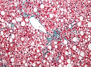
Non-alcoholic fatty liver disease1.jpg
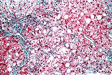
Micrograph showing a fatty liver (macrovesicular steatosis), as seen in non-alcoholic fatty liver disease. Trichrome stain.

SpecialtyGastroenterology
SymptomsNone, tiredness, pain in the upper right side of the abdomen
ComplicationsCirrhosis, liver cancer, esophageal varices
TypesNon-alcoholic fatty liver disease (NAFLD), alcoholic liver disease
CausesAlcohol, diabetes, obesity
Diagnostic methodBased on the medical history supported by blood tests, medical imaging, liver biopsy
Differential diagnosisViral hepatitis, Wilson disease, primary sclerosing cholangitis
TreatmentNo alcohol, weight loss
PrognosisGood if treated early
FrequencyNAFLD: 30% (Western countries)
ALD: >90% of heavy drinkers

Fatty liver disease (FLD), also known as hepatic steatosis, is a condition where excess fat builds up in the liver. Often there are no or few symptoms. Occasionally there may be tiredness or pain in the upper right side of the abdomen. Complications may include cirrhosis, liver cancer, and esophageal varices.

There are two types of fatty liver disease: non-alcoholic fatty liver disease (NAFLD) and alcoholic liver disease.[1] NAFLD is made up of simple fatty liver and non-alcoholic steatohepatitis (NASH). The primary risks include alcohol, type 2 diabetes, and obesity. Other risk factors include certain medications such as glucocorticoids, and hepatitis C. It is unclear why some people with NAFLD develop simple fatty liver and others develop NASH. Diagnosis is based on the medical history supported by blood tests, medical imaging, and occasionally liver biopsy.

Treatment of NAFLD is generally by dietary changes and exercise to bring about weight loss. In those who are severely affected, liver transplantation may be an option. More than 90% of heavy drinkers develop fatty liver while about 25% develop the more severe alcoholic hepatitis. NAFLD affects about 30% of people in Western countries and 10% of people in Asia. NAFLD affects about 10% of children in the United States. It occurs more often in older people and males.

Signs and symptoms

Often there are no or few symptoms. Occasionally there may be tiredness or pain in the upper right side of the abdomen.

Complications

Fatty liver can develop into a fibrosis or a liver cancer. For people affected by NAFLD, the 10-year survival rate was about 80%. The rate of progression of fibrosis is estimated to be one per 7 years in NASH and one per 14 years in NAFLD, with an increasing speed There is a strong relationship between these pathologies and metabolic illnesses (diabetes type II, metabolic syndrome). These pathologies can also affect non-obese people, who are then at a higher risk.

Less than 10% of people with cirrhotic alcoholic FLD will develop hepatocellular carcinoma, the most common type of primary liver cancer in adults, but up to 45% people with NASH without cirrhosis can develop hepatocellular carcinoma.

The condition is also associated with other diseases that influence fat metabolism.

Causes

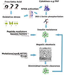
Different stages of liver damage

Fatty liver (FL) is commonly associated with metabolic syndrome (diabetes, hypertension, obesity, and dyslipidemia), but can also be due to any one of many causes:

Alcohol
Alcohol use disorder is one of the causes of fatty liver due to production of toxic metabolites like aldehydes during metabolism of alcohol in the liver. This phenomenon most commonly occurs with chronic alcohol use disorder.
Metabolic
abetalipoproteinemia, glycogen storage diseases, Weber–Christian disease, acute fatty liver of pregnancy, lipodystrophy
Nutritional
obesity, malnutrition, total parenteral nutrition, severe weight loss, refeeding syndrome, jejunoileal bypass, gastric bypass, jejunal diverticulosis with bacterial overgrowth
Drugs and toxins
amiodarone, methotrexate, diltiazem, expired tetracycline, highly active antiretroviral therapy, glucocorticoids, tamoxifen, environmental hepatotoxins (e.g., phosphorus, mushroom poisoning)
Other
celiac disease, inflammatory bowel disease, HIV, hepatitis C (especially genotype 3), and alpha 1-antitrypsin deficiency

Pathology

Micrograph of periportal hepatic steatosis, as may be seen due to steroid use, trichrome stain

Fatty change represents the intracytoplasmatic accumulation of triglycerides (neutral fats). At the beginning, the hepatocytes present small fat vacuoles (liposomes) around the nucleus (microvesicular fatty change). In this stage, liver cells are filled with multiple fat droplets that do not displace the centrally located nucleus. In the late stages, the size of the vacuoles increases, pushing the nucleus to the periphery of the cell, giving characteristic signet ring appearance (macrovesicular fatty change). These vesicles are well-delineated and optically "empty" because fats dissolve during tissue processing. Large vacuoles may coalesce and produce fatty cysts, which are irreversible lesions. Macrovesicular steatosis is the most common form and is typically associated with alcohol, diabetes, obesity, and corticosteroids. Acute fatty liver of pregnancy and Reye's syndrome are examples of severe liver disease caused by microvesicular fatty change. The diagnosis of steatosis is made when fat in the liver exceeds 5–10% by weight.

Mechanism leading to hepatic steatosis

Defects in fatty acid metabolism are responsible for pathogenesis of FLD, which may be due to imbalance in energy consumption and its combustion, resulting in lipid storage, or can be a consequence of peripheral resistance to insulin, whereby the transport of fatty acids from adipose tissue to the liver is increased. Impairment or inhibition of receptor molecules (PPAR-α, PPAR-γ and SREBP1) that control the enzymes responsible for the oxidation and synthesis of fatty acids appears to contribute to fat accumulation. In addition, alcohol use disorder is known to damage mitochondria and other cellular structures, further impairing cellular energy mechanism. On the other hand, non-alcoholic FLD may begin as excess of unmetabolised energy in liver cells. Hepatic steatosis is considered reversible and to some extent nonprogressive if the underlying cause is reduced or removed.

Micrograph of inflamed fatty liver (steatohepatitis)

Severe fatty liver is sometimes accompanied by inflammation, a situation referred to as steatohepatitis. Progression to alcoholic steatohepatitis (ASH) or non-alcoholic steatohepatitis (NASH) depends on the persistence or severity of the inciting cause. Pathological lesions in both conditions are similar. However, the extent of inflammatory response varies widely and does not always correlate with degree of fat accumulation. Steatosis (retention of lipid) and onset of steatohepatitis may represent successive stages in FLD progression.

Liver disease with extensive inflammation and a high degree of steatosis often progresses to more severe forms of the disease. Hepatocyte ballooning and necrosis of varying degrees are often present at this stage. Liver cell death and inflammatory responses lead to the activation of hepatic stellate cells, which play a pivotal role in hepatic fibrosis. The extent of fibrosis varies widely. Perisinusoidal fibrosis is most common, especially in adults, and predominates in zone 3 around the terminal hepatic veins.

The progression to cirrhosis may be influenced by the amount of fat and degree of steatohepatitis and by a variety of other sensitizing factors. In alcoholic FLD, the transition to cirrhosis related to continued alcohol consumption is well-documented, but the process involved in non-alcoholic FLD is less clear.

Diagnosis

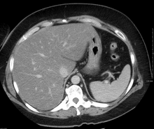
Liver steatosis (fatty liver disease) as seen on CT
 
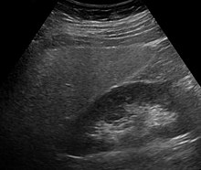
Ultrasound showing diffuse increased echogenicity of the liver.

Flow chart for diagnosis


Elevated liver enzyme


Serology to exclude viral hepatitis


Imaging study showing
fatty infiltrate


Alcohol intake


Less than two drinks per day‡
More than two drinks per day‡


Nonalcoholic fatty liver disease likely
Alcoholic liver disease likely

Criteria for nonalcoholic fatty liver disease:
consumption of ethanol less than 20 g/day for women and 30 g/day for men

Most individuals are asymptomatic and are usually discovered incidentally because of abnormal liver function tests or hepatomegaly noted in unrelated medical conditions. Elevated liver enzymes are found in 50% of patients with simple steatosis. The serum alanine transaminase (ALT) level usually is greater than the aspartate transaminase (AST) level in the nonalcoholic variant and the opposite in alcoholic FLD (AST:ALT more than 2:1). Simple blood test may help to determine the magnitude of the disease by assessing the degree of liver fibrosis. For example, AST-to-platelets ratio index (APRI score) and several other score, calculated from the results of blood test, can detect the degree of liver fibrosis and predict the future formation of liver cancer.

Imaging studies are often obtained during the evaluation process. Ultrasonography reveals a "bright" liver with increased echogenicity. Medical imaging can aid in diagnosis of fatty liver; fatty livers have lower density than spleens on computed tomography (CT), and fat appears bright in T1-weighted magnetic resonance images (MRIs). Magnetic resonance elastography, a variant of magnetic resonance imaging, is investigated as a non-invasive method to diagnose fibrosis progression. Histological diagnosis by liver biopsy is the most accurate measure of fibrosis and liver fat progression as of 2018.

Treatment

Decreasing caloric intake by at least 30% or by approximately 750–1,000 kcal/day results in improvement in hepatic steatosis. For people with NAFLD or NASH, weight loss via a combination of diet and exercise was shown to improve or resolve the disease. In more serious cases, medications that decrease insulin resistance, hyperlipidemia, and those that induce weight loss such as bariatric surgery as well as Vitamin E have been shown to improve or resolve liver function.

Bariatric surgery, while not recommended in 2017 as a treatment for fatty liver disease (FLD) alone, has been shown to revert FLD, NAFLD, NASH and advanced steatohepatitis in over 90% of people who have undergone this surgery for the treatment of obesity.

In the case of long-term total parenteral nutrition induced fatty liver disease, choline has been shown to alleviate symptoms. This may be due to a deficiency in the methionine cycle.

Epidemiology

NAFLD affects about 30% of people in Western countries and 10% of people in Asia. In the United States rates are around 35% with about 7% having the severe form NASH. NAFLD affects about 10% of children in the United States. Recently the term Metabolic dysfunction-associated fatty liver disease (MAFLD) has been proposed to replace NAFLD. MAFLD is a more inclusionary diagnostic name as it is based on the detection of fatty liver by histology (biopsy),medical imaging or blood biomarkers but should be accompanied by either overweight/obesity, type 2 diabetes mellitus, or metabolic dysregulation. The new definition no longer excludes alcohol consumption or coexistence of other liver diseases such as viral hepatitis. Using this more inclusive definition, the global prevalence of MAFLD is an astonishingly high 50.7%. Indeed, also using the old NAFLD definition, the disease is observed in up to 80% of obese people, 35% of whom progress to NASH, and in up to 20% of normal weight people, despite no evidence of excessive alcohol consumption. FLD is the most common cause of abnormal liver function tests in the United States. Fatty liver is more prevalent in Hispanic people than white, with black people having the lowest susceptibility.

In the study of Children of the 90s, 2.5% born in 1991 and 1992 were found by ultrasound at the age of 18 to have non-alcoholic fatty liver disease; five years later transient elastography (fibroscan) found over 20% to have the fatty deposits on the liver, indicating non-alcoholic fatty liver disease; half of those were classified as severe. The scans also found that 2.4% had the liver scarring, which can lead to cirrhosis.

In animals

Fatty liver disease can occur in pets such as reptiles (particularly turtles) and birds as well as mammals like cats and dogs. The most common cause is overnutrition. A distinct sign in birds is a misshapen beak. Fatty livers can be induced via gavage in geese or ducks to produce foie gras.


Septic shock

From Wikipedia, the free encyclopedia
 
Septic shock
Gabriel Metsu - La Fille malade.jpg
Sepsis is one of the most common causes of death in critically ill patients in Intensive Care Units. (Oil by Gabriël Metsu).


Thrombocytopenia with purpura on right hand in patient with septic shock

Septic shock is a potentially fatal medical condition that occurs when sepsis, which is organ injury or damage in response to infection, leads to dangerously low blood pressure and abnormalities in cellular metabolism. The Third International Consensus Definitions for Sepsis and Septic Shock (Sepsis-3) defines septic shock as a subset of sepsis in which particularly profound circulatory, cellular, and metabolic abnormalities are associated with a greater risk of mortality than with sepsis alone. Patients with septic shock can be clinically identified by requiring a vasopressor to maintain a mean arterial pressure of 65 mm Hg or greater and having serum lactate level greater than 2 mmol/L (>18 mg/dL) in the absence of hypovolemia. This combination is associated with hospital mortality rates greater than 40%.

The primary infection is most commonly caused by bacteria, but also may be by fungi, viruses or parasites. It may be located in any part of the body, but most commonly in the lungs, brain, urinary tract, skin or abdominal organs. It can cause multiple organ dysfunction syndrome (formerly known as multiple organ failure) and death.

Frequently, people with septic shock are cared for in intensive care units. It most commonly affects children, immunocompromised individuals, and the elderly, as their immune systems cannot deal with infection as effectively as those of healthy adults. The mortality rate from septic shock is approximately 25–50%.

Causes

Septic shock is a result of a systemic response to infection or multiple infectious causes. The precipitating infections that may lead to septic shock if severe enough include but are not limited to appendicitis, pneumonia, bacteremia, diverticulitis, pyelonephritis, meningitis, pancreatitis, necrotizing fasciitis, MRSA and mesenteric ischemia.

According to the earlier definitions of sepsis updated in 2001, sepsis is a constellation of symptoms secondary to an infection that manifests as disruptions in heart rate, respiratory rate, temperature, and white blood cell count. If sepsis worsens to the point of end-organ dysfunction (kidney failure, liver dysfunction, altered mental status, or heart damage), then the condition is called severe sepsis. Once severe sepsis worsens to the point where blood pressure can no longer be maintained with intravenous fluids alone, then the criterion has been met for septic shock.

Pathophysiology

The pathophysiology of septic shock is not entirely understood, but it is known that a key role in the development of severe sepsis is played by an immune and coagulation response to an infection. Both pro-inflammatory and anti-inflammatory responses play a role in septic shock. Septic shock involves a widespread inflammatory response that produces a hypermetabolic effect. This is manifested by increased cellular respiration, protein catabolism, and metabolic acidosis with a compensatory respiratory alkalosis.

Most cases of septic shock are caused by gram-positive bacteria, followed by endotoxin-producing gram-negative bacteria, although fungal infections are an increasingly prevalent cause of septic shock. Toxins produced by pathogens cause an immune response; in gram-negative bacteria these are endotoxins, which are bacterial membrane lipopolysaccharides (LPS).

Gram-positive

In gram-positive bacteria, these are exotoxins or enterotoxins, which may vary depending on the species of bacteria. These are divided into three types. Type I, cell surface-active toxins, disrupt cells without entering, and include superantigens and heat-stable enterotoxins. Type II, membrane-damaging toxins, destroy cell membranes in order to enter and include hemolysins and phospholipases. Type III, intracellular toxins or A/B toxins interfere with internal cell function and include shiga toxin, cholera toxin, and anthrax lethal toxin. (note that Shigella and Vibrio cholerae are Gram negative organisms).

Gram-negative

In gram-negative sepsis, free LPS attaches to a circulating LPS-binding protein, and the complex then binds to the CD14 receptor on monocytes, macrophages, and neutrophils. Engagement of CD14 (even at doses as minute as 10 pg/mL) results in intracellular signaling via an associated "Toll-like receptor" protein 4 (TLR-4). This signaling results in the activation of nuclear factor kappaB (NF-κB), which leads to transcription of a number of genes that trigger a proinflammatory response. It was the result of significant activation of mononuclear cells and synthesis of effector cytokines. It also results in profound activation of mononuclear cells and the production of potent effector cytokines such as IL-1, IL-6, and TNF-α. TLR-mediated activation helps to trigger the innate immune system to efficiently eradicate invading microbes, but the cytokines they produce also act on endothelial cells. There, they have a variety of effects, including reduced synthesis of anticoagulation factors such as tissue factor pathway inhibitor and thrombomodulin. The effects of the cytokines may be amplified by TLR-4 engagement on endothelial cells.

In response to inflammation, a compensatory reaction of production of anti-inflammatory substances such as IL-4, IL-10 antagonists, IL-1 receptor, and cortisol occurs. This is called compensatory anti-inflammatory response syndrome (CARS). Both the inflammatory and anti-inflammatory reactions are responsible for the course of sepsis and are described as MARS (Mixed Antagonist Response Syndrome). The aim of these processes is to keep inflammation at an appropriate level. CARS often leads to suppression of the immune system, which leaves patients vulnerable to secondary infection. It was once thought that SIRS or CARS could predominate in a septic individual, and it was proposed that CARS follows SIRS in a two-wave process. It is now believed that the systemic inflammatory response and the compensatory anti-inflammatory response occur simultaneously.

At high levels of LPS, the syndrome of septic shock supervenes; the same cytokine and secondary mediators, now at high levels, result in systemic vasodilation (hypotension), diminished myocardial contractility, widespread endothelial injury, activation causing systemic leukocyte adhesion and diffuse alveolar capillary damage in the lung, and activation of the coagulation system culminating in disseminated intravascular coagulation (DIC).

The hypoperfusion from the combined effects of widespread vasodilation, myocardial pump failure, and DIC causes multiorgan system failure that affects the liver, kidneys, and central nervous system, among other organ systems. Recently, severe damage to liver ultrastructure has been noticed from treatment with cell-free toxins of Salmonella. Unless the underlying infection (and LPS overload) is rapidly brought under control, the patient usually dies.

The ability of TLR4 to respond to a distinct LPS species are clinically important. Pathogenic bacteria may employ LPS with low biological activity to evade proper recognition by the TLR4/MD-2 system, dampening the host immune response and increasing the risk of bacterial dissemination. On the other hand, such LPS would not be able to induce septic shock in susceptible patients, rendering septic complications more manageable. Yet, defining and understanding how even the smallest structural differences between the very similar LPS species may affect the activation of the immune response may provide the mechanism for the fine tuning of the latter and new insights to immunomodulatory processes.

Diagnosis

According to current guidelines, requirements for diagnosis with sepsis are "the presence (probable or documented) of infection together with systemic manifestations of infection". These manifestations may include:

  • Tachypnea (fast rate of breathing), which is defined as more than 20 breaths per minute, or when testing blood gas, a PaCO
    2
    less than 32 mm Hg, which signifies hyperventilation
  • White blood cell count either significantly low (< 4000 cells/mm3), or elevated (> 12000 cells/mm3)
  • Tachycardia (rapid heart rate), which in sepsis is defined as a rate greater than 90 beats per minute
  • Altered body temperature: Fever > 38.0 °C (100.4 °F) or hypothermia < 36.0 °C (96.8 °F)

Documented evidence of infection, may include positive blood culture, signs of pneumonia on chest x-ray, or other radiologic or laboratory evidence of infection. Signs of end-organ dysfunction are present in septic shock, including kidney failure, liver dysfunction, changes in mental status, or elevated serum lactate.

Septic shock is diagnosed if there is low blood pressure (BP) that does not respond to treatment. This means that intravenous fluid administration alone is not enough to maintain a patient's BP. Diagnosis of septic shock is made when systolic blood pressure is less than 90 mm Hg, a mean arterial pressure (MAP) is less than 70 mm Hg, or a systolic BP decrease of 40 mm Hg or more without other causes for low BP.

Definition

Septic shock is a subclass of distributive shock, a condition in which abnormal distribution of blood flow in the smallest blood vessels results in inadequate blood supply to the body tissues, resulting in ischemia and organ dysfunction. Septic shock refers specifically to distributive shock due to sepsis as a result of infection.

Septic shock may be defined as sepsis-induced low blood pressure that persists despite treatment with intravenous fluids. Low blood pressure reduces tissue perfusion pressure, causing the tissue hypoxia that is characteristic of shock. Cytokines released in a large scale inflammatory response result in massive vasodilation, increased capillary permeability, decreased systemic vascular resistance, and low blood pressure. Finally, in an attempt to offset decreased blood pressure, ventricular dilatation and myocardial dysfunction occur.

Septic shock may be regarded as a stage of SIRS (Systemic Inflammatory Response Syndrome), in which sepsis, severe sepsis and multiple organ dysfunction syndrome (MODS) represent different stages of a pathophysiological process. If an organism cannot cope with an infection, it may lead to a systemic response - sepsis, which may further progress to severe sepsis, septic shock, organ failure, and eventually, result in death.

Treatment

Treatment primarily consists of the following:

  1. Giving intravenous fluids
  2. Early antibiotic administration 
  3. Early goal directed therapy
  4. Rapid source identification and control
  5. Support of major organ dysfunction

Fluids

Because lowered blood pressure in septic shock contributes to poor perfusion, fluid resuscitation is an initial treatment to increase blood volume. Patients demonstrating sepsis-induced hypoperfusion should be initially resuscitated with at least 30 ml/kg of intravenous crystalloid within the first three hours. Crystalloids such as normal saline and lactated Ringer's solution are recommended as the initial fluid of choice, while the use of colloid solutions such as hydroxyethyl starch have not shown any advantage or decrease in mortality. When large quantities of fluids are given, administering albumin has shown some benefit.

Antibiotics

Treatment guidelines call for the administration of broad-spectrum antibiotics within the first hour following recognition of septic shock. Prompt antimicrobial therapy is important, as risk of dying increases by approximately 10% for every hour of delay in receiving antibiotics. Time constraints do not allow the culture, identification, and testing for antibiotic sensitivity of the specific microorganism responsible for the infection. Therefore, combination antimicrobial therapy, which covers a wide range of potential causative organisms, is tied to better outcomes. Antibiotics should be continued for 7–10 days in most patients, though treatment duration may be shorter or longer depending on clinical response.

Vasopressors

Among the choices for vasopressors, norepinephrine is superior to dopamine in septic shock. Norepinephrine is the preferred vasopressor, while epinephrine may be added to norepinephrine when needed. Low-dose vasopressin also may be used as an addition to norepinephrine, but is not recommended as a first-line treatment. Dopamine may cause rapid heart rate and arrhythmias, and is only recommended in combination with norepinephrine in those with slow heart rate and low risk of arrhythmia. In the initial treatment of low blood pressure in septic shock, the goal of vasopressor treatment is a mean arterial pressure (MAP) of 65 mm Hg. In 2017, the FDA approved angiotensin II injection for intravenous infusion to increase blood pressure in adults with septic or other distributive shock.

Methylene blue

Methylene blue has been found to be useful for this condition. Although use of methylene blue has mostly been in adults it has also been shown to work in children. Its mechanism of action is thought to be via the inhibition of the nitric oxide-cyclic guanosine monophosphate pathway. This pathway is excessively activated in septic shock. Methylene blue has been found to work in cases resistant to the usual agents. This effect was first reported in the early 1990s.

Other

While there is tentative evidence for β-Blocker therapy to help control heart rate, evidence is not significant enough for its routine use. There is tentative evidence that steroids may be useful in improving outcomes.

Tentative evidence exists that Polymyxin B-immobilized fiber column hemoperfusion may be beneficial in treatment of septic shock. Trials are ongoing and it is currently being used in Japan and Western Europe.

Recombinant activated protein C (drotrecogin alpha) in a 2011 Cochrane review was found not to decrease mortality and to increase bleeding, and thus, was not recommended for use. Drotrecogin alfa (Xigris), was withdrawn from the market in October 2011.

Epidemiology

Sepsis has a worldwide incidence of more than 20 million cases a year, with mortality due to septic shock reaching up to 50 percent even in industrialized countries.

According to the U.S. Centers for Disease Control, septic shock is the thirteenth leading cause of death in the United States and the most frequent cause of death in intensive care units. There has been an increase in the rate of septic shock deaths in recent decades, which is attributed to an increase in invasive medical devices and procedures, increases in immunocompromised patients, and an overall increase in elderly patients.

Tertiary care centers (such as hospice care facilities) have 2-4 times the rate of bacteremia than primary care centers, 75% of which are hospital-acquired infections.[citation needed]

The process of infection by bacteria or fungi may result in systemic signs and symptoms that are variously described. Approximately 70% of septic shock cases were once traceable to gram-negative bacteria that produce endotoxins, however, with the emergence of MRSA and the increased use of arterial and venous catheters, gram-positive bacteria are implicated approximately as commonly as bacilli. In rough order of increasing severity these are, bacteremia or fungemia; sepsis, severe sepsis or sepsis syndrome; septic shock, refractory septic shock, multiple organ dysfunction syndrome, and death.

35% of septic shock cases derive from urinary tract infections, 15% from the respiratory tract, 15% from skin catheters (such as IVs), and more than 30% of all cases are idiopathic in origin.

The mortality rate from sepsis, especially if it is not treated rapidly with the needed medications in a hospital, is approximately 40% in adults and 25% in children. It is significantly greater when sepsis is left untreated for more than seven days.


Operator (computer programming)

From Wikipedia, the free encyclopedia https://en.wikipedia.org/wiki/Operator_(computer_programmin...