Search This Blog

Monday, November 29, 2021

Cancer immunotherapy

From Wikipedia, the free encyclopedia
 
Cancer immunotherapy
Peptide bound to Rituximab FAB.png
SpecialtyImmuno-oncology

Cancer immunotherapy (sometimes called immuno-oncology) is the artificial stimulation of the immune system to treat cancer, improving on the immune system's natural ability to fight the disease. It is an application of the fundamental research of cancer immunology and a growing subspeciality of oncology.

Cancer immunotherapy exploits the fact that cancer cells often have tumor antigens, molecules on their surface that can be detected by the antibody proteins of the immune system, binding to them. The tumor antigens are often proteins or other macromolecules (e.g., carbohydrates). Normal antibodies bind to external pathogens, but the modified immunotherapy antibodies bind to the tumor antigens marking and identifying the cancer cells for the immune system to inhibit or kill. Clinical success of cancer immunotherapy is highly variable between different forms of cancer; for instance, certain subtypes of gastric cancer react well to the approach whereas immunotherapy is not effective for other subtypes. 

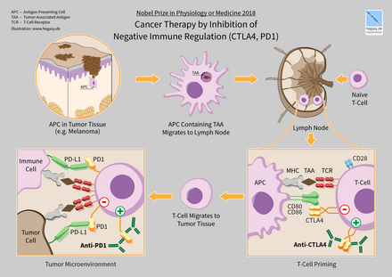
In 2018, American immunologist James P. Allison and Japanese immunologist Tasuku Honjo received the Nobel Prize in Physiology or Medicine for their discovery of cancer therapy by inhibition of negative immune regulation.

History

"During the 17th and 18th centuries, various forms of immunotherapy in cancer became widespread... In the 18th and 19th centuries, septic dressings enclosing ulcerative tumours were used for the treatment of cancer. Surgical wounds were left open to facilitate the development of infection, and purulent sores were created deliberately... One of the most well-known effects of microorganisms on...cancer was reported in 1891, when an American surgeon, William Coley, inoculated patients having inoperable tumours with [ Streptococcus pyogenes ]." "Coley [had] thoroughly reviewed the literature available at that time and found 38 reports of cancer patients with accidental or iatrogenic feverish erysipelas. In 12 patients, the sarcoma or carcinoma had completely disappeared; the others had substantially improved. Coley decided to attempt the therapeutic use of iatrogenic erysipelas…" "Coley developed a toxin that contained heat-killed bacteria [ Streptococcus pyogenes and Serratia marcescens ]. Until 1963, this treatment was used for the treatment of sarcoma." "Coley injected more than 1000 cancer patients with bacteria or bacterial products." 51.9% of [Coley's] patients with inoperable soft-tissue sarcomas showed complete tumour regression and survived for more than 5 years, and 21.2% of the patients had no clinical evidence of tumour at least 20 years after this treatment…" Research continued in the 20th Century under Dr. Maria O'Connor Hornung at Tulane Medical School

Categories

Immunotherapies can be categorized as active or passive. Active immunotherapy specifically targets tumor cells via the immune system. Examples include therapeutic cancer vaccines (also known as treatment vaccines, which are designed to boost the body's immune system to fight cancer) and CAR-T cell, and targeted antibody therapies. In contrast, passive immunotherapy does not directly target tumor cells, but enhances the ability of the immune system to attack cancer cells. Examples include checkpoint inhibitors and cytokines.

Active cellular therapies aim to destroy cancer cells by recognition of distinct markers known as antigens. In cancer vaccines, the goal is to generate an immune response to these antigens through a vaccine. Currently, only one vaccine (sipuleucel-T for prostate cancer) has been approved. In cell-mediated therapies like CAR-T cell therapy, immune cells are extracted from the patient, genetically engineered to recognize tumor specific antigens, and returned to the patient. Cell types that can be used in this way are natural killer (NK) cells, lymphokine-activated killer cells, cytotoxic T cells and dendritic cells. Finally, specific antibodies can be developed that recognize cancer cells and target them for destruction by the immune system. Examples of such antibodies include rituximab (targeting CD-20), trastuzumab (targeting HER-2), and cetuximab (targeting EGFR).

Passive antibody therapies aim to increase the activity of the immune system without specifically targeting cancer cells. For example, cytokines directly stimulate the immune system and increase immune activity. Checkpoint inhibitors target proteins (immune checkpoints) that normally dampen the immune response. This enhances the ability of the immune system to attack cancer cells. Current research is identifying new potential targets to enhance immune function. Approved checkpoint inhibitors include antibodies such as ipilimumab, nivolumab, and pembrolizumab.

Cellular immunotherapy

Dendritic cell therapy

Blood cells are removed from the body, incubated with tumour antigen(s) and activated. Mature dendritic cells are then returned to the original cancer-bearing donor to induce an immune response.

Dendritic cell therapy provokes anti-tumor responses by causing dendritic cells to present tumor antigens to lymphocytes, which activates them, priming them to kill other cells that present the antigen. Dendritic cells are antigen presenting cells (APCs) in the mammalian immune system. In cancer treatment they aid cancer antigen targeting. The only approved cellular cancer therapy based on dendritic cells is sipuleucel-T.

One method of inducing dendritic cells to present tumor antigens is by vaccination with autologous tumor lysates or short peptides (small parts of protein that correspond to the protein antigens on cancer cells). These peptides are often given in combination with adjuvants (highly immunogenic substances) to increase the immune and anti-tumor responses. Other adjuvants include proteins or other chemicals that attract and/or activate dendritic cells, such as granulocyte macrophage colony-stimulating factor (GM-CSF). The most common source of antigens used for dendritic cell vaccine in Glioblastoma (GBM) as an aggressive brain tumor were whole tumor lysate, CMV antigen RNA and tumor associated peptides like EGFRvIII.

Dendritic cells can also be activated in vivo by making tumor cells express GM-CSF. This can be achieved by either genetically engineering tumor cells to produce GM-CSF or by infecting tumor cells with an oncolytic virus that expresses GM-CSF.

Another strategy is to remove dendritic cells from the blood of a patient and activate them outside the body. The dendritic cells are activated in the presence of tumor antigens, which may be a single tumor-specific peptide/protein or a tumor cell lysate (a solution of broken down tumor cells). These cells (with optional adjuvants) are infused and provoke an immune response.

Dendritic cell therapies include the use of antibodies that bind to receptors on the surface of dendritic cells. Antigens can be added to the antibody and can induce the dendritic cells to mature and provide immunity to the tumor. Dendritic cell receptors such as TLR3, TLR7, TLR8 or CD40 have been used as antibody targets. Dendritic cell-NK cell interface also has an important role in immunotherapy. The design of new dendritic cell-based vaccination strategies should also encompass NK cell-stimulating potency. It is critical to systematically incorporate NK cells monitoring as an outcome in antitumor DC-based clinical trials.

Approved drugs

Sipuleucel-T (Provenge) was approved for treatment of asymptomatic or minimally symptomatic metastatic castration-resistant prostate cancer in 2010. The treatment consists of removal of antigen-presenting cells from blood by leukapheresis and growing them with the fusion protein PA2024 made from GM-CSF and prostate-specific prostatic acid phosphatase (PAP) and reinfused. This process is repeated three times.

CAR-T cell therapy

The premise of CAR-T immunotherapy is to modify T cells to recognize cancer cells in order to more effectively target and destroy them. Scientists harvest T cells from people, genetically alter them to add a chimeric antigen receptor (CAR) that specifically recognizes cancer cells, then infuse the resulting CAR-T cells into patients to attack their tumors.

Approved drugs

Tisagenlecleucel (Kymriah), a chimeric antigen receptor (CAR-T) therapy, was approved by FDA in 2017 to treat acute lymphoblastic leukemia (ALL). This treatment removes CD19 positive cells (B-cells) from the body (including the diseased cells, but also normal antibody producing cells).

Axicabtagene ciloleucel (Yescarta) is another CAR-T therapeutic, approved in 2017 for treatment of diffuse large B-cell lymphoma (DLBCL).

Antibody therapy

Many forms of antibodies can be engineered.

Antibodies are a key component of the adaptive immune response, playing a central role in both recognizing foreign antigens and stimulating an immune response. Antibodies are Y-shaped proteins produced by some B cells and are composed of two regions: an antigen-binding fragment (Fab), which binds to antigens, and a Fragment crystallizable (Fc) region, which interacts with so-called Fc receptors that are expressed on the surface of different immune cell types including macrophages, neutrophils and NK cells. Many immunotherapeutic regimens involve antibodies. Monoclonal antibody technology engineers and generates antibodies against specific antigens, such as those present on tumor surfaces. These antibodies that are specific to the antigens of the tumor, can then be injected into a tumor.

Antibody types

Conjugation

Two types are used in cancer treatments:

  • Naked monoclonal antibodies are antibodies without added elements. Most antibody therapies use this antibody type.
  • Conjugated monoclonal antibodies are joined to another molecule, which is either cytotoxic or radioactive. The toxic chemicals are those typically used as chemotherapy drugs, but other toxins can be used. The antibody binds to specific antigens on cancer cell surfaces, directing the therapy to the tumor. Radioactive compound-linked antibodies are referred to as radiolabelled. Chemolabelled or immunotoxins antibodies are tagged with chemotherapeutic molecules or toxins, respectively. Research has also demonstrated conjugation of a TLR agonist to an anti-tumor monoclonal antibody.

Fc regions

Fc's ability to bind Fc receptors is important because it allows antibodies to activate the immune system. Fc regions are varied: they exist in numerous subtypes and can be further modified, for example with the addition of sugars in a process called glycosylation. Changes in the Fc region can alter an antibody's ability to engage Fc receptors and, by extension, will determine the type of immune response that the antibody triggers. For example, immune checkpoint blockers targeting PD-1 are antibodies designed to bind PD-1 expressed by T cells and reactivate these cells to eliminate tumors.[23] Anti-PD-1 drugs contain not only an Fab region that binds PD-1 but also an Fc region. Experimental work indicates that the Fc portion of cancer immunotherapy drugs can affect the outcome of treatment. For example, anti-PD-1 drugs with Fc regions that bind inhibitory Fc receptors can have decreased therapeutic efficacy. Imaging studies have further shown that the Fc region of anti-PD-1 drugs can bind Fc receptors expressed by tumor-associated macrophages. This process removes the drugs from their intended targets (i.e. PD-1 molecules expressed on the surface of T cells) and limits therapeutic efficacy. Furthermore, antibodies targeting the co-stimulatory protein CD40 require engagement with selective Fc receptors for optimal therapeutic efficacy. Together, these studies underscore the importance of Fc status in antibody-based immune checkpoint targeting strategies.

Human/non-human antibodies

Antibodies can come from a variety of sources, including human cells, mice, and a combination of the two (chimeric antibodies). Different sources of antibodies can provoke different kinds of immune responses. For example, the human immune system can recognize mouse antibodies (also known as murine antibodies) and trigger an immune response against them. This could reduce the effectiveness of the antibodies as a treatment and cause an immune reaction. Chimeric antibodies attempt to reduce murine antibodies' immunogenicity by replacing part of the antibody with the corresponding human counterpart. Humanized antibodies are almost completely human; only the complementarity determining regions of the variable regions are derived from murine sources. Human antibodies have been produced using unmodified human DNA.

Antibody-dependent cell-mediated cytotoxicity. When the Fc receptors on natural killer (NK) cells interact with Fc regions of antibodies bound to cancer cells, the NK cell releases perforin and granzyme, leading to cancer cell apoptosis.

Mechanism of Action

Antibody-dependent cell-mediated cytotoxicity (ADCC)

Antibody-dependent cell-mediated cytotoxicity (ADCC) requires antibodies to bind to target cell surfaces. Antibodies are formed of a binding region (Fab) and the Fc region that can be detected by immune system cells via their Fc surface receptors. Fc receptors are found on many immune system cells, including NK cells. When NK cells encounter antibody-coated cells, the latter's Fc regions interact with their Fc receptors, releasing perforin and granzyme B to kill the tumor cell. Examples include Rituximab, Ofatumumab, Elotuzumab, and Alemtuzumab. Antibodies under development have altered Fc regions that have higher affinity for a specific type of Fc receptor, FcγRIIIA, which can dramatically increase effectiveness.

Complement Activation

The complement system includes blood proteins that can cause cell death after an antibody binds to the cell surface (the classical complement pathway, among the ways of complement activation). Generally the system deals with foreign pathogens, but can be activated with therapeutic antibodies in cancer. The system can be triggered if the antibody is chimeric, humanized or human; as long as it contains the IgG1 Fc region. Complement can lead to cell death by activation of the membrane attack complex, known as complement-dependent cytotoxicity; enhancement of antibody-dependent cell-mediated cytotoxicity; and CR3-dependent cellular cytotoxicity. Complement-dependent cytotoxicity occurs when antibodies bind to the cancer cell surface, the C1 complex binds to these antibodies and subsequently protein pores are formed in the cancer cell membrane.

Blocking

Antibody therapies can also function by binding to proteins and physically blocking them from interacting with other proteins. Checkpoint inhibitors (CTLA-4, PD-1, and PD-L1) operate by this mechanism. Briefly, checkpoint inhibitors are proteins that normally help to slow immune responses and prevent the immune system from attacking normal cells. Checkpoint inhibitors bind these proteins and prevent them from functioning normally, which increases the activity of the immune system. Examples include durvalumab, ipilimumab, nivolumab, and pembrolizumab.

FDA-approved antibodies

Cancer immunotherapy:Monoclonal antibodies
Antibody Brand name Type Target Approval date Approved treatment(s)
Alemtuzumab Campath humanized CD52 2001 B-cell chronic lymphocytic leukemia (CLL)
Atezolizumab Tecentriq humanized PD-L1 2016 bladder cancer
Avelumab Bavencio human PD-L1 2017 metastatic Merkel cell carcinoma
Ipilimumab Yervoy human CTLA4 2011 metastatic melanoma
Elotuzumab Empliciti humanized SLAMF7 2015 Multiple myeloma
Ofatumumab Arzerra human CD20 2009 refractory CLL
Nivolumab Opdivo human PD-1 2014 unresectable or metastatic melanoma, squamous non-small cell lung cancer, Renal cell carcinoma, colorectal cancer, hepatocellular carcinoma, classical hodgkin lymphoma
Pembrolizumab Keytruda humanized PD-1 2014 unresectable or metastatic melanoma, squamous non-small cell lung cancer (NSCLC), Hodgkin's lymphoma, Merkel-cell carcinoma (MCC), primary mediastinal B-cell lymphoma (PMBCL), stomach cancer, cervical cancer
Rituximab Rituxan, Mabthera chimeric CD20 1997 non-Hodgkin lymphoma
Durvalumab Imfinzi human PD-L1 2017 bladder cancer non-small cell lung cancer

Alemtuzumab

Alemtuzumab (Campath-1H) is an anti-CD52 humanized IgG1 monoclonal antibody indicated for the treatment of fludarabine-refractory chronic lymphocytic leukemia (CLL), cutaneous T-cell lymphoma, peripheral T-cell lymphoma and T-cell prolymphocytic leukemia. CD52 is found on >95% of peripheral blood lymphocytes (both T-cells and B-cells) and monocytes, but its function in lymphocytes is unknown. It binds to CD52 and initiates its cytotoxic effect by complement fixation and ADCC mechanisms. Due to the antibody target (cells of the immune system) common complications of alemtuzumab therapy are infection, toxicity and myelosuppression.

Durvalumab

Durvalumab (Imfinzi) is a human immunoglobulin G1 kappa (IgG1κ) monoclonal antibody that blocks the interaction of programmed cell death ligand 1 (PD-L1) with the PD-1 and CD80 (B7.1) molecules. Durvalumab is approved for the treatment of patients with locally advanced or metastatic urothelial carcinoma who:

  • have disease progression during or following platinum-containing chemotherapy.
  • have disease progression within 12 months of neoadjuvant or adjuvant treatment with platinum-containing chemotherapy.

On 16 February 2018, the Food and Drug Administration approved durvalumab for patients with unresectable stage III non-small cell lung cancer (NSCLC) whose disease has not progressed following concurrent platinum-based chemotherapy and radiation therapy.

Ipilimumab

Ipilimumab (Yervoy) is a human IgG1 antibody that binds the surface protein CTLA4. In normal physiology T-cells are activated by two signals: the T-cell receptor binding to an antigen-MHC complex and T-cell surface receptor CD28 binding to CD80 or CD86 proteins. CTLA4 binds to CD80 or CD86, preventing the binding of CD28 to these surface proteins and therefore negatively regulates the activation of T-cells.

Active cytotoxic T-cells are required for the immune system to attack melanoma cells. Normally inhibited active melanoma-specific cytotoxic T-cells can produce an effective anti-tumor response. Ipilimumab can cause a shift in the ratio of regulatory T-cells to cytotoxic T-cells to increase the anti-tumor response. Regulatory T-cells inhibit other T-cells, which may benefit the tumor.

Nivolumab

Nivolumab is a human IgG4 antibody that prevents T-cell inactivation by blocking the binding of programmed cell death 1 ligand 1 or programmed cell death 1 ligand 2 (PD-L1 or PD-L2), a protein expressed by cancer cells, with PD-1, a protein found on the surface of activated T-cells. Nivolumab is used in advanced melanoma, metastatic renal cell carcinoma, advanced lung cancer, advanced head and neck cancer, and Hodgkin's lymphoma.

Ofatumumab

Ofatumumab is a second generation human IgG1 antibody that binds to CD20. It is used in the treatment of chronic lymphocytic leukemia (CLL) because the cancerous cells of CLL are usually CD20-expressing B-cells. Unlike rituximab, which binds to a large loop of the CD20 protein, ofatumumab binds to a separate, small loop. This may explain their different characteristics. Compared to rituximab, ofatumumab induces complement-dependent cytotoxicity at a lower dose with less immunogenicity.

Pembrolizumab

As of 2019, pembrolizumab, which blocks PD-1, programmed cell death protein 1, has been used via intravenous infusion to treat inoperable or metastatic melanoma, metastatic non-small cell lung cancer (NSCLC) in certain situations, as a second-line treatment for head and neck squamous cell carcinoma (HNSCC), after platinum-based chemotherapy, and for the treatment of adult and pediatric patients with refractory classic Hodgkin's lymphoma (cHL). It is also indicated for certain patients with urothelial carcinoma, stomach cancer and cervical cancer.

Rituximab

Rituximab is a chimeric monoclonal IgG1 antibody specific for CD20, developed from its parent antibody Ibritumomab. As with ibritumomab, rituximab targets CD20, making it effective in treating certain B-cell malignancies. These include aggressive and indolent lymphomas such as diffuse large B-cell lymphoma and follicular lymphoma and leukemias such as B-cell chronic lymphocytic leukemia. Although the function of CD20 is relatively unknown, CD20 may be a calcium channel involved in B-cell activation. The antibody's mode of action is primarily through the induction of ADCC and complement-mediated cytotoxicity. Other mechanisms include apoptosis and cellular growth arrest. Rituximab also increases the sensitivity of cancerous B-cells to chemotherapy.

Cytokine therapy

Cytokines are proteins produced by many types of cells present within a tumor. They can modulate immune responses. The tumor often employs them to allow it to grow and reduce the immune response. These immune-modulating effects allow them to be used as drugs to provoke an immune response. Two commonly used cytokines are interferons and interleukins.

Interleukin-2 and interferon-α are cytokines, proteins that regulate and coordinate the behavior of the immune system. They have the ability to enhance anti-tumor activity and thus can be used as passive cancer treatments. Interferon-α is used in the treatment of hairy-cell leukaemia, AIDS-related Kaposi's sarcoma, follicular lymphoma, chronic myeloid leukaemia and malignant melanoma. Interleukin-2 is used in the treatment of malignant melanoma and renal cell carcinoma.

Interferon

Interferons are produced by the immune system. They are usually involved in anti-viral response, but also have use for cancer. They fall in three groups: type I (IFNα and IFNβ), type II (IFNγ) and type III (IFNλ). IFNα has been approved for use in hairy-cell leukaemia, AIDS-related Kaposi's sarcoma, follicular lymphoma, chronic myeloid leukaemia and melanoma. Type I and II IFNs have been researched extensively and although both types promote anti-tumor immune system effects, only type I IFNs have been shown to be clinically effective. IFNλ shows promise for its anti-tumor effects in animal models.

Unlike type I IFNs, Interferon gamma is not approved yet for the treatment of any cancer. However, improved survival was observed when Interferon gamma was administrated to patients with bladder carcinoma and melanoma cancers. The most promising result was achieved in patients with stage 2 and 3 of ovarian carcinoma. The in vitro study of IFN-gamma in cancer cells is more extensive and results indicate anti-proliferative activity of IFN-gamma leading to the growth inhibition or cell death, generally induced by apoptosis but sometimes by autophagy.

Interleukin

Interleukins have an array of immune system effects. Interleukin-2 is used in the treatment of malignant melanoma and renal cell carcinoma. In normal physiology it promotes both effector T cells and T-regulatory cells, but its exact mechanism of action is unknown.

Combination immunotherapy

Combining various immunotherapies such as PD1 and CTLA4 inhibitors can enhance anti-tumor response leading to durable responses.

Combining ablation therapy of tumors with immunotherapy enhances the immunostimulating response and has synergistic effects for curative metastatic cancer treatment.

Combining checkpoint immunotherapies with pharmaceutical agents has the potential to improve response, and such combination therapies are a highly investigated area of clinical investigation. Immunostimulatory drugs such as CSF-1R inhibitors and TLR agonists have been particularly effective in this setting.

Polysaccharide-K

Japan's Ministry of Health, Labour and Welfare approved the use of polysaccharide-K extracted from the mushroom, Coriolus versicolor, in the 1980s, to stimulate the immune systems of patients undergoing chemotherapy. It is a dietary supplement in the US and other jurisdictions.

Genetic pre-testing for therapeutic significance

Because of the high cost of many of the immunotherapy medications and the reluctance of medical insurance companies to prepay for their prescriptions various test methods have been proposed, to attempt to forecast the effectiveness of these medications. The detection of PD-L1 protein seemed to be an indication of cancer susceptible to several immunotherapy medications, but research found that both the lack of this protein or its inclusion in the cancerous tissue was inconclusive, due to the little-understood varying quantities of the protein during different times and locations within the infected cells and tissue.

In 2018 some genetic indications such as Tumor Mutational Burden (TMB, the number of mutations within a targeted genetic region in the cancerous cell's DNA), and Microsatellite instability (MSI, the quantity of impaired DNA mismatch leading to probable mutations), have been approved by the FDA as good indicators for the probability of effective treatment of immunotherapy medication for certain cancers, but research is still in progress. The patient prioritization for immunotherapy based on TMB is still highly controversial.

In some cases the FDA has approved genetic tests for medication that is specific to certain genetic markers. For example, the FDA approved BRAF associated medication for metastatic melanoma, to be administered to patients after testing for the BRAF genetic mutation.

Tests of this sort are being widely advertised for general cancer treatment and are expensive. In the past, some genetic testing for cancer treatment has been involved in scams such as the Duke University Cancer Fraud scandal, or claimed to be hoaxes.

Research

Adoptive T-cell therapy

Cancer specific T-cells can be obtained by fragmentation and isolation of tumour infiltrating lymphocytes, or by genetically engineering cells from peripheral blood. The cells are activated and grown prior to transfusion into the recipient (tumor bearer).

Adoptive T cell therapy is a form of passive immunization by the transfusion of T-cells (adoptive cell transfer). They are found in blood and tissue and usually activate when they find foreign pathogens. Specifically they activate when the T-cell's surface receptors encounter cells that display parts of foreign proteins on their surface antigens. These can be either infected cells, or Antigen-presenting cells (APCs). They are found in normal tissue and in tumor tissue, where they are known as tumor infiltrating lymphocytes (TILs). They are activated by the presence of APCs such as dendritic cells that present tumor antigens. Although these cells can attack the tumor, the environment within the tumor is highly immunosuppressive, preventing immune-mediated tumour death.

Multiple ways of producing and obtaining tumour targeted T-cells have been developed. T-cells specific to a tumor antigen can be removed from a tumor sample (TILs) or filtered from blood. Subsequent activation and culturing is performed ex vivo, with the results reinfused. Activation can take place through gene therapy, or by exposing the T cells to tumor antigens.

As of 2014, multiple ACT clinical trials were underway. Importantly, one study from 2018 showed that clinical responses can be obtained in patients with metastatic melanoma resistant to multiple previous immunotherapies.

The first 2 adoptive T-cell therapies, tisagenlecleucel and axicabtagene ciloleucel, were approved by the FDA in 2017.

Another approach is adoptive transfer of haploidentical γδ T cells or NK cells from a healthy donor. The major advantage of this approach is that these cells do not cause GVHD. The disadvantage is frequently impaired function of the transferred cells.

Anti-CD47 therapy

Many tumor cells overexpress CD47 to escape immunosurveilance of host immune system. CD47 binds to its receptor signal regulatory protein alpha (SIRPα) and downregulate phagocytosis of tumor cell. Therefore, anti-CD47 therapy aims to restore clearance of tumor cells. Additionally, growing evidence supports the employment of tumor antigen-specific T cell response in response to anti-CD47 therapy. A number of therapeutics are being developed, including anti-CD47 antibodies, engineered decoy receptors, anti-SIRPα antibodies and bispecific agents. As of 2017, wide range of solid and hematologic malignancies were being clinically tested.

Anti-GD2 antibodies

The GD2 ganglioside

Carbohydrate antigens on the surface of cells can be used as targets for immunotherapy. GD2 is a ganglioside found on the surface of many types of cancer cell including neuroblastoma, retinoblastoma, melanoma, small cell lung cancer, brain tumors, osteosarcoma, rhabdomyosarcoma, Ewing's sarcoma, liposarcoma, fibrosarcoma, leiomyosarcoma and other soft tissue sarcomas. It is not usually expressed on the surface of normal tissues, making it a good target for immunotherapy. As of 2014, clinical trials were underway.

Immune checkpoints

Immune checkpoints in the tumour microenvironment
 
Cancer therapy by inhibition of negative immune regulation (CTLA4, PD1)

Immune checkpoints affect immune system function. Immune checkpoints can be stimulatory or inhibitory. Tumors can use these checkpoints to protect themselves from immune system attacks. Currently approved checkpoint therapies block inhibitory checkpoint receptors. Blockade of negative feedback signaling to immune cells thus results in an enhanced immune response against tumors. Immune checkpoint blockade therapies have varied effectiveness. In Hodgkin lymphoma and natural killer T-cell lymphoma, response rates are high, at 50–60%. Response rates are quite low for breast and prostate cancers, however.

One ligand-receptor interaction under investigation is the interaction between the transmembrane programmed cell death 1 protein (PDCD1, PD-1; also known as CD279) and its ligand, PD-1 ligand 1 (PD-L1, CD274). PD-L1 on the cell surface binds to PD1 on an immune cell surface, which inhibits immune cell activity. Among PD-L1 functions is a key regulatory role on T cell activities. It appears that (cancer-mediated) upregulation of PD-L1 on the cell surface may inhibit T cells that might otherwise attack. PD-L1 on cancer cells also inhibits FAS- and interferon-dependent apoptosis, protecting cells from cytotoxic molecules produced by T cells. Antibodies that bind to either PD-1 or PD-L1 and therefore block the interaction may allow the T-cells to attack the tumor.

CTLA-4 blockade

The first checkpoint antibody approved by the FDA was ipilimumab, approved in 2011 for treatment of melanoma. It blocks the immune checkpoint molecule CTLA-4. Clinical trials have also shown some benefits of anti-CTLA-4 therapy on lung cancer or pancreatic cancer, specifically in combination with other drugs. In on-going trials the combination of CTLA-4 blockade with PD-1 or PD-L1 inhibitors is tested on different types of cancer.

However, patients treated with check-point blockade (specifically CTLA-4 blocking antibodies), or a combination of check-point blocking antibodies, are at high risk of suffering from immune-related adverse events such as dermatologic, gastrointestinal, endocrine, or hepatic autoimmune reactions. These are most likely due to the breadth of the induced T-cell activation when anti-CTLA-4 antibodies are administered by injection in the blood stream.

Using a mouse model of bladder cancer, researchers have found that a local injection of a low dose anti-CTLA-4 in the tumour area had the same tumour inhibiting capacity as when the antibody was delivered in the blood. At the same time the levels of circulating antibodies were lower, suggesting that local administration of the anti-CTLA-4 therapy might result in fewer adverse events.

PD-1 inhibitors

Initial clinical trial results with IgG4 PD1 antibody Nivolumab were published in 2010. It was approved in 2014. Nivolumab is approved to treat melanoma, lung cancer, kidney cancer, bladder cancer, head and neck cancer, and Hodgkin's lymphoma. A 2016 clinical trial for non-small cell lung cancer failed to meet its primary endpoint for treatment in the first line setting, but is FDA approved in subsequent lines of therapy.

Pembrolizumab is another PD1 inhibitor that was approved by the FDA in 2014. Keytruda (Pembrolizumab) is approved to treat melanoma and lung cancer.

Antibody BGB-A317 is a PD-1 inhibitor (designed to not bind Fc gamma receptor I) in early clinical trials.

PD-L1 inhibitors

In May 2016, PD-L1 inhibitor atezolizumab was approved for treating bladder cancer.

Anti-PD-L1 antibodies currently in development include avelumab and durvalumab, in addition to an inhibitory affimer.

CISH

Other modes of enhancing [adoptive] immuno-therapy include targeting so-called intrinsic checkpoint blockades e.g. CISH. Many cancer patients do not respond to immune checkpoint blockade. Response rate may be improved by combining immune checkpoint blockade with additional rationally selected anticancer therapies, including those that stimulate T cell infiltration. For example, targeted therapies such as radiotherapy, vasculature targeting agents, and immunogenic chemotherapy can improve immune checkpoint blockade response in animal models.

Oncolytic virus

An oncolytic virus is a virus that preferentially infects and kills cancer cells. As the infected cancer cells are destroyed by oncolysis, they release new infectious virus particles or virions to help destroy the remaining tumour. Oncolytic viruses are thought not only to cause direct destruction of the tumour cells, but also to stimulate host anti-tumour immune responses for long-term immunotherapy.

The potential of viruses as anti-cancer agents was first realized in the early twentieth century, although coordinated research efforts did not begin until the 1960s. A number of viruses including adenovirus, reovirus, measles, herpes simplex, Newcastle disease virus and vaccinia have now been clinically tested as oncolytic agents. T-Vec is the first FDA-approved oncolytic virus for the treatment of melanoma. A number of other oncolytic viruses are in Phase II-III development.

Polysaccharides

Certain compounds found in mushrooms, primarily polysaccharides, can up-regulate the immune system and may have anti-cancer properties. For example, beta-glucans such as lentinan have been shown in laboratory studies to stimulate macrophage, NK cells, T cells and immune system cytokines and have been investigated in clinical trials as immunologic adjuvants.

Neoantigens

Many tumors express mutations. These mutations potentially create new targetable antigens (neoantigens) for use in T cell immunotherapy. The presence of CD8+ T cells in cancer lesions, as identified using RNA sequencing data, is higher in tumors with a high mutational burden. The level of transcripts associated with cytolytic activity of natural killer cells and T cells positively correlates with mutational load in many human tumors. In non–small cell lung cancer patients treated with lambrolizumab, mutational load shows a strong correlation with clinical response. In melanoma patients treated with ipilimumab, long-term benefit is also associated with a higher mutational load, although less significantly. The predicted MHC binding neoantigens in patients with a long-term clinical benefit were enriched for a series of tetrapeptide motifs that were not found in tumors of patients with no or minimal clinical benefit. However, human neoantigens identified in other studies do not show the bias toward tetrapeptide signatures.

Cancer biomarker

From Wikipedia, the free encyclopedia
 
text
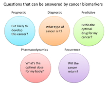
Questions that can be answered by biomarkers

A cancer biomarker refers to a substance or process that is indicative of the presence of cancer in the body. A biomarker may be a molecule secreted by a tumor or a specific response of the body to the presence of cancer. Genetic, epigenetic, proteomic, glycomic, and imaging biomarkers can be used for cancer diagnosis, prognosis, and epidemiology. Ideally, such biomarkers can be assayed in non-invasively collected biofluids like blood or serum.

Cancer is a disease that affects society at a world-wide level. By testing for biomarkers, early diagnosis can be given to prevent deaths.

While numerous challenges exist in translating biomarker research into the clinical space; a number of gene and protein based biomarkers have already been used at some point in patient care; including, AFP (liver cancer), BCR-ABL (chronic myeloid leukemia), BRCA1 / BRCA2 (breast/ovarian cancer), BRAF V600E (melanoma/colorectal cancer), CA-125 (ovarian cancer), CA19.9 (pancreatic cancer), CEA (colorectal cancer), EGFR (Non-small-cell lung carcinoma), HER-2 (Breast Cancer), KIT (gastrointestinal stromal tumor), PSA (prostate specific antigen) (prostate cancer), S100 (melanoma), and many others. Mutant proteins themselves detected by selected reaction monitoring (SRM) have been reported to be the most specific biomarkers for cancers because they can only come from an existing tumor. About 40% of cancers can be cured if detected early through examinations.

Definitions of cancer biomarkers

Organizations and publications vary in their definition of biomarker. In many areas of medicine, biomarkers are limited to proteins identifiable or measurable in the blood or urine. However, the term is often used to cover any molecular, biochemical, physiological, or anatomical property that can be quantified or measured.

The National Cancer Institute (NCI), in particular, defines biomarker as a: “A biological molecule found in blood, other body fluids, or tissues that is a sign of a normal or abnormal process, or of a condition or disease. A biomarker may be used to see how well the body responds to a treatment for a disease or condition. Also called molecular marker and signature molecule."

In cancer research and medicine, biomarkers are used in three primary ways:

  1. To help diagnose conditions, as in the case of identifying early stage cancers (diagnostic)
  2. To forecast how aggressive a condition is, as in the case of determining a patient's ability to fare in the absence of treatment (prognostic)
  3. To predict how well a patient will respond to treatment (predictive)

Role of biomarkers in cancer research and medicine

Uses of biomarkers in cancer medicine

Risk assessment

Cancer biomarkers, particular those associated with genetic mutations or epigenetic alterations, often offer a quantitative way to determine when individuals are predisposed to particular types of cancers. Notable examples of potentially predictive cancer biomarkers include mutations on genes KRAS, p53, EGFR, erbB2 for colorectal, esophageal, liver, and pancreatic cancer; mutations of genes BRCA1 and BRCA2 for breast and ovarian cancer; abnormal methylation of tumor suppressor genes p16, CDKN2B, and p14ARF for brain cancer; hypermethylation of MYOD1, CDH1, and CDH13 for cervical cancer; and hypermethylation of p16, p14, and RB1, for oral cancer.

Diagnosis

Cancer biomarkers can also be useful in establishing a specific diagnosis. This is particularly the case when there is a need to determine whether tumors are of primary or metastatic origin. To make this distinction, researchers can screen the chromosomal alterations found on cells located in the primary tumor site against those found in the secondary site. If the alterations match, the secondary tumor can be identified as metastatic; whereas if the alterations differ, the secondary tumor can be identified as a distinct primary tumor. For example, people with tumors have high levels of circulating tumor DNA (ctDNA) due to tumor cells that have gone through apoptosis. This tumor marker can be detected in the blood, saliva, or urine. The possibility of identifying an effective biomarker for early cancer diagnosis has recently been questioned, in light of the high molecular heterogeneity of tumors observed by next-generation sequencing studies.

Prognosis and treatment predictions

Another use of biomarkers in cancer medicine is for disease prognosis, which take place after an individual has been diagnosed with cancer. Here biomarkers can be useful in determining the aggressiveness of an identified cancer as well as its likelihood of responding to a given treatment. In part, this is because tumors exhibiting particular biomarkers may be responsive to treatments tied to that biomarker's expression or presence. Examples of such prognostic biomarkers include elevated levels of metallopeptidase inhibitor 1 (TIMP1), a marker associated with more aggressive forms of multiple myeloma, elevated estrogen receptor (ER) and/or progesterone receptor (PR) expression, markers associated with better overall survival in patients with breast cancer; HER2/neu gene amplification, a marker indicating a breast cancer will likely respond to trastuzumab treatment; a mutation in exon 11 of the proto-oncogene c-KIT, a marker indicating a gastrointestinal stromal tumor (GIST) will likely respond to imatinib treatment; and mutations in the tyrosine kinase domain of EGFR1, a marker indicating a patient's non-small-cell lung carcinoma (NSCLC) will likely respond to gefitinib or erlotinib treatment.

Pharmacodynamics and pharmacokinetics

Cancer biomarkers can also be used to determine the most effective treatment regime for a particular person's cancer. Because of differences in each person's genetic makeup, some people metabolize or change the chemical structure of drugs differently. In some cases, decreased metabolism of certain drugs can create dangerous conditions in which high levels of the drug accumulate in the body. As such, drug dosing decisions in particular cancer treatments can benefit from screening for such biomarkers. An example is the gene encoding the enzyme thiopurine methyl-transferase (TPMPT). Individuals with mutations in the TPMT gene are unable to metabolize large amounts of the leukemia drug, mercaptopurine, which potentially causes a fatal drop in white blood count for such patients. Patients with TPMT mutations are thus recommended to be given a lower dose of mercaptopurine for safety considerations.

Monitoring treatment response

Cancer biomarkers have also shown utility in monitoring how well a treatment is working over time. Much research is going into this particular area, since successful biomarkers have the potential of providing significant cost reduction in patient care, as the current image-based tests such as CT and MRI for monitoring tumor status are highly costly.

One notable biomarker garnering significant attention is the protein biomarker S100-beta in monitoring the response of malignant melanoma. In such melanomas, melanocytes, the cells that make pigment in our skin, produce the protein S100-beta in high concentrations dependent on the number of cancer cells. Response to treatment is thus associated with reduced levels of S100-beta in the blood of such individuals.

Similarly, additional laboratory research has shown that tumor cells undergoing apoptosis can release cellular components such as cytochrome c, nucleosomes, cleaved cytokeratin-18, and E-cadherin. Studies have found that these macromolecules and others can be found in circulation during cancer therapy, providing a potential source of clinical metrics for monitoring treatment.

Recurrence

Cancer biomarkers can also offer value in predicting or monitoring cancer recurrence. The Oncotype DX® breast cancer assay is one such test used to predict the likelihood of breast cancer recurrence. This test is intended for women with early-stage (Stage I or II), node-negative, estrogen receptor-positive (ER+) invasive breast cancer who will be treated with hormone therapy. Oncotype DX looks at a panel of 21 genes in cells taken during tumor biopsy. The results of the test are given in the form of a recurrence score that indicates likelihood of recurrence at 10 years.

Uses of biomarkers in cancer research

Developing drug targets

In addition to their use in cancer medicine, biomarkers are often used throughout the cancer drug discovery process. For instance, in the 1960s, researchers discovered the majority of patients with chronic myelogenous leukemia possessed a particular genetic abnormality on chromosomes 9 and 22 dubbed the Philadelphia chromosome. When these two chromosomes combine they create a cancer-causing gene known as BCR-ABL. In such patients, this gene acts as the principle initial point in all of the physiological manifestations of the leukemia. For many years, the BCR-ABL was simply used as a biomarker to stratify a certain subtype of leukemia. However, drug developers were eventually able to develop imatinib, a powerful drug that effectively inhibited this protein and significantly decreased production of cells containing the Philadelphia chromosome.

Surrogate endpoints

Another promising area of biomarker application is in the area of surrogate endpoints. In this application, biomarkers act as stand-ins for the effects of a drug on cancer progression and survival. Ideally, the use of validated biomarkers would prevent patients from having to undergo tumor biopsies and lengthy clinical trials to determine if a new drug worked. In the current standard of care, the metric for determining a drug's effectiveness is to check if it has decreased cancer progression in humans and ultimately whether it prolongs survival. However, successful biomarker surrogates could save substantial time, effort, and money if failing drugs could be eliminated from the development pipeline before being brought to clinical trials.

Some ideal characteristics of surrogate endpoint biomarkers include:

  • Biomarker should be involved in process that causes the cancer
  • Changes in biomarker should correlate with changes in the disease
  • Levels of biomarkers should be high enough that they can be measured easily and reliably
  • Levels or presence of biomarker should readily distinguish between normal, cancerous, and precancerous tissue
  • Effective treatment of the cancer should change the level of the biomarker
  • Level of the biomarker should not change spontaneously or in response to other factors not related to the successful treatment of the cancer

Two areas in particular that are receiving attention as surrogate markers include circulating tumor cells (CTCs) and circulating miRNAs. Both these markers are associated with the number of tumor cells present in the blood, and as such, are hoped to provide a surrogate for tumor progression and metastasis. However, significant barriers to their adoption include the difficulty of enriching, identifying, and measuring CTC and miRNA levels in blood. New technologies and research are likely necessary for their translation into clinical care.

Types of cancer biomarkers

Molecular cancer biomarkers

Tumor type Biomarker
Breast ER/PR (estrogen receptor/progesteron receptor)
HER-2/neu
Colorectal EGFR
KRAS
UGT1A1
Gastric HER-2/neu 
GIST c-KIT
Leukemia/lymphoma CD20
CD30
FIP1L1-PDGFRalpha
PDGFR
Philadelphia chromosome (BCR/ABL
PML/RAR-alpha
TPMT
UGT1A1
Lung EML4/ALK
EGFR 
KRAS 
Melanoma BRAF
Pancreas Elevated levels of leucine, isoleucine and valine
Ovaries CA-125

Other examples of biomarkers:

Cancer biomarkers without specificity

Not all cancer biomarkers have to be specific to types of cancer. Some biomarkers found in the circulatory system can be used to determine an abnormal growth of cells present in the body. All these types of biomarkers can be identified through diagnostic blood tests, which is one of the main reasons to get regularly health tested. By getting regularly tested, many health issues such as cancer can be discovered at an early stage, preventing many deaths.

The neutrophil-to-lymphocyte ratio has been shown to be a non-specific determinant for many cancers. This ratio focuses on the activity of two components of the immune system that are involved in inflammatory response which is shown to be higher in presence of malignant tumors. Additionally, basic fibroblast growth factor (bFGF) is a protein that is involved in the proliferation of cells. Unfortunately, it has been shown that in the presence of tumors it is highly active which has led to the conclusion that it may help malignant cells reproduce at faster rates. Research has shown that anti-bFGF antibodies can be used to help treat tumors from many origins. Moreover, insulin-like growth factor (IGF-R) is involved in cell proliferation and growth. It has is possible that it is involved in inhibiting apoptosis, programmed cell death due to some defect. Due to this, the levels of IGF-R can be increased when cancer such as breast, prostate, lung, and colorectum is present.


Biomarker Description Biosensor used
NLR (neutrophil-to-lymphocyte ratio) Elevates with inflammation caused by cancer No
Basic Fibroblast Growth Factor (bFGF) This level increases when a tumor is present, helps with the fast reproduction of tumor cells Electrochemical
Insulin-like Growth Factor (IGF-R) High activity in cancer cells, help reproduction Electrochemical Impedance Spectroscopy Sensor


Maximum life span

From Wikipedia, the free encyclopedia

Maximum life span (or, for humans, maximum reported age at death) is a measure of the maximum amount of time one or more members of a population have been observed to survive between birth and death. The term can also denote an estimate of the maximum amount of time that a member of a given species could survive between birth and death, provided circumstances that are optimal to that member's longevity.

Most living species have at least one upper limit on the number of times the cells of a member can divide. This is called the Hayflick limit, although the number of cell divisions does not strictly control lifespan.

Definition

In animal studies, maximum span is often taken to be the mean life span of the most long-lived 10% of a given cohort. By another definition, however, maximum life span corresponds to the age at which the oldest known member of a species or experimental group has died. Calculation of the maximum life span in the latter sense depends upon the initial sample size.

Maximum life span contrasts with mean life span (average life span, life expectancy), and longevity. Mean life span varies with susceptibility to disease, accident, suicide and homicide, whereas maximum life span is determined by "rate of aging". Longevity refers only to the characteristics of the especially long lived members of a population, such as infirmities as they age or compression of morbidity, and not the specific life span of an individual.

If age x is subtracted from the hypothetical upper limit w for the species and logs are taken, then the resulting variable log(w – x) is normally distributed similarly as all natural quantitative variables resulting from gene expression. This is due to the law of large numbers, the Central Limit Theorem.

In humans

Demographic evidence

The longest living person whose dates of birth and death were verified according to the modern norms of Guinness World Records and the Gerontology Research Group was Jeanne Calment (1875–1997), a French woman who is verified to have lived to 122. Reduction of infant mortality has accounted for most of the increased average life span longevity, but since the 1960s mortality rates among those over 80 years have decreased by about 1.5% per year. "The progress being made in lengthening lifespans and postponing senescence is entirely due to medical and public-health efforts, rising standards of living, better education, healthier nutrition and more salubrious lifestyles." Animal studies suggest that further lengthening of median human lifespan as well as maximum lifespan could be achieved through "calorie restriction mimetic" drugs or by directly reducing food consumption. Although calorie restriction has not been proven to extend the maximum human life span, as of 2014, results in ongoing primate studies have demonstrated that the assumptions derived from rodents are valid in primates as well [Reference: Nature 1 April 2014].

It has been proposed that no fixed theoretical limit to human longevity is apparent today. Studies in the biodemography of human longevity indicate a late-life mortality deceleration law: that death rates level off at advanced ages to a late-life mortality plateau. That is, there is no fixed upper limit to human longevity, or fixed maximal human lifespan. This law was first quantified in 1939, when researchers found that the one-year probability of death at advanced age asymptotically approaches a limit of 44% for women and 54% for men.

However, this evidence depends on the existence of a late-life plateaus and deceleration that can be explained, in humans and other species, by the existence of very rare errors. Age-coding error rates below 1 in 10,000 are sufficient to make artificial late-life plateaus, and errors below 1 in 100,000 can generate late-life mortality deceleration. These error rates cannot be ruled out by examining documents, the standard, because of successful pension fraud, identity theft, forgeries and errors that leave no documentary evidence. This capacity for errors to explain late-life plateaus solves the "fundamental question in aging research is whether humans and other species possess an immutable life-span limit" and suggests that a limit to human life span exists. A theoretical study suggested the maximum human lifespan to be around 125 years using a modified stretched exponential function for human survival curves. The analysis of dynamics of the body mass in human population indicates extremums, which correspond to mean (70–75 years), the commonly accepted maximum (100–110 years) and maximum known (140–160 years) lifespan[citation needed]. In another study, researchers claimed that there exists a maximum lifespan for humans, and that the human maximal lifespan has been declining since the 1990s. A theoretical study also suggested that the maximum human life expectancy at birth is limited by the human life characteristic value δ, which is around 104 years.

The United Nations has undertaken an important Bayesian sensitivity analysis of global population burden based on life expectancy projection at birth in future decades. The 2017 95% prediction interval of 2090 average life expectancy rises as high as +6 (106, in Century Representation Form) by 2090, with dramatic, ongoing, layered consequences on world population and demography should that happen. The prediction interval is extremely wide, and the United Nations can not be certain. Organizations like the Methuselah Foundation are working toward an end to senescence and practically unlimited human lifespan. If successful, the demographic implications for human population will be greater in effective multiplier terms than any experienced in the last five centuries if maximum lifespan or the birthrate remain unlimited by law. Modern Malthusian predictions of overpopulation based on increased longevity have been criticized on the same basis as general population alarmism (see Malthusianism).

Non-demographic evidence

Evidence for maximum lifespan is also provided by the dynamics of physiological indices with age. For example, scientists have observed that a person's VO2max value (a measure of the volume of oxygen flow to the cardiac muscle) decreases as a function of age. Therefore, the maximum lifespan of a person could be determined by calculating when the person's VO2max value drops below the basal metabolic rate necessary to sustain life, which is approximately 3 ml per kg per minute. On the basis of this hypothesis, athletes with a VO2max value between 50 and 60 at age 20 would be expected "to live for 100 to 125 years, provided they maintained their physical activity so that their rate of decline in VO2max remained constant".

Longitudinal variations of physiological indices, as diverse as complete blood counts (CBC) or physical activity records collected by wearable devices, along individual aging trajectories revealed a linear increase of the organism state fluctuations range with age. The broadening could be explained by a progressive loss of physiological resilience measured by the inverse auto-correlation times of the organism state fluctuations. Extrapolation of this data suggested that organism state recovery time and variance could simultaneously diverge at a critical point of 120 – 150 years of age corresponding to a complete loss of resilience and hence should be incompatible with survival. The criticality resulting in the end of life is an intrinsic biological property of an organism that is independent of stress factors and signifies a fundamental or absolute limit of human lifespan

Average and commonly accepted maximum lifespans correspond to the extremums of the body mass (1, 2) and mass normalized to height (3, 4) of men (1, 3) and women (2, 4).

In other animals

Small animals such as birds and squirrels rarely live to their maximum life span, usually dying of accidents, disease or predation.

The maximum life span of most species is documented in the Anage repository.

Maximum life span is usually longer for species that are larger or have effective defenses against predation, such as bird flight, chemical defenses or living in social groups.

The differences in life span between species demonstrate the role of genetics in determining maximum life span ("rate of aging"). The records (in years) are these:

The longest-lived vertebrates have been variously described as

  • Large parrots (macaws and cockatoos can live up to 80–100 years in captivity)
  • Koi (a Japanese species of fish, allegedly living up to 200 years, though generally not exceeding 50 – a specimen named Hanako was reportedly 226 years old upon her death)
  • Tortoises (Galápagos tortoise) (190 years)
  • Tuataras (a New Zealand reptile species, 100–200+ years)
  • Eels, the so-called Brantevik Eel (Swedish: Branteviksålen) is thought to have lived in a water well in southern Sweden since 1859, which makes it over 150 years old. It was reported that it had died in August 2014 at an age of 155.
  • Whales (bowhead whale) (Balaena mysticetus about 200 years)—Although this idea was unproven for a time, recent research has indicated that bowhead whales recently killed still had harpoons in their bodies from about 1890, which, along with analysis of amino acids, has indicated a maximum life span of "177 to 245 years old".
  • Greenland sharks are currently the vertebrate species with the longest known lifespan. An examination of 28 specimens in one study published in 2016 determined by radiocarbon dating that the oldest of the animals that they sampled had lived for about 392 ± 120 years (a minimum of 272 years and a maximum of 512 years). The authors further concluded that the species reaches sexual maturity at about 150 years of age.

Invertebrate species which continue to grow as long as they live (e.g., certain clams, some coral species) can on occasion live hundreds of years:

Exceptions

  • Some jellyfish species, including Turritopsis dohrnii, Laodicea undulata, and Aurelia sp.1, are able to revert to the polyp stage even after reproducing (so-called reversible life cycle), rather than dying as in other jellyfish. Consequently, these species are considered biologically immortal and have no maximum lifespan.
  • There may be no natural limit to the Hydra's life span, but it is not yet clear how to estimate the age of a specimen.
  • Flatworms, or Platyhelminthes, are known to be "almost immortal" as they have a great regeneration capacity, continuous growth and binary fission type cellular division.
  • Lobsters are sometimes said to be biologically immortal because they don't seem to slow down, weaken, or lose fertility with age. However, due to the energy needed for moulting, they cannot live indefinitely.
  • Tardigrades can live indefinitely in a state of suspended animation, a state which they enter when they are not hydrated. In this state, they can withstand an extremely large number of environmental pressures, including intense radioactivity and heat, and being sent into space. Despite this, they can only live in a hydrated state for a few months.

In plants

Plants are referred to as annuals which live only one year, biennials which live two years, and perennials which live longer than that. The longest-lived perennials, woody-stemmed plants such as trees and bushes, often live for hundreds and even thousands of years (one may question whether or not they may die of old age). A giant sequoia, General Sherman is alive and well in its third millennium. A Great Basin Bristlecone Pine called Methuselah is 4,853 years old and the Bristlecone Pine called Prometheus was a little older still, at least 4,844 years (and possibly as old as 5,000 years), when it was cut down in 1964. The oldest known plant (possibly oldest living thing) is a clonal Quaking Aspen (Populus tremuloides) tree colony in the Fishlake National Forest in Utah called Pando at about 80,000 years. Lichen, a symbiotic algae and fungal proto-plant, such as Rhizocarpon geographicum can live upwards of 10,000 years.

Increasing maximum life span

"Maximum life span" here means the mean life span of the most long-lived 10% of a given cohort. Caloric restriction has not yet been shown to break mammalian world records for longevity. Rats, mice, and hamsters experience maximum life-span extension from a diet that contains all of the nutrients but only 40–60% of the calories that the animals consume when they can eat as much as they want. Mean life span is increased 65% and maximum life span is increased 50%, when caloric restriction is begun just before puberty. For fruit flies the life extending benefits of calorie restriction are gained immediately at any age upon beginning calorie restriction and ended immediately at any age upon resuming full feeding.

A few transgenic strains of mice have been created that have maximum life spans greater than that of wild-type or laboratory mice. The Ames and Snell mice, which have mutations in pituitary transcription factors and hence are deficient in Gh, LH, TSH, and secondarily IGF1, have extensions in maximal lifespan of up to 65%. To date, both in absolute and relative terms, these Ames and Snell mice have the maximum lifespan of any mouse not on caloric restriction (see below on GhR). Mutations/knockout of other genes affecting the GH/IGF1 axis, such as Lit, Ghr and Irs1 have also shown extension in lifespan, but much more modest both in relative and absolute terms. The longest lived laboratory mouse ever was a Ghr knockout mouse, which lived to ≈1800 days in the lab of Andrzej Bartke at Southern Illinois University. The maximum for normal B6 mice under ideal conditions is 1200 days.

Most biomedical gerontologists believe that biomedical molecular engineering will eventually extend maximum lifespan and even bring about rejuvenation. Anti-aging drugs are a potential tool for extending life.

Aubrey de Grey, a theoretical gerontologist, has proposed that aging can be reversed by strategies for engineered negligible senescence. De Grey has established The Methuselah Mouse Prize to award money to researchers who can extend the maximum life span of mice. So far, three Mouse Prizes have been awarded: one for breaking longevity records to Dr. Andrzej Bartke of Southern Illinois University (using GhR knockout mice); one for late-onset rejuvenation strategies to Dr. Stephen Spindler of the University of California (using caloric restriction initiated late in life); and one to Dr. Z. Dave Sharp for his work with the pharmaceutical rapamycin.

Correlation with DNA repair capacity

Accumulated DNA damage appears to be a limiting factor in the determination of maximum life span. The theory that DNA damage is the primary cause of aging, and thus a principal determinant of maximum life span, has attracted increased interest in recent years. This is based, in part, on evidence in human and mouse that inherited deficiencies in DNA repair genes often cause accelerated aging. There is also substantial evidence that DNA damage accumulates with age in mammalian tissues, such as those of the brain, muscle, liver and kidney (reviewed by Bernstein et al. and see DNA damage theory of aging and DNA damage (naturally occurring)). One expectation of the theory (that DNA damage is the primary cause of aging) is that among species with differing maximum life spans, the capacity to repair DNA damage should correlate with lifespan. The first experimental test of this idea was by Hart and Setlow who measured the capacity of cells from seven different mammalian species to carry out DNA repair. They found that nucleotide excision repair capability increased systematically with species longevity. This correlation was striking and stimulated a series of 11 additional experiments in different laboratories over succeeding years on the relationship of nucleotide excision repair and life span in mammalian species (reviewed by Bernstein and Bernstein). In general, the findings of these studies indicated a good correlation between nucleotide excision repair capacity and life span. The association between nucleotide excision repair capability and longevity is strengthened by the evidence that defects in nucleotide excision repair proteins in humans and rodents cause features of premature aging, as reviewed by Diderich.

Further support for the theory that DNA damage is the primary cause of aging comes from study of Poly ADP ribose polymerases (PARPs). PARPs are enzymes that are activated by DNA strand breaks and play a role in DNA base excision repair. Burkle et al. reviewed evidence that PARPs, and especially PARP-1, are involved in maintaining mammalian longevity. The life span of 13 mammalian species correlated with poly(ADP ribosyl)ation capability measured in mononuclear cells. Furthermore, lymphoblastoid cell lines from peripheral blood lymphocytes of humans over age 100 had a significantly higher poly(ADP-ribosyl)ation capability than control cell lines from younger individuals.

Research data

  • A comparison of the heart mitochondria in rats (7-year maximum life span) and pigeons (35-year maximum life span) showed that pigeon mitochondria leak fewer free-radicals than rat mitochondria, despite the fact that both animals have similar metabolic rate and cardiac output
  • For mammals there is a direct relationship between mitochondrial membrane fatty acid saturation and maximum life span
  • Studies of the liver lipids of mammals and a bird (pigeon) show an inverse relationship between maximum life span and number of double bonds
  • Selected species of birds and mammals show an inverse relationship between telomere rate of change (shortening) and maximum life span
  • Maximum life span correlates negatively with antioxidant enzyme levels and free-radicals production and positively with rate of DNA repair
  • Female mammals express more Mn−SOD and glutathione peroxidase antioxidant enzymes than males. This has been hypothesized as the reason they live longer However, mice entirely lacking in glutathione peroxidase 1 do not show a reduction in lifespan.
  • The maximum life span of transgenic mice has been extended about 20% by overexpression of human catalase targeted to mitochondria
  • A comparison of 7 non-primate mammals (mouse, hamster, rat, guinea-pig, rabbit, pig and cow) showed that the rate of mitochondrial superoxide and hydrogen peroxide production in heart and kidney were inversely correlated with maximum life span
  • A study of 8 non-primate mammals showed an inverse correlation between maximum life span and oxidative damage to mtDNA (mitochondrial DNA) in heart & brain
  • A study of several species of mammals and a bird (pigeon) indicated a linear relationship between oxidative damage to protein and maximum life span
  • There is a direct correlation between DNA repair and maximum life span for mammalian species
  • Drosophila (fruit-flies) bred for 15 generations by only using eggs that were laid toward the end of reproductive life achieved maximum life spans 30% greater than that of controls
  • Overexpression of the enzyme which synthesizes glutathione in long-lived transgenic Drosophila (fruit-flies) extended maximum lifespan by nearly 50%
  • A mutation in the age−1 gene of the nematode worm Caenorhabditis elegans increased mean life span 65% and maximum life span 110%. However, the degree of lifespan extension in relative terms by both the age-1 and daf-2 mutations is strongly dependent on ambient temperature, with ≈10% extension at 16 °C and 65% extension at 27 °C.
  • Fat-specific Insulin Receptor KnockOut (FIRKO) mice have reduced fat mass, normal calorie intake and an increased maximum life span of 18%.
  • The capacity of mammalian species to detoxify the carcinogenic chemical benzo(a)pyrene to a water-soluble form also correlates well with maximum life span.
  • Short-term induction of oxidative stress due to calorie restriction increases life span in Caenorhabditis elegans by promoting stress defense, specifically by inducing an enzyme called catalase. As shown by Michael Ristow and co-workers nutritive antioxidants completely abolish this extension of life span by inhibiting a process called mitohormesis.


Operator (computer programming)

From Wikipedia, the free encyclopedia https://en.wikipedia.org/wiki/Operator_(computer_programmin...