Search This Blog

Saturday, December 11, 2021

Haematopoiesis

From Wikipedia, the free encyclopedia
 
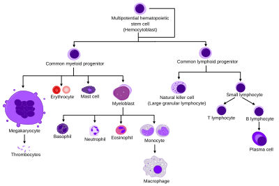
Diagram showing the development of different blood cells from haematopoietic stem cell to mature cells

Haematopoiesis (/hɪˌmætpɔɪˈsɪs, ˈhmət-, ˌhɛmə-/, from Greek αἷμα, 'blood' and ποιεῖν 'to make'; also hematopoiesis in American English; sometimes also h(a)emopoiesis) is the formation of blood cellular components. All cellular blood components are derived from haematopoietic stem cells. In a healthy adult person, approximately 1011–1012 new blood cells are produced daily in order to maintain steady state levels in the peripheral circulation.

Process

Haematopoietic stem cells (HSCs)

Haematopoietic stem cells (HSCs) reside in the medulla of the bone (bone marrow) and have the unique ability to give rise to all of the different mature blood cell types and tissues. HSCs are self-renewing cells: when they differentiate, at least some of their daughter cells remain as HSCs, so the pool of stem cells is not depleted. This phenomenon is called asymmetric division. The other daughters of HSCs (myeloid and lymphoid progenitor cells) can follow any of the other differentiation pathways that lead to the production of one or more specific types of blood cell, but cannot renew themselves. The pool of progenitors is heterogeneous and can be divided into two groups; long-term self-renewing HSC and only transiently self-renewing HSC, also called short-terms. This is one of the main vital processes in the body.

Cell types

All blood cells are divided into three lineages.

Granulopoiesis (or granulocytopoiesis) is haematopoiesis of granulocytes, except of mast cells which are granulocytes but with an extramedullar maturation.

Megakaryocytopoiesis is haematopoiesis of megakaryocytes.

Terminology

Between 1948 and 1950, the Committee for Clarification of the Nomenclature of Cells and Diseases of the Blood and Blood-forming Organs issued reports on the nomenclature of blood cells. An overview of the terminology is shown below, from earliest to final stage of development:

  • [root]blast
  • pro[root]cyte
  • [root]cyte
  • meta[root]cyte
  • mature cell name

The root for erythrocyte colony-forming units (CFU-E) is "rubri", for granulocyte-monocyte colony-forming units (CFU-GM) is "granulo" or "myelo" and "mono", for lymphocyte colony-forming units (CFU-L) is "lympho" and for megakaryocyte colony-forming units (CFU-Meg) is "megakaryo". According to this terminology, the stages of red blood cell formation would be: rubriblast, prorubricyte, rubricyte, metarubricyte, and erythrocyte. However, the following nomenclature seems to be, at present, the most prevalent:

Committee "lympho" "rubri" "granulo" or "myelo" "mono" "megakaryo"
Lineage Lymphoid Myeloid Myeloid Myeloid Myeloid
CFU CFU-L CFU-GEMMCFU-E CFU-GEMM→CFU-GMCFU-G CFU-GEMM→CFU-GMCFU-M CFU-GEMM→CFU-Meg
Process lymphocytopoiesis erythropoiesis granulocytopoiesis monocytopoiesis thrombocytopoiesis
[root]blast Lymphoblast Proerythroblast Myeloblast Monoblast Megakaryoblast
pro[root]cyte Prolymphocyte Polychromatophilic erythrocyte Promyelocyte Promonocyte Promegakaryocyte
[root]cyte Normoblast Eosino/neutro/basophilic myelocyte
Megakaryocyte
meta[root]cyte Large lymphocyte Reticulocyte Eosinophilic/neutrophilic/basophilic metamyelocyte, Eosinophilic/neutrophilic/basophilic band cell Early monocyte -
mature cell name Small lymphocyte Erythrocyte granulocytes (Eosino/neutro/basophil) Monocyte thrombocytes (Platelets)

Osteoclasts also arise from hemopoietic cells of the monocyte/neutrophil lineage, specifically CFU-GM.

Location

Sites of haematopoesis (human) in pre- and postnatal periods

In developing embryos, blood formation occurs in aggregates of blood cells in the yolk sac, called blood islands. As development progresses, blood formation occurs in the spleen, liver and lymph nodes. When bone marrow develops, it eventually assumes the task of forming most of the blood cells for the entire organism. However, maturation, activation, and some proliferation of lymphoid cells occurs in the spleen, thymus, and lymph nodes. In children, haematopoiesis occurs in the marrow of the long bones such as the femur and tibia. In adults, it occurs mainly in the pelvis, cranium, vertebrae, and sternum.

Extramedullary

In some cases, the liver, thymus, and spleen may resume their haematopoietic function, if necessary. This is called extramedullary haematopoiesis. It may cause these organs to increase in size substantially. During fetal development, since bones and thus the bone marrow develop later, the liver functions as the main haematopoetic organ. Therefore, the liver is enlarged during development. Extramedullary hematopoiesis and myelopoiesis may supply leukocytes in cardiovascular disease and inflammation during adulthood. Splenic macrophages and adhesion molecules may be involved in regulation of extramedullary myeloid cell generation in cardiovascular disease.

Maturation

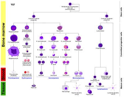
More detailed and comprehensive diagram that shows the development of different blood cells in humans.
  • The morphological characteristics of the hematopoietic cells are shown as seen in a Wright’s stain, May-Giemsa stain or May-Grünwald-Giemsa stain. Alternative names of certain cells are indicated between parentheses.
  • Certain cells may have more than one characteristic appearance. In these cases, more than one representation of the same cell has been included.
  • Together, the monocyte and the lymphocytes comprise the agranulocytes, as opposed to the granulocytes (basophil, neurtophil and eosinophil) that are produced during granulopoiesis.
  • B., N. and E. stand for Basophilic, Neutrophilic and Eosinophilic, respectively – as in Basophilic promyelocyte. For lymphocytes, the T and B are actual designations.
  1. The polychromatic erythrocyte (reticulocyte) at the right shows its characteristic appearance when stained with methylene blue or Azure B.
  2. The erythrocyte at the right is a more accurate representation of its appearance in reality when viewed through a microscope.
  3. Other cells that arise from the monocyte: osteoclast, microglia (central nervous system), Langerhans cell (epidermis), Kupffer cell (liver).
  4. For clarity, the T and B lymphocyte are split to better indicate that the plasma cell arises from the B-cell. Note that there is no difference in the appearance of B- and T-cells unless specific staining is applied.

As a stem cell matures it undergoes changes in gene expression that limit the cell types that it can become and moves it closer to a specific cell type (cellular differentiation). These changes can often be tracked by monitoring the presence of proteins on the surface of the cell. Each successive change moves the cell closer to the final cell type and further limits its potential to become a different cell type.

Cell fate determination

Two models for hematopoiesis have been proposed: determinism and stochastic theory. For the stem cells and other undifferentiated blood cells in the bone marrow, the determination is generally explained by the determinism theory of haematopoiesis, saying that colony stimulating factors and other factors of the haematopoietic microenvironment determine the cells to follow a certain path of cell differentiation. This is the classical way of describing haematopoiesis. In stochastic theory, undifferentiated blood cells differentiate to specific cell types by randomness. This theory has been supported by experiments showing that within a population of mouse haematopoietic progenitor cells, underlying stochastic variability in the distribution of Sca-1, a stem cell factor, subdivides the population into groups exhibiting variable rates of cellular differentiation. For example, under the influence of erythropoietin (an erythrocyte-differentiation factor), a subpopulation of cells (as defined by the levels of Sca-1) differentiated into erythrocytes at a sevenfold higher rate than the rest of the population. Furthermore, it was shown that if allowed to grow, this subpopulation re-established the original subpopulation of cells, supporting the theory that this is a stochastic, reversible process. Another level at which stochasticity may be important is in the process of apoptosis and self-renewal. In this case, the haematopoietic microenvironment prevails upon some of the cells to survive and some, on the other hand, to perform apoptosis and die. By regulating this balance between different cell types, the bone marrow can alter the quantity of different cells to ultimately be produced.

Growth factors

Diagram including some of the important cytokines that determine which type of blood cell will be created. SCF= Stem cell factor; Tpo= Thrombopoietin; IL= Interleukin; GM-CSF= Granulocyte Macrophage-colony stimulating factor; Epo= Erythropoietin; M-CSF= Macrophage-colony stimulating factor; G-CSF= Granulocyte-colony stimulating factor; SDF-1= Stromal cell-derived factor-1; FLT-3 ligand= FMS-like tyrosine kinase 3 ligand; TNF-a = Tumour necrosis factor-alpha; TGFβ = Transforming growth factor beta

Red and white blood cell production is regulated with great precision in healthy humans, and the production of leukocytes is rapidly increased during infection. The proliferation and self-renewal of these cells depend on growth factors. One of the key players in self-renewal and development of haematopoietic cells is stem cell factor (SCF), which binds to the c-kit receptor on the HSC. Absence of SCF is lethal. There are other important glycoprotein growth factors which regulate the proliferation and maturation, such as interleukins IL-2, IL-3, IL-6, IL-7. Other factors, termed colony-stimulating factors (CSFs), specifically stimulate the production of committed cells. Three CSFs are granulocyte-macrophage CSF (GM-CSF), granulocyte CSF (G-CSF) and macrophage CSF (M-CSF). These stimulate granulocyte formation and are active on either progenitor cells or end product cells.

Erythropoietin is required for a myeloid progenitor cell to become an erythrocyte. On the other hand, thrombopoietin makes myeloid progenitor cells differentiate to megakaryocytes (thrombocyte-forming cells). The diagram to the right provides examples of cytokines and the differentiated blood cells they give rise to.

Transcription factors

Growth factors initiate signal transduction pathways, which lead to activation of transcription factors. Growth factors elicit different outcomes depending on the combination of factors and the cell's stage of differentiation. For example, long-term expression of PU.1 results in myeloid commitment, and short-term induction of PU.1 activity leads to the formation of immature eosinophils. Recently, it was reported that transcription factors such as NF-κB can be regulated by microRNAs (e.g., miR-125b) in haematopoiesis.

The first key player of differentiation from HSC to a multipotent progenitor (MPP) is transcription factor CCAAT-enhancer binding protein α (C/EBPα). Mutations in C/EBPα are associated with acute myeloid leukaemia. From this point, cells can either differentiate along the Erythroid-megakaryocyte lineage or lymphoid and myeloid lineage, which have common progenitor, called lymphoid-primed multipotent progenitor. There are two main transcription factors. PU.1 for Erythroid-megakaryocyte lineage and GATA-1, which leads to a lymphoid-primed multipotent progenitor.

Other transcription factors include Ikaros (B cell development), and Gfi1 (promotes Th2 development and inhibits Th1) or IRF8 (basophils and mast cells). Significantly, certain factors elicit different responses at different stages in the haematopoiesis. For example, CEBPα in neutrophil development or PU.1 in monocytes and dendritic cell development. It is important to note that processes are not unidirectional: differentiated cells may regain attributes of progenitor cells.

An example is PAX5 factor, which is important in B cell development and associated with lymphomas. Surprisingly, pax5 conditional knock out mice allowed peripheral mature B cells to de-differentiate to early bone marrow progenitors. These findings show that transcription factors act as caretakers of differentiation level and not only as initiators.

Mutations in transcription factors are tightly connected to blood cancers, as acute myeloid leukemia (AML) or acute lymphoblastic leukemia (ALL). For example, Ikaros is known to be regulator of numerous biological events. Mice with no Ikaros lack B cells, Natural killer and T cells. Ikaros has six zinc fingers domains, four are conserved DNA-binding domain and two are for dimerization. Very important finding is, that different zinc fingers are involved in binding to different place in DNA and this is the reason for pleiotropic effect of Ikaros and different involvement in cancer, but mainly are mutations associated with BCR-Abl patients and it is bad prognostic marker.

Other animals

In some vertebrates, haematopoiesis can occur wherever there is a loose stroma of connective tissue and slow blood supply, such as the gut, spleen or kidney.
Prenatal development

From Wikipedia, the free encyclopedia

Prenatal development (from Latin natalis 'relating to birth') includes the development of the embryo and of the foetus during a viviparous animal's gestation. Prenatal development starts with fertilization, in the germinal stage of embryonic development, and continues in fetal development until birth.

In human pregnancy, prenatal development is also called antenatal development. The development of the human embryo follows fertilization, and continues as fetal development. By the end of the tenth week of gestational age the embryo has acquired its basic form and is referred to as a fetus. The next period is that of fetal development where many organs become fully developed. This fetal period is described both topically (by organ) and chronologically (by time) with major occurrences being listed by gestational age.

The very early stages of embryonic development are the same in all mammals. Later stages of development across all taxa of animals and the length of gestation vary.

Terminology

In the human:

Stages during pregnancy. Embryonic development is marked in green. Weeks and months are numbered by gestation.

Different terms are used to describe prenatal development, meaning development before birth. A term with the same meaning is the "antepartum" (from Latin ante "before" and parere "to give birth") Sometimes "antepartum" is however used to denote the period between the 24th/26th week of gestational age until birth, for example in antepartum hemorrhage.

The perinatal period (from Greek peri, "about, around" and Latin nasci "to be born") is "around the time of birth". In developed countries and at facilities where expert neonatal care is available, it is considered from 22 completed weeks (usually about 154 days) of gestation (the time when birth weight is normally 500 g) to 7 completed days after birth. In many of the developing countries the starting point of this period is considered 28 completed weeks of gestation (or weight more than 1000 g).

Fertilization

Fertilization marks the first germinal stage of embryonic development. When semen is released into the vagina, the spermatozoa travel through the cervix and body of the uterus and into the fallopian tubes where fertilization usually takes place. Many sperm cells are released with the possibility of just one managing to adhere to and enter the thick protective layer surrounding the egg cell (ovum). The first sperm cell to successfully penetrate the egg cell donates its genetic material (DNA) to combine with the DNA of the egg cell resulting in a new organism called the zygote. The term "conception" refers variably to either fertilization or to formation of the conceptus after its implantation in the uterus, and this terminology is controversial.

The zygote will develop into a male if the egg is fertilized by a sperm that carries a Y chromosome, or a female if the sperm carries an X chromosome. The Y chromosome contains a gene, SRY, which will switch on androgen production at a later stage leading to the development of a male body type. In contrast, the mitochondrial DNA of the zygote comes entirely from the egg cell.

Development of the embryo

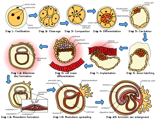
The initial stages of human embryogenesis.

Following fertilization, the embryonic stage of development continues until the end of the 10th week (gestational age) (8th week fertilization age). The first two weeks from fertilization is also referred to as the germinal stage or preembryonic stage.

The zygote spends the next few days traveling down the fallopian tube dividing several times to form a ball of cells called a morula. Further cellular division is accompanied by the formation of a small cavity between the cells. This stage is called a blastocyst. Up to this point there is no growth in the overall size of the embryo, as it is confined within a glycoprotein shell, known as the zona pellucida. Instead, each division produces successively smaller cells.

The blastocyst reaches the uterus at roughly the fifth day after fertilization. It is here that lysis of the zona pellucida occurs. This process is analogous to zona hatching, a term that refers to the emergence of the blastocyst from the zona pellucida, when incubated in vitro. This allows the trophectoderm cells of the blastocyst to come into contact with, and adhere to, the endometrial cells of the uterus. The trophectoderm will eventually give rise to extra-embryonic structures, such as the placenta and the membranes. The embryo becomes embedded in the endometrium in a process called implantation. In most successful pregnancies, the embryo implants 8 to 10 days after ovulation. The embryo, the extra-embryonic membranes, and the placenta are collectively referred to as a conceptus, or the "products of conception".

Rapid growth occurs and the embryo's main features begin to take form. This process is called differentiation, which produces the varied cell types (such as blood cells, kidney cells, and nerve cells). A spontaneous abortion, or miscarriage, in the first trimester of pregnancy is usually due to major genetic mistakes or abnormalities in the developing embryo. During this critical period (most of the first trimester), the developing embryo is also susceptible to toxic exposures, such as:

Nutrition

The embryo passes through 3 phases of acquisition of nutrition from the mother:

  1. Absorption phase: Zygote is nourished by cellular cytoplasm and secretions in fallopian tubes and uterine cavity.
  2. Histoplasmic transfer: After nidation and before establishment of uteroplacental circulation, embryonic nutrition is derived from decidual cells and maternal blood pools that open up as a result of eroding activity of trophoblasts.
  3. Hematotrophic phase: After third week of gestation, substances are transported passively via intervillous space.

Development of the fetus

Fetal development is the third of the three stages of prenatal development, following from the initial germinal stage (preembryonic stage), and stage of embryonic development. These stages are also referred to in pregnancy as terms or trimesters.

From the 10th week of gestation (8th week of development), the developing organism is called a fetus.

All major structures are already formed in the fetus, but they continue to grow and develop. Since the precursors of all the major organs are created by this time, the fetal period is described both by organ and by a list of changes by weeks of gestational age.

Because the precursors of the organs are now formed, the fetus is not as sensitive to damage from environmental exposure as the embryo was. Instead, toxic exposure often causes physiological abnormalities or minor congenital malformation.

Development of organ systems

Development continues throughout the life of the fetus and through into life after birth. Significant changes occur to many systems in the period after birth as they adapt to life outside the uterus.

Fetal blood

Hematopoiesis first takes place in the yolk sac. The function is transferred to the liver by the 10th week of gestation and to the spleen and bone marrow beyond that. The total blood volume is about 125 ml/kg of fetal body weight near term.

Red blood cells

Megaloblastic red blood cells are produced early in development, which become normoblastic near term. Life span of prenatal RBCs is 80 days. Rh antigen appears at about 40 days of gestation.

White blood cells

The fetus starts producing leukocytes at 2 months gestational age, mainly from the thymus and the spleen. Lymphocytes derived from the thymus are called T lymphocytes (T cells), whereas those derived from bone marrow are called B lymphocytes (B cells). Both of these populations of lymphocytes have short-lived and long-lived groups. Short-lived T cells usually reside in thymus, bone marrow and spleen; whereas long-lived T cells reside in the blood stream. Plasma cells are derived from B cells and their life in fetal blood is 0.5 to 2 days.

Glands

The thyroid is the first gland to develop in the embryo at the 4th week of gestation. Insulin secretion in the fetus starts around the 12th week of gestation.

Cognitive development

Initial knowledge of the effects of prenatal experience on later neuropsychological development originates from the Dutch Famine Study, which researched the cognitive development of individuals born after the Dutch famine of 1944–45. The first studies focused on the consequences of the famine to cognitive development, including the prevalence of intellectual disability. Such studies predate David Barker's hypothesis about the association between the prenatal environment and the development of chronic conditions later in life. The initial studies found no association between malnourishment and cognitive development, but later studies found associations between malnourishment and increased risk for schizophrenia, antisocial disorders, and affective disorders.

There is evidence that the acquisition of language begins in the prenatal stage. After 26 weeks of gestation, the peripheral auditory system is already fully formed. Also, most low-frequency sounds (less than 300 Hz) can reach the fetal inner ear in the womb of mammals. Those low-frequency sounds include pitch, rhythm, and phonetic information related to language. Studies have indicated that fetuses react to and recognize differences between sounds. Such ideas are further reinforced by the fact that newborns present a preference for their mother's voice, present behavioral recognition of stories only heard during gestation, and (in monolingual mothers) present preference for their native language. A more recent study with EEG demonstrated different brain activation in newborns hearing their native language compared to when they were presented with a different language, further supporting the idea that language learning starts while in gestation.

Growth rate

Growth rate of fetus is linear up to 37 weeks of gestation, after which it plateaus. The growth rate of an embryo and infant can be reflected as the weight per gestational age, and is often given as the weight put in relation to what would be expected by the gestational age. A baby born within the normal range of weight for that gestational age is known as appropriate for gestational age (AGA). An abnormally slow growth rate results in the infant being small for gestational age, and, on the other hand, an abnormally large growth rate results in the infant being large for gestational age. A slow growth rate and preterm birth are the two factors that can cause a low birth weight. Low birth weight (below 2000 grams) can slightly increase the likelihood of schizophrenia.

The growth rate can be roughly correlated with the fundal height which can be estimated by abdominal palpation. More exact measurements can be performed with obstetric ultrasonography.

Factors influencing development

Intrauterine growth restriction is one of the causes of low birth weight associated with over half of neonatal deaths.

Poverty

Poverty has been linked to poor prenatal care and has been an influence on prenatal development. Women in poverty are more likely to have children at a younger age, which results in low birth weight. Many of these expecting mothers have little education and are therefore less aware of the risks of smoking, drinking alcohol, and drug use – other factors that influence the growth rate of a fetus.

Mother's age

Women between the ages of 16 and 35 have a healthier environment for a fetus than women under 16 or over 35. Women between this age gap are more likely to have fewer complications. Women over 35 are more inclined to have a longer labor period, which could potentially result in death of the mother or fetus. Women under 16 and over 35 have a higher risk of preterm labor (premature baby), and this risk increases for women in poverty, women who take drugs, and women who smoke. Young mothers are more likely to engage in high risk behaviors, such as using alcohol, drugs, or smoking, resulting in negative consequences for the fetus. Premature babies from young mothers are more likely to have neurological defects that will influence their coping capabilities – irritability, trouble sleeping, constant crying for example. There is an increased risk of Down syndrome for infants born to those aged over 40 years. Young teenaged mothers (younger than 16) and mothers over 35 are more exposed to the risks of miscarriages, premature births, and birth defects.

Drug use

An estimated 5 percent of fetuses in the United States are exposed to illicit drug use during pregnancy. Maternal drug use occurs when drugs ingested by the pregnant woman are metabolized in the placenta and then transmitted to the fetus. Resent research display that there is a correlation between fine motor skills and prenatal risk factors such as the use of psychoactive substances and signs of abortion during pregnancy. As well as perinatal risk factors such as gestation time, duration of delivery, birth weight and postnatal risk factors such as constant falls.

Cannabis

When using cannabis, there is a greater risk of birth defects, low birth weight, and a higher rate of death in infants or stillbirths. Drug use will influence extreme irritability, crying, and risk for SIDS once the fetus is born. Marijuana will slow the fetal growth rate and can result in premature delivery. It can also lead to low birth weight, a shortened gestational period and complications in delivery. Cannabis use during pregnancy was unrelated to risk of perinatal death or need for special care, but, the babies of women who used cannabis at least once per week before and throughout pregnancy were 216g lighter than those of non‐users, had significantly shorter birth lengths and smaller head circumferences.

Opioids

Opioids including heroin will cause interrupted fetal development, stillbirths, and can lead to numerous birth defects. Heroin can also result in premature delivery, creates a higher risk of miscarriages, result in facial abnormalities and head size, and create gastrointestinal abnormalities in the fetus. There is an increased risk for SIDS, dysfunction in the central nervous system, and neurological dysfunctions including tremors, sleep problems, and seizures. The fetus is also put at a great risk for low birth weight and respiratory problems.

Cocaine

Cocaine use results in a smaller brain, which results in learning disabilities for the fetus. Cocaine puts the fetus at a higher risk of being stillborn or premature. Cocaine use also results in low birthweight, damage to the central nervous system, and motor dysfunction. The vasoconstriction of the effects of cocaine lead to a decrease in placental blood flow to the fetus that results in fetal hypoxia that is oxygen deficiency and decreased fetal nutrition these vasoconstrictive effects on the placenta have been linked to the number of complications in malformations that are evident in the newborn. 

Methamphetamine

Prenatal methamphetamine exposure has shown to negatively impact brain development and behavioral functioning. A 2019 study further investigated neurocognitive and neurodevelopmental effects of prenatal methamphetamine exposure. This study had two groups, one containing children who were prenatally exposed to methamphetamine but no other illicit drugs and one containing children who met diagnosis criteria for ADHD but were not prenatally exposed to any illicit substance. Both groups of children completed intelligence measures to compute an IQ. Study results showed that the prenatally exposed children performed lower on the intelligence measures than their non-exposed peers with ADHD. The study results also suggest that prenatal exposure to methamphetamine may negatively impact processing speed as children develop.

Alcohol

Maternal alcohol use leads to disruptions of the fetus's brain development, interferes with the fetus's cell development and organization, and affects the maturation of the central nervous system. Even small amounts of alcohol use can cause lower height, weight and head size at birth and higher aggressiveness and lower intelligence during childhood. Fetal alcohol spectrum disorder is a developmental disorder that is a consequence of heavy alcohol intake by the mother during pregnancy. Children with FASD have a variety of distinctive facial features, heart problems, and cognitive problems such as developmental disabilities, attention difficulties, and memory deficits.

Tobacco use

Tobacco smoking during pregnancy exposes the fetus to nicotine, tar, and carbon monoxide. Nicotine results in less blood flow to the fetus because it constricts the blood vessels. Carbon monoxide reduces the oxygen flow to the fetus. The reduction of blood and oxygen flow may result in miscarriage, stillbirth, low birth weight, and premature births. Exposure to secondhand smoke leads to higher risks of low birth weight and childhood cancer.

Infections

If a mother is infected with a disease, the placenta cannot always filter out the pathogens. Viruses such as rubella, chicken pox, mumps, herpes, and human immunodeficiency virus (HIV) are associated with an increased risk of miscarriage, low birth weight, prematurity, physical malformations, and intellectual disabilities. HIV can lead to acquired immune deficiency syndrome (AIDS). Untreated HIV carries a risk of between 10 and 20 per cent of being passed on to the fetus. Bacterial or parasitic diseases may also be passed on to the fetus, and include chlamydia, syphilis, tuberculosis, malaria, and commonly toxoplasmosis. Toxoplasmosis can be acquired through eating infected undercooked meat or contaminated food, and by drinking contaminated water. The risk of fetal infection is lowest during early pregnancy, and highest during the third trimester. However, in early pregnancy the outcome is worse, and can be fatal.

Maternal nutrition

Adequate nutrition is needed for a healthy fetus. Mothers who gain less than 20 pounds during pregnancy are at increased risk for having a preterm or low birth weight infant. Iron and iodine are especially important during prenatal development. Mothers who are deficient in iron are at risk for having a preterm or low birth weight infant. Iodine deficiencies increase the risk of miscarriage, stillbirth, and fetal brain abnormalities Adequate prenatal care gives an improved result in the newborn.

Low birth weight

Low birth weight increases an infants risk of long-term growth and cognitive and language deficits. It also results in a shortened gestational period and can lead to prenatal complications.

Stress

Stress during pregnancy can impact the development of the embryo. Reilly (2017) states that stress can come from many forms of life events such as community, family, financial issues, and natural causes. While a woman is pregnant, stress from outside sources can take a toll on the growth in the womb that may affect the child's learning and relationships when born. For instance, they may have behavioral problems and might be antisocial. The stress that the mother experiences affects the fetus and the fetus' growth which can include the fetus' nervous system (Reilly, 2017). Stress can also lead to low birth weight. Even after avoiding other factors like alcohol, drugs, and being healthy, stress can have its impacts whether families know it or not. Many women who deal with maternal stress do not seek treatment. Similar to stress, Reilly stated that in recent studies, researchers have found that pregnant women who show depressive symptoms are not as attached and bonded to their child while it is in the womb (2017).

Environmental toxins

Exposure to environmental toxins in pregnancy lead to higher rates of miscarriage, sterility, and birth defects. Toxins include fetal exposure to lead, mercury, and ethanol or hazardous environments. Prenatal exposure to mercury may lead to physical deformation, difficulty in chewing and swallowing, and poor motoric coordination. Exposure to high levels of lead prenatally is related to prematurity, low birth weight, brain damage, and a variety of physical defects. Exposure to persistent air pollution from traffic and smog may lead to reduced infant head size, low birth weight, increased infant death rates, impaired lung and immune system development.

Prenatal memory

From Wikipedia, the free encyclopedia

Prenatal memory, also called fetal memory, is important for the development of memory in humans. Many factors can impair fetal memory and its functions, primarily maternal actions. There are multiple techniques available not only to demonstrate the existence of fetal memory but to measure it. Fetal memory is vulnerable to certain diseases so much so that exposure can permanently damage the development of the fetus and even terminate the pregnancy by aborting the fetus. Maternal nutrition and the avoidance of drugs, alcohol and other substances during all nine months of pregnancy (especially the critical period when the nervous system is developing) is important to the development of the fetus and its memory systems. The use of certain substances can entail long-term permanent effects on the fetus that can carry on throughout their lifespan.

Background information and functions

Fetal memory is integral to mother-infant attachment.

There is some evidence that fetal memory may begin within the second trimester after conception. Substantial evidence for fetal memories has been found at around 30 weeks after conception. Fetal memory is important for parental recognition, and facilitates the bond between child and parents. One of the most important types of memory is that which stores information contributing to the maternal bond between infant and mother. This form of memory is important for a type of development known as attachment. Fetal memory is thus critical to the survival of the fetus both prenatally (in the womb) and after birth as an infant.

Development

The Central Nervous System (CNS) and memory in the fetus develop from the ectoderm following fertilization via a process called neurulation. The ectoderm is the outermost layer of the embryo. This happens towards the end of the third week of gestation (time period when the embryo is carried in the women's uterus) and ends with the start of the development of the neural tube, an important structure crucial to development of the central nervous system. Some evidence suggests memory is actually responsible for carrying out the development of the CNS during neurulation. However, much more research needs to be done on this. Fetal memory and brain development can be impaired by a number of maternal implications. Rubella, intrauterine hypoxia and hypothyroidism are some of the more researched examples. Alcohol and other substances such as hard drugs can affect this process as well.

Functions

Once neurulation is complete and has given rise to the nervous system, fetal memory becomes responsible for a variety of tasks. One of its main functions at this point is to control breathing in the fetus. Also noted, was its ability to control eye movement and coordination during all nine months of development. There is evidence that these are practiced in the womb and carried out similarly after birth. Learning language as an infant also requires fetal memory. It is now known that the mother's voice is clearly heard from inside the womb and that the fetus can differentiate speech sounds, particularly the phonemes (a single segment of sound) in speech. This is evident in the baby when born, showing many signs of early language comprehension. It has also been shown that infants prefer their mother's native language after being exposed to it in the womb. Evidence also exists that the infant, when born, prefers its mother's smell from having memorized her scent as a fetus. Memory is critical for the recognition process that takes place between the mother and infant through breastfeeding. Breast milk contains contents recognizable by the infant that they were exposed to through amniotic fluid (fluid that encompasses the fetus and is responsible for its nutrition in the womb) in the fetal stage. Since the baby is so dependent upon the mother, maternal nutrition also plays a large role in the infant developing healthy brain functioning; including memory function, which the infant cannot live without. Thus, fetal memory is critical to the survival and healthy development of the infant before and after birth. Many of these functions are measured through methods such as classical conditioning, habituation and exposure learning, being the most popular.

Measurement techniques

There are considered to be three paradigms used to investigate fetal learning and memory. They are: classical conditioning, habituation and exposure learning.

Classical conditioning

Classical conditioning is described as the pairing of a conditioned stimulus (CS) (such as a vibration) with an unconditioned stimulus (US) (such as a loud noise) to evoke a conditioned response (CR) (agitation). In this pairing, the vibration will be presented immediately followed by a loud noise. Initially, the presentation of the loud noise (US) would cause the unconditioned response (UR) (natural agitation) without prior classical conditioning. However, the continuous pairing of the loud noise (US) with the vibration (CS) converts the unconditioned response (UR) into a (CR) as the fetus learns that the presentation of a vibration will be followed by a loud noise. Eventually, the fetus will respond to the vibration (CS) without being exposed to the loud noise (US); this is when conditioning has occurred. Conditioning has been demonstrated in as few as 12-15 pairings of the vibration (CS) with the loud noise (US) in fetuses as early as 32 weeks of gestation. Another study replicated these findings.

Fetuses between 32 and 39 weeks gestation were presented a pure tone (CS), which was paired with a vibroacoustic stimulus (US). A vibroacoustic stimulus is a low bass sound frequency that is felt by the fetus as a mechanical vibration. After 10-20 pairings, approximately 50% of the fetuses showed successful conditioning, unrelated to age or sex of the fetus. It is suggested that poorly prepared experimental set up, inaccurate or inappropriate response measures and unsuitable stimuli could all contribute to failed conditioning, as opposed to lack of fetal memory. Reasons for some fetuses demonstrating conditioning, while others do not, remains undetermined.

Habituation

Evidence shows that newborns in the neonatal period, like above, are habituated to auditory stimuli experienced while a fetus.

The second paradigm, habituation, is one of the most successful ways of investigating fetal memory. Habituation has been demonstrated in fetuses as early as 22 weeks and corresponds to the onset of fetal auditory abilities. Both auditory and vibroacoustic stimulation have been used in habituation. Vibroacoustic stimulation is a technique involving the repetitive stimulation of the fetus, by applying a vibroacoustic stimulus (in predetermined intervals) to the abdomen of the mother. The movement and reaction of the fetus, in response to the stimulus, is recorded using ultrasound technology. This process is repeated until habituation, defined as a lack of response to the stimulus by the fetus, is reached. Stimulation trials continue into the neonatal period (first 28 days after birth) by presenting the same auditory stimulus, to test whether or not the fetus has memory of the stimulation events. A scientific control group of babies in the neonatal period, having not been exposed to the stimulus as a fetus, are used in neonatal trials to serve as a comparison.

Results from another recent study suggest that fetuses were able to form both short and long-term memories. This conclusion was drawn from the fact that habituation rates (number of stimuli needed to habituate) were higher in babies in the neonatal stage that had not previously undergone fetal stimulations when compared to those who had: therefore demonstrating the memory of the stimulus in its fetal stage being carried into the neonatal stage.

Exposure learning

The final experimental technique used to investigate fetal learning and memory is exposure learning. This technique allows the experimenter a lot of control over the presentation of the stimulus and following testing. Exposure learning is the act of presenting the fetus with a stimulus, such as a television theme tune, repeatedly while in the womb and then testing recognition (learning) of that tune after birth. One experiment was conducted where fetuses were exposed to the television theme tune from the show "Neighbours" while in the womb. In the first condition of the experiment, newborns age 2–4 days who were exposed to the tune while in the womb were presented with the tune after birth. Upon hearing the tune, these newborns showed physiological changes, such as a decrease in heart rate. This observed change did not happen with unfamiliar tunes, or to newborns that were not exposed to the tune in the womb; so the tune had to be learned in the womb. Recognition of the tune was strong 2–4 days after birth, however, diminished after the age of 21 days without repeated exposure.

A second exposure learning experiment, using the television theme tune, was conducted to determine when learning and memory could first take place in utero. It was determined that by 30–37 weeks of gestation, fetuses previously exposed to the theme tune were more active when presented with the tune than those who had no previous experience with the tune. This demonstrates that stimulus recognition begins no earlier than 30 weeks of gestation.

Implications

Overall, studies indicate that there is an ability for fetal learning and memory, and through classical conditioning, habituation and exposure learning that memory can be measured. It is important to note that certain periods in fetal development allow for different learning and memory abilities, which should be taken into consideration when determining if fetal memory exists. Auditory stimuli presented in the womb can be retained and recognized (learned) into the days following birth and that learning is specific to familiar auditory stimuli. Measuring learning and memory in the fetus has only been discussed in terms of healthy pregnancies; however, many factors such as disease affect these delicate processes.

Diseases and conditions affecting fetal memory

Much research and literature has shown that endocrine, neurological and most other diseases a mother or father carries can have adverse effects on a fetus's development. The majority of the research done regarding fetal brain development, and consequently its memory after birth, has focused on one condition or state and two main diseases: intrauterine hypoxia, hypothyroidism and rubella.

Intrauterine hypoxia

The frontal lobe (highlighted in red) is one part of the fetus's brain that can be negatively affected by decreased levels of oxygen due to intrauterine hypoxia.

Intrauterine hypoxia is a condition or state caused by insufficient oxygen levels reaching a fetus during gestation, having detrimental effects on the development of its central nervous system (CNS). In many cases, intrauterine hypoxia results in the death of the fetus. Commonly known, the CNS is vital to the communication and response transmissions between the brain and all of the body parts within an organism. Due to dysfunction in this system such things as cognitive functioning and attention capacity are impeded, resulting in a poor ability to decode or encode information and form memories. It has also been discovered that lower levels of oxygen reaching the developing fetus may, in fact, decrease the amount of grey matter produced within its brain and increase the amount of sulcal (referring to a sulcus: a fissure within the surface of the brain) cerebrospinal fluid (CSF); importantly in the frontal lobe and temporal lobe, which are critical memory centers. The later point regarding sulcal CSF has been linked to schizophrenia (a mental disorder affecting thought processes). Grey matter is a large component of the CNS and is related to: muscle control, sensory perceptions, memory, emotions and speech; please follow this link for more information regarding the different brain structures and their effects on human function.

Hypothyroidism

Hypothyroidism is a disease that may have severe, adverse effects on the brain development in a fetus. These problems are often caused by a "passing-down" from the mother or from an external neurotoxin causing impaired cognitive ability and, in extreme cases, mental retardation.

Hypothyroidism is usually caused by an iodine deficiency that results in the under production of thyroid hormones or an underdeveloped thyroid gland with similar effects. Thyroid hormone release is regulated by a stimulating hormone called thyrotropin (TSH) in a normal functioning person. In abnormal cases when there are lowered levels of thyroid hormone, TSH levels increase to compensate, thus doctors and medical researchers can measure TSH levels to predict hypothyroidism. If interested, a good explanation of this process and the consequences of abnormal levels of TSH can be found under this link. Reduced levels of thyroid hormones have many physical and cognitive implications for a fetus once fully developed. Because of ethical reasons, most research has been carried out on rats and other mammals. However, in the hypothyroid rat brain, numerous malformations were found: reduced myelin of neurons in the CNS, deficiency of neurons in the cerebral cortex, the visual cortex and auditory cortex, hippocampus and cerebellum, which relate to general learning and motor skill acquisition.

Rubella

Rubella, synonymous with German measles, is a disease caused by a virus with the same name and is highly contagious. It is often combated using preventative measures, typically through vaccination. For children and adults it can be overcome quite easily once vaccinated, however, if the fetus is exposed to the virus, especially during the first trimester (the first three months of pregnancy), major implications can occur.

Fetal nutrition and memory

Fetal nutrition has implications for both the short term and long-term development of the brain. Due to ethical reasons, studies, which may result in diminished physical functioning or short/long-term damage, are generally done on animals before being deemed safe for human trials.

There are two points in rodent brain development during which treatment with choline, a neurotransmitter, produces lifelong enhancement of spatial memory.

Choline, a neurotransmitter important for spatial memory.

The first point is at 12–17 days into embryo development, and the second is between 16 and 30 days after the rat has been born. Baby rats, from mothers fed a diet lacking in choline during these two periods of pregnancy, have poorer memory function than baby rats from mothers who received choline. Choline, when given during these critical periods, causes a major improvement in memory performance when rats are being trained in a maze. Even in older rats, these memory changes persist and can be used to easily identify which rats came from mothers that received enough choline. Supplementation with choline appears to reduce the speed at which memory declines with age. Choline before pregnancy is also related to changes in the birth, death, and migration of cells in the hippocampus during the development of the baby rats in the womb. Choline is also associated with the different location and shape of neurons involved in memory storage within the brain.

In another study using rats, it was found that the size of the hippocampus (the central region in memory functioning) was affected by protein malnutrition. More specifically, only the CA1 region of the hippocampus seemed to demonstrate a significant reduction in size. The CA1 subsection of the hippocampus was 20% smaller in offspring from mothers who were fed a protein deficient diet while pregnant. Because the region of the hippocampus affected by protein malnutrition is so specific, global hippocampal function is not impaired, but rather just the function that would appear to be associated with the CA1. Rats with the CA1 volume deficit were found to perform poorly in a tasks requiring behavioural inhibition and accurate response timing.

As both of these studies have only been done on rats, it is still unknown for certain whether the same effects of choline would be seen in humans. Further research in this area is needed.

Longitudinal memory effects of prenatal drug exposure

Crystal Methamphetamine is an example of a recreational drug that can have serious negative consequences on fetal memory development if used during pregnancy.

Similarly to nutritional intake, drugs consumed by the mother during pregnancy can affect the brain development of her fetus. There has been a great deal of research concerned with the damaging effects of prenatal drug use, and how exactly this use impairs future memory functioning of the child. Research has focused on a variety of recreational drugs, primarily alcohol, cocaine, heroin, and methamphetamine.

Pregnancy category

Most drugs are rated by the Food and Drug Administration to a pregnancy category, which is a government assessment of the risks to the fetus that drug use by the mother incurs. The pregnancy category levels (from least to most dangerous) are A, B, C, D and X and are described as follows:

  • Category A: "Adequate and well-controlled studies have failed to demonstrate a risk to the fetus in the first trimester of pregnancy (and there is no evidence of risk in later trimesters)"
  • Category B: "Animal reproduction studies have failed to demonstrate a risk to the fetus and there are no adequate and well-controlled studies in pregnant women"
  • Category C: "Animal reproduction studies have shown an adverse effect on the fetus and there are no adequate and well-controlled studies in humans, but potential benefits may warrant use of the drug in pregnant women despite potential risks"
  • Category D: "There is positive evidence of human fetal risk based on adverse reaction data from investigational or marketing experience or studies in humans, but potential benefits may warrant use of the drug in pregnant women despite potential risks"
  • Category X: "Studies in animals or humans have demonstrated fetal abnormalities and/or there is positive evidence of human fetal risk based on adverse reaction data from investigational or marketing experience, and the risks involved in use of the drug in pregnant women clearly outweigh potential benefits"

Alcohol

Alcoholic beverages are the most commonly abused recreational drug and include beer, wine and spirits.

Alcohol is the most widely used of these drugs, and for that reason, the majority research on prenatal drug use has been focused on it. Research shows that prenatal exposure to alcohol can have many negative consequences, and is significantly associated with memory problems, attention problems and decreased cognitive functioning (mental processes involved in memory, perception, thinking etc.) for the offspring later in life. Also, it can lead to the development of alcohol-related problems in later years, such as alcohol dependence.

One study compared data about maternal drinking during pregnancy (alcohol consumption by the pregnant mother), to observations gathered about the offspring many years after birth. The mother's alcoholic drinking levels during mid pregnancy were measured via self-report measures (a questionnaire). In this longitudinal study (a study which measures participant changes over time through repeated measures), the offspring also reported their drinking habits at 21 years of age, and completed the Alcohol Dependence Scale (a self-report questionnaire). The results suggested that there is a significant correlation between prenatal alcohol exposure and the presence of dangerous alcohol problems at age 21. Episodic drinking (multiple drinks during drinking occasions) by the mother significantly increased subsequent Alcohol Dependence scores for their children.

One of the items on the Alcohol Dependence scale most positively related to their prenatal alcohol exposure includes "blacking out". Blackouts are alcohol-related amnesia, occurring when long-term memory creation is impaired during a drinking episode, resulting in an inability to remember. The frequency of blackouts in young adults while drinking is strongly related to prenatal alcohol exposure; those exposed to alcohol as a fetus are more vulnerable to experiencing blackouts as an adult. Prenatal alcohol exposure can also lead to decreased problem solving skills and/or attention deficits. Attention deficits result in an inability to maintain focus on one task for a length of time, and being prone to distraction.

Another study examined the relationship between prenatal alcohol exposure and executive functioning performance through a number of tests. Executive functions are a group of processes that serve to regulate more basic brain functions such as memory, motor skills and attention. Patients who had been exposed prenatally to alcohol show decreased ability to hold and manipulate information in working memory (the memory system that is used to keep things in mind during complex tasks). Those with Fetal Alcohol Syndrome (FAS) and Fetal Alcohol Effects (FAE) (defects caused by a mother's alcohol consumption during pregnancy), have poor problem solving skills compared to control participants (participants that do not have either FAS or FAE). The memory tests used to assess the participants in this study included the following:

  • Consonant Trigrams Test (CTT) – a measure of working memory. The participant attempts to remember three consonants, while having to count backwards by three through various delays.
  • Digit Span – another measure of working memory. The participant is given a set of numbers and is asked to recount as many as possibly afterwards.
  • California Verbal Learning Test (CVLT) – tests for lists which are learned over repeated trials.

Prenatal alcohol exposure directly affects the ability to switch tasks, maintain attention as well as keep and manipulate information in working memory despite distraction. Therefore, in almost all cases in this study, the patients with FAS or FAE had inferior performance on the tests when compared to the control group. Patterns show that longitudinal memory effects of prenatal alcohol exposure manifest themselves both directly and also indirectly through lower IQ.

Cocaine

Cocaine.

Cocaine is an addictive stimulant, and although there is not a comparable academic research base to alcohol, there are a fair number of studies which show adverse effects to memory performance after prenatal exposure. The pregnancy category level of cocaine is C, as described above.

Prenatal exposure to cocaine has also been linked to decreased cognitive functioning in school aged children, including lower scores in short-term memory assessments. Short-term memory is the memory system responsible for holding information in an easily accessible state for a short period of time. One particular study examined the effects of prenatal cocaine exposure, among other factors, on cognitive ability in children. In this specific assessment, the Stanford-Binet IV Test, an IQ test, was given to children who were exposed to cocaine in utero (before birth) as well as a control group of children (not exposed to cocaine in utero). Overall, those children who were exposed to cocaine during pregnancy had lower scores on the Short Term Memory scale than unexposed children at all age levels, especially boys. Cocaine exposure also predicted lower IQ scores in general. Exposing prenatal offspring to cocaine can cause irreversible damage and increase developmental risks, especially for boys.

A review of 42 follow up studies of prenatal drug exposed children suggests that cocaine affects the areas concerned with behavior problems, attention, language and cognition for children tested between 4 and 13 years of age. Specifically, short-term memory, visual spatial short-term memory (short-term memory for visual information specifically) and working memory were negatively affected in a number of studies.

Heroin

Prenatal exposure research is less abundant for opiates, such as heroin. Despite this, heroin is given a pregnancy risk level of X, the highest rating. One study suggested that children exposed to prenatal heroin performed worse in memory subscales of the McCarthy Scales. In other words, youth who had been prenatally exposed to heroin performed worse on general cognitive tasks, including those associated with memory.

Methamphetamine

The hippocampus (red), which is vital to attention and verbal memory, has been shown to be reduced in size in patients with prenatal methamphetamine exposure.

Methamphetamine is another stimulant that has been demonstrated to have negative effects on the offspring of a pregnant woman, and is considered a level C pregnancy category drug. One study attempted to determine the neurotoxic effects (harm to nerve cells) of prenatal methamphetamine exposure on brain development, as well as on cognitive functioning. Children exposed to methamphetamine in utero scored lower on key measures of memory performance, including attention, verbal memory and long term spatial memory.

The reduced size of brain structures associated with memory and attention was also noted through magnetic resonance imaging (MRI), a process used to produce images of the brain for observation. For example, those with prenatal methamphetamine exposure had, on average, a smaller hippocampus (a brain structure involved in many things, including memory) than control participants. This reduction in size was correlated with poorer sustained attention (decreased ability to focus on a single task for a period of time) and delayed verbal memory (memory of words read or heard). The conclusion from this study is that prenatal methamphetamine exposure can be neurotoxic to the developing fetus brain.

Thermodynamic diagrams

From Wikipedia, the free encyclopedia https://en.wikipedia.org/wiki/Thermodynamic_diagrams Thermodynamic diagrams are diagrams used to repr...