Search This Blog

Friday, June 16, 2023

Childhood cancer

From Wikipedia, the free encyclopedia
 
Childhood cancer
Other namesPediatric cancer
Trying out hats to wear after chemotherapy - cropped.jpg
A girl trying out hats to wear after chemotherapy against a Wilms' tumor
SpecialtyPediatrics, oncology

Childhood cancer is cancer in a child. About 80% of childhood cancer cases can be successfully treated. thanks to the modern medical treatments and optimal patient care. However, only about 10% of children diagnosed with cancer reside in high-income countries where the necessary treatments and care is available. Childhood cancer represents only about 1% of all types of cancers diagnosed in children and adults. For this reason, childhood cancer is often ignored in control planning, contributing to the burden of missed opportunities for its diagnoses and management in countries that are low- and mid-income.

In the United States, an arbitrarily adopted standard of the ages used are 0–14 years inclusive, that is, up to 14 years 11.9 months of age. However, the definition of childhood cancer sometimes includes adolescents between 15 and 19 years old. Pediatric oncology is the branch of medicine concerned with the diagnosis and treatment of cancer in children.

Signs and symptoms

Leukemia

This is the most common type of cancer during childhood, and acute lymphoblastic leukemia (ALL) is most common in children. ALL usually develops in children between the ages of 1 and 10 (it could occur at any age). This type of cancer is more prevalent in males and in whites.

Signs & Symptoms:

Frequent delayed diagnosis (early symptoms are nonspecific)

Physical examination:

Important: It is recommended that a complete blood count is obtained (CBC) if any suspicious finding arise.

Central nervous system tumors

This is the second most common malignancy diagnosed during childhood.

Signs and Symptoms

  • Ataxia
  • Other gait disturbances (hydrocephalus due to aqueduct compression)
  • Cranial nerve abnormalities as a result of brainstem compression

Hodgkin's disease

The likelihood of developing Hodgkin's disease increases during childhood and it peaks in adolescence.  

Signs and Symptoms

  • Painless mass in the neck
  • Persistent cough secondary to a mediastinal mass
  • Less commonly: splenomegaly or enlarged axillary or inguinal lymph nodes
  • Intermittent fever
  • Drenching night sweats
  • Loss of greater than 10 percent of total body weight.
  • Anorexia
  • Fatigue
  • Pruritus
  • Persistent painless mass

Non-Hodgkin's lymphoma

This type of cancer is more common in older children, and it is less prevalent than Hodgkin's disease.

Signs and Symptoms

If abdomen is affected

If mediastinum is affected

If head and neck masses are affected

  • Palpable mass
  • Cranial nerve palsies
  • Nasal obstruction

Neuroblastoma

This cancer is an extracranial solid tumor commonly diagnosed in childhood.  

Signs and Symptoms

  • Dysfunction of the location of the primary tumor
  • Anorexia
  • Abdominal pain
  • Distention.

Wilms' tumor

This malignancy presents as an abdominal mass in a child.

Signs and Symptoms

Malignancies of the musculoskeletal system

A tumor that arises in the musculoskeletal system often presents as a mass, a painful extremity or, occasionally, a pathologic fracture.

Signs and Symptoms

  • Pain awakens a child at night
  • Significant extremity dysfunction (when trauma is not involved)

Genetic syndromes associated with cancer

The cause of cancer is not yet well understood. Several chromosomal disorders and constitutional syndromes are associated with it.

Learning problems

Children with cancer are at risk for developing various cognitive or learning problems. These difficulties may be related to brain injury stemming from the cancer itself, such as a brain tumor or central nervous system metastasis or from side effects of cancer treatments such as chemotherapy and radiation therapy. Studies have shown that chemo and radiation therapies may damage brain white matter and disrupt brain activity.

This cognitive problem is known as post-chemotherapy cognitive impairment (PCCI) or "chemo brain." This term is commonly use by cancer survivors who describe having thinking and memory problems after cancer treatment. Researchers are unsure what exactly causes chemo brain, however, they say it is likely to be linked to either the cancer itself, the cancer treatment, or be an emotional reaction to both.

This cognitive impairment is commonly noticed a few years after a child endures cancer treatment. When a childhood cancer survivor goes back to school, they might experience lower test scores, problems with memory, attention, and behavior, as well as poor hand-eye coordination and slowed development over time. Children with cancer should be monitored and assessed for these neuropsychological deficits during and after treatment. Patients with brain tumors can have cognitive impairments before treatment  and radiation therapy is associated with increased risk of cognitive impairment. Parents can apply their children for special educational services at school if their cognitive learning disability affects their educational success.

Risk factors

Risk factors are any genetic or environmental exposure that increase the chances of developing a pathological condition. Some examples are age, family history, environmental factors, genetics, and economic status among others.

Demographic risk factors

  • Childhood cancer varies by age, sex, ethnicity, and race. Its incidence peaks in infancy with about 240 cases/million/year.
  • This rate decreases to 128 cases per million from 5–9 years of age, and it rises again to 220 cases/million.
  • Slight male dominance for most childhood cancers.

Environmental factors

  • High dose ionizing radiation and prior chemotherapy are accepted causes of childhood cancer, each raising risk several fold (4-7).

Genetic factors

Identified Cancer Predisposition Syndromes

  • Li-Fraumeni syndrome (TP53)
  • Hereditary breast or ovarian cancer (BRCA 12)
  • Colorectal cancer/polyposis syndromes
  • Familial retinoblastoma (RB1)
  • Familial and genetic factors are identified in 5-15% of childhood cancer cases. In <5-10% of cases, there are known environmental exposures and exogenous factors, such as prenatal exposure to tobacco, X-rays, or certain medications. For the remaining 75-90% of cases, however, the individual causes remain unknown. In most cases, as in carcinogenesis in general, the cancers are assumed to involve multiple risk factors and variables.

Aspects that make the risk factors of childhood cancer different from those seen in adult cancers include:

  • Different, and sometimes unique, exposures to environmental hazards. Children must often rely on adults to protect them from toxic environmental agents.
  • Immature physiological systems to clear or metabolize environmental substances
  • The growth and development of children in phases known as "developmental windows" result in certain "critical windows of vulnerability".

Also, a longer life expectancy in children avails for a longer time to manifest cancer processes with long latency periods, increasing the risk of developing some cancer types later in life.

Advanced parental age has been associated with increased risk of childhood cancer in the offspring. There are preventable causes of childhood malignancy, such as delivery overuse and misuse of ionizing radiation through computed tomography scans when the test is not indicated or when adult protocols are used.

Diagnosis

Types

The most common cancers in children are (childhood) leukemia (32%), brain tumors (18%), and lymphomas (11%). In 2005, 4.1 of every 100,000 young people under 20 years of age in the U.S. were diagnosed with leukemia, and 0.8 per 100,000 died from it. The number of new cases was highest among the 1–4 age group, but the number of deaths was highest among the 10–14 age group.

In 2005, 2.9 of every 100,000 people 0–19 years of age were found to have cancer of the brain or central nervous system, and 0.7 per 100,000 died from it. These cancers were found most often in children between 1 and 4 years of age, but the most deaths occurred among those aged 5–9. The main subtypes of brain and central nervous system tumors in children are: astrocytoma, brain stem glioma, craniopharyngioma, desmoplastic infantile ganglioglioma, ependymoma, high-grade glioma, medulloblastoma and atypical teratoid rhabdoid tumor.

Other, less common childhood cancer types are:

Medical specialties

Overall, treating childhood cancer requires a multidisciplinary team of doctors, nurses, social workers, therapists, and other members of the community. Here is a brief list of doctors that can treat childhood cancer:

  • Pediatric oncologist: These doctors specialize in treating childhood cancers.
  • Pediatric hematology-oncologist: These doctors specialize in treating blood diseases in children.
  • Pediatric surgeon: These doctors specialize in performing surgery on children.
  • Adolescent and young adult oncology (AYA): AYA is a branch of medicine that deals with the prevention, diagnosis, and treatment of cancer in adolescents and young adults, often defined as those aged 13–30. Studies have continuously shown that while pediatric cancer survival rates have gone up, the survival rate for adolescents and young adults has remained stagnant. Additionally, AYA helps patients with college concerns, fertility, and sense of aloneness. Studies have often shown that treating young adults with the same protocols used in pediatrics is more effective than adult-oriented treatments.

Other specialties that can assist in the treatment process include radiology, neurosurgery, orthopedic surgery, psychiatry, and endocrinology.

Treatment

Childhood cancer treatment is individualized and varies based on the severity & type of cancer. In general, treatment can include surgical resection, chemotherapy, radiation therapy, or immunotherapy.

Recent medical advances have improved our understanding of the genetic basis of childhood cancers. Treatment options are expanding, and precision medicine for childhood cancers is a rapidly growing area of research.

The side effects of chemotherapy can result in immediate and long-term treatment-related comorbidities. For children undergoing treatment for high-risk cancer, more than 80% experience life-threatening or fatal toxicity as a result of their treatment.

Psychosocial care of children with cancer is also important during the cancer journey, but the implementation of evidence-based interventions need to be further spread across pediatric cancer centers. In general, psychosocial care can include therapy with a psychologist or psychiatrist, referral to a social worker, or referral to a pastoral counselor. Family-centered psychosocial care is one approach that can be used to not only support the patient's psychosocial well-being but also support the parents and any caregivers of the patient.

Prognosis

With the advancement of new treatments for childhood cancer, 85% of individuals who had childhood cancer now survive 5 years or more. This is an increase from the mid-1970s where only 58% of children with childhood cancer survived 5 years or more. However, this survival rate is dependent on many factors such as the type of cancer, age of onset, location of the cancer, cancer stage, and if there is any genetic component to the cancer. Survival rate is also impacted by socioeconomic status and access to resources during treatment.

Since adult survivors of childhood cancer are living longer, these individuals may experience long-term complications that are associated with their cancer treatment. This can include problems with organ function, growth and development, neurocognitive function and academic achievement, and risk for additional cancers.

Premature heart disease is one example of a major long-term consequence seen in adult survivors of childhood cancer. These individuals are eight times more likely to die of heart disease than other people, and more than half of the children treated for cancer develop some type of cardiac abnormality, although this may be asymptomatic or too mild to qualify for a clinical diagnosis of heart disease.

Childhood cancer survivors are also at risk of sustaining adverse effects on the kidneys and the liver. Specific cancer treatments such as cisplatin, carboplatin, and radiotherapy are known to cause kidney damage. The risk of liver damage is increased in those who have had radiotherapy to the liver and in those with other risk factors, such as a higher body mass index or chronic viral hepatitis. Certain treatments and liver surgery may also increase the risk of adverse liver effects in childhood cancer survivors.

To help monitor for these long-term consequences, a set of guidelines have been created to facilitate long term follow up for childhood, adolescent, and young adult cancer survivors. This provides guidance for healthcare professionals on how to provide high quality follow-up care and appropriate monitoring. These guidelines also help healthcare providers collaborate with oncology specialists, in order to create recommendations specific to an individual patient.

Epidemiology

Epidemiology is the study of the distribution and determinants of disease frequency in the human population and the study of how to control health problems. Internationally, the greatest variation in childhood cancer incidence occurs when comparing high-income countries to low-income ones. This may result from differences in being able to diagnose cancer, differences in risk among different ethnic or racial population subgroups, as well as differences in risk factors. An example of differing risk factors is in cases of pediatric Burkitt lymphoma, a form of non-Hodgkin lymphoma that sickens 6 to 7 children out of every 100,000 annually in parts of sub-Saharan Africa, where it is associated with a history of infection by both Epstein-Barr virus and malaria. In industrialized countries, Burkitt lymphoma is not associated with these infectious diseases. Non-Hispanic white children often have a better chance of survival compared to other racial and ethnic groups. Where an individual lives is one of the biggest determinants of health in the world, as illness and healthcare options can vary by an individual's postal code.

United States

In the United States, cancer is the second most common cause of death among children between the ages of 1 and 14 years, exceeded only by unintentional injuries such as injuries sustained in a car wreck. More than 16 out of every 100,000 children and teens in the U.S. were diagnosed with cancer, and nearly 3 of every 100,000 died from the disease. In the United States in 2012, it was estimated that there was an incidence of 12,000 new cases, and 1,300 deaths, from cancer among children 0 to 14 years of age. Cancer is the second leading cause of death in males and fourth in women under the age of 20 in the United States. The survival rate of children with cancer has improved since the late 1960s which is due to improved treatment and public health measures. The estimated proportion surviving 5 years from diagnosis increased from 77.8 percent to 82.7 percent to 85.4 percent for those diagnosed in the 1990s, 2000s, and 2010–2016.

Statistics from the 2014 American Cancer Society report:

Ages birth to 14
Sex Incidence Mortality Observed Survival %
Boys 178.0 23.3 81.3
Girls 160.1 21.1 82.0
Ages 15 to 19
Sex Incidence Mortality Observed Survival %
Boys 237.7 34.5 80.0
Girls 235.5 24.7 85.4

Note: Incidence and mortality rates are per 1,000,000 and age-adjusted to the 2000 US standard population. Observed survival percentage is based on data from 2003-2009.

Sub-Saharan Africa

A large number of children in Africa live in low- and middle-income countries where there is limited access to prevention or treatment of cancer. The under-five mortality rate (U5MR), a robust indicator of child health, is at 109 per 1,000 live births. The proportion of childhood cancer is higher in Africa than in developed countries, at 4.8%. Kids with cancer are disadvantaged compared to kids in developed countries; therefore their statistic for childhood cancer is higher. In sub-Saharan Africa, 10% of children die before their 5th birthday, yet it is not due to cancer; communicable diseases such as malaria, cholera, and other infections are the leading cause of death. Children with cancer are often exposed to these preventable infections and diseases. Tumor registries only cover 11% of the African population, and there is a significant absence in death registration, making the mortality database unreliable. Overall, there is a lack of reliable data, as there is limited funding and many diseases are largely unknown to this population.

United Kingdom

Cancer in children is rare in the UK, with an average of 1,800 diagnoses every year but contributing to less than 1% of all cancer-related deaths. Age is not a confounding factor in mortality from the disease in the UK. From 2014 to 2016, approximately 230 children died from cancer, with brain/CNS cancers being the most commonly fatal type.

Foundations and fundraising

Part of the proceeds from the sale of yellow silage wrappings goes to childhood cancer research, Brastad, Sweden

Currently, there are various organizations whose main focus is fighting childhood cancer. Organizations focused on childhood cancer through cancer research and/or support programs include: Childhood Cancer Canada, Young Lives vs Cancer and the Children's Cancer and Leukaemia Group (in United Kingdom), Child Cancer Foundation (in New Zealand), Children's Cancer Recovery Foundation (in United States), American Childhood Cancer Organization (in United States), Childhood Cancer Support (Australia) and the Hayim Association (in Israel). Alex's Lemonade Stand Foundation allows people across the US to raise money for pediatric cancer research by organizing lemonade stands. The National Pediatric Cancer Foundation focuses on finding less toxic and more effective treatments for pediatric cancers. This foundation works with 24 different hospitals across the US in search of treatments effective in practice. Childhood Cancer International is the largest global pediatric cancer foundation. It focuses on early access to care for childhood cancers, focusing on patient support and patient advocacy.

According to estimates by experts in the field of pediatric cancer, by 2020, cancer will cost $158 million annually for both research and treatment which marks a 27% increase since 2010. Ways in which the foundations are helped by people include writing checks, collecting spare coins, bake/lemonade sales, donating portions of purchases from stores or restaurants, or Paid Time Off donations as well as auctions, bike rides, dance-a-thons. Additionally, many of the major foundations have donation buttons on their respective websites.

In addition to advancing research focusing on cancer, the foundations also offer support to families whose children are affected by the disease. The estimated total cost for one child with cancer (medical costs and lost parental wages) is $833,000. Organizations such as the National Children's Cancer Society and the Leukemia and Lymphoma Society can provide financial assistance for the costs associated with childhood cancer like medical care, home care, child care, and transportation.

Importance of family support

The emotional challenges that a parent may encounter can disrupt their child's treatment, parenting and support for the child who is ill and their siblings, and impact overall family stability. Therefore, having a support network during this time is important. Different foundations fund support groups within hospitals and online for parents and families to aid in the coping process. Targeted support for siblings of children with cancer is also warranted. Resources that account for family context, age, and gender can help siblings process cancer-related emotional reactions. These targeted resources help promote family activities and normal family functioning, while enhancing family adjustment over time.

The foundations for pediatric cancer all fall under the 501(c)3 designation which means that they are non-profit organizations that are tax-exempt. The "International Childhood Cancer Day" occurs annually on February 15.

Epidemiology of cancer

From Wikipedia, the free encyclopedia
 
The age-adjusted death rate from cancer per 100,000 inhabitants in 2004.
  no data
  less than 55
  55–80
  80–105
  105–130
  130–155
  155–180
  180–205
  205–230
  230–255
  255–280
  280–305
  more than 305
Age adjusted, new cases of cancer in 2017

The epidemiology of cancer is the study of the factors affecting cancer, as a way to infer possible trends and causes. The study of cancer epidemiology uses epidemiological methods to find the cause of cancer and to identify and develop improved treatments.

This area of study must contend with problems of lead time bias and length time bias. Lead time bias is the concept that early diagnosis may artificially inflate the survival statistics of a cancer, without really improving the natural history of the disease. Length bias is the concept that slower growing, more indolent tumors are more likely to be diagnosed by screening tests, but improvements in diagnosing more cases of indolent cancer may not translate into better patient outcomes after the implementation of screening programs. A related concern is overdiagnosis, the tendency of screening tests to diagnose diseases that may not actually impact the patient's longevity. This problem especially applies to prostate cancer and PSA screening.

Some cancer researchers have argued that negative cancer clinical trials lack sufficient statistical power to discover a benefit to treatment. This may be due to fewer patients enrolled in the study than originally planned.

Organizations

State and regional cancer registries are organizations that abstract clinical data about cancer from patient medical records. These institutions provide information to state and national public health groups to help track trends in cancer diagnosis and treatment. One of the largest and most important cancer registries is Surveillance Epidemiology and End Results (SEER), administered by the US Federal government.

Health information privacy concerns have led to the restricted use of cancer registry data in the United States Department of Veterans Affairs and other institutions. The American Cancer Society predicts that approximately 1,690,000 new cancer cases will be diagnosed and 577,000 Americans will ultimately die of cancer in 2012.

Studies

Observational epidemiological studies that show associations between risk factors and specific cancers mostly serve to generate hypotheses about potential interventions that could reduce cancer incidence or morbidity. Randomized controlled trials then test whether hypotheses generated by epidemiological studies and laboratory research actually result in reduced cancer incidence and mortality. In many cases, findings from observational epidemiological studies are not confirmed by randomized controlled trials.

Risk factors

The approximate relative levels of the preventable causes of cancer in the United States, taken from the article Cancer prevention.

The most significant risk factor is age. According to cancer researcher Robert A. Weinberg, "If we lived long enough, sooner or later we all would get cancer." Essentially all of the increase in cancer rates between prehistoric times and people who died in England between 1901 and 1905 is due to increased lifespans.

Although the age-related increase in cancer risk is well-documented, the age-related patterns of cancer are complex. Some types of cancer, like testicular cancer, have early-life incidence peaks, for reasons unknown. Besides, the rate of age-related increase in cancer incidence varies between cancer types with, for instance, prostate cancer incidence accelerating much faster than brain cancer. It has been proposed that the age distribution of cancer incidence can be viewed as the distribution of probability to accumulate the required number of driver events by the given age.

Over a third of cancer deaths worldwide (and about 75-80% of cancers in the United States) are due to potentially modifiable risk factors. The leading modifiable risk factors worldwide are:

  • tobacco smoking, which is strongly associated with lung cancer, mouth, and throat cancer;
  • drinking alcohol, which is associated with a small increase in oral, esophageal, breast, liver and other cancers;
  • a diet low in fruit and vegetables,
  • physical inactivity, which is associated with increased risk of colon, breast, and possibly other cancers
  • obesity, which is associated with colon, breast, endometrial, and possibly other cancers
  • sexual transmission of human papillomavirus, which causes cervical cancer and some forms of anal cancer, vaginal cancer, vulvar cancer, penile cancer, rectal cancer, and oropharyngeal cancer.

Men with cancer are twice as likely as women to have a modifiable risk factor for their disease.

Other lifestyle and environmental factors known to affect cancer risk (either beneficially or detrimentally) include the use of exogenous hormones (e.g., hormone replacement therapy causes breast cancer), exposure to ionizing radiation and ultraviolet radiation, and certain occupational and chemical exposures. A Western diet is associated with increased exposure of the gastrointestinal tract to bile acids that are produced by the body to digest fatty foods. Bile acids are likely endogenous etiologic agents in gastrointestinal cancer.

Every year, at least 200,000 people die worldwide from cancer related to their workplace. Millions of workers run the risk of developing cancers such as pleural and peritoneal mesothelioma from inhaling asbestos fibers, or leukemia from exposure to benzene at their workplaces. Currently, most cancer deaths caused by occupational risk factors occur in the developed world. It is estimated that approximately 20,000 cancer deaths and 40,000 new cases of cancer each year in the U.S. are attributable to occupation.

Rates and mortality

New cancer diagnosis in the England in 2012

In the U.S. cancer is second only to cardiovascular disease as the leading cause of death; in the UK it is the leading cause of death. In many developing countries cancer incidence (insofar as this can be measured) appears much lower, most likely because of the higher death rates due to infectious disease or injury. With the increased control over malaria and tuberculosis in some Third World countries, incidence of cancer is expected to rise; in the Eastern Mediterranean region, for example, cancer incidence is expected to increase by 100% to 180% in the next 15 years due to increases in life expectancy, an increasing proportion of elderly people, and the successful control of childhood disease. This is termed the epidemiologic transition in epidemiological terminology.

Cancer epidemiology closely mirrors risk factor spread in various countries. Hepatocellular carcinoma (liver cancer) is rare in the West but is the main cancer in China and neighbouring countries, most likely due to the endemic presence of hepatitis B and aflatoxin in that population. Similarly, with tobacco smoking becoming more common in various Third World countries, lung cancer incidence has increased in a parallel fashion.

India

According to the National Cancer Registry Programme of the India Council of Medical Research (ICMR), more than 1300 Indians die every day due to cancer. Between 2012 and 2014, the mortality rate due to cancer increased by approximately 6%. In 2012, there were 478,180 deaths out of 2,934,314 cases reported. In 2013 there were 465,169 deaths out of 3,016,628 cases. In 2014, 491,598 people died in out of 2,820,179 cases. According to the Population Cancer Registry of Indian Council of Medical Research, the incidence and mortality of cancer is highest in the north-eastern region of the country. Breast cancer is the most common, and stomach cancer is the leading cause of death by cancer for the population as a whole. Breast cancer and lung cancer kill the most women and men respectively.

Canada

In Canada, as of 2007, cancer is the number one cause of death, contributing to 29.6% of all deaths in the country. The second highest cause of death is cardiovascular diseases resulting in 21.5% of deaths. As of 2011, prostate cancer was the most common form of cancer among males (about 28% of all new cases) and breast cancer the most common in females (also about 28% of all new cases).

The leading cause of death in both males and females is lung cancer, which contributes to 26.8% of all cancer deaths. Statistics indicate that between the ages of 20 and 50 years, the incidence rate of cancer is higher amongst women whereas after 50 years of age, the incidence rate increases in men. Predictions by the Canadian Cancer Society indicate that with time, there will be an increase in the rates of incidence of cancer for both males and females. Cancer will thus continue to be a persistent issue in years to come.

United States

In the United States, cancer is responsible for 25% of all deaths with 30% of these from lung cancer. The most commonly occurring cancer in men is prostate cancer (about 25% of new cases) and in women is breast cancer (also about 25%). Cancer can occur in children and adolescents, but it is uncommon (about 150 cases per million in the U.S.), with leukemia the most common. In the first year of life the incidence is about 230 cases per million in the U.S., with the most common being neuroblastoma. Data from 2004 to 2008 in the United States indicates that the overall age-adjusted incidence of cancer was approximately 460 per 100,000 men and women per year.

Cancer is responsible for about 25% of all deaths in the U.S., and is a major public health problem in many parts of the world. The statistics below are estimates for the U.S. in 2008, and may vary substantially in other countries. They exclude basal and squamous cell skin cancers, and carcinoma in situ in locations other than the urinary bladder. As seen, breast/prostate cancer, lung cancer and colorectal cancer are responsible for approximately half of cancer incidence. The same applies for cancer mortality, but with lung cancer replacing breast/prostate cancer as the main cause.

In 2016, an estimated 1,685,210 new cases of cancer will be diagnosed in the United States and 595,690 people will die from the disease.

Male
Female
most common (by occurrence) most common (by mortality) most common (by occurrence) most common (by mortality)
prostate cancer (25%) lung cancer (31%) breast cancer (26%) lung cancer (26%)
lung cancer (15%) prostate cancer (10%) lung cancer (14%) breast cancer (15%)
colorectal cancer (10%) colorectal cancer (8%) colorectal cancer (10%) colorectal cancer (9%)
bladder cancer (7%) pancreatic cancer (6%) endometrial cancer (7%) pancreatic cancer (6%)
non-Hodgkin lymphoma (5%) liver & intrahepatic bile duct (4%) non-Hodgkin lymphoma (4%) ovarian cancer (6%)
skin melanoma (5%) leukemia (4%) thyroid cancer (4%) non-Hodgkin lymphoma (3%)
kidney cancer (4%) esophageal cancer (4%) Skin melanoma (4%) leukemia (3%)
oral and pharyngeal cancer (3%) bladder cancer (3%) ovarian cancer (3%) uterine cancer (3%)
leukemia (3%) non-Hodgkin lymphoma (3%) kidney cancer (3%) liver & intrahepatic bile duct (2%)
pancreatic cancer (3%) kidney cancer (3%) leukemia (3%) brain and other nervous system (2%)
other (20%) other (24%) other (22%) other (25%)

Incidence of a second cancer in survivors

In the developed world, one in three people will develop cancer during their lifetimes. If all cancer patients survived and cancer occurred randomly, the normal lifetime odds of developing a second primary cancer (not the first cancer spreading to a new site) would be one in nine. However, cancer survivors have an increased risk of developing a second primary cancer, and the odds are about two in nine. About half of these second primaries can be attributed to the normal one-in-nine risk associated with random chance.

The increased risk is believed to be primarily due to the same risk factors that produced the first cancer, such as the person's genetic profile, alcohol and tobacco use, obesity, and environmental exposures, and partly due, in some cases, to the treatment for the first cancer, which might have included mutagenic chemotherapeutic drugs or radiation. Cancer survivors may also be more likely to comply with recommended screening, and thus may be more likely than average to detect cancers.

Children

Childhood cancer and cancer in adolescents is rare (about 150 cases per million yearly in the US). Leukemia (usually acute lymphoblastic leukemia) is the most common cancer in children aged 1–14 in the U.S., followed by the central nervous system cancers, neuroblastoma, Wilms' tumor, and non-Hodgkin's lymphoma. Statistics from the SEER program of the US NCI demonstrate that childhood cancers increased 19% between 1975 and 1990, mainly due to an increased incidence in acute leukemia. Since 1990, incidence rates have decreased.

Infants

The age of peak incidence of cancer in children occurs during the first year of life, in infants. The average annual incidence in the United States, 1975–1995, was 233 per million infants. Several estimates of incidence exist. According to SEER, in the United States:

  • Neuroblastoma comprised 28% of infant cancer cases and was the most common malignancy among these young children (65 per million infants).
  • The leukemias as a group (41 per million infants) represented the next most common type of cancer, comprising 17% of all cases.
  • Central nervous system malignancies comprised 13% of infant cancer, with an average annual incidence rate of nearly 30 per million infants.
  • The average annual incidence rates for malignant germ cell and malignant soft tissue tumors were essentially the same at 15 per million infants. Each comprised about 6% of infant cancer.

Teratoma (a germ cell tumor) often is cited as the most common tumor in this age group, but most teratomas are surgically removed while still benign, hence not necessarily cancer. Prior to the widespread routine use of prenatal ultrasound examinations, the incidence of sacrococcygeal teratomas diagnosed at birth was 25 to 29 per million births.

Female and male infants have essentially the same overall cancer incidence rates, a notable difference compared to older children.

White infants have higher cancer rates than black infants. Leukemias accounted for a substantial proportion of this difference: the average annual rate for white infants (48.7 per million) was 66% higher than for black infants (29.4 per million).

Relative survival for infants is very good for neuroblastoma, Wilms' tumor and retinoblastoma, and fairly good (80%) for leukemia, but not for most other types of cancer.

Cancer cell

From Wikipedia, the free encyclopedia

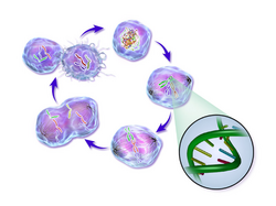
Cancer cells are cells that divide continually, forming solid tumors or flooding the blood or lymph with abnormal cells. Cell division is a normal process used by the body for growth and repair. A parent cell divides to form two daughter cells, and these daughter cells are used to build new tissue or to replace cells that have died because of aging or damage. Healthy cells stop dividing when there is no longer a need for more daughter cells, but cancer cells continue to produce copies. They are also able to spread from one part of the body to another in a process known as metastasis.

Classification

There are different categories of cancer cell, defined according to the cell type from which they originate.

Histology

Histological features of normal cells and cancer cells

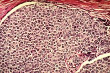
Cancer cells have distinguishing histological features visible under the microscope. The nucleus is often large and irregular, and the cytoplasm may also display abnormalities.

Nucleus

The shape, size, protein composition, and texture of the nucleus are often altered in malignant cells. The nucleus may acquire grooves, folds or indentations, chromatin may aggregate or disperse, and the nucleolus can become enlarged. In normal cells, the nucleus is often round or solid in shape, but in cancer cells the outline is often irregular. Different combinations of abnormalities are characteristic of different cancer types, to the extent that nuclear appearance can be used as a marker in cancer diagnostics and staging.

Causes

Cancer cells are created when the genes responsible for regulating cell division are damaged. Carcinogenesis is caused by mutation and epimutation of the genetic material of normal cells, which upsets the normal balance between proliferation and cell death. This results in uncontrolled cell division in the body. The uncontrolled and often rapid proliferation of cells can lead to benign or malignant tumours (cancer). Benign tumors do not spread to other parts of the body or invade other tissues. Malignant tumors can invade other organs, spread to distant locations (metastasis) and become life-threatening.

More than one mutation is necessary for carcinogenesis. In fact, a series of several mutations to certain classes of genes is usually required before a normal cell will transform into a cancer cell.

Damage to DNA can be caused by exposure to radiation, chemicals, and other environmental sources, but mutations also accumulate naturally over time through uncorrected errors in DNA transcription, making age another risk factor. Oncoviruses can cause certain types of cancer, and genetics are also known to play a role.

Stem cell research suggests that excess SP2 protein may turn stem cells into cancer cells. However, a lack of particular co-stimulated molecules that aid in the way antigens react with lymphocytes can impair the natural killer cells' function, ultimately leading to cancer.

DNA repair and mutation

When a cell is deficient in the capacity to repair DNA damages, such damages tend to be retained within the cell at an increased level. These damages, upon replication of the cell’s DNA, may cause replication errors, including mutations that lead to cancer. Numerous inherited DNA repair disorders have been described that increase cancer risk (see Wikipedia article DNA repair-deficiency disorder). In addition, particular DNA repair enzymes have been found to be deficient in multiple cancers. For example, deficient expression of the DNA repair enzyme O-6-methylguanine-DNA methyltransferase is observed in several different kinds of cancer (see Wikipedia article O-6-methylguanine-DNA methyltransferase). Although a DNA repair deficiency can predispose a cell lineage to develop cancer, increased (rather than decreased) expression of a repair capability may also emerge in the progression of cancer cell lineages, and this capability may be clinically important as reviewed by Lingg et al. For instance, the DNA repair gene DMC1 encodes a protein that is normally expressed only in cells undergoing meiosis where it helps maintain an undamaged germ-line. However, DMC1 is also expressed in various cancer cell lines including cervical, breast, and lymphoma cancer cell lines. Expression of meiotic DNA repair genes such as DMC1 may promote tumor cell growth by dealing with endogenous DNA damage within the tumor, and may also diminish the effectiveness of anticancer therapy, such as radiation therapy.

Pathology

Cells playing roles in the immune system, such as T-cells, are thought to use a dual receptor system when they determine whether or not to kill sick or damaged human cells. If a cell is under stress, turning into tumors, or infected, molecules including MIC-A and MIC-B are produced so that they can attach to the surface of the cell. These work to help macrophages detect and kill cancer cells.

Discovery

Early evidence of human cancer can be interpreted from Egyptian papers (1538 BCE) and mummified remains. In 2016, a 1.7 million year old osteosarcoma was reported by Edward John Odes (a doctoral student in Anatomical Sciences from Witwatersrand Medical School, South Africa) and colleagues, representing the oldest documented malignant hominin cancer.

The understanding of cancer was significantly advanced during the Renaissance period and in to the Age of Discovery. Sir Rudolf Virchow, a German biologist and politician, studied microscopic pathology, and linked his observations to illness. He is described as "the founder of cellular pathology". In 1845, Virchow and John Hughes Bennett independently observed abnormal increase in white blood cells in patients. Virchow correctly identified the condition as blood disease, and named it leukämie in 1847 (later anglicised to leukemia). In 1857, he was the first to describe a type of tumour called chordoma that originated from the clivus (at the base of the skull).

Telomerase

Cancer cells have unique features that make them "immortal" according to some researchers. The enzyme telomerase is used to extend the cancer cell's life span. While the telomeres of most cells shorten after each division, eventually causing the cell to die, telomerase extends the cell's telomeres. This is a major reason that cancer cells can accumulate over time, creating tumors.

Cancer stem cells and drug resistance

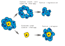
A diagram illustrating the distinction between cancer stem cell targeted and conventional cancer therapies

Scientists have discovered a molecule on the surface of tumors that appears to promote drug resistance—by converting the tumor cells back into a stem cell-like state.

When the tumor cells began to exhibit drug resistance, the cells were simultaneously transforming into a stem cell-like state, which made them impervious to the drugs. It appeared that the treatment itself was driving this transformation by activating a specific molecular pathway. Luckily, several existing drugs, such as Bortezomib for example, can attack this pathway and reverse the cellular transformation, thus 're-sensitizing' the tumor to treatment.

Treatment

In February 2019, medical scientists announced that iridium attached to albumin, creating a photosensitized molecule, can penetrate cancer cells and, after being irradiated with light (a process called photodynamic therapy), destroy the cancer cells.

Neoplasm

From Wikipedia, the free encyclopedia
 
Neoplasm
Other namesTumor, tumour, carcinocytes
Colon cancer 2.jpg
Colectomy specimen containing a malignant neoplasm, namely an invasive example of colorectal cancer (the crater-like, reddish, irregularly shaped tumor at top-center)
SpecialtyOncology
SymptomsLump
ComplicationsCancer
CausesRadiation, environmental factor, certain infections

A neoplasm (/ˈn.ˌplæzəm, ˈn.ə-/) is a type of abnormal and excessive growth of tissue. The process that occurs to form or produce a neoplasm is called neoplasia. The growth of a neoplasm is uncoordinated with that of the normal surrounding tissue, and persists in growing abnormally, even if the original trigger is removed. This abnormal growth usually forms a mass, when it may be called a tumour or tumor.

ICD-10 classifies neoplasms into four main groups: benign neoplasms, in situ neoplasms, malignant neoplasms, and neoplasms of uncertain or unknown behavior. Malignant neoplasms are also simply known as cancers and are the focus of oncology.

Prior to the abnormal growth of tissue, as neoplasia, cells often undergo an abnormal pattern of growth, such as metaplasia or dysplasia. However, metaplasia or dysplasia does not always progress to neoplasia and can occur in other conditions as well. The word is from Ancient Greek νέος- neo 'new' and πλάσμα plasma 'formation, creation'.

Types

A neoplasm can be benign, potentially malignant, or malignant (cancer).

  • Benign tumors include uterine fibroids, osteophytes and melanocytic nevi (skin moles). They are circumscribed and localized and do not transform into cancer.
  • Potentially-malignant neoplasms include carcinoma in situ. They are localised, do not invade and destroy but in time, may transform into a cancer.
  • Malignant neoplasms are commonly called cancer. They invade and destroy the surrounding tissue, may form metastases and, if untreated or unresponsive to treatment, will generally prove fatal.
  • Secondary neoplasm refers to any of a class of cancerous tumor that is either a metastatic offshoot of a primary tumor, or an apparently unrelated tumor that increases in frequency following certain cancer treatments such as chemotherapy or radiotherapy.
  • Rarely there can be a metastatic neoplasm with no known site of the primary cancer and this is classed as a cancer of unknown primary origin.

Clonality

Neoplastic tumors are often heterogeneous and contain more than one type of cell, but their initiation and continued growth is usually dependent on a single population of neoplastic cells. These cells are presumed to be monoclonal – that is, they are derived from the same cell, and all carry the same genetic or epigenetic anomaly – evident of clonality. For lymphoid neoplasms, e.g. lymphoma and leukemia, clonality is proven by the amplification of a single rearrangement of their immunoglobulin gene (for B cell lesions) or T cell receptor gene (for T cell lesions). The demonstration of clonality is now considered to be necessary to identify a lymphoid cell proliferation as neoplastic.

It is tempting to define neoplasms as clonal cellular proliferations but the demonstration of clonality is not always possible. Therefore, clonality is not required in the definition of neoplasia.

Neoplasm vs. tumor

The word tumor or tumour comes from the Latin word for swelling, which is one of the cardinal signs of inflammation. The word originally referred to any form of swelling, neoplastic or not. In modern English, tumor is used as a synonym for neoplasm (a solid or fluid-filled cystic lesion that may or may not be formed by an abnormal growth of neoplastic cells) that appears enlarged in size. Some neoplasms do not form a tumor - these include leukemia and most forms of carcinoma in situ. Tumor is also not synonymous with cancer. While cancer is by definition malignant, a tumor can be benign, precancerous, or malignant.

The terms mass and nodule are often used synonymously with tumor. Generally speaking, however, the term tumor is used generically, without reference to the physical size of the lesion. More specifically, the term mass is often used when the lesion has a maximal diameter of at least 20 millimeters (mm) in greatest direction, while the term nodule is usually used when the size of the lesion is less than 20 mm in its greatest dimension (25.4 mm = 1 inch).

Causes

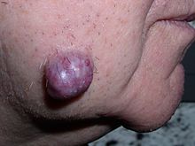
Neoplastic tumor of the cheek skin, here a benign neoplasm of the sweat glands called hidradenoma, which is not solid but is fluid-filled
 
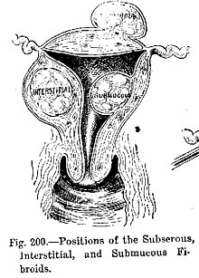
Diagram illustrating benign neoplasms, namely fibroids of the uterus

Tumors in humans occur as a result of accumulated genetic and epigenetic alterations within single cells, which cause the cell to divide and expand uncontrollably. A neoplasm can be caused by an abnormal proliferation of tissues, which can be caused by genetic mutations. Not all types of neoplasms cause a tumorous overgrowth of tissue, however (such as leukemia or carcinoma in situ) and similarities between neoplasmic growths and regenerative processes, e.g., dedifferentiation and rapid cell proliferation, have been pointed out.

Tumor growth has been studied using mathematics and continuum mechanics. Vascular tumors such as hemangiomas and lymphangiomas (formed from blood or lymph vessels) are thus looked at as being amalgams of a solid skeleton formed by sticky cells and an organic liquid filling the spaces in which cells can grow. Under this type of model, mechanical stresses and strains can be dealt with and their influence on the growth of the tumor and the surrounding tissue and vasculature elucidated. Recent findings from experiments that use this model show that active growth of the tumor is restricted to the outer edges of the tumor and that stiffening of the underlying normal tissue inhibits tumor growth as well.

Benign conditions that are not associated with an abnormal proliferation of tissue (such as sebaceous cysts) can also present as tumors, however, but have no malignant potential. Breast cysts (as occur commonly during pregnancy and at other times) are another example, as are other encapsulated glandular swellings (thyroid, adrenal gland, pancreas).

Encapsulated hematomas, encapsulated necrotic tissue (from an insect bite, foreign body, or other noxious mechanism), keloids (discrete overgrowths of scar tissue) and granulomas may also present as tumors.

Discrete localized enlargements of normal structures (ureters, blood vessels, intrahepatic or extrahepatic biliary ducts, pulmonary inclusions, or gastrointestinal duplications) due to outflow obstructions or narrowings, or abnormal connections, may also present as a tumor. Examples are arteriovenous fistulae or aneurysms (with or without thrombosis), biliary fistulae or aneurysms, sclerosing cholangitis, cysticercosis or hydatid cysts, intestinal duplications, and pulmonary inclusions as seen with cystic fibrosis. It can be dangerous to biopsy a number of types of tumor in which the leakage of their contents would potentially be catastrophic. When such types of tumors are encountered, diagnostic modalities such as ultrasound, CT scans, MRI, angiograms, and nuclear medicine scans are employed prior to (or during) biopsy or surgical exploration/excision in an attempt to avoid such severe complications.

Malignant neoplasms

DNA damage

The central role of DNA damage and epigenetic defects in DNA repair genes in malignant neoplasms

DNA damage is considered to be the primary underlying cause of malignant neoplasms known as cancers. Its central role in progression to cancer is illustrated in the figure in this section, in the box near the top. (The central features of DNA damage, epigenetic alterations and deficient DNA repair in progression to cancer are shown in red.) DNA damage is very common. Naturally occurring DNA damages (mostly due to cellular metabolism and the properties of DNA in water at body temperatures) occur at a rate of more than 60,000 new damages, on average, per human cell, per day also see article DNA damage (naturally occurring) ]. Additional DNA damages can arise from exposure to exogenous agents. Tobacco smoke causes increased exogenous DNA damage, and these DNA damages are the likely cause of lung cancer due to smoking. UV light from solar radiation causes DNA damage that is important in melanoma. Helicobacter pylori infection produces high levels of reactive oxygen species that damage DNA and contributes to gastric cancer. Bile acids, at high levels in the colons of humans eating a high fat diet, also cause DNA damage and contribute to colon cancer. Katsurano et al. indicated that macrophages and neutrophils in an inflamed colonic epithelium are the source of reactive oxygen species causing the DNA damages that initiate colonic tumorigenesis. Some sources of DNA damage are indicated in the boxes at the top of the figure in this section.

Individuals with a germline mutation causing deficiency in any of 34 DNA repair genes (see article DNA repair-deficiency disorder) are at increased risk of cancer. Some germline mutations in DNA repair genes cause up to 100% lifetime chance of cancer (e.g., p53 mutations). These germline mutations are indicated in a box at the left of the figure with an arrow indicating their contribution to DNA repair deficiency.

About 70% of malignant neoplasms have no hereditary component and are called "sporadic cancers". Only a minority of sporadic cancers have a deficiency in DNA repair due to mutation in a DNA repair gene. However, a majority of sporadic cancers have deficiency in DNA repair due to epigenetic alterations that reduce or silence DNA repair gene expression. For example, of 113 sequential colorectal cancers, only four had a missense mutation in the DNA repair gene MGMT, while the majority had reduced MGMT expression due to methylation of the MGMT promoter region (an epigenetic alteration). Five reports present evidence that between 40% and 90% of colorectal cancers have reduced MGMT expression due to methylation of the MGMT promoter region.

Similarly, out of 119 cases of mismatch repair-deficient colorectal cancers that lacked DNA repair gene PMS2 expression, PMS2 was deficient in 6 due to mutations in the PMS2 gene, while in 103 cases PMS2 expression was deficient because its pairing partner MLH1 was repressed due to promoter methylation (PMS2 protein is unstable in the absence of MLH1). In the other 10 cases, loss of PMS2 expression was likely due to epigenetic overexpression of the microRNA, miR-155, which down-regulates MLH1.

In further examples, epigenetic defects were found at frequencies of between 13%-100% for the DNA repair genes BRCA1, WRN, FANCB, FANCF, MGMT, MLH1, MSH2, MSH4, ERCC1, XPF, NEIL1 and ATM. These epigenetic defects occurred in various cancers (e.g. breast, ovarian, colorectal and head and neck). Two or three deficiencies in expression of ERCC1, XPF or PMS2 occur simultaneously in the majority of the 49 colon cancers evaluated by Facista et al. Epigenetic alterations causing reduced expression of DNA repair genes is shown in a central box at the third level from the top of the figure in this section, and the consequent DNA repair deficiency is shown at the fourth level.

When expression of DNA repair genes is reduced, DNA damages accumulate in cells at a higher than normal level, and these excess damages cause increased frequencies of mutation or epimutation. Mutation rates strongly increase in cells defective in DNA mismatch repair or in homologous recombinational repair (HRR).

During repair of DNA double strand breaks, or repair of other DNA damages, incompletely cleared sites of repair can cause epigenetic gene silencing. DNA repair deficiencies (level 4 in the figure) cause increased DNA damages (level 5 in the figure) which result in increased somatic mutations and epigenetic alterations (level 6 in the figure).

Field defects, normal appearing tissue with multiple alterations (and discussed in the section below), are common precursors to development of the disordered and improperly proliferating clone of tissue in a malignant neoplasm. Such field defects (second level from bottom of figure) may have multiple mutations and epigenetic alterations.

Once a cancer is formed, it usually has genome instability. This instability is likely due to reduced DNA repair or excessive DNA damage. Because of such instability, the cancer continues to evolve and to produce sub clones. For example, a renal cancer, sampled in 9 areas, had 40 ubiquitous mutations, demonstrating tumor heterogeneity (i.e. present in all areas of the cancer), 59 mutations shared by some (but not all areas), and 29 "private" mutations only present in one of the areas of the cancer.

Field defects

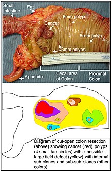
Longitudinally opened freshly resected colon segment showing a cancer and four polyps, plus a schematic diagram indicating a likely field defect (a region of tissue that precedes and predisposes to the development of cancer) in this colon segment. The diagram indicates sub-clones and sub-sub-clones that were precursors to the tumors.

Various other terms have been used to describe this phenomenon, including "field effect", "field cancerization", and "field carcinogenesis". The term "field cancerization" was first used in 1953 to describe an area or "field" of epithelium that has been preconditioned by (at that time) largely unknown processes so as to predispose it towards development of cancer. Since then, the terms "field cancerization" and "field defect" have been used to describe pre-malignant tissue in which new cancers are likely to arise.

Field defects are important in progression to cancer. However, in most cancer research, as pointed out by Rubin "The vast majority of studies in cancer research has been done on well-defined tumors in vivo, or on discrete neoplastic foci in vitro. Yet there is evidence that more than 80% of the somatic mutations found in mutator phenotype human colorectal tumors occur before the onset of terminal clonal expansion. Similarly, Vogelstein et al. point out that more than half of somatic mutations identified in tumors occurred in a pre-neoplastic phase (in a field defect), during growth of apparently normal cells. Likewise, epigenetic alterations present in tumors may have occurred in pre-neoplastic field defects.

An expanded view of field effect has been termed "etiologic field effect", which encompasses not only molecular and pathologic changes in pre-neoplastic cells but also influences of exogenous environmental factors and molecular changes in the local microenvironment on neoplastic evolution from tumor initiation to patient death.

In the colon, a field defect probably arises by natural selection of a mutant or epigenetically altered cell among the stem cells at the base of one of the intestinal crypts on the inside surface of the colon. A mutant or epigenetically altered stem cell may replace the other nearby stem cells by natural selection. Thus, a patch of abnormal tissue may arise. The figure in this section includes a photo of a freshly resected and lengthwise-opened segment of the colon showing a colon cancer and four polyps. Below the photo, there is a schematic diagram of how a large patch of mutant or epigenetically altered cells may have formed, shown by the large area in yellow in the diagram. Within this first large patch in the diagram (a large clone of cells), a second such mutation or epigenetic alteration may occur so that a given stem cell acquires an advantage compared to other stem cells within the patch, and this altered stem cell may expand clonally forming a secondary patch, or sub-clone, within the original patch. This is indicated in the diagram by four smaller patches of different colors within the large yellow original area. Within these new patches (sub-clones), the process may be repeated multiple times, indicated by the still smaller patches within the four secondary patches (with still different colors in the diagram) which clonally expand, until stem cells arise that generate either small polyps or else a malignant neoplasm (cancer).

In the photo, an apparent field defect in this segment of a colon has generated four polyps (labeled with the size of the polyps, 6mm, 5mm, and two of 3mm, and a cancer about 3 cm across in its longest dimension). These neoplasms are also indicated, in the diagram below the photo, by 4 small tan circles (polyps) and a larger red area (cancer). The cancer in the photo occurred in the cecal area of the colon, where the colon joins the small intestine (labeled) and where the appendix occurs (labeled). The fat in the photo is external to the outer wall of the colon. In the segment of colon shown here, the colon was cut open lengthwise to expose the inner surface of the colon and to display the cancer and polyps occurring within the inner epithelial lining of the colon.

If the general process by which sporadic colon cancers arise is the formation of a pre-neoplastic clone that spreads by natural selection, followed by formation of internal sub-clones within the initial clone, and sub-sub-clones inside those, then colon cancers generally should be associated with, and be preceded by, fields of increasing abnormality reflecting the succession of premalignant events. The most extensive region of abnormality (the outermost yellow irregular area in the diagram) would reflect the earliest event in formation of a malignant neoplasm.

In experimental evaluation of specific DNA repair deficiencies in cancers, many specific DNA repair deficiencies were also shown to occur in the field defects surrounding those cancers. The Table, below, gives examples for which the DNA repair deficiency in a cancer was shown to be caused by an epigenetic alteration, and the somewhat lower frequencies with which the same epigenetically caused DNA repair deficiency was found in the surrounding field defect.

Some of the small polyps in the field defect shown in the photo of the opened colon segment may be relatively benign neoplasms. Of polyps less than 10mm in size, found during colonoscopy and followed with repeat colonoscopies for 3 years, 25% were unchanged in size, 35% regressed or shrank in size while 40% grew in size.

Genome instability

Cancers are known to exhibit genome instability or a mutator phenotype. The protein-coding DNA within the nucleus is about 1.5% of the total genomic DNA. Within this protein-coding DNA (called the exome), an average cancer of the breast or colon can have about 60 to 70 protein altering mutations, of which about 3 or 4 may be "driver" mutations, and the remaining ones may be "passenger" mutations. However, the average number of DNA sequence mutations in the entire genome (including non-protein-coding regions) within a breast cancer tissue sample is about 20,000. In an average melanoma tissue sample (where melanomas have a higher exome mutation frequency) the total number of DNA sequence mutations is about 80,000. This compares to the very low mutation frequency of about 70 new mutations in the entire genome between generations (parent to child) in humans.

The high frequencies of mutations in the total nucleotide sequences within cancers suggest that often an early alteration in the field defects giving rise to a cancer (e.g. yellow area in the diagram in this section) is a deficiency in DNA repair. The large field defects surrounding colon cancers (extending to at about 10 cm on each side of a cancer) were shown by Facista et al. to frequently have epigenetic defects in 2 or 3 DNA repair proteins (ERCC1, XPF or PMS2) in the entire area of the field defect. Deficiencies in DNA repair cause increased mutation rates. A deficiency in DNA repair, itself, can allow DNA damages to accumulate, and error-prone translesion synthesis past some of those damages may give rise to mutations. In addition, faulty repair of these accumulated DNA damages may give rise to epimutations. These new mutations or epimutations may provide a proliferative advantage, generating a field defect. Although the mutations/epimutations in DNA repair genes do not, themselves, confer a selective advantage, they may be carried along as passengers in cells when the cells acquire additional mutations/epimutations that do provide a proliferative advantage.

Etymology

The term neoplasm is a synonym of tumor. Neoplasia denotes the process of the formation of neoplasms/tumors, and the process is referred to as a neoplastic process. The word neoplastic itself comes from Greek neo 'new' and plastic 'formed, molded'.

The term tumor derives from the Latin noun tumor 'a swelling', ultimately from the verb tumēre 'to swell'. In the British Commonwealth, the spelling tumour is commonly used, whereas in the U.S. the word is usually spelled tumor.

In its medical sense, tumor has traditionally meant an abnormal swelling of the flesh. The Roman medical encyclopedist Celsus (c. 30 BC–38 AD) described the four cardinal signs of acute inflammation as tumor, dolor, calor, and rubor (swelling, pain, increased heat, and redness). (His treatise, De Medicina, was the first medical book printed in 1478 following the invention of the movable-type printing press.)

In contemporary English, the word tumor is often used as a synonym for a cystic (liquid-filled) growth or solid neoplasm (cancerous or non-cancerous), with other forms of swelling often referred to as "swellings".

Related terms occur commonly in the medical literature, where the nouns tumefaction and tumescence (derived from the adjective tumescent) are current medical terms for non-neoplastic swelling. This type of swelling is most often caused by inflammation caused by trauma, infection, and other factors.

Tumors may be caused by conditions other than an overgrowth of neoplastic cells, however. Cysts (such as sebaceous cysts) are also referred to as tumors, even though they have no neoplastic cells. This is standard in medical-billing terminology (especially when billing for a growth whose pathology has yet to be determined).

Archetype

From Wikipedia, the free encyclopedia https://en.wikipedia.org/wiki/Archetype The concept of an archetyp...