Search This Blog

Tuesday, June 20, 2023

Cranial nerves

From Wikipedia, the free encyclopedia
 
Cranial nerves
Skull brain human normal.svg
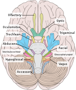
Left View of the human brain from below, showing origins of cranial nerves.
Right Juxtaposed skull base with foramina in which many nerves exit the skull.
 
Skull and brainstem inner ear.svg
Cranial nerves as they pass through the skull base to the brain
Details
Identifiers
Latinnervus cranialis
(pl: nervi craniales)
MeSHD003391
TA98A14.2.01.001
A14.2.00.038
TA26142, 6178
FMA5865

Cranial nerves are the nerves that emerge directly from the brain (including the brainstem), of which there are conventionally considered twelve pairs. Cranial nerves relay information between the brain and parts of the body, primarily to and from regions of the head and neck, including the special senses of vision, taste, smell, and hearing.

The cranial nerves emerge from the central nervous system above the level of the first vertebra of the vertebral column. Each cranial nerve is paired and is present on both sides. There are conventionally twelve pairs of cranial nerves, which are described with Roman numerals I–XII. Some considered there to be thirteen pairs of cranial nerves, including cranial nerve zero. The numbering of the cranial nerves is based on the order in which they emerge from the brain and brainstem, from front to back.

The terminal nerves (0), olfactory nerves (I) and optic nerves (II) emerge from the cerebrum, and the remaining ten pairs arise from the brainstem, which is the lower part of the brain.

The cranial nerves are considered components of the peripheral nervous system (PNS), although on a structural level the olfactory (I), optic (II), and trigeminal (V) nerves are more accurately considered part of the central nervous system (CNS).

The cranial nerves are in contrast to spinal nerves, which emerge from segments of the spinal cord.

Anatomy

View of the human brain from below showing the cranial nerves on an autopsy specimen
 
View from below of the brain and brainstem showing the cranial nerves, numbered from olfactory to hypoglossal after the order in which they emerge
 
The brainstem, with cranial nerve nuclei and tracts shown in red

Most typically, humans are considered to have twelve pairs of cranial nerves (I–XII), with the terminal nerve (0) more recently canonized. The nerves are: the olfactory nerve (I), the optic nerve (II), oculomotor nerve (III), trochlear nerve (IV), trigeminal nerve (V), abducens nerve (VI), facial nerve (VII), vestibulocochlear nerve (VIII), glossopharyngeal nerve (IX), vagus nerve (X), accessory nerve (XI), and the hypoglossal nerve (XII).

Terminology

Cranial nerves are generally named according to their structure or function. For example, the olfactory nerve (I) supplies smell, and the facial nerve (VII) supplies the muscles of the face. Because Latin was the lingua franca of the study of anatomy when the nerves were first documented, recorded, and discussed, many nerves maintain Latin or Greek names, including the trochlear nerve (IV), named according to its structure, as it supplies a muscle that attaches to a pulley (Greek: trochlea). The trigeminal nerve (V) is named in accordance with its three components (Latin: trigeminus meaning triplets), and the vagus nerve (X) is named for its wandering course (Latin: vagus).

Cranial nerves are numbered based on their position from front to back (rostral-caudal) of their position on the brain, as, when viewing the forebrain and brainstem from below, they are often visible in their numeric order. For example, the olfactory nerves (I) and optic nerves (II) arise from the base of the forebrain, and the other nerves, III to XII, arise from the brainstem.

Cranial nerves have paths within and outside the skull. The paths within the skull are called "intracranial" and the paths outside the skull are called "extracranial". There are many holes in the skull called "foramina" by which the nerves can exit the skull. All cranial nerves are paired, which means they occur on both the right and left sides of the body. The muscle, skin, or additional function supplied by a nerve, on the same side of the body as the side it originates from, is an ipsilateral function. If the function is on the opposite side to the origin of the nerve, this is known as a contralateral function.

Intracranial course

Nuclei

Grossly, all cranial nerves have a Nucleus. With the exception of the olfactory nerve (I) and optic nerve (II), all the nuclei are present in the brainstem.

The midbrain of the brainstem has the nuclei of the oculomotor nerve (III) and trochlear nerve (IV); the pons has the nuclei of the trigeminal nerve (V), abducens nerve (VI), facial nerve (VII) and vestibulocochlear nerve (VIII); and the medulla has the nuclei of the glossopharyngeal nerve (IX), vagus nerve (X), accessory nerve (XI) and hypoglossal nerve (XII). The olfactory nerve (I) emerges from the olfactory bulb, and depending slightly on division the optic nerve (II) is considered to emerge from the lateral geniculate nuclei.

Because each nerve may have several functions, the nerve fibres that make up the nerve may collect in more than one nucleus. For example, the trigeminal nerve (V), which has a sensory and a motor role, has at least four nuclei.

Exiting the brainstem

With the exception of the olfactory nerve (I) and optic nerve (II), the cranial nerves emerge from the brainstem. The oculomotor nerve (III) and trochlear nerve (IV) emerge from the midbrain, the trigeminal (V), abducens (VI), facial (VII) and vestibulocochlear (VIII) from the pons, and the glossopharyngeal (IX), vagus (X), accessory (XI) and hypoglossal (XII) emerge from the medulla.

The olfactory nerve (I) and optic nerve (II) emerge separately. The olfactory nerves emerge from the olfactory bulbs on either side of the crista galli, a bony projection below the frontal lobe, and the optic nerves (II) emerge from the lateral colliculus, swellings on either side of the temporal lobes of the brain.

Ganglia

The cranial nerves give rise to a number of ganglia, collections of the cell bodies of neurons in the nerves that are outside of the brain. These ganglia are both parasympathetic and sensory ganglia.

The sensory ganglia of the cranial nerves, directly correspond to the dorsal root ganglia of spinal nerves and are known as cranial nerve ganglia. Sensory ganglia exist for nerves with sensory function: V, VII, VIII, IX, X. There are also a number of parasympathetic cranial nerve ganglia. Sympathetic ganglia supplying the head and neck reside in the upper regions of the sympathetic trunk, and do not belong to the cranial nerves.

The ganglion of the sensory nerves, which are similar in structure to the dorsal root ganglion of the spinal cord, include:

Additional ganglia for nerves with parasympathetic function exist, and include the ciliary ganglion of the oculomotor nerve (III), the pterygopalatine ganglion of the maxillary nerve (V2), the submandibular ganglion of the lingual nerve, a branch of the facial nerve (VII), and the otic ganglion of the glossopharyngeal nerve (IX).

Exiting the skull and extracranial course

Exits of cranial nerves from the skull.
Location Nerve
cribriform plate Terminal nerve (0)
cribriform plate Olfactory nerve (I)
optic foramen Optic nerve (II)
superior orbital fissure Oculomotor (III)
Trochlear (IV)
Abducens (VI)
Trigeminal V1
(ophthalmic)
foramen rotundum Trigeminal V2
(maxillary)
foramen ovale Trigeminal V3
(mandibular)
stylomastoid foramen Facial nerve (VII)
internal auditory canal Vestibulocochlear (VIII)
jugular foramen Glossopharyngeal (IX)
Vagus (X)
Accessory (XI)
hypoglossal canal Hypoglossal (XII)

After emerging from the brain, the cranial nerves travel within the skull, and some must leave it in order to reach their destinations. Often the nerves pass through holes in the skull, called foramina, as they travel to their destinations. Other nerves pass through bony canals, longer pathways enclosed by bone. These foramina and canals may contain more than one cranial nerve and may also contain blood vessels.

  • The terminal nerve (0), is a thin network of fibers associated with the dura and lamina terminalis running rostral to the olfactory nerve, with projections through the cribriform plate.
  • The olfactory nerve (I), passes through perforations in the cribriform plate part of the ethmoid bone. The nerve fibres end in the upper nasal cavity.
  • The optic nerve (II) passes through the optic foramen in the sphenoid bone as it travels to the eye.
  • The oculomotor nerve (III), trochlear nerve (IV), abducens nerve (VI) and the ophthalmic branch of the trigeminal nerve (V1) travel through the cavernous sinus into the superior orbital fissure, passing out of the skull into the orbit.
  • The maxillary division of the trigeminal nerve (V2) passes through foramen rotundum in the sphenoid bone.
  • The mandibular division of the trigeminal nerve (V3) passes through foramen ovale of the sphenoid bone.
  • The facial nerve (VII) and vestibulocochlear nerve (VIII) both enter the internal auditory canal in the temporal bone. The facial nerve then reaches the side of the face by using the stylomastoid foramen, also in the temporal bone. Its fibers then spread out to reach and control all of the muscles of facial expression. The vestibulocochlear nerve reaches the organs that control balance and hearing in the temporal bone and therefore does not reach the external surface of the skull.
  • The glossopharyngeal (IX), vagus (X) and accessory nerve (XI) all leave the skull via the jugular foramen to enter the neck. The glossopharyngeal nerve provides sensation to the upper throat and the back of the tongue, the vagus supplies the muscles in the larynx and continues downward to supply parasympathetic supply to the chest and abdomen. The accessory nerve controls the trapezius and sternocleidomastoid muscles in the neck and shoulder.
  • The hypoglossal nerve (XII) exits the skull using the hypoglossal canal in the occipital bone.

Development

The cranial nerves are formed from the contribution of two specialized embryonic cell populations, cranial neural crest and ectodermal placodes. The components of the sensory nervous system of the head are derived from the neural crest and from an embryonic cell population developing in close proximity, the cranial sensory placodes (the olfactory, lens, otic, trigeminal, epibranchial and paratympanic placodes). The dual origin cranial nerves are summarized in the following Table:

Contributions of neural crest cells and placodes to ganglia and cranial nerves

Cranial nerve Ganglion and type Origin of neurons
CNI – olfactory

(Ensheating glia of olfactory nerves)


Telencephalon/olfactory placode; NCCs at forebrain
CNIII – oculomotor

(m)

Ciliary, visceral efferent NCCs at forebrain-midbrain junction (caudal diencephalon and the anterior mesencephalon)
CNV – trigeminal

(mix)

Trigeminal, general afferent NCCs at forebrain-midbrain junction (from r2 into 1st PA), trigeminal placode
CNVII – facial

(mix)

-Superior, general and special afferent

-Inferior: geniculate, general and special afferent

-Sphenopalatine, visceral efferent

-Submandibular, visceral efferent

-Hindbrain NCCs (from r4 into 2nd PA), 1st epibranchial placode

-1st epibranchial placode (geniculate)

-Hindbrain NCCs (2nd PA)

-Hindbrain NCCs (2nd PA)

CNVIII – Vestibulocochlear

(s)

-Acoustic: cochlear, special afferent; and vestibular, special afferent -Otic placode and hindbrain (from r4) NCCs
CNIX – glossopharyngeal

(mix)

-Superior, general and special afferent

-Inferior, petrosal, general and special afferent

-Otic, visceral efferent

-Hindbrain NCCs (from r6 into 3rd PA)

-2nd epibranchial placode (petrosal)

-Hindbrain NCCs (from r6 into 3rd PA)

CNX – vagus

(mix)

Superior laryngeal branch; and recurrent laryngeal branch

-Superior, general afferent

-Inferior: nodose, general and special afferent

-Vagal: parasympathetic, visceral efferent

-Hindbrain NCCs (from r7-r8 to 4th & 6th PA)

-Hindbrain NCCs (4th& 6th PA); 3rd (nodose) and 4th epibranchial placodes

-Hindbrain NCCs (4th & 6th PA)

CNXI – accessory

(m)

No ganglion * Hindbrain (from r7-r8 to PA 4); NCCs (4th PA)

Abbreviations: CN, cranial nerve; m, purely motor nerve; mix, mixed nerve (sensory and motor); NC, neural crest; PA, pharyngeal (branchial) arch; r, rhombomere; s, purely sensory nerve. * There is no known ganglion of the accessory nerve. The cranial part of the accessory nerve sends occasional branches to the superior ganglion of the vagus nerve.

Function

The cranial nerves provide motor and sensory supply mainly to the structures within the head and neck. The sensory supply includes both "general" sensation such as temperature and touch, and "special" senses such as taste, vision, smell, balance and hearing. The vagus nerve (X) provides sensory and autonomic (parasympathetic) supply to structures in the neck and also to most of the organs in the chest and abdomen.

Terminal nerve (0)

The terminal nerve (0) may not have a role in humans, although it has been implicated in hormonal responses to smell, sexual response and mate selection.

Smell (I)

The olfactory nerve (I) conveys information giving rise to the sense of smell.

Damage to the olfactory nerve (I) can cause an inability to smell (anosmia), a distortion in the sense of smell (parosmia), or a distortion or lack of taste.

Vision (II)

The optic nerve (II) transmits visual information.

Damage to the optic nerve (II) affects specific aspects of vision that depend on the location of the damage. A person may not be able to see objects on their left or right sides (homonymous hemianopsia), or may have difficulty seeing objects from their outer visual fields (bitemporal hemianopsia) if the optic chiasm is involved. Inflammation (optic neuritis) may impact the sharpness of vision or colour detection.

Eye movement (III, IV, VI)

The oculomotor (III), troclear (IV) and abducens (VI) nerves supply the muscle of the eye. Damage will affect the movement of the eye in various ways, shown here.

The oculomotor nerve (III), trochlear nerve (IV) and abducens nerve (VI) coordinate eye movement. The oculomotor nerve controls all muscles of the eye except for the superior oblique muscle controlled by the trochlear nerve (IV), and the lateral rectus muscle controlled by the abducens nerve (VI). This means the ability of the eye to look down and inwards is controlled by the trochlear nerve (IV), the ability to look outwards is controlled by the abducens nerve (VI), and all other movements are controlled by the oculomotor nerve (III)

Damage to these nerves may affect the movement of the eye. Damage may result in double vision (diplopia) because the movements of the eyes are not synchronized. Abnormalities of visual movement may also be seen on examination, such as jittering (nystagmus).

Damage to the oculomotor nerve (III) can cause double vision and inability to coordinate the movements of both eyes (strabismus), also eyelid drooping (ptosis) and pupil dilation (mydriasis). Lesions may also lead to inability to open the eye due to paralysis of the levator palpebrae muscle. Individuals suffering from a lesion to the oculomotor nerve may compensate by tilting their heads to alleviate symptoms due to paralysis of one or more of the eye muscles it controls.

Damage to the trochlear nerve (IV) can also cause double vision with the eye adducted and elevated. The result will be an eye which can not move downwards properly (especially downwards when in an inward position). This is due to impairment in the superior oblique muscle.

Damage to the abducens nerve (VI) can also result in double vision. This is due to impairment in the lateral rectus muscle, supplied by the abducens nerve.

Trigeminal nerve (V)

The trigeminal nerve (V) and its three main branches the ophthalmic (V1), maxillary (V2), and mandibular (V3) provide sensation to the skin of the face and also controls the muscles of chewing.

Damage to the trigeminal nerve leads to loss of sensation in an affected area. Other conditions affecting the trigeminal nerve (V) include trigeminal neuralgia, herpes zoster, sinusitis pain, presence of a dental abscess, and cluster headaches.

The facial nerve (VII) supplies the muscles of facial expression. Damage to the nerve causes a lack of muscle tone on the affected side, as can be seen on the right side of the face here.

Facial expression (VII)

The facial nerve (VII) controls most muscles of facial expression, supplies the sensation of taste from the front two-thirds of the tongue, and controls the stapedius muscle. Most muscles are supplied by the cortex on the opposite side of the brain; the exception is the frontalis muscle of the forehead, in which the left and the right side of the muscle both receive inputs from both sides of the brain.

Damage to the facial nerve (VII) may cause facial palsy. This is where a person is unable to move the muscles on one or both sides of their face. The most common cause of this is Bell's palsy, the ultimate cause of which is unknown. Patients with Bell's palsy often have a drooping mouth on the affected side and often have trouble chewing because the buccinator muscle is affected. The facial nerve is also the most commonly affected cranial nerve in blunt trauma.

Hearing and balance (VIII)

The vestibulocochlear nerve (VIII) supplies information relating to balance and hearing via its two branches, the vestibular and cochlear nerves. The vestibular part is responsible for supplying sensation from the vestibules and semicircular canal of the inner ear, including information about balance, and is an important component of the vestibuloocular reflex, which keeps the head stable and allows the eyes to track moving objects. The cochlear nerve transmits information from the cochlea, allowing sound to be heard.

When damaged, the vestibular nerve may give rise to the sensation of spinning and dizziness (vertigo). Function of the vestibular nerve may be tested by putting cold and warm water in the ears and watching eye movements caloric stimulation. Damage to the vestibulocochlear nerve can also present as repetitive and involuntary eye movements (nystagmus), particularly when the eye is moving horizontally. Damage to the cochlear nerve will cause partial or complete deafness in the affected ear.

Oral sensation, taste, and salivation (IX)

A damaged glossopharyngeal nerve (IX) may cause the uvula to deviate to the affected side.

The glossopharyngeal nerve (IX) supplies the stylopharyngeus muscle and provides sensation to the oropharynx and back of the tongue. The glossopharyngeal nerve also provides parasympathetic input to the parotid gland.

Damage to the nerve may cause failure of the gag reflex; a failure may also be seen in damage to the vagus nerve (X).

Vagus nerve (X)

The vagus nerve (X) provides sensory and parasympathetic supply to structures in the neck and also to most of the organs in the chest and abdomen.

Loss of function of the vagus nerve (X) will lead to a loss of parasympathetic supply to a very large number of structures. Major effects of damage to the vagus nerve may include a rise in blood pressure and heart rate. Isolated dysfunction of only the vagus nerve is rare, but – if the lesion is located above the point at which the vagus first branches off – can be indicated by a hoarse voice, due to dysfunction of one of its branches, the recurrent laryngeal nerve.

Damage to this nerve may result in difficulties swallowing.

Shoulder elevation and head-turning (XI)

The accessory nerve (XI) supplies the sternocleidomastoid and trapezius muscles. Damage to the nerve may cause a winged scapula, shown here.
 
The hypoglossal nerve (XII) supplies the muscles of the tongue. A damaged hypoglossal nerve will result in an inability to stick the tongue out straight; here seen in an injury resulting from branchial cyst surgery. 

The accessory nerve (XI) supplies the sternocleidomastoid and trapezius muscles.

Damage to the accessory nerve (XI) will lead to weakness in the trapezius muscle on the same side as the damage. The trapezius lifts the shoulder when shrugging, so the affected shoulder will not be able to shrug and the shoulder blade (scapula) will protrude into a winged position. Depending on the location of the lesion there may also be weakness present in the sternocleidomastoid muscle, which acts to turn the head so that the face points to the opposite side.

Tongue movement (XII)

The hypoglossal nerve (XII) supplies the intrinsic muscles of the tongue, controlling tongue movement. The hypoglossal nerve (XII) is unique in that it is supplied by the motor cortices of both hemispheres of the brain.

Damage to the nerve may lead to fasciculations or wasting (atrophy) of the muscles of the tongue. This will lead to weakness of tongue movement on that side. When damaged and extended, the tongue will move towards the weaker or damaged side, as shown in the image. The fasciculations of the tongue are sometimes said to look like a "bag of worms". Damage to the nerve tract or nucleus will not lead to atrophy or fasciculations, but only weakness of the muscles on the same side as the damage.

Clinical significance

Examination

Doctors, neurologists and other medical professionals may conduct a cranial nerve examination as part of a neurological examination to examine the cranial nerves. This is a highly formalised series of steps involving specific tests for each nerve. Dysfunction of a nerve identified during testing may point to a problem with the nerve or of a part of the brain.

A cranial nerve exam starts with observation of the patient, as some cranial nerve lesions may affect the symmetry of the eyes or face. Vision may be tested by examining the visual fields, or by examining the retina with an ophthalmoscope, using a process known as funduscopy. Visual field testing may be used to pin-point structural lesions in the optic nerve, or further along the visual pathways. Eye movement is tested and abnormalities such as nystagmus are observed for. The sensation of the face is tested, and patients are asked to perform different facial movements, such as puffing out of the cheeks. Hearing is checked by voice and tuning forks. The patient's uvula is examined. After performing a shrug and head turn, the patient's tongue function is assessed by various tongue movements.

Smell is not routinely tested, but if there is suspicion of a change in the sense of smell, each nostril is tested with substances of known odors such as coffee or soap. Intensely smelling substances, for example ammonia, may lead to the activation of pain receptors of the trigeminal nerve (V) located in the nasal cavity and this can confound olfactory testing.

Damage

Compression

Nerves may be compressed because of increased intracranial pressure, a mass effect of an intracerebral haemorrhage, or tumour that presses against the nerves and interferes with the transmission of impulses along the nerve. Loss of function of a cranial nerve may sometimes be the first symptom of an intracranial or skull base cancer.

An increase in intracranial pressure may lead to impairment of the optic nerves (II) due to compression of the surrounding veins and capillaries, causing swelling of the eyeball (papilloedema). A cancer, such as an optic nerve glioma, may also impact the optic nerve (II). A pituitary tumour may compress the optic tracts or the optic chiasm of the optic nerve (II), leading to visual field loss. A pituitary tumour may also extend into the cavernous sinus, compressing the oculomotor nerve (III), trochlear nerve (IV) and abducens nerve (VI), leading to double-vision and strabismus. These nerves may also be affected by herniation of the temporal lobes of the brain through the falx cerebri.

The cause of trigeminal neuralgia, in which one side of the face is exquisitely painful, is thought to be compression of the nerve by an artery as the nerve emerges from the brain stem. An acoustic neuroma, particularly at the junction between the pons and medulla, may compress the facial nerve (VII) and vestibulocochlear nerve (VIII), leading to hearing and sensory loss on the affected side.

Stroke

Occlusion of blood vessels that supply the nerves or their nuclei, an ischemic stroke, may cause specific signs and symptoms relating to the damaged area. If there is a stroke of the midbrain, pons or medulla, various cranial nerves may be damaged, resulting in dysfunction and symptoms of a number of different syndromes. Thrombosis, such as a cavernous sinus thrombosis, refers to a clot (thrombus) affecting the venous drainage from the cavernous sinus, affects the optic (II), oculomotor (III), trochlear (IV), opthalmic branch of the trigeminal nerve (V1) and the abducens nerve (VI).

Inflammation

Inflammation of a cranial nerve can occur as a result of infection, such as viral causes like reactivated herpes simplex virus, or can occur spontaneously. Inflammation of the facial nerve (VII) may result in Bell's palsy.

Multiple sclerosis, an inflammatory process resulting in a loss of the myelin sheathes which surround the cranial nerves, may cause a variety of shifting symptoms affecting multiple cranial nerves. Inflammation may also affect other cranial nerves. Other rarer inflammatory causes affecting the function of multiple cranial nerves include sarcoidosis, miliary tuberculosis, and inflammation of arteries, such as granulomatosis with polyangiitis.

Other

Trauma to the skull, disease of bone, such as Paget's disease, and injury to nerves during surgery are other causes of nerve damage.

History

The Graeco-Roman anatomist Galen (AD 129–210) named seven pairs of cranial nerves. Much later, in 1664, English anatomist Sir Thomas Willis suggested that there were actually 9 pairs of nerves. Finally, in 1778, German anatomist Samuel Soemmering named the 12 pairs of nerves that are generally accepted today. However, because many of the nerves emerge from the brain stem as rootlets, there is continual debate as to how many nerves there actually are, and how they should be grouped. For example, there is reason to consider both the olfactory (I) and optic (II) nerves to be brain tracts, rather than cranial nerves.

Other animals

Dog-fish brain in two projections.
top; ventral bottom; lateral
The accessory nerve (XI) and hypoglossal nerve (XII) cannot be seen, as they are not always present in all vertebrates.

Cranial nerves are also present in other vertebrates. Other amniotes (non-amphibian tetrapods) have cranial nerves similar to those of humans. In anamniotes (fishes and amphibians), the accessory nerve (XI) and hypoglossal nerve (XII) do not exist, with the accessory nerve (XI) being an integral part of the vagus nerve (X); the hypoglossal nerve (XII) is represented by a variable number of spinal nerves emerging from vertebral segments fused into the occiput. These two nerves only became discrete nerves in the ancestors of amniotes. The very small terminal nerve (nerve N or O) exists in humans but may not be functional. In other animals, it appears to be important to sexual receptivity based on perceptions of pheromones.

Psychotic depression

From Wikipedia, the free encyclopedia
 
Psychotic depression
Other namesDepressive psychosis
Grieving shadow.jpg
A drawing that attempts to capture the sadness, loneliness, and detachment from reality, as described by patients with psychotic depression
SpecialtyPsychiatry
SymptomsHallucinations, delusions, anhedonia, psychomotor retardation, sleep problems,
ComplicationsSuicide, self-harm, risk of relapse of psychotic depression
Usual onset20-40 years old
DurationDays to weeks, sometimes longer
Diagnostic methodClinical interview
Differential diagnosisSchizoaffective disorder, schizophrenia, personality disorders, dissociative disorders
TreatmentMedication, cognitive behavioral therapy
MedicationAnti-depressants, anti-psychotics

Psychotic depression, also known as depressive psychosis, is a major depressive episode that is accompanied by psychotic symptoms. It can occur in the context of bipolar disorder or major depressive disorder. It can be difficult to distinguish from schizoaffective disorder, a diagnosis that requires the presence of psychotic symptoms for at least two weeks without any mood symptoms present. Unipolar psychotic depression requires that psychotic symptoms occur during severe depressive episodes, although residual psychotic symptoms may also be present in between episodes (eg: during remission, mild depression…). Diagnosis using the DSM-5 involves meeting the criteria for a major depressive episode, along with the criteria for "mood-congruent or mood-incongruent psychotic features" specifier.

Signs and symptoms

Individuals with psychotic depression experience the symptoms of a major depressive episode, along with one or more psychotic symptoms, including delusions and/or hallucinations. Delusions can be classified as mood congruent or incongruent, depending on whether or not the nature of the delusions is in keeping with the individual's mood state. Common themes of mood congruent delusions include guilt, persecution, punishment, personal inadequacy, or disease. Half of patients experience more than one kind of delusion. Delusions occur without hallucinations in about one-half to two-thirds of patients with psychotic depression. Hallucinations can be auditory, visual, olfactory (smell), or tactile (touch), and are congruent with delusional material. Affect is sad, not flat. Severe anhedonia, loss of interest, and psychomotor retardation are typically present.

Cause

Psychotic symptoms tend to develop after an individual has already had several episodes of depression without psychosis. However, once psychotic symptoms have emerged, they tend to reappear with each future depressive episode. The prognosis for psychotic depression is not considered to be as poor as for schizoaffective disorders or primary psychotic disorders. Still, those who have experienced a depressive episode with psychotic features have an increased risk of relapse and suicide compared to those without psychotic features, and they tend to have more pronounced sleep abnormalities.

Family members of those who have experienced psychotic depression are at increased risk for both psychotic depression and schizophrenia.

Most patients with psychotic depression report having an initial episode between the ages of 20 and 40. As with other depressive episodes, psychotic depression tends to be episodic, with symptoms lasting for a certain amount of time and then subsiding. While psychotic depression can be chronic (lasting more than 2 years), most depressive episodes last less than 24 months. A study conducted by Kathleen S. Bingham found that patients receiving appropriate treatment for psychotic depression went into "remission". They reported a quality of life similar to that of people without PD.

Pathophysiology

There are a number of biological features that may distinguish psychotic depression from non-psychotic depression. The most significant difference may be the presence of an abnormality in the hypothalamic pituitary adrenal axis (HPA). The HPA axis appears to be dysregulated in psychotic depression, with dexamethasone suppression tests demonstrating higher levels of cortisol following dexamethasone administration (i.e. lower cortisol suppression). Those with psychotic depression also have higher ventricular-brain ratios than those with non-psychotic depression.

Diagnosis

Differential diagnosis

Psychotic symptoms are often missed in psychotic depression, either because patients do not think their symptoms are abnormal or they attempt to conceal their symptoms from others. On the other hand, psychotic depression may be confused with schizoaffective disorder. Due to overlapping symptoms, differential diagnosis includes also dissociative disorders.

Treatment

Several treatment guidelines recommend pharmaceutical treatments that include either the combination of a second-generation antidepressant and atypical antipsychotic or tricyclic antidepressant monotherapy or electroconvulsive therapy (ECT) as the first-line treatment for unipolar psychotic depression.

There is no evidence for or against the use of mifepristone.

Combined antidepressant and antipsychotic medications

There is some evidence indicating that combination therapy with an antidepressant plus an antipsychotic is more effective in treating psychotic depression than either antidepressant treatment alone or placebo. In the context of psychotic depression, the following are the most well-studied antidepressant/antipsychotic combinations:

First-generation

Second-generation

Antidepressant medications

There is insufficient evidence to determine if treatment with an antidepressant alone is effective. Tricyclic antidepressants may be particularly dangerous, because overdosing has the potential to cause fatal cardiac arrhythmias.

Antipsychotic medications

There is insufficient evidence to determine if treatment with antipsychotic medications alone is effective. Olanzapine may be an effective monotherapy in psychotic depression, although there is evidence that it is ineffective for depressive symptoms as a monotherapy; and olanzapine/fluoxetine is more effective. Quetiapine monotherapy may be particularly helpful in psychotic depression since it has both antidepressant and antipsychotic effects and a reasonable tolerability profile compared to other atypical antipsychotics. The current drug-based treatments of psychotic depression are reasonably effective but can cause side effects, such as nausea, headaches, dizziness, and weight gain.

Electroconvulsive therapy (ECT)

In modern practice of ECT a therapeutic clonic seizure is induced by electric current via electrodes placed on a person under general anesthesia. Despite much research the exact mechanism of action of ECT is still not known. ECT carries the risk of temporary cognitive deficits (e.g., confusion, memory problems), in addition to the burden of repeated exposures to general anesthesia.

Research

Efforts are made to find a treatment which targets the proposed specific underlying pathophysiology of psychotic depression. A promising candidate was mifepristone, which by competitively blocking certain neuro-receptors, renders cortisol less able to directly act on the brain and was thought to therefore correct an overactive HPA axis. However, a Phase III clinical trial, which investigated the use of mifepristone in PMD, was terminated early due to lack of efficacy.

Transcranial magnetic stimulation (TMS) is being investigated as an alternative to ECT in the treatment of depression. TMS involves the administration of a focused electromagnetic field to the cortex to stimulate specific nerve pathways.

Research has shown that psychotic depression differs from non-psychotic depression in a number of ways: potential precipitating factors, underlying biology, symptomatology beyond psychotic symptoms, long-term prognosis, and responsiveness to psychopharmacological treatment and ECT.

Prognosis

The long-term outcome for psychotic depression is generally poorer than for non-psychotic depression.

Brain asymmetry

From Wikipedia, the free encyclopedia

In human neuroanatomy, brain asymmetry can refer to at least two quite distinct findings:

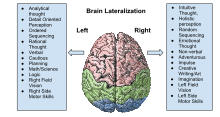
A stereotypical image of brain lateralisation - demonstrated to be false in neuroscientific research.

Neuroanatomical differences themselves exist on different scales, from neuronal densities, to the size of regions such as the planum temporale, to—at the largest scale—the torsion or "wind" in the human brain, reflected shape of the skull, which reflects a backward (posterior) protrusion of the left occipital bone and a forward (anterior) protrusion of the right frontal bone. In addition to gross size differences, both neurochemical and structural differences have been found between the hemispheres. Asymmetries appear in the spacing of cortical columns, as well as dendritic structure and complexity. Larger cell sizes are also found in layer III of Broca's area.

The human brain has an overall leftward posterior and rightward anterior asymmetry (or brain torque). There are particularly large asymmetries in the frontal, temporal and occipital lobes, which increase in asymmetry in the antero-posterior direction beginning at the central region. Leftward asymmetry can be seen in the Heschl gyrus, parietal operculum, Silvian fissure, left cingulate gyrus, temporo-parietal region and planum temporale. Rightward asymmetry can be seen in the right central sulcus (potentially suggesting increased connectivity between motor and somatosensory cortices in the left side of the brain), lateral ventricle, entorhinal cortex, amygdala and temporo-parieto-occipital area. Sex-dependent brain asymmetries are also common. For example, human male brains are more asymmetrically lateralized than those of females. However, gene expression studies done by Hawrylycz and colleagues and Pletikos and colleagues, were not able to detect asymmetry between the hemispheres on the population level.

People with autism have much more symmetrical brains than people without it.

History

In the mid-19th century scientists first began to make discoveries regarding lateralization of the brain, or differences in anatomy and corresponding function between the brain's two hemispheres. Franz Gall, a German anatomist, was the first to describe what is now known as the Doctrine of Cerebral Localization. Gall believed that, rather than the brain operating as a single, whole entity, different mental functions could be attributed to different parts of the brain. He was also the first to suggest language processing happened in the frontal lobes. However, Gall's theories were controversial among many scientists at the time. Others were convinced by experiments such as those conducted by Marie-Jean-Pierre Flourens, in which he demonstrated lesions to bird brains caused irreparable damage to vital functions. Flourens's methods, however, were not precise; the crude methodology employed in his experiments actually caused damage to several areas of the tiny brains of the avian models.

Paul Broca was among the first to offer compelling evidence for localization of function when he identified an area of the brain related to speech.

In 1861 surgeon Paul Broca provided evidence that supported Gall's theories. Broca discovered that two of his patients who had had speech loss had similar lesions in the same area of the left frontal lobe. While this was compelling evidence for localization of function, the connection to "sidedness" was not made immediately. As Broca continued to study similar patients, he made the connection that all of the cases involved damage to the left hemisphere, and in 1864 noted the significance of these findings—that this must be a specialized region. He also—incorrectly—proposed theories about the relationship of speech areas to "handedness".

Accordingly, some of the most famous early studies on brain asymmetry involved speech processing. Asymmetry in the Sylvian fissure (also known as the lateral sulcus), which separates the frontal and parietal lobes from the temporal lobe, was one of the first incongruencies to be discovered. Its anatomical variances are related to the size and location of two areas of the human brain that are important for language processing, Broca's area and Wernicke's area, both in the left hemisphere.

Around the same time that Broca and Wernicke made their discoveries, neurologist Hughlings Jackson suggested the idea of a "leading hemisphere"—or, one side of the brain that played a more significant role in overall function—which would eventually pave the way for understanding hemispheric "dominance" for various processes. Several years later, in the mid-20th century, critical understanding of hemispheric lateralization for visuospatial, attention and perception, auditory, linguistic and emotional processing came from patients who underwent split-brain procedures to treat disorders such as epilepsy. In split-brain patients, the corpus callosum is cut, severing the main structure for communication between the two hemispheres. The first modern split-brain patient was a war veteran known as Patient W.J., whose case contributed to further understanding of asymmetry.

Brain asymmetry is not unique to humans. In addition to studies on human patients with various diseases of the brain, much of what is understood today about asymmetries and lateralization of function has been learned through both invertebrate and vertebrate animal models, including zebrafish, pigeons, rats, and many others. For example, more recent studies revealing sexual dimorphism in brain asymmetries in the cerebral cortex and hypothalamus of rats show that sex differences emerging from hormonal signaling can be an important influence on brain structure and function. Work with zebrafish has been especially informative because this species provides the best model for directly linking asymmetric gene expression with asymmetric morphology, and for behavioral analyses.

In humans

Lateralized functional differences

The left and right hemispheres operate the contralateral sides of the body. Each hemisphere contains sections of all 4 lobes: the frontal lobe, parietal lobe, temporal lobe, and occipital lobe. The two hemispheres are separated along the mediated longitudinal fissure and are connected by the corpus callosum which allows for communication and coordination of stimuli and information. The corpus callosum is the largest collective pathway of white matter tissue in the body that is made of more than 200 million nerve fibers. The left and right hemispheres are associated with different functions and specialize in interpreting the same data in different ways, referred to as lateralization of the brain. The left hemisphere is associated with language and calculations, while the right hemisphere is more closely associated with visual-spatial recognition and facial recognition. This lateralization of brain function results in some specialized regions being only present in a certain hemisphere or being dominant in one hemisphere versus the other. Some of the significant regions included in each hemisphere are listed below.

Left hemisphere

Broca's Area
Broca's area is located in the left hemisphere prefrontal cortex above the cingulate gyrus in the third frontal convolution. Broca's area was discovered by Paul Broca in 1865. This area handles speech production. Damage to this area would result in Broca aphasia which causes the patient to become unable to formulate coherent appropriate sentences.
Wernicke's Area
Wernicke's area was discovered in 1976 by Carl Wernicke and was found to be the site of language comprehension. Wernicke's area is also found in the left hemisphere in the temporal lobe. Damage to this area of the brain results in the individual losing the ability to understand language. However, they are still able to produce sounds, words, and sentence although they are not used in the appropriate context.

Right hemisphere

Fusiform Face Area
The Fusiform face area (FFA) is an area that has been studied to be highly active when faces are being attended to in the visual field. A FFA is found to be present in both hemispheres, however, studies have found that the FFA is predominantly lateralized in the right hemisphere where a more in-depth cognitive processing of faces is conducted. The left hemisphere FFA is associated with rapid processing of faces and their features.

Other regions and associated diseases

Some significant regions that can present as asymmetrical in the brain can result in either of the hemispheres due to factors such as genetics. An example would include handedness. Handedness can result from asymmetry in the motor cortex of one hemisphere. For right handed individuals, since the brain operates the contralateral side of the body, they could have a more induced motor cortex in the left hemisphere.

Several diseases have been found to exacerbate brain asymmetries that are already present in the brain. Researchers are starting to look into the effect and relationship of brain asymmetries to diseases such as schizophrenia and dyslexia.

Schizophrenia
Schizophrenia is a complex long-term mental disorder that causes hallucinations, delusions and a lack of concentration, thinking, and motivation in an individual. Studies have found that individuals with schizophrenia have a lack in brain asymmetry thus reducing the functional efficiency of affected regions such as the frontal lobe. Conditions include leftward functional hemispheric lateralization, loss of laterality for language comprehension, a reduction in gyrification, brain torsion etc.
Dyslexia
As studied earlier, language is usually dominant in the left hemisphere. Developmental language disorders, such as dyslexia, have been researched using brain imaging techniques to understand the neuronal or structural changes associated with the disorder. Past research has exhibited that hemispheric asymmetries that are usually found in healthy adults such as the size of the temporal lobe is not present in adult patients with dyslexia. In conjunction, past research has exhibited that patients with dyslexia lack a lateralization of language in their brain compared to healthy patients. Instead patients with dyslexia showed to have a bilateral hemispheric dominance for language.

Current research

Lateralization of function and asymmetry in the human brain continues to propel a popular branch of neuroscientific and psychological inquiry. Technological advancements for brain mapping have enabled researchers to see more parts of the brain more clearly, which has illuminated previously undetected lateralization differences that occur during different life stages. As more information emerges, researchers are finding insights into how and why early human brains may have evolved the way that they did to adapt to social, environmental and pathological changes. This information provides clues regarding plasticity, or how different parts of the brain can sometimes be recruited for different functions.

Continued study of brain asymmetry also contributes to the understanding and treatment of complex diseases. Neuroimaging in patients with Alzheimer's disease, for example, shows significant deterioration in the left hemisphere, along with a rightward hemispheric dominance—which could relate to recruitment of resources to that side of the brain in the face of damage to the left. These hemispheric changes have been connected to performance on memory tasks.

As has been the case in the past, studies on language processing and the implications of left- and right- handedness also dominate current research on brain asymmetry.

Dual consciousness

From Wikipedia, the free encyclopedia

Dual consciousness is a theoretical concept in neuroscience. It is proposed that it is possible that a person may develop two separate conscious entities within their one brain after undergoing a corpus callosotomy. The idea first began circulating in the neuroscience community after some split-brain patients exhibited the alien hand syndrome, which led some scientists to believe that there must be two separate consciousnesses within the brain's left and right hemispheres in competition with one another once the corpus callosum is severed.

The idea of dual consciousness has caused controversy in the neuroscience community. No conclusive evidence of the proposed phenomenon has been discovered.

Background

During the first half of the 20th century, some neurosurgeons concluded that the best option of treating severe epilepsy was by severing the patient's corpus callosum. The corpus callosum is the primary communication mechanism between the brain's two cerebral hemispheres. For example, communication across the callosum allows information from both the left and right visual fields to be interpreted by the brain in a way that makes sense to comprehend the person's actual experience (visual inputs from both eyes are interpreted by the brain to make sense of the experience that you are looking at a computer that is directly in front of you). The procedure of surgically removing the corpus callosum is called a corpus callosotomy. Patients who have undergone a corpus callosotomy are colloquially referred to as "split-brain patients". They are called so because now their brain's left and right hemispheres are no longer connected by the corpus callosum.

Split-brain patients have been subjects for numerous psychological experiments that sought to discover what occurs in the brain now that the primary interhemispheric pathways have been disrupted. Notable researchers in the field include Roger Sperry, one of the first to publish ideas involving a dual consciousness, and his famous graduate student, Michael Gazzaniga. Their results found a pattern among patients: severing the entire corpus callosum stops the interhemispheric transfer of perceptual, sensory, motor, and other forms of information. For most cases, corpus callosotomies did not in any way affect patients' real-world functioning; however, those psychology experiments have demonstrated some interesting differences between split-brain patients and normal subjects.

Split-brain patients and the corpus callosotomy

The first successful corpus callosotomies on humans were performed in the 1930s. The purpose of the procedure was to alleviate the effects of epilepsy when other forms of treatment (medications) had failed to stop the violent convulsions associated with the disorder. Epileptic seizures occur because of abnormal electrical discharges that spread across areas of the brain. William Van Wagenen proposed the idea of severing the corpus callosum to eliminate transcortical electrical signals across the brain's hemispheres. If this could be achieved, then the seizures should be reduced or even eliminated.

The general procedure of a corpus callosotomy is as follows. The patient is put under anesthesia. Once the patient is in deep sleep, a craniotomy is performed. This procedure removes a section of the skull, leaving the brain exposed and accessible to the surgeon. The dura mater is pulled back so the deeper areas of the brain, including the corpus callosum, can be seen. Specialized instruments are placed into the brain that allows safe severing of the corpus. Initially, a partial callosotomy is performed, which only severs the front two-thirds of the callosum. It is important to note that because the back section of the callosum is preserved, visual information is still sent across both hemispheres. Though the corpus callosum loses a majority of its functioning during a partial callosotomy, it does not completely lose its capabilities. If this operation does not succeed in reducing the seizures, a complete callosotomy is needed to reduce the severity of the seizures. A similar type of procedure, known as a commissurotomy, involves severing a number of interhemispheric tracts (such as the anterior commissure, the hippocampal commissure and the massa intermedia of the thalamus) in addition to the corpus callosum.

After surgery, the split-brain patients are often given extensive neuropsychological assessments. An interesting finding among split-brain patients is many of them claim to feel normal after the surgery and do not feel that their brains are "split". The corpus callosotomy and commissurotomy have been successful in reducing, and in some cases, eliminating epileptic seizures. Van Wagenen's theory was correct.

Alien hand syndrome

Alien hand syndrome, sometimes used synonymously with anarchic hand is a neurological disorder in which the afflicted person's hand appears to take on a mind of its own. Alien hand syndrome has been documented in some split brain patients.

Symptoms

The classic sign of Alien Hand Syndrome is that the affected person cannot control one of their hands. For example, if a split-brain patient with Alien Hand Syndrome is asked to pick up a glass with their right hand, as the right hand moves over to the glass, the left hand will interfere with the action, thwarting the right hand's task. The interference from the left hand is completely out of the control of the patient and is not being done "on purpose". Affected patients at times cannot control the movements of their hands. Another example included patients unbuttoning a shirt with one hand, and the other hand simultaneously re-buttoning the shirt (although some reported feeling normal after their surgery).

Relationship to dual consciousness

When scientists first started observing the alien hand syndrome in split-brain patients, they began to question the nature of consciousness and began to theorize that perhaps when the corpus callosum is cut, consciousness also is split into two separate entities. This development added to the general appeal of split-brain research.

Gazzaniga and LeDoux's experiment

Procedure and results

In 1978, Michael Gazzaniga and Joseph DeLoux discovered a unique phenomenon among split-brain patients who were asked to perform a simultaneous concept task. The patient was shown 2 pictures: of a house in the winter time and of a chicken's claw. The pictures were positioned so they would exclusively be seen in only one visual field of the brain (the winter house was positioned so it would only be seen in the patient's left visual field (LVF), which corresponds to the brain's right hemisphere, and the chicken's claw was placed so it would only be seen in the patient's right visual field (RVF), which corresponds to the brain's left hemisphere).

A series of pictures was placed in front of the patients. Gazzaniga and LeDoux then asked the patient to choose a picture with his right hand and a picture with his left hand. The paradigm was set up so the choices would be obvious for the patients. A snow shovel is used for shoveling the snowy driveway of the winter house and a chicken's head correlates to the chicken's claw. The other pictures do not in any way correlate with the 2 original pictures. In the study, a patient chose the snow shovel with his left hand (corresponding to his brain's right hemisphere) and his right hand chose the chicken's head (corresponding to the brain's left hemisphere). When the patient was asked why he had chosen the pictures he had chosen, the answer he gave was "The chicken claw goes with the chicken head, and you need a snow shovel to clean out the chicken shed."

Why would he say this? Wouldn't it be obvious that the shovel goes with the winter house? For people with an intact corpus callosum, yes it is obvious, but not for a split-brain patient. Both the winter house and the shovel are being projected to the patient from his LVF, so his right hemisphere is receiving and processing the information and this input is completely independent from what is going on in the RVF, which involves the chicken's claw and head (the information being processed in the left hemisphere). The human brain's left hemisphere is primarily responsible interpreting the meaning of the sensory input it receives from both fields; however, the left hemisphere has no knowledge of the winter house. Because it has no knowledge of the winter house, it must invent a logical reason for why the shovel was chosen. Since the only objects it has to work with are the chicken's claw and head, the left hemisphere interprets the meaning of choosing the shovel as "it is an object necessary to help the chicken, which lives in a shed, therefore, the shovel is used to clean the chicken’s shed." Gazzaniga famously coined the term left brain interpreter to explain this phenomenon.

Interpreting Gazzaniga's "left brain interpreter"

What does the results of Gazzaniga and LeDoux's work suggest about the existence of a dual consciousness? There are varying possibilities.

  • The left hemisphere dominates all interpretation of the split-brain patient's perceptual field, with the right hemisphere having little importance in these processes.
  • If so, one could by extension claim there are 2 separate conscious entities that do not interact with each other or are in competition with each other and have separate interpretations of the stimuli, the left hemisphere winning the struggle.
  • Or perhaps the right hemisphere is unconscious of the snow house and shovel while the left hemisphere retains a conscious perception of its objects.

Other experiments

Sperry–Gazzaniga

The Gazzaniga–LeDoux studies were based on previous studies done by Sperry and Gazzaniga. Sperry examined split-brain patients. Sperry's experiment included a subject being seated at a table, with a shield blocking the visions from the subject's hands, including the objects on the table and the examiner seated across. The shield was also used as a viewing screen. On the shield, the examiner can select to present the visual material to both hemispheres or to selective hemispheres by means of having the patient look at certain points on the viewing screen. The patient is briefly exposed to the stimuli on the viewing screen. The stimuli shown to the left eye goes to the right hemisphere and the visual material shown to the right eye will be projected to the left hemisphere. During the experiment, when the stimulus was shown to the left side of the screen, the patient indicated he did not see anything. Patients have shown the inability to describe in writing or in speech the stimuli that was shown briefly to the left side. The speaking hemisphere, which in most people is the left hemisphere, would not have awareness of stimulus being shown to the right hemisphere (left visual field), except the left hand was able to point to the correct object. Based on his observations and data, Sperry concluded each hemisphere possessed its own consciousness.

Revonsuo

Revonsuo explains a procedure that was similar in nature to the Sperry–Gazzaniga design. Split-brain patients are shown a picture with two objects: a flower and a rabbit. The flower is exclusively shown in the right visual field, which is interpreted by the left hemisphere and the rabbit is exclusively shown in the left visual field, which is interpreted by the right hemisphere. The left brain is seeing the flower as the right brain is simultaneously viewing the rabbit. When the patients were asked what they saw, patients said they only saw the flower and did not see the rabbit. The flower is in the right visual field and the left hemisphere can only see the flower. The left hemisphere dominates the interpretation of the stimulus and since it cannot see the rabbit (only being represented in the right hemisphere), patients do not believe they saw a rabbit. They can, however, still point to the rabbit with their left hand. Revonsuo stated that it seemed that one consciousness saw the flower and another consciousness saw the rabbit independently from one another.

Joseph

Rhawn Joseph observed two patients who had both undergone a complete corpus callosotomy. Joseph observed that the right hemisphere of one of the patients is able to gather, comprehend, and express information. The right hemisphere was able to direct activity to the patient's left arm and leg. The execution of the left arm and leg's action as was inhibited by the left hemisphere. Joseph found that the patient's left leg would attempt to move forward as if to walk straight but the right leg would either refuse to move or begin to walk in the opposite direction. After observing the struggles of the execution of activities involving the left and right arms and legs, led Joseph to believe that the two hemispheres possessed their own consciousness.

Joseph also noted that the patient had other specific instances of conflict between the right and left hemispheres including, the left hand (right hemisphere) carrying out actions contrary to the left hemisphere's motives such as the left hand turning off the television immediately after the right hand turned it on. Joseph found that the patient's left leg would only allow the patient to return home when the patient was going for a walk and would reject continuing to go for that walk.

Further observations by Joseph

In the laboratory, a patient was given two different fabrics: a wire screen in his left hand and a piece of sandpaper in his right hand. The patient received two different fabrics out of his view so that neither eye nor hemisphere visually seen what his hands were given. When the patient was indicating what fabric was in the left hand, he was able to correctly indicate and point with the left hand to the wire screen after it had been set on a table. As he pointed with his left hand, however, the right hand tried to stop the left hand and make the left hand point to the fabric that the right hand was holding. The left hand continued to point at the correct fabric, even though the right hand tried to forcefully move the left hand. During the struggle, the patient also verbalized feelings of animosity by saying, "That’s wrong!" and "I hate this hand." Joseph concluded that the left hemisphere did not understand at all why the left hand (right hemisphere) would point to a different material.

Controversy and alternative explanations

Proponents of the dual consciousness theory have caused a great amount of controversy and debate within the neuroscience community. The magnitude of such a claim: that consciousness can be split into two entities within the one brain are considered by some scientists to be audacious. There is no concrete evidence to validate the theory and the current evidence provided is, at best, anecdotal.

The most powerful claims against the dual consciousness theory are:

  • There is no universally accepted definition of "consciousness".
  • Split-brain patients are not the only people to exhibit the Alien Hand Syndrome. People with intact brains who have suffered a stroke may also have the Alien Hand Syndrome. It also has been observed in patients with Alzheimer's disease or in patients who have brain tumors.
  • Other existing and established neurological mechanisms can account for an explanation of the same phenomena.

Gonzalo Munevar has proposed an alternative explanation to demonstrate that these strange behaviors are spawned from areas in the brain and not by a dual consciousness. Two cortical areas in particular, the supplementary motor area (SMA) and the premotor cortex (PMC), are crucial in the planning of executing motor tasks to external stimuli presented in the person's perceptual field. For example, a person may pick up a glass of water with his right hand and put it up to his lips for a drink. The person may have picked up the glass with his right hand, but well before this action takes place, the PMC and SMA consider a variety of different possibilities of how this action could be performed. He could have picked it up with his left hand, his mouth, even his foot! He could have done it quickly or slowly. Many possibilities are entertained, but few are actually executed. These actions are sent from the PMC to the Motor Cortex for execution. The rest are inhibited by the SMA and are not performed.

It is also important to understand that the processes of the SMA and PMC are done unconsciously. The SMA and PMC consider the many alternative actions many milliseconds before the chosen action takes place. The person is never consciously aware of these alternative possibilities the brain has juggled with before he picks it up with the right hand; he just does it. The action of picking up the glass with the right hand is also performed unconsciously. It may be preferable to use his right hand because he is right handed and doing so is therefore more comfortable or perhaps the glass is placed on his right side and the possibility that expends the least amount of energy is using the right hand to pick it up.

Another important fact about the PMC is that its activation is bilateral. When it is activated, it is activated in both hemispheres of the brain. Gazzaniga observed and wrote about this phenomenon. When the corpus callosum is severed, many interhemispheric interactions are disrupted. Many areas of the brain become compromised, including the SMA. If the SMA has trouble regulating and inhibiting the actions of the PMC, it is very possible that conflicting sets of actions may be sent to the MC and performed (accounting for both hands reaching for the glass, even if only one hand is intended to grab it). It would make the appearance that there is a dual consciousness competing for dominance over the other for control of the brain, but it is not the case.

The fact that the Alien Hand Syndrome eventually goes away in some split-brain patients is not evidence of one consciousness "defeating" the other and taking complete control of the brain. It is likely that the plasticity of the brain may be the cause for alleviating the disorder. Eventually the split patient's brain may find adaptive routes to compensate for the lost interhemispheric communication, such as alternative pathways involving subcortical structures that perform subcortical interhemispheric inhibition to regain a sense of normalcy between the two hemispheres.

Models of multiple consciousnesses

Michael Gazzaniga, while working on the model of dual consciousness, came to the conclusion that simple dual consciousness (i.e. right-brain/left-brain model of the mind) is a gross oversimplification and the brain is organized into hundreds maybe even thousands of modular-processing systems.

The theory of a division of consciousness was touched upon by Carl Jung in 1935 when he stated, "The so-called unity of consciousness is an illusion... we like to think that we are one but we are not."

Similar models (which also claim that mind is formed from many little agents, i.e. the brain is made up of a constellation of independent or semi-independent agents) were also described by:

  • Marvin Minsky's "Society of Mind" model claims that mind is built up from the interactions of simple parts called agents, which are themselves mindless.
  • Thomas R. Blakeslee described the brain model which claims that brain is composed of hundreds of independent centers of thought called "modules".
  • Neurocluster Brain Model describes the brain as a massively parallel computing machine in which huge number of neuroclusters process information independently from each other. The neurocluster which most of the time has the access to actuators (i.e. neurocluster which most of the time acts upon an environment using actuators) is called the main personality. Other neuroclusters which do not have access to actuators or which have only short duration and limited access to actuators are called "autonomous neuroclusters".
  • Michio Kaku described the brain model using the analogy of large corporation which is controlled by CEO.
  • Robert E. Ornstein claimed that the mind is a squadron of simpletons. Ernest Hilgard described neodissociationist theory which claims that a "hidden observer" is created in the mind while hypnosis is taking place and this "hidden observer" has his own separate consciousness.
  • George Ivanovich Gurdjieff in year 1915 taught his students that man has no single, big I; man is divided into a multiplicity of small I’s.

Trikaya

From Wikipedia, the free encyclopedia The Trikāya ( Sanskrit : त्रिकाय , lit...