Search This Blog

Friday, June 23, 2023

Structure and genome of HIV

The genome and proteins of HIV (human immunodeficiency virus) have been the subject of extensive research since the discovery of the virus in 1983. "In the search for the causative agent, it was initially believed that the virus was a form of the Human T-cell leukemia virus (HTLV), which was known at the time to affect the human immune system and cause certain leukemias. However, researchers at the Pasteur Institute in Paris isolated a previously unknown and genetically distinct retrovirus in patients with AIDS which was later named HIV." Each virion comprises a viral envelope and associated matrix enclosing a capsid, which itself encloses two copies of the single-stranded RNA genome and several enzymes. The discovery of the virus itself occurred two years following the report of the first major cases of AIDS-associated illnesses.

Structure

Diagram of HIV
 
Structure of the immature HIV-1 capsid in intact virus particles
 
A diagram of the HIV spike protein (green), with the fusion peptide epitope highlighted in red, and a broadly neutralizing antibody (yellow) binding to the fusion peptide

The complete sequence of the HIV-1 genome, extracted from infectious virions, has been solved to single-nucleotide resolution. The HIV genome encodes a small number of viral proteins, invariably establishing cooperative associations among HIV proteins and between HIV and host proteins, to invade host cells and hijack their internal machineries. HIV is different in structure from other retroviruses. The HIV virion is ~100 nm in diameter. Its innermost region consists of a cone-shaped core that includes two copies of the (positive sense) ssRNA genome, the enzymes reverse transcriptase, integrase and protease, some minor proteins, and the major core protein. The genome of human immunodeficiency virus (HIV) encodes 8 viral proteins playing essential roles during the HIV life cycle.

HIV-1 is composed of two copies of noncovalently linked, unspliced, positive-sense single-stranded RNA enclosed by a conical capsid composed of the viral protein p24, typical of lentiviruses. The two copies of RNA strands are vital in contributing to HIV-1 recombination, which occurs during reverse transcription of viral replication. The containment of two copies of single-stranded RNA within a virion but the production of only a single DNA provirus is called pseudodiploidy. The RNA component is 9749 nucleotides long and bears a 5’ cap (Gppp), a 3’ poly(A) tail, and many open reading frames (ORFs). Viral structural proteins are encoded by long ORFs, whereas smaller ORFs encode regulators of the viral life cycle: attachment, membrane fusion, replication, and assembly.

The single-strand RNA is tightly bound to p7 nucleocapsid proteins, late assembly protein p6, and enzymes essential to the development of the virion, such as reverse transcriptase and integrase. Lysine tRNA is the primer of the magnesium-dependent reverse transcriptase. The nucleocapsid associates with the genomic RNA (one molecule per hexamer) and protects the RNA from digestion by nucleases. Also enclosed within the virion particle are Vif, Vpr, Nef, and viral protease. The envelope of the virion is formed by a plasma membrane of host cell origin, which is supported by a matrix composed of the viral p17 protein, ensuring the integrity of the virion particle. At the surface of the virion can be found a limited number of the envelope glycoprotein (Env) of HIV, a trimer formed by heterodimers of gp120 and gp41. Env is responsible for binding to its primary host receptor, CD4, and its co-receptor (mainly CCR5 or CXCR4), leading to viral entry into its target cell.

As the only proteins on the surface of the virus, the envelope glycoproteins (gp120 and gp41) are the major targets for HIV vaccine efforts. Over half of the mass of the trimeric envelope spike is N-linked glycans. The density is high as the glycans shield underlying viral protein from neutralisation by antibodies. This is one of the most densely glycosylated molecules known and the density is sufficiently high to prevent the normal maturation process of glycans during biogenesis in the endoplasmic reticulum and Golgi apparatus. The majority of the glycans are therefore stalled as immature 'high-mannose' glycans not normally present on secreted or cell surface human glycoproteins. The unusual processing and high density means that almost all broadly neutralising antibodies that have so far been identified (from a subset of patients that have been infected for many months to years) bind to or, are adapted to cope with, these envelope glycans.

The molecular structure of the viral spike has now been determined by X-ray crystallography and cryo-electron microscopy. These advances in structural biology were made possible due to the development of stable recombinant forms of the viral spike by the introduction of an intersubunit disulphide bond and an isoleucine to proline mutation in gp41. The so-called SOSIP trimers not only reproduce the antigenic properties of the native viral spike but also display the same degree of immature glycans as presented on the native virus. Recombinant trimeric viral spikes are promising vaccine candidates as they display less non-neutralising epitopes than recombinant monomeric gp120 which act to suppress the immune response to target epitopes.

Genome organization

Structure of the RNA genome of HIV-1

HIV has several major genes coding for structural proteins that are found in all retroviruses as well as several nonstructural ("accessory") genes unique to HIV. The HIV genome contains nine genes that encode fifteen viral proteins. These are synthesized as polyproteins which produce proteins for virion interior, called Gag, group specific antigen; the viral enzymes (Pol, polymerase) or the glycoproteins of the virion env (envelope). In addition to these, HIV encodes for proteins which have certain regulatory and auxiliary functions as well. HIV-1 has two important regulatory elements: Tat and Rev and few important accessory proteins such as Nef, Vpr, Vif and Vpu which are not essential for replication in certain tissues. The gag gene provides the basic physical infrastructure of the virus, and pol provides the basic mechanism by which retroviruses reproduce, while the others help HIV to enter the host cell and enhance its reproduction. Though they may be altered by mutation, all of these genes except tev exist in all known variants of HIV; see Genetic variability of HIV.

HIV employs a sophisticated system of differential RNA splicing to obtain nine different gene products from a less than 10kb genome. HIV has a 9.2kb unspliced genomic transcript which encodes for gag and pol precursors; a singly spliced, 4.5 kb encoding for env, Vif, Vpr and Vpu and a multiply spliced, 2 kb mRNA encoding for Tat, Rev and Nef.

Proteins encoded by the HIV genome
Class Gene name Primary protein products Processed protein products
Viral structural proteins gag Gag polyprotein MA, CA, SP1, NC, SP2, P6
pol Pol polyprotein RT, RNase H, IN, PR
env gp160 gp120, gp41
Essential regulatory elements tat Tat
rev Rev
Accessory regulatory proteins nef Nef
vpr Vpr
vif Vif
vpu Vpu

Viral structural proteins

The HIV capsid consists of roughly 200 copies of the p24 protein. The p24 structure is shown in two representations: cartoon (top) and isosurface (bottom)
  • gag (group-specific antigen) codes for the precursor gag polyprotein which is processed by viral protease during maturation to MA (matrix protein, p17); CA (capsid protein, p24); SP1 (spacer peptide 1, p2); NC (nucleocapsid protein, p7); SP2 (spacer peptide 2, p1) and P6 protein.
  • pol codes for viral enzymes reverse transcriptase (RT) and RNase H, integrase (IN), and HIV protease (PR). HIV protease is required to cleave the precursor Gag polyprotein to produce structural proteins, RT is required to transcribe DNA from RNA template, and IN is necessary to integrate the double-stranded viral DNA into the host genome.
  • env (for "envelope") codes for gp160, which is cleaved by a host protease, furin, within the endoplasmic reticulum of the host cell. The post-translational processing produces a surface glycoprotein, gp120 or SU, which attaches to the CD4 receptors present on lymphocytes, and gp41 or TM, which embeds in the viral envelope to enable the virus to attach to and fuse with target cells.

Essential regulatory elements

  • tat (HIV trans-activator) plays an important role in regulating the reverse transcription of viral genome RNA, ensuring efficient synthesis of viral mRNAs and regulating the release of virions from infected cells. Tat is expressed as 72-amino acid one-exon Tat as well as the 86–101-amino-acid two-exon Tat, and plays an important role early in HIV infection. Tat (14–15 kDa) binds to the bulged genomic RNA stem-loop secondary structure near the 5' LTR region forming the trans-activation response element (TAR).
  • rev (regulator of expression of virion proteins): The Rev protein binds to the viral genome via an arginine-rich RNA-binding motif that also acts as a NLS (nuclear localization signals), required for the transport of Rev to the nucleus from cytosol during viral replication. Rev recognizes a complex stem-loop structure of the mRNA env located in the intron separating coding exon of Tat and Rev, known as the HIV Rev response element (RRE). Rev is important for the synthesis of major viral proteins and is hence essential for viral replication.

Accessory regulatory proteins

  • vpr (lentivirus protein R): Vpr is a virion-associated, nucleocytoplasmic shuttling regulatory protein. It is believed to play an important role in replication of the virus, specifically, nuclear import of the preintegration complex. Vpr also appears to cause its host cells to arrest their cell cycle in the G2 phase. This arrest activates the host DNA repair machinery which may enable integration of the viral DNA. HIV-2 and SIV encode an additional Vpr related protein called Vpx which functions in association with Vpr.
  • vif - Vif is a highly conserved, 23 kDa phosphoprotein important for the infectivity of HIV-1 virions depending on the cell type. HIV-1 has been found to require Vif to synthesize infectious viruses in lymphocytes, macrophages, and certain human cell lines. It does not appear to require Vif for the same process in HeLa cells or COS cells, among others.
  • nef- Nef, negative factor, is a N-terminal myristoylated membrane-associated phosphoprotein. It is involved in multiple functions during the replication cycle of the virus. It is believed to play an important role in cell apoptosis and increase virus infectivity.
  • vpu (Virus protein U) - Vpu is specific to HIV-1. It is a class I oligomeric integral membrane phosphoprotein with numerous biological functions. Vpu is involved in CD4 degradation involving the ubiquitin proteasome pathway as well as in the successful release of virions from infected cells.
  • tev: This gene is only present in a few HIV-1 isolates. It is a fusion of parts of the tat, env, and rev genes, and codes for a protein with some of the properties of tat, but little or none of the properties of rev.

RNA secondary structure

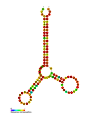
HIV pol-1 stem loop
RF01418.png
Predicted secondary structure of the HIV pol-1 stem loop
 
Identifiers
Symbolpol
RfamRF01418
Other data
RNA typeCis-reg
PDB structuresPDBe

Several conserved secondary structure elements have been identified within the HIV RNA genome. The HIV viral RNA structures regulates the progression of reverse transcription.  The 5'UTR structure consists of series of stem-loop structures connected by small linkers. These stem-loops (5' to 3') include the trans-activation region (TAR) element, the 5' polyadenylation signal [poly(A)], the PBS, the DIS, the major SD and the ψ hairpin structure located within the 5' end of the genome and the HIV Rev response element (RRE) within the env gene. Another RNA structure that has been identified is gag stem loop 3 (GSL3), thought to be involved in viral packaging. RNA secondary structures have been proposed to affect the HIV life cycle by altering the function of HIV protease and reverse transcriptase, although not all elements identified have been assigned a function.

An RNA secondary structure determined by SHAPE analysis has shown to contain three stem loops and is located between the HIV protease and reverse transcriptase genes. This cis regulatory RNA has been shown to be conserved throughout the HIV family and is thought to influence the viral life cycle.

V3 loop

The third variable loop or V3 loop is a part or region of the Human Immunodeficiency Virus. The V3 loop of the viron's envelope glycoprotein, gp120, allows it to infect human immune cells by binding to a cytokine receptor on the target human immune cell, such as a CCR5 cell or CXCR4 cell, depending on the strain of HIV. The envelope glycoprotein (Env) gp 120/41 is essential for HIV-1 entry into cells. Env serves as a molecular target of a medicine treating individuals with HIV-1 infection, and a source of immunogen to develop AIDS vaccine. However, the structure of the functional Env trimer has remained elusive.

Thursday, June 22, 2023

Bioavailability

From Wikipedia, the free encyclopedia

In pharmacology, bioavailability is a subcategory of absorption and is the fraction (%) of an administered drug that reaches the systemic circulation.

By definition, when a medication is administered intravenously, its bioavailability is 100%. However, when a medication is administered via routes other than intravenous, its bioavailability is generally lower than that of intravenous due to intestinal endothelium absorption and first-pass metabolism. Thereby, mathematically, bioavailability equals the ratio of comparing the area under the plasma drug concentration curve versus time (AUC) for the extravascular formulation to the AUC for the intravascular formulation. AUC is used because AUC is proportional to the dose that has entered the systemic circulation.

Bioavailability of a drug is an average value; to take population variability into account. To ensure that the drug taker who has poor absorption is dosed appropriately, the bottom value of the deviation range is employed to represent real bioavailability and to calculate the drug dose needed for the drug taker to achieve systemic concentrations similar to the intravenous formulation. To dose without knowing the drug taker's absorption rate, the bottom value of the deviation range is used in order to ensure the intended efficacy, unless the drug is associated with a narrow therapeutic window.

For dietary supplements, herbs and other nutrients in which the route of administration is nearly always oral, bioavailability generally designates simply the quantity or fraction of the ingested dose that is absorbed.

Definitions

In pharmacology

Bioavailability is a term used to describe the percentage of an administered dose of a xenobiotic that reaches the systemic circulation. It is denoted by the letter f (or, if expressed in percent, by F).

In nutritional science

In nutritional science, which covers the intake of nutrients and non-drug dietary ingredients, the concept of bioavailability lacks the well-defined standards associated with the pharmaceutical industry. The pharmacological definition cannot apply to these substances because utilization and absorption is a function of the nutritional status and physiological state of the subject, resulting in even greater differences from individual to individual (inter-individual variation). Therefore, bioavailability for dietary supplements can be defined as the proportion of the administered substance capable of being absorbed and available for use or storage.

In both pharmacology and nutrition sciences, bioavailability is measured by calculating the area under curve (AUC) of the drug concentration time profile.

In environmental sciences or science

Bioavailability is the measure by which various substances in the environment may enter into living organisms. It is commonly a limiting factor in the production of crops (due to solubility limitation or absorption of plant nutrients to soil colloids) and in the removal of toxic substances from the food chain by microorganisms (due to sorption to or partitioning of otherwise degradable substances into inaccessible phases in the environment). A noteworthy example for agriculture is plant phosphorus deficiency induced by precipitation with iron and aluminum phosphates at low soil pH and precipitation with calcium phosphates at high soil pH. Toxic materials in soil, such as lead from paint may be rendered unavailable to animals ingesting contaminated soil by supplying phosphorus fertilizers in excess. Organic pollutants such as solvents or pesticides may be rendered unavailable to microorganisms and thus persist in the environment when they are adsorbed to soil minerals or partition into hydrophobic organic matter.

Absolute bioavailability

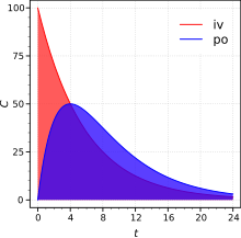
Absolute bioavailability is a ratio of areas under the curves. IV, intravenous; PO, oral route. C is plasma concentration (arbitrary units).

Absolute bioavailability compares the bioavailability of the active drug in systemic circulation following non-intravenous administration (i.e., after oral, buccal, ocular, nasal, rectal, transdermal, subcutaneous, or sublingual administration), with the bioavailability of the same drug following intravenous administration. It is the fraction of the drug absorbed through non-intravenous administration compared with the corresponding intravenous administration of the same drug. The comparison must be dose normalized (e.g., account for different doses or varying weights of the subjects); consequently, the amount absorbed is corrected by dividing the corresponding dose administered.

In pharmacology, in order to determine absolute bioavailability of a drug, a pharmacokinetic study must be done to obtain a plasma drug concentration vs time plot for the drug after both intravenous (iv) and extravascular (non-intravenous, i.e., oral) administration. The absolute bioavailability is the dose-corrected area under curve (AUC) non-intravenous divided by AUC intravenous. The formula for calculating the absolute bioavailability, F, of a drug administered orally (po) is given below (where D is dose administered).

Therefore, a drug given by the intravenous route will have an absolute bioavailability of 100% (f = 1), whereas drugs given by other routes usually have an absolute bioavailability of less than one. If we compare the two different dosage forms having same active ingredients and compare the two drug bioavailability is called comparative bioavailability.

Although knowing the true extent of systemic absorption (referred to as absolute bioavailability) is clearly useful, in practice it is not determined as frequently as one may think. The reason for this is that its assessment requires an intravenous reference; that is, a route of administration that guarantees all of the administered drug reaches systemic circulation. Such studies come at considerable cost, not least of which is the necessity to conduct preclinical toxicity tests to ensure adequate safety, as well as potential problems due to solubility limitations. These limitations may be overcome, however, by administering a very low dose (typically a few micrograms) of an isotopically labelled drug concomitantly with a therapeutic non-isotopically labelled oral dose (the isotopically-labelled intravenous dose is sufficiently low so as not to perturb the systemic drug concentrations achieved from the non-labelled oral dose). The intravenous and oral concentrations can then be deconvoluted by virtue of their different isotopic constitution, and can thus be used to determine the oral and intravenous pharmacokinetics from the same dose administration. This technique eliminates pharmacokinetic issues with non-equivalent clearance as well as enabling the intravenous dose to be administered with a minimum of toxicology and formulation. The technique was first applied using stable-isotopes such as 13C and mass-spectrometry to distinguish the isotopes by mass difference. More recently, 14C labelled drugs are administered intravenously and accelerator mass spectrometry (AMS) used to measure the isotopically labelled drug along with mass spectrometry for the unlabelled drug.

There is no regulatory requirement to define the intravenous pharmacokinetics or absolute bioavailability however regulatory authorities do sometimes ask for absolute bioavailability information of the extravascular route in cases in which the bioavailability is apparently low or variable and there is a proven relationship between the pharmacodynamics and the pharmacokinetics at therapeutic doses. In all such cases, to conduct an absolute bioavailability study requires that the drug be given intravenously.

Intravenous administration of a developmental drug can provide valuable information on the fundamental pharmacokinetic parameters of volume of distribution (V) and clearance (CL).

Relative bioavailability and bioequivalence

In pharmacology, relative bioavailability measures the bioavailability (estimated as the AUC) of a formulation (A) of a certain drug when compared with another formulation (B) of the same drug, usually an established standard, or through administration via a different route. When the standard consists of intravenously administered drug, this is known as absolute bioavailability (see above).

Relative bioavailability is one of the measures used to assess bioequivalence (BE) between two drug products. For FDA approval, a generic manufacturer must demonstrate that the 90% confidence interval for the ratio of the mean responses (usually of AUC and the maximum concentration, Cmax) of its product to that of the "brand name drug" is within the limits of 80% to 125%. Where AUC refers to the concentration of the drug in the blood over time t = 0 to t = ∞, Cmax refers to the maximum concentration of the drug in the blood. When Tmax is given, it refers to the time it takes for a drug to reach Cmax.

While the mechanisms by which a formulation affects bioavailability and bioequivalence have been extensively studied in drugs, formulation factors that influence bioavailability and bioequivalence in nutritional supplements are largely unknown. As a result, in nutritional sciences, relative bioavailability or bioequivalence is the most common measure of bioavailability, comparing the bioavailability of one formulation of the same dietary ingredient to another.

Factors influencing bioavailability

The absolute bioavailability of a drug, when administered by an extravascular route, is usually less than one (i.e., F< 100%). Various physiological factors reduce the availability of drugs prior to their entry into the systemic circulation. Whether a drug is taken with or without food will also affect absorption, other drugs taken concurrently may alter absorption and first-pass metabolism, intestinal motility alters the dissolution of the drug and may affect the degree of chemical degradation of the drug by intestinal microflora. Disease states affecting liver metabolism or gastrointestinal function will also have an effect.

Other factors may include, but are not limited to:

Each of these factors may vary from patient to patient (inter-individual variation), and indeed in the same patient over time (intra-individual variation). In clinical trials, inter-individual variation is a critical measurement used to assess the bioavailability differences from patient to patient in order to ensure predictable dosing.

Bioavailability of drugs versus dietary supplements

In comparison to drugs, there are significant differences in dietary supplements that impact the evaluation of their bioavailability. These differences include the following: the fact that nutritional supplements provide benefits that are variable and often qualitative in nature; the measurement of nutrient absorption lacks the precision; nutritional supplements are consumed for prevention and well-being; nutritional supplements do not exhibit characteristic dose-response curves; and dosing intervals of nutritional supplements, therefore, are not critical in contrast to drug therapy.

In addition, the lack of defined methodology and regulations surrounding the consumption of dietary supplements hinders the application of bioavailability measures in comparison to drugs. In clinical trials with dietary supplements, bioavailability primarily focuses on statistical descriptions of mean or average AUC differences between treatment groups, while often failing to compare or discuss their standard deviations or inter-individual variation. This failure leaves open the question of whether or not an individual in a group is likely to experience the benefits described by the mean-difference comparisons. Further, even if this issue were discussed, it would be difficult to communicate meaning of these inter-subject variances to consumers and/or their physicians.

Nutritional science: reliable and universal bioavailability

One way to resolve this problem is to define "reliable bioavailability" as positive bioavailability results (an absorption meeting a predefined criterion) that include 84% of the trial subjects and "universal bioavailability" as those that include 98% of the trial subjects. This reliable-universal framework would improve communications with physicians and consumers such that, if it were included on products labels for example, make educated choices as to the benefits of a formulation for them directly. In addition, the reliable-universal framework is similar to the construction of confidence intervals, which statisticians have long offered as one potential solution for dealing with small samples, violations of statistical assumptions or large standard deviations.

CCR5 receptor antagonist

From Wikipedia, the free encyclopedia

CCR5 receptor antagonists are a class of small molecules that antagonize the CCR5 receptor. The C-C motif chemokine receptor CCR5 is involved in the process by which HIV, the virus that causes AIDS, enters cells. Hence antagonists of this receptor are entry inhibitors and have potential therapeutic applications in the treatment of HIV infections.

The life cycle of the HIV presents potential targets for drug therapy, one of them being the viral entry pathway. CCR5 and CXCR4 are the main receptors involved in the HIV entry process. These receptors belong to the seven transmembrane G-protein-coupled receptor (GPCR) family and are predominantly expressed on human T-cells, dendritic cells and macrophages, Langerhans cells. They play an important role as co-receptors that HIV type 1 (HIV-1) uses to attach to cells before viral fusion and entry into host cells. HIV isolates can be divided into R5 and X4 strains. R5 strain is when the virus uses the co-receptor CCR5 and X4 strain is when it uses CXCR4. The location of CCR5 receptors at the cell surface, both large and small molecules have the potential to interfere with the CCR5-viral interaction and inhibit viral entry into human cells.

History

Since the discovery of HIV in the 1980s, remarkable progress has been made in the development of novel antiviral drugs. The trigger for the discovery of the CCR5 antagonists was the observation that a small percentage of high-risk populations showed either resistance or delayed development of the disease. This population was found to have a mutation (CCR5-Δ32) in the gene that codes for the CCR5 receptor which results in almost complete resistance against HIV-1 infection and scientists then discovered the key role of the cell surface receptors CCR5 and CXCR4 in successful viral fusion and infection. In 1996, it was demonstrated that CCR5 serves as a co-receptor for the most commonly transmitted HIV-1 strains, R5. This type of virus is predominant during the early stages of infection and remains the dominant form in over 50% of late stage HIV-1 infected patients, however R5 strains can eventually evolve into X4 as the disease progresses. This information led to the development of a new class of HIV drugs called CCR5 antagonists.

Mechanism of action

Figure 1 HIV entry into CD4+ cell via CCR5 co-receptor.

HIV enters host cells in the blood by attaching itself to receptors on the surface of the CD4+ cell. Viral entry to the CD4+ cell begins with attachment of the R5 HIV-1 glycoprotein 120 (gp120) to the CD4+ T-cell receptor, which produces a conformational change in gp120 and allows it to bind to CCR5, thereby triggering glycoprotein 41 (gp41) mediated fusion of the viral envelope with the cell membrane and the nucleocapsid enters the host cell (Figure 1). CCR5 co-receptor antagonists prevent HIV-1 from entering and infecting immune cells by blocking CCR5 cell-surface receptor. Small molecule antagonists of CCR5 bind to a hydrophobic pocket formed by the transmembrane helices of the CCR5 receptor. They are thought to interact with the receptor in an allosteric manner locking the receptor in a conformation that prohibits its co-receptor function.

Drug development

As mentioned, the CCR5 receptor is a G-protein coupled receptor (GPCR). Before the discovery of CCR5's role in HIV infection, many pharmaceutical companies had already built a substantial collection of compounds that target GPCRs. Some of these compounds would prove to be a starting point for CCR5 antagonist medicinal chemistry, but would need optimization to improve CCR5 selectivity and potency, and to improve pharmacokinetic properties. A significant problem was the affinity of available screening hits for the hERG ion channel; inhibition of hERG leads to QT interval prolongation, which can increase the risk of developing fatal ventricular arrhythmias. Many CCR5 antagonists have been studied by pharmaceutical companies, but few of them have actually reached human efficacy studies; for example AstraZeneca, Novartis, Merck, and Takeda have used their GPRC-targeting compound collections to develop a potent CCR5 antagonist, but none of them have reached clinical trials. Three pharmaceutical companies were in competition to be the first to have a small molecule CCR5 antagonist approved: GlaxoSmithKline (GSK) with their compound aplaviroc, Schering-Plough with vicriviroc, and Pfizer with maraviroc. All of the compounds reached clinical trials in humans; only maraviroc has been approved by the U.S. Food and Drug Administration (FDA).

Leronlimab

Leronlimab is a humanized monoclonal antibody targeted against the CCR5 receptor found on T lymphocytes of the human immune system and many types of cancers. It is being investigated as a potential therapy in the treatment of HIV infection, graft versus host disease (NCT02737306) and metastatic cancer (NCT03838367). The United States Food and Drug Administration (FDA) has designated leronlimab for fast-track approval. In February 2008, the drug entered Phase 2 clinical trials and a phase 3 trial was begun in 2015. In February 2018 CytoDyn Inc reported that the primary endpoint has been achieved in the PRO 140 pivotal combination therapy trial in HIV infection.

Leronlimab is being developed by CytoDyn Inc. In May 2007, results from the phase I clinical trial of the drug demonstrated "potent, rapid, prolonged, dose-dependent, highly significant antiviral activity" for leronlimab. Participants in the highest-dosing group received 5 milligrams per kilogram and showed an average viral load decrease of -1.83 log10. On average, reductions of greater than -1 log10 per millilitre were maintained for between two and three weeks, from only a single dose of the drug. The largest individual HIV RNA reductions ranged up to -2.5 log10 among patients receiving both the 2 and 5 mg/kg doses.

Leronlimab is a lab-made antibody that functions as an entry inhibitor. Leronlimab binds to the CCR5 receptor on the CD4 cells, and interferes with HIV's ability to enter the cell. Leronlimab, a humanized form of a PA14 antibody, is a chemokine-receptor CCR5 monoclonal antibody and can inhibit CCR5 tropic HIV-1 at concentrations that do not antagonize the natural activity of CCR5 in vitro. HIV-1 entry is mediated by the HIV-1 envelope glycoproteins gp120 and gp41. The gp120 will bind CD4 and the CCR5co receptor molecule, and this triggers gp41-mediated fusion of the viral and cellular membranes. CCR5 is hence needed for the entry of the virus and this infection of healthy cells. Leronlimab, the anti-CCR5 monoclonal antibody, can stop HIV from entering the cell and stop viral replication. It prevents the virus-cell binding at a distinct site in the CCR5 co-receptor without interfering with its natural activity. Unlike other entry inhibitors, PRO 140 is a monoclonal antibody. The mechanism of inhibition is competitive rather than allosteric. As such, it must be injected to be effective. However, once inside the body, PRO 140 binds to CCR5 for >60 days, which may allow for dosing as infrequently as every other week. Compared to highly-active antiretroviral therapy which has been shown to have treatment-related toxicities for HIV-infected patients, PRO140 has no multi-drug resistance or toxicities.

In February 2018, CytoDyn reported that the primary endpoint has been achieved in the PRO 140 pivotal combination therapy trial in HIV infection and will continue for an additional 24 weeks (end of August 2018) with PRO 140 weekly subcutaneous injections and optimized ART. The report discloses that a single 350 mg subcutaneous injection of PRO 140 resulted in a HIV-1 RNA viral load reduction greater than 0.5log or 68% within one week compared with those who received a placebo. The primary efficacy endpoint results were presented at ASM Microbe 2018. In the pivotal trial of leronlimab in combination with standard anti-retroviral therapies in HIV-infected treatment-experienced patients, 81% of patients completing trial achieved HIV viral load suppression of < 50 cp/mL. Recent approved drugs for this population range from 43% after 24 weeks to 45% after 48 weeks with viral load suppression of < 50 cp/mL. In March 2019, CytoDyn filed with the US FDA the first part of the BLA for leronlimab (PRO140) as a combination therapy with HAART in HIV. In May 2020, the company filed its BLA with potential FDA approval in 4Q'20. CytoDyn is conducting an investigative monotherapy trial of leronlimab (PRO140) for HIV. If successful, once per week self-administered leronlimab would represent a paradigm shift in treatment of HIV.

CytoDyn is investigating the use of leronlimab in various solid tumors. On February 18, 2019, CytoDyn announced it will begin 8 pre-clinical studies on melanoma cancer, pancreatic, breast, prostate, colon, lung, liver, and stomach cancer. This has the potential to lead to 8 phase II clinical studies with leronlimab in the cancer arena. On November 23, 2018, CytoDyn received FDA approval of its IND submission and allowed to initiate a Phase Ib/II clinical trial for metastatic triple-negative breast cancer (mTNBC) patients. On February 20, 2019, CytoDyn announced that leronlimab was able to reduce by more than 98% the incidence of human breast cancer metastasis in a mouse xenograft model for cancer through six weeks with leronlimab. The temporal equivalency of the murine 6 weeks study may be up to 6 years in humans. In May 2019, the U.S. Food and Drug Administration (FDA) granted fast track designation for leronlimab for use in combination with carboplatin for the treatment of patients with CCR5-positive mTNBC. In July 2019, CytoDyn announced the dosing of first mTNBC patient under compassionate use. Simultaneously, the Phase Ib/II trial for treatment-naïve mTNBC patients is active and anticipates top line data in 2020. If successful, the data from treatment-naïve mTNBC patients could serve as the basis for potentially seeking accelerated US FDA approval.

A study demonstrated leronlimab reduced the number and size of new human breast cancer metastasis in a mouse model and reduced the size of established metastasis thereby extending survival.

In May 2019, CytoDyn initiated pre-clinical study of leronlimab to prevent NASH.

Aplaviroc

Figure 2. Molecular structure of aplaviroc and its lead compound
 
Figure 3 Molecular structure of vicriviroc and its lead compounds

Aplaviroc is originated from a class of spirodiketopiperazine derivatives. Figure 2 shows the molecular structure of the lead compound and the final compound aplaviroc. The lead compound showed good potency in blocking CCR5 in a number of R5 HIV strains and against multi-drug resistant strains. The problem with this compound was not its CCR5 selectivity but the oral bioavailability. This led to further development of the molecule and the result was a compound named aplaviroc. Unfortunately, despite the promising preclinical and early clinical results, some severe liver toxicity was observed in the treatment of naïve and treatment-experienced patients that led to the discontinuation in further development of aplaviroc.

Vicriviroc

Schering-Plough identified an active compound during screening. Figure 3 shows the molecular structure of the lead compound, intermediate compound, and the final compound vicriviroc. The lead compound contained a piperazine scaffold and was a potent muscarinic acetylcholine receptor (M2) antagonist with modest CCR5 activity. The changes that were made on the left hand side of the lead compound and the addition of a methyl group on the piperazine group ((S)-methylpiperazine) resulted in the intermediate compound that had good affinity for CCR5 receptors but very little affinity for muscarinic activity, however, the compound did show affinity for the hERG ion channel. Further reconstruction led to the development of the final compound vicriviroc, when Schering discovered that the pyridyl N-oxide on the intermediate could be replaced by 4,6-dimethylpyrimidine carboxamide. Vicriviroc had an excellent selectivity for CCR5 receptors over muscarinic and hERG affinity was greatly reduced. Phase I clinical trial of vicriviroc gave promising results, so a phase II study in the treatment of naïve patients was initiated. The phase II study was discontinued since there was a viral breakthrough in the vicriviroc group compared to the control group. These results suggested that vicriviroc was not effective in the treatment of treatment-naïve patients compared to current treatments. Another phase II clinical study was performed in treatment-experienced patients. The results were that vicriviroc did have strong antiviral activity but five instances of cancer among the participants were reported, however, the study was continued since there was lack of causal association of the malignancies and vicriviroc. In late 2009, vicriviroc was reported by the company to have entered phase II studies in treatment for naïve patients and phase III studies in treatment-experienced patients.

Maraviroc

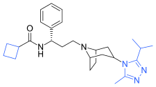
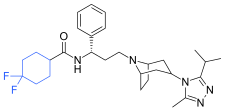
Pfizer turned to high-throughput screening in their search for a good starting point for a small molecule CCR5 antagonist. Their screening resulted in a compound that presented weak affinity and no antiviral activity but represented a good starting point for further optimization. Compounds 1–9 in Table 1 show the development of maraviroc in few steps. The chemical structure of the starting molecule (UK-107,543) is presented as compound 1. Their first focus was to minimize CYP2D6 activity of the molecule and to reduce its lipophilicity. They replaced the imidazopyridine with benzimidazole and the benzhydril group was swapped out for a benzamide. The outcome was compound 2. That compound showed good binding potency and the start of an antiviral activity. Further structure–activity relationship (SAR) optimization of the amide region and identifying the enantiomeric preference led to the cyclobutyl amide structure in compound 3. However, the problem with the CYP2D6 activity of the compound was still unacceptable so they had to perform further SAR optimization that determined that the [3.2.1]-azabicycloamine (tropane) could replace the aminopiperidine moiety. This change in the chemical structure led to compound 4. Compound 4 had no CYP2D6 activity while preserving excellent binding affinity and antiviral activity. Although compound 4 showed promising results, it demonstrated 99% inhibition on the hERG ion channel. That inhibition was unacceptable since it can lead to QTc interval prolongation. The research team then did a few modifications to see which part of the molecule played a role in the hERG affinity. Compound 5 shows an analogue that they synthesized which contained an oxygen bridgehead in the tropane ring; however, that reconstruction did not have an effect on the hERG affinity. They then focused on the polar surface area in the molecule to dial out the hERG affinity. These efforts resulted in compound 6. That compound preserved desired antiviral activity and was selective against the hERG inhibition but the problem was its bioavailability. Reduction in the lipophilicity, by replacing the benzimidazol group with a substituted triazole group gave compound 7. Compound 7 had shown a significant reduction in lipophilicity and maintained the antiviral activity but again, with the introduction of a cyclobutyl group, the compound showed hERG inhibition. Changing the ring size in compound 7 from a cyclobutyl unit to a cyclopentyl unit in compound 8 led to a significant increase in antiviral activity and loss of hERG affinity. Further development led to discovery of a 4,4'-difluorocyclohexylamide also known as maraviroc. Maraviroc preserved excellent antiviral activity, whilst demonstrating no significant hERG binding affinity. The lack of hERG binding affinity was predicted to be because of the large size of the cyclohexyl group and the high polarity of the fluoro substituents. In August 2007 the FDA approved the first CCR5 antagonist, maraviroc, discovered and developed by Pfizer.

Table 1 represents the molecular structures in the development of maraviroc.
Blue indicates differences from the previous step
Compound 1 (maraviroc development).svg Compound 2 (maraviroc development).svg Compound 3 (maraviroc development).svg
Compound 1 Compound 2 Compound 3
Compound 4 (maraviroc development).svg Compound 5 (maraviroc development).svg Compound 6 (maraviroc development).svg
Compound 4 Compound 5 Compound 6
Compound 7 (maraviroc development).svg Compound 8 (maraviroc development).svg Maraviroc (maraviroc development).svg
Compound 7 Compound 8 Compound 9 (maraviroc)

Pharmacophore

Figure 4 Predictive pharmacophore model for piperidine- and piperazine-based CCR5 antagonists

The predictive pharmacophore model was developed for a large series of piperidine- and piperazine-based CCR5 antagonists by Schering-Plough Research Institute. Their hypothesis consisted of mostly five features, two hydrogen bond acceptors, marked C and D in figure 4 and three hydrophobic groups, A, B and E in figure 4. Part B usually has a basic nitrogen group. The model was validated using diverse set of six CCR5 antagonists from five different pharmaceutical companies. The best model correctly predicted these compounds as being highly active. It is possible to use the model as a tool in virtual screening for new small molecular CCR5 antagonists and also to predict biological activities of compounds prior to undertaking their costly synthesis.

Binding

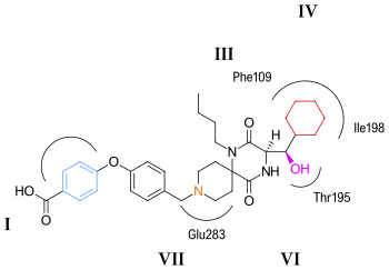
Figure 5. Putative binding of aplaviroc to the CCR5 receptor
 
Figure 6. Putative binding of maraviroc to the CCR5 receptor

CCR5 is a member of G protein-coupled, seven transmembrane segment receptors. The structure of the receptor comprises seven-helix bundle in the transmembrane region, these regions are labeled I–VII in figures 5 and 6. The CCR5 antagonists are predicted to bind to a putative binding pocket which is buried inside the transmembrane domain, enclosed by the seven transmembrane helices. The binding pocket is very hydrophobic with multiple aromatic residues lining the pocket. The key residues are tryptophan 86 and 248 (Trp86, Trp248), tyrosine 108 and 251 (Tyr108, Tyr251), phenylalanine 109 (Phe109), threonine 195 (Thr195), isoleucine 198 (Ile198), glutamic acid 283 (Glu283). CCR5 antagonists are very different in shape and electrostatic potential although they all share the same binding pocket. The interesting thing about the binding of these molecules is that they exhibit significantly different binding modes, although they all establish an extensive interaction network with CCR5.

Aplaviroc

The putative binding mode for aplaviroc is shown in figure 5. The key saltbridge interaction between aplaviroc and Glu283 is predicted to be quite weak compared to other CCR5 antagonists. The hydroxyl group on aplaviroc forms a strong hydrogen bond to the polar residue Thr195. This H-bond interaction is the strongest with aplaviroc compared to other CCR5 antagonists. The cyclohexyl group in the aplaviroc structure is predicted to interact with the receptor in a hydrophobic pocket formed by Ile198, Thr195 and Phe109 and is thought to show quite strong hydrophobic interactions. The researchers predict that the butyl group of aplaviroc is buried within the helical bundle through strong hydrophobic interaction with multiple aromatic residues of the CCR5 receptor. Aplaviroc has a unique feature of preserving two of the natural chemokine protein ligands binding to CCR5 and subsequent activation, whereas maraviroc and the other antagonists almost fully block chemokine-CCR5 interactions. This kind of interference is so far considered to be safe, and individuals that naturally lack CCR5 do not show any obvious health problems. However, to limit the toxicity and side effects of CCR5 antagonists it would be ideal to be able to preserve the chemokine receptor function. Consequently, it should be of interest to design inhibitors that specifically disrupt CCR5–gp120 binding but do not affect the CCR5 chemokine activation.

Maraviroc

The putative binding mode for maraviroc is shown in figure 6. The strongest interaction is estimated to be between maraviroc and glutamic acid (Glu283) through a strong salt bridge interaction. The interaction between tryptophan (Trp86) and maraviroc involves T-shaped π-π stacking while the interaction with phenylalanine (Phe109) is predicted to be hydrophobic. Tyrosine (Tyr108) is thought to interact with the phenyl group on maraviroc through a parallel displaced interaction. The interaction between maraviroc and isoleucine (Ile198) is predicted to be mostly hydrophobic in nature and the interaction between maraviroc and tyrosine (Tyr251) is very limited.

Other CCR5 antagonists

Figure 7. Molecular structure of compound A

Development of new CCR5 antagonists continues, both for their antiviral effects and also for potential utility in a variety of autoimmune indications. Researchers at Roche Palo Alto discovered a novel series of potent CCR5 small-molecule antagonists. Lead optimization was pursued by balancing opposing trends of metabolic stability and potency. Combination of the spiropiperidine template with pharmacophore elements from both aplaviroc, and Schering's CCR5 antagonist program, led to the initial lead compound in this series. Further development of that lead compound led to the discovery of compound A in figure 7 — a compound that possesses a good selectivity and pharmacokinetic properties.

The CCR5 antagonist INCB009471 has nanomolar activity against HIV-1 in vitro. This compound demonstrated potent and prolonged antiviral activity against R5-tropic HIV-1 when given 200 mg once daily dose for 14 days. These findings supported further clinical development of INCB009471 and they have since progressed to phase IIb clinical trials. As of 2009 the study of this compound is inactive and no further studies are planned at this time.

Not only small molecules but also proteins delivered by gene therapy have been suggested to ablate CCR5 function, an approach that has also been employed for other HIV targets.

HIV/AIDS research

From Wikipedia, the free encyclopedia
https://en.wikipedia.org/wiki/HIV/AIDS_research

A large round blue object with a smaller red object attached to it. Multiple small green spots are speckled over both.
Scanning electron micrograph of HIV-1, colored green, budding from a cultured lymphocyte
 
Diagram of HIV

HIV/AIDS research includes all medical research that attempts to prevent, treat, or cure HIV/AIDS, as well as fundamental research about the nature of HIV as an infectious agent and AIDS as the disease caused by HIV.

Transmission

A body of scientific evidence has shown that men who are circumcised are less likely to contract HIV than men who are uncircumcised. Research published in 2014 concludes that the sex hormones estrogen and progesterone selectively impact HIV transmission.

Pre- and post-exposure prophylaxis

"Pre-exposure prophylaxis" refers to the practice of taking some drugs before being exposed to HIV infection, and having a decreased chance of contracting HIV as a result of taking that drug. Post-exposure prophylaxis refers to taking some drugs quickly after being exposed to HIV, while the virus is in a person's body but before the virus has established itself. In both cases, the drugs would be the same as those used to treat persons with HIV, and the intent of taking the drugs would be to eradicate the virus before the person becomes irreversibly infected.

Post-exposure prophylaxis is recommended in anticipated cases of HIV exposure, such as if a nurse somehow has blood-to-blood contact with a patient in the course of work, or if someone without HIV requests the drugs immediately after having unprotected sex with a person who might have HIV. Pre-exposure prophylaxis is sometimes an option for HIV-negative persons who feel that they are at increased risk of HIV infection, such as an HIV-negative person in a serodiscordant relationship with an HIV-positive partner.

Current research in these agents include drug development, efficacy testing, and practice recommendations for using drugs for HIV prevention.

Progression of HIV

The progression of HIV infection is analyzed by measuring the concentration of  HIV virions (or viral load) and the concentration of CD4 T cells in the patient's bloodstream and lymphoid tissues. An untreated infection will progress in the following phases: Acute phase, chronic phase, and AIDs phase. In the Acute phase, the virions invade the host body and replicate expeditiously. The concentration of the virions increase vastly, while the concentration of CD4 T cells declines. After a spiked replication of HIV, the  viral load and CD4 T cell count drops back down. Symptoms of acute HIV infection include fever, chills, rash, night sweats, muscle aches, and swollen lymph nodes. Acute symptoms occur usually 2–4 weeks after initial HIV infection and can last between a few days and several weeks.

During the chronic phase, HIV will continue to replicate, but the concentration of virions tend to stabilize for a period of time before rising again. The CD4 T cell count continues to fall. Individuals in the chronic phase may not experience any symptoms. Left untreated, the chronic stage can last between 10 and 15 years. However, some individuals can move through this stage quickly to the AIDS phase.

An untreated HIV infection ultimately progresses to AIDS (acquired immunodeficiency syndrome). In the AIDS phase, the CD4 T-cell count significantly drops to below 200 cells per cubic millimeter. Individuals with AIDS become immunocompromised due to irreversible  damage to the immune system and lymph nodes. The immune system does not have the ability to generate new T cells. Opportunistic infections, that a robust immune system could fight off, now are capable of causing severe symptoms and illnesses. Without a comprehensive anti-HIV drug therapy, an individual diagnosed with AIDS is expected to have less than three years to live.

Immune System Response to HIV

Once the retrovirus invades the body, the immune system mobilizes to fight against HIV infection. The first line of defense for the immune system utilizes dendritic cells. These cells actively patrol vulnerable tissue (i.e. lining of the digestive and reproductive tracts). Once a dendritic cell apprehends the virion invader, it will transport the virus to lymphoid tissue and introduce parts of the virus's proteins to Naive helper T cells (which are specialized white blood cells). The transported viral protein binds to the naive helper T cell's receptor, and the T cell activates. As the helper T cells grow and divide, they produce effecter helper T cells (which help coordinate the immune system response to HIV). The effector T cells utilize cytokines to mobilize other immune cells to join the combat against HIV. The cytokines promote the maturation of B cells into plasma cells. Then the plasma cells secrete antibodies that will bind to the HIV virions and target them for destruction. Finally, activated killer T-cells come in to eradicate the infected host cells.

Within-host dynamics

The within-host dynamics of HIV infection include the spread of the virus in vivo, the establishment of latency, the effects of immune response on the virus, etc. Early studies used simple models and only considered the cell-free spreading of HIV, in which virus particles bud from an infected T cell, enter the blood/extracellular fluid, and then infect another T cell. A 2015 study proposes a more realistic model of HIV dynamics that also incorporates the viral cell-to-cell spreading mechanism, where the virus is directly transited from one cell to another, as well as the T cell activation, the cellular immune response, and the immune exhaustion as the infection progresses.

Virus characteristics

HIV binds to immune cell surface receptors, including CD 4 and CXCR4 or CD4 and CCR5. The binding causes conformation changes and results in the membrane fusion between HIV and cell membrane. Active infection occurs in most cells, while latent infection occurs in much fewer cells 1, 2 and at very early stages of HIV infection. 9, 35 In active infection, HIV pro virus is active and HIV virus particles are actively replicated; and the infected cells continuously release viral progeny; while in latent infection, HIV pro virus is transcriptionally silenced and no viral progeny is produced.

Management of HIV/AIDS

Research to improve current treatments includes decreasing side effects of current drugs, further simplifying drug regimens to improve adherence, and determining better sequences of regimens to manage drug resistance. There are variations in the health community in recommendations on what treatment doctors should recommend for people with HIV. One question, for example, is determining when a doctor should recommend that a patient take antiretroviral drugs and what drugs a doctor may recommend. This field also includes the development of antiretroviral drugs.

Age acceleration effects due to HIV-1 infection

Infection with the Human Immunodeficiency Virus-1 (HIV) is associated with clinical symptoms of accelerated aging, as evidenced by increased incidence and diversity of age-related illnesses at relatively young ages. A significant age acceleration effect could be detected in brain (7.4 years) and blood (5.2 years) tissue due to HIV-1 infection with the help of a biomarker of aging, which is known as epigenetic clock.

Long-term nonprogressor

A long-term nonprogressor is a person who is infected with HIV, but whose body, for whatever reason, naturally controls the virus so that the infection does not progress to the AIDS stage. Such persons are of great interest to researchers, who feel that a study of their physiologies could provide a deeper understanding of the virus and disease. There are also two cases where HIV was apparently entirely cleared by a person's immune system without a therapy.

HIV vaccine

An HIV vaccine is a vaccine that would be given to a person who does not have HIV, in order to confer protection against subsequent exposures to HIV, thus reducing the likelihood that the person would become infected by HIV. Currently, no effective HIV vaccine exists. Various HIV vaccines have been tested in clinical trials almost since the discovery of HIV.

Only a vaccine is thought to be able to halt the pandemic. This is because a vaccine would cost less, thus being affordable for developing countries, and would not require daily treatment. However, after over 20 years of research, HIV-1 remains a difficult target for a vaccine.

In 2003 a clinical trial in Thailand tested an HIV vaccine called RV 144. In 2009, the researchers reported that this vaccine showed some efficacy in protecting recipients from HIV infection (31% efficiency). Results of this trial give the first supporting evidence of any vaccine being effective in lowering the risk of contracting HIV. Other vaccine trials continue worldwide including a mosaic vaccine using an adenovirus 26 vector as well as a newer formulation of RV144 called HVTN 702.

One recent trial was conducted by scientists at The Scripps Research Institute (TSRI) who found a way to attach HIV-fighting antibodies to immune cells, creating a HIV-resistant cell population.

HIV cure

Three people have been reported cured of AIDS. In 2019, the NIH and Bill & Melinda Gates Foundation announced making $200 million available for broad-based, multi-prong scientific efforts focused on developing a global cure for AIDS as well as for sickle cell disease, with NIH Director Francis S. Collins saying, "We aim to go big or we go home." In 2020, Tony Fauci's division at NIH, NIAID, issued its first solicitation exclusively focused on methods to cure HIV infection. These announcements from NIH are not limited to stem cell therapies.

Excision is a biotechnology company with a first-in-human CRISPR-based one-time gene therapy to be evaluated in individuals with HIV. Research Foundation to Cure AIDS is the first 501(c)(3) non-for-profit organization with a royalty-free license to research, develop and commercialize a cell engineering technology in the field of curing AIDS on a pro bono basis.

Microbicides for sexually transmitted diseases

A microbicide for sexually transmitted diseases is a gel which would be applied to the skin – perhaps a rectal microbicide for persons who engage in anal sex or a vaginal microbicide for persons who engage in vaginal sex – and if infected body fluid such as blood or semen were to touch the gel, then HIV in that fluid would be destroyed and the people having sex would be less likely to spread infection between themselves.

On March 7, 2013, the Washington University in St. Louis website published a report by Julia Evangelou Strait, in which it was reported that ongoing nanoparticle research showed that nanoparticles loaded with various compounds could be used to target infectious agents whilst leaving healthy cells unaffected. In the study detailed by this report, it was found that nanoparticles loaded with Mellitin, a compound found in Bee venom, could deliver the agent to the HIV, causing the breakdown of the outer protein envelope of the virus. This, they say, could lead to the production of a vaginal gel which could help prevent infection by disabling the virus. Dr Joshua Hood goes on to explain that beyond preventive measures in the form of a topical gel, he sees "potential for using nanoparticles with melittin as therapy for existing HIV infections, especially those that are drug-resistant. The nanoparticles could be injected intravenously and, in theory, would be able to clear HIV from the blood stream."

Initial stem cell cures of HIV/AIDS

In 2007, Timothy Ray Brown, a 40-year-old HIV-positive man, also known as "the Berlin Patient", was given a stem cell transplant as part of his treatment for acute myeloid leukemia (AML). A second transplant was made a year later after a relapse. The donor was chosen not only for genetic compatibility but also for being homozygous for a CCR5-Δ32 mutation that confers resistance to HIV infection. After 20 months without antiretroviral drug treatment, it was reported that HIV levels in Brown's blood, bone marrow, and bowel were below the limit of detection. The virus remained undetectable over three years after the first transplant. Although the researchers and some commentators have characterized this result as a cure, others suggest that the virus may remain hidden in tissues such as the brain (which acts as a viral reservoir). Stem cell treatment remains investigational because of its anecdotal nature, the disease and mortality risk associated with stem cell transplants, and the difficulty of finding suitable donors. As of 2022, there have been four patients cured by stem cell transplant.

Strategies to develop broadly-applicable cures

Scientists have been using different approaches of stem cell based gene therapy in an attempt to develop a cure as well as to propose an alternative to the conventional antiretroviral therapy (ART). Specifically, advances had been made with a cure to HIV.

A cellular receptor, generally CCR5 or CXCR4 is required in order for HIV entry into CD4 cells. Cells of individuals homozygous for the CCR5 gene variant Δ32 (CCR5Δ32/Δ32) lack the CCR5 cell-surface expression, meaning that they are naturally resistant to infection with CCR5 tropic HIV strains (R5 HIV). One study done in 2011 achieves successful CD4+ T-cell reconstitution as a result of CCR5Δ32/Δ32 stem cell transplantation at the systemic level and in the gut mucosal immune system in a patient with HIV. Additionally, it provides evidence for the reduction in the size of the potential HIV reservoir over time. The patient in this study even remained HIV free without any evidence of having it for more than 3.5 years.

Other theoretical cures to HIV-1 have been proposed. One supposed cure to HIV-1 involves the creation of a disease-resistant immune system through transplantation of autologous, gene-modified (HIV-1-resistant) hematopoietic stem cells and progenitor cells (GM-HSPC). Though this study does involve several early stage clinical trials that have demonstrated the safety and feasibility of this technique only for HIV-1, none have resulted in improvement of the disease state itself. Therefore, this strategy is intended to go alongside already existing treatment techniques such as drugs and vaccines. However, future technology regarding this approach of single treatment cell therapy could potentially replace current therapy altogether as a functional or sterilizing cure to HIV-1.

An additional study involves the use of genetically engineered CD34+ hematopoietic stem and progenitor cells. Experimental long-term in vivo HIV gene therapy have had huge issues due to both transduction ending in multiple copies of heterologous DNA in target cells as well as low efficacy of cell transduction at the time of transplantation. This study demonstrated the efficacy of a transplantation approach that ultimately allows for an enriched population of HSPCs expressing a single copy of a CCR5 miRNA. Since positive selection of modified cells is likely to be insufficient below the threshold they found of at least 70% of the HIV target cells resulting in gene modification from efficient maintenance of CD34+ T cell and a low viral titer, the findings show evidence that clinical protocols of HIV gene therapy require a selective enrichment of genetically targeted cells.

Immunomodulatory agents

Complementing efforts to control viral replication, immunotherapies that may assist in the recovery of the immune system have been explored in past and ongoing trials, including IL-2 and IL-7.

The failure of vaccine candidates to protect against HIV infection and progression to AIDS has led to a renewed focus on the biological mechanisms responsible for HIV latency. A limited period of therapy combining anti-retrovirals with drugs targeting the latent reservoir may one day allow for total eradication of HIV infection. Researchers have discovered an abzyme that can destroy the protein gp120 CD4 binding site. This protein is common to all HIV variants as it is the attachment point for B lymphocytes and subsequent compromising of the immune system.

New developments

A turning point for HIV research occurred in 2007, following the bone marrow transplant of HIV sufferer Timothy Ray Brown. Brown underwent the procedure after he developed leukaemia and the donor of the bone marrow possessed a rare genetic mutation that caused Brown's cells to become resistant to HIV. Brown attained the title of the "Berlin Patient" in the HIV research field and is the first man to have been cured of the virus. As of April 2013, two primary approaches are being pursued in the search for a HIV cure: The first is gene therapy that aims to develop a HIV-resistant immune system for patients, and the second is being led by Danish scientists, who are conducting clinical trials to strip the HIV from human DNA and have it destroyed permanently by the immune system.

Three more cases with similarities to the Brown case have occurred since the 2007 discovery; however, they differ because the transplanted marrow has not been confirmed as mutated. Two of the cases were publicized in a July 2013 CNN story that relayed the experience of two patients who had taken antiretroviral therapy for years before they developed lymphoma, a cancer of the lymph nodes. They then underwent lymphoma chemotherapy and bone marrow transplantation, while remaining on an antiretroviral regimen; while they retained traces of HIV four months afterwards, six to nine months after the transplant, the two patients had no detectable trace of HIV in their blood. However, the managing clinician Dr. Timothy Heinrich stated at the Malaysian International AIDS Society Conference where the findings were presented:

It's possible, again, that the virus could return in a week, it could return in a month—in fact, some mathematical modeling predicts that virus could even return one to two years after we stop antiretroviral therapy, so we really don't know what the long-term or full effects of stem cell transplantation and viral persistence is.

In 2014, Dr Warner C. Greene and Dr Gilad Doitsh at the Gladstone Institutes identified pyroptosis as the predominant mechanism that causes the two signature pathogenic events in HIV infection––CD4 T-cell depletion and chronic inflammation. Identifying pyroptosis may provide novel therapeutic opportunities targeting caspase-1, which controls the pyroptotic cell death pathway. Specifically, these findings could open the door to an entirely new class of "anti-AIDS" therapies that act by targeting the host rather than the virus. Recently, pyroptosis and downstream pathways were also identified as promising targets for treatment of severe coronavirus disease 2019–associated diseases.

In March 2016, researchers at Temple University, Philadelphia, reported that they have used genome editing to delete HIV from T cells. According to the researchers, this approach could lead to a dramatic reduction of the viral load in patient cells.

In April 2016, it was announced the publication of a preclinical animal study using SupT1 cells as a decoy target for the HIV virus, aiming to move infection from the patient's cells to the inoculated cells, and therefore to induce the virus to become less aggressive by replicating in such permissive cells.

In March 2019, a patient with Hodgkin's lymphoma was also reported to possibly have been cured using similar treatment to Brown.

In 2022, Moderna announced that the first participants have been vaccinated in a Phase 1 clinical trial of an experimental HIV vaccine that utilizes Moderna's mRNA technology.

Operator (computer programming)

From Wikipedia, the free encyclopedia https://en.wikipedia.org/wiki/Operator_(computer_programmin...