Search This Blog

Saturday, October 14, 2023

Diabetic foot ulcer

From Wikipedia, the free encyclopedia
 
Diabetic foot ulcer
Neuropathic diabetic foot ulcer
Causesdiabetes

Diabetic foot ulcer is a major complication of diabetes mellitus, and probably the major component of the diabetic foot.

Wound healing is an innate mechanism of action that works reliably most of the time. A key feature of wound healing is stepwise repair of lost extracellular matrix (ECM) that forms the largest component of the dermal skin layer. But in some cases, certain disorders or physiological insult disturbs the wound healing process. Diabetes mellitus is one such metabolic disorder that impedes the normal steps of the wound healing process. Many studies show a prolonged inflammatory phase in diabetic wounds, which causes a delay in the formation of mature granulation tissue and a parallel reduction in wound tensile strength.

Treatment of diabetic foot ulcers should include: blood sugar control, removal of dead tissue from the wound, wound dressings, and removing pressure from the wound through techniques such as total contact casting. Surgery in some cases may improve outcomes. Hyperbaric oxygen therapy may also help but is expensive.

It occurs in 15% of people with diabetes, and precedes 84% of all diabetes-related lower-leg amputations.

Risk factors

Risk factors implicated in the development of diabetic foot ulcers are infection, older age, diabetic neuropathy, peripheral vascular disease, cigarette smoking, poor glycemic control, previous foot ulcerations or amputations, and ischemia of small and large blood vessels. Prior history of foot  disease, foot deformities that produce abnormally high forces of pressure, callus at pressure areas  renal  failure, oedema, impaired ability to look after personal care (e.g. visual impairment) are further risk factors for diabetic foot ulcer.

People with diabetes often develop diabetic neuropathy due to several metabolic and neurovascular factors. Peripheral neuropathy causes loss of pain or feeling in the toes, feet, legs, and arms due to distal nerve damage and low blood flow. Autonomic neuropathy causes Sudomotor dysfunction and dryness of the skin. Blisters and sores may appear on numb areas of the feet and legs, such as metatarsophalangeal joints and the heel region, as a result of pressure or injury which may go unnoticed and eventually become a portal of entry for bacteria and infection.

Pathophysiology

Extracellular matrix

Extra cellular matrix (or "ECM") is the external structural framework that cells attach to in multicellular organisms. The dermis lies below the epidermis, and these two layers are collectively known as the skin. Dermal skin is primarily a combination of fibroblasts growing in this matrix. The specific species of ECM of connective tissues often differ chemically, but collagen generally forms the bulk of the structure.

Through the interaction of a cell with its extracellular matrix (transmitted through the anchoring molecules classed as integrins) there forms a continuous association between the cell interior, cell membrane and its extracellular matrix components that helps drive various cellular events in a regulated fashion. Wound healing is a localized event involving the reaction of cells to the damage sustained.

The cells break down damaged ECM and replace it, generally increasing in number to react to the harm. The process is activated, though perhaps not exclusively, by cells responding to fragments of damaged ECM, and the repairs are made by reassembling the matrix by cells growing on and through it. Because of this, extracellular matrix is often considered as a 'conductor of the wound healing symphony'. In the Inflammatory phase, neutrophils and macrophages recruit and activate fibroblasts which in subsequent granulation phase migrate into the wound, laying down new collagen of the subtypes I and III.

In the initial events of wound healing, collagen III predominates in the granulation tissue which later on in remodeling phase gets replaced by collagen I giving additional tensile strength to the healing tissue. It is evident from the known collagen assembly that the tensile strength is basically due to fibrillar arrangement of collagen molecules, which self-assemble into microfibrils in a longitudinal as well as lateral manner producing extra strength and stability to the collagen assembly. Metabolically altered collagen is known to be highly inflexible and prone to break down, particularly over pressure areas. Fibronectin is the major glycoprotein secreted by fibroblasts during initial synthesis of extracellular matrix proteins. It serves important functions, being a chemo-attractant for macrophages, fibroblasts and endothelial cells.

The basement membrane that separates the epidermis from the dermal layer and the endothelial basement membrane mainly contains collagen IV that forms a sheet and binds to other extracellular matrix molecules like laminin and proteoglycans. In addition to collagen IV, the epidermal and endothelial basement membrane also contains laminin, perlecan and nidogen. Hyaluronic acid, a pure glycosaminoglycan component, is found in high amounts in damaged or growing tissues. It stimulates cytokine production by macrophages and thus promotes angiogenesis. In normal skin chondroitin sulfate proteoglycan is mainly found in the basement membrane, but in healing wounds they are up-regulated throughout the granulation tissue especially during the second week of wound repair where they provide a temporary matrix with highly hydrative capacity. Binding of growth factors is clearly an important role of perlecan in wound healing and angiogenesis. Poor wound healing in diabetes mellitus may be related to perlecan expression. High levels of glucose can decrease perlecan expression in some cells, probably through transcriptional and post-transcriptional modification. Wound healing phases especially, granulation, re-epithelization and remodelling exhibit controlled turnover of extracellular matrix components.

Altered metabolism

Diabetes mellitus is a metabolic disorder and hence the defects observed in diabetic wound healing are thought to be the result of altered protein and lipid metabolism and thereby abnormal granulation tissue formation. Increased glucose levels in the body end up in uncontrolled covalent bonding of aldose sugars to a protein or lipid without any normal glycosylation enzymes. These stable products then accumulate over the surface of cell membranes, structural proteins and circulating proteins. These products are called advanced glycation endproducts (AGEs) or Amadori products. Formation of AGEs occurs on extracellular matrix proteins with slow turnover rate. AGEs alter the properties of matrix proteins such as collagen, vitronectin, and laminin through AGE-AGE intermolecular covalent bonds or cross-linking. AGE cross-linking on type I collagen and elastin results in increased stiffness. AGEs are also known to increase synthesis of type III collagen that forms the granulation tissue. AGEs on laminin result in reduced binding to type IV collagen in the basement membrane, reduced polymer elongation and reduced binding of heparan sulfate proteoglycan.

Impaired NO synthesis
Nitric oxide is known as an important stimulator of cell proliferation, maturation and differentiation. Thus, nitric oxide increases fibroblast proliferation and thereby collagen production in wound healing. Also, L-arginine and nitric oxide are required for proper cross linking of collagen fibers, via proline, to minimize scarring and maximize the tensile strength of healed tissue. Endothelial cell specific nitric oxide synthase (EcNOS) is activated by the pulsatile flow of blood through vessels. Nitric oxide produced by EcNOS, maintains the diameter of blood vessels and proper blood flow to tissues. In addition to this, nitric oxide also regulates angiogenesis, which plays a major role in wound healing. Thus, diabetic patients exhibit reduced ability to generate nitric oxide from L-arginine. Reasons that have been postulated in the literature include accumulation of nitric oxide synthase inhibitor due to high glucose associated kidney dysfunction and reduced production of nitric oxide synthase due to ketoacidosis observed in diabetic patients and pH dependent nature of nitric oxide synthase.
Structural and functional changes in fibroblasts
Diabetic ulcer fibroblasts show various morphological differences compared to fibroblasts from age matched controls. Diabetic ulcer fibroblasts are usually large and widely spread in the culture flask compared to the spindle shaped morphology of the fibroblasts in age-matched controls. They often show dilated endoplasmic reticulum, numerous vesicular bodies and lack of microtubular structure in transmission electron microscopy study. Therefore, interpretation of these observations would be that in spite of high protein production and protein turnover in diabetic ulcer fibroblasts, vesicles containing secretory proteins could not travel along the microtubules to release the products outside. Fibroblasts from diabetic ulcer exhibit proliferative impairment that probably contributes to a decreased production of extracellular matrix proteins and delayed wound contraction and impaired wound healing.
Increased matrix metalloproteinases (MMP) activity
In order for a wound to heal, extracellular matrix not only needs to be laid down but also must be able to undergo degradation and remodeling to form a mature tissue with appropriate tensile strength. Proteases, namely matrix metalloproteinases are known to degrade almost all the extracellular matrix components. They are known to be involved in fibroblast and keratinocyte migration, tissue re-organization, inflammation and remodeling of the wounded tissue. Due to persistently high concentrations of pro-inflammatory cytokines in diabetic ulcers, MMP activity is known to increase by 30 fold when compared to acute wound healing. MMP-2 and MMP-9 show sustained overexpression in chronic non-healing diabetic ulcers. Balance in the MMP activity is usually achieved by tissue inhibitor of metalloproteinases (TIMP). Rather than absolute concentrations of either two, it is the ratio of MMP and TIMP that maintains the proteolytic balance and this ratio is found to be disturbed in diabetic ulcer. In spite of these findings, the exact mechanism responsible for increased MMP activity in diabetes is not known yet. One possible line of thought considers Transforming growth factor beta (TGF-β) as an active player. Most MMP genes have TGF-β inhibitory element in their promoter regions and thus TGF–β regulates the expression of both MMP and their inhibitor TIMP. In addition to the importance of cell-cell and cell-matrix interactions, all phases of wound healing are controlled by a wide variety of different growth factors and cytokines. To mention precisely, growth factors promote switching of early inflammatory phase to the granulation tissue formation. Decrease in growth factors responsible for tissue repair such as TGF-β is documented in diabetic wounds. Thus, reduced levels of TGFβ in diabetes cases lower down the effect of inhibitory regulatory effect on MMP genes and thus cause MMPs to over express.

Biomechanics

Complications in the diabetic foot and foot-ankle complex are wider and more destructive than expected and may compromise the structure and function of several systems: vascular, nervous, somatosensory, musculoskeletal. Thus, deeper comprehension of the alteration of gait and foot biomechanics in the diabetic foot is of great interest and may play a role in the design and onset of preventive as well as therapeutic actions.

Briefly, the effect of diabetes on the main structures of the foot-ankle complex can be summarised as:

  • effects on the skin: skin – and the soft tissues immediately underneath the skin – undergo greater compression and shear loading than usual, thus explaining the onset of tissue damage so deeply correlated to traumatic ulceration processes. Besides this, skin of the diabetic foot loses autonomic nervous control and consequently reduced hydration, making it less elastic and thus more vulnerable to the action of increased mechanical stress;
  • effects on tendons and ligaments: protein glycosylation and the resulting collagen abnormalities lead to greater transversal section – i.e. thickening – of tendons and ligaments and a greater coefficient of elasticity. Particularly affected by this process are Plantar Fascia and Achilles Tendon. Both causes lead to increased stiffness of those structures;
  • effects on cartilage: similar to what happens to tendons and ligaments, cartilage changes its composition mainly due to the modification of collagen fibers. This increases its stiffness and decreases the range of motion of all joints in the foot and ankle.
  • effects on muscles: Diabetes mellitus causes severe damage to nerve conduction, thus causing a worsening in the management of the related muscle fibers. As a consequence, both intrinsic and extrinsic muscles of the foot-ankle complex are damaged in structure (reduction of muscle volume) and function (reduction of muscle strength);
  • effects on the peripheral sensory system: impaired nerve conduction has a dramatic effect on the peripheral sensory system since it leads to loss of protective sensation under the sole of the foot. This exposes the diabetic foot to thermal or mechanical trauma, and to the late detection of infection processes or tissue breakdown;
  • effects on foot morphology (deformities): due to most of the above alterations, a significant imbalance of peripheral musculature and soft tissue occur in the foot which seriously alters its morphology and determines the onset of foot deformities. Most common deformities of the diabetic foot are represented by a high longitudinal arch (rigid cavus foot), hammer toes and hallux valgus. A completely different morphologic degeneration is represented by neuropathic arthropathy, whose analysis is not part of this discussion.

Diagnosis

Assessment of diabetic foot ulcer includes identifying risk factors such as diabetic peripheral neuropathy, noting that 50 percent of people are asymptomatic, and ruling out other causes of peripheral neuropathy such as alcohol use disorder and spinal injury. Diabetic foot ulcers are often misdiagnosed in patients with undiagnosed skin malignancies, especially high-risk in elderly patients.

The location of the ulcer, its size, shape, depth and whether the tissue is granulating or sloughy needs to be considered. Further considerations include whether there is malodour, condition of the border of the wound and palpable bone and sinus formation should be investigated. Signs of infection require to be considered such as development of grey or yellow tissue, purulent discharge, unpleasant smell, sinus, undermined edges and exposure of bone or tendon.

Identification of diabetic foot in medical databases, such as commercial claims and prescription data, is complicated by the lack of a specific ICD-9 code for diabetic foot and variation in coding practices. The following codes indicate ulcer of the lower limb or foot:

  • 707.1 Ulcer of lower limbs, except pressure ulcer
  • 707.14 Ulcer of heel and midfoot
  • 707.15 Ulcer of other part of foot
  • 707.19 Ulcer of other part of lower limb

One or more codes, in combination with a current or prior diagnosis of diabetes may be sufficient to conclude diabetic foot:

  • 250.0 Diabetes Mellitus
  • 250.8 Diabetes with other specified manifestations

Classification

Diabetic foot ulcer is a complication of diabetes. Diabetic foot ulcers are classified as either neuropathic, neuroischaemic or ischaemic.

Doctors also use the Wagner Grades to describe the severity of an ulcer. The purpose of the Wagner Grades is to allow specialists to better monitor and treat diabetic foot ulcers. This grading system classifies Diabetic foot ulcers using numbers, from 0 to 5.

Wagner Grades 0 through 5 are as follows:

  • 0. No diabetic foot ulcer is present, but there is a high risk of developing one.
  • 1. A surface ulcer involves full skin thickness, but does not yet involve the underlying tissues.
  • 2. A deep ulcer penetrates past the surface, down to the ligaments and muscle. There is no abscess or bone involved yet.
  • 3. A deep ulcer occurs with inflammation of subcutaneous connective tissue or an abscess. This can include infections in the muscle, tendon, joint, and/or bone.
  • 4. The tissue around the area of the ulcer (limited to the toes and forefoot) has begun to decay. This condition is called gangrene.
  • 5. Gangrene has spread from the localized area of the ulcer to become extensive. This involves the whole foot.

Prevention

Many diabetic shoes have velcro closures for ease of application and removal.

Steps to prevent diabetic foot ulcers include frequent review by a foot specialist and multidisciplinary team, good foot hygiene, diabetic socks and shoes, as well as avoiding injury. Foot-care education combined with increased surveillance can reduce the incidence of serious foot lesions.

There is no high quality researches that evaluate complex intervention of combining two or more preventive strategies in preventing diabetic foot ulcer.

Monitoring and prediction

People with loss of feeling in their feet should inspect their feet on a daily basis, to ensure that there are no wounds starting to develop. Monitoring a person's feet can help in predicting the likelihood of developing ulcers.

A common method for this is using a special thermometer to look for spots on the foot that have higher temperature which indicate the possibility of an ulcer developing. At the same time there is no strong scientific evidence supporting the effectiveness of at-home foot temperature monitoring.

The current guideline in the United Kingdom recommends collecting 8-10 pieces of information for predicting the development of foot ulcers. A simpler method proposed by researchers provides a more detailed risk score based on three pieces of information (insensitivity, foot pulse, previous history of ulcers or amputation). This method is not meant to replace people regularly checking their own feet but complement it.

Footwear

Diabetic shoes, insoles and socks are personalised products that relieve pressure on the foot in order to prevent ulcers. The evidence for special footwear to treat foot ulcers is poor but their effectiveness for prevention is well-established. Design features of footwear that are effective in reducing pressure are arch supports, cushioned cut-outs around points at risk of damage, and cushioning at the ball of the foot. Technology for measuring the pressure within the shoes is recommended during designing diabetic footwear.

People with loss of feeling in their feet should not walk around barefoot, but use proper footwear at all times.

Treatment

Foot ulcers in diabetes require multidisciplinary assessment, usually by diabetes nurse specialist, a tissue viability nurse, podiatrists, diabetes specialists and surgeons. An aim to improve glycaemic  control, if poor, forms part of the management, to slow disease progression. Individuals who have sausage shaped toes, a positive 'probe to bone' test, evidence suggesting osteomyelitis, suspected charcot neuroarthropathy, or those whose ulcers do not improve within 4 weeks of standard care and where there is evidence that exudate is of synovial membrane in origin. When osteomyelitis is suspected to be involved in the foot ulcer, but not evidenced on an x-ray, an MRI scan should be obtained.

With regards to infected foot ulcers, the presence of microorganisms is not in itself enough to determine whether an infection is present. Signs such as inflammation and purulence are the best indicators of an active infection. The most common organism causing infection is staphylococcus. The treatment consists of debridement, appropriate bandages, managing peripheral arterial disease and appropriate use of antibiotics (against pseudomonas aeruginosa, staphylococcus, streptococcus and anaerobe strains), and arterial revascularisation.

Antibiotics

The length of antibiotic courses depend on the severity of the infection and whether bone infection is involved but can range from 1 week to 6 weeks or more. Current recommendations are that antibiotics are only used when there is evidence of infection and continued until there is evidence that the infection has cleared, instead of evidence of ulcer healing. Choice of antibiotic depends on common local bacterial strains known to infect ulcers. Microbiological swabs are believed to be of limited value in identifying causative strain. Microbiological investigation is of value in cases of osteomyelitis. Most ulcer infections involve multiple microorganisms.

There is limited safety and efficacy data of topical antibiotics in treating diabetic foot ulcers.

Wound dressings

There are many types of dressings used to treat diabetic foot ulcers such as absorptive fillers, hydrogel dressings, and hydrocolloids. There is no good evidence that one type of dressing is better than another for diabetic foot ulcers. In selecting dressings for chronic non healing wounds it is recommended that the cost of the product be taken into account.

Hydrogel dressings may have shown a slight advantage over standard dressings, but the quality of the research is of concern. Dressings and creams containing silver have not been properly studied nor have alginate dressings. Biologically active bandages that combine hydrogel and hydrocolloid traits are available, however more research needs to be conducted as to the efficacy of this option over others.

Total contact casting

Total contact casting (TCC) is a specially designed cast designed to take weight of the foot (off-loading) in patients with DFUs. Reducing pressure on the wound by taking weight of the foot has proven to be very effective in DFU treatment. DFUs are a major factor leading to lower leg amputations among the diabetic population in the US with 85% of amputations in diabetics being preceded by a DFU. Furthermore, the 5 year post-amputation mortality rate among diabetics is estimated at 45% for those with neuropathic DFUs.

TCC has been used for off-loading DFUs in the US since the mid-1960s and is regarded by many practitioners as the "reference standard" for off-loading the bottom surface (sole) of the foot.

TCC helps patients to maintain their quality of life. By encasing the patient's complete foot — including the toes and lower leg — in a specialist cast to redistribute weight and pressure from the foot to the lower leg during everyday movements, patients can remain mobile. The manner in which TCC redistributes pressure protects the wound, letting damaged tissue regenerate and heal. TCC also keeps the ankle from rotating during walking, which helps prevent shearing and twisting forces that can further damage the wound.

Effective off loading is a key treatment modality for DFUs, particularly those where there is damage to the nerves in the feet (peripheral neuropathy). Along with infection management and vascular assessment, TCC is vital aspect to effectively managing DFUs. TCC is the most effective and reliable method for off-loading DFUs.

A 2013 meta-analysis by the Cochrane Collaboration compared the effectiveness of non-removable pressure relieving interventions, such as casts, with therapeutic shoes, dressings, removable pressure relieving orthotic devices, and surgical interventions. Non-removable pressure relieving interventions, including non-removable casts with an Achilles tendon lengthening component, were found to be more effective at healing foot ulcers related to diabetes that therapeutic shoes and other pressure relieving approaches.

TCC systems include TCC-EZ (Integra LifeSciences) and Cutimed Off-loader (BSN Medical).

Hyperbaric oxygen

In 2015, a Cochrane review concluded that for people with diabetic foot ulcers, hyperbaric oxygen therapy reduced the risk of amputation and may improve the healing at 6 weeks. However, there was no benefit at one year and the quality of the reviewed trials was inadequate to draw strong conclusions.

Negative pressure wound therapy

This treatment uses vacuum to remove excess fluid and cellular waste that usually prolong the inflammatory phase of wound healing. Despite a straightforward mechanism of action, results of negative pressure wound therapy studies have been inconsistent. Research needs to be carried out to optimize the parameters of pressure intensity, treatment intervals and exact timing to start negative pressure therapy in the course of chronic wound healing.

There is low-certainty evidence that negative pressure wound therapy would improve wound healing in diabetic foot ulcers.

Other treatments

Ozone therapy – there is only limited and poor-quality information available regarding the effectiveness of ozone therapy for treating foot ulcers in people with diabetes.

Growth factors - there is some low-quality evidence that growth factors may increase the likelihood that diabetic foot ulcers will heal completely.

Continuous diffusion of oxygen (CDO) - CDO delivers continuous oxygen to an occluded, moist wound site at much lower flow rates of 3–12 mL/h for 24 h 7 days a week for up to several weeks or months, depending on the wound status.

Phototherapy - there is very weak evidence to suggest that people with foot ulcers due to diabetes may have improved healing. There is no evidence to suggest that phototherapy improves the quality of life for people with foot ulcers caused by diabetes.

Sucrose-octasulfate impregnated dressing is recommended by the International Working Group on the Diabetic Foot Ulcer (IWGDF) for the treatment of non-infected, neuro-ischaemic diabetic foot ulcers that do not show an improvement with a standard of care regimen

Autologous combined leucocyte, platelet and fibrin as an adjunctive treatment, in addition to best standard of care is also recommended by IWGDF However, there is only low quality evidence that such treatment is effective in treating diabetic foot ulcer.

There is limited evidence that granulocyte colony-stimulating factor may not hasten the resolution of diabetic foot ulcer infection. However, it may reduce the need for surgical interventions such as amputations and hospitalizations.

It is unknown that whether intensive or conventional blood glucose control is better for diabetic foot ulcer healing.

A 2020 Cochrane systematic review evaluated the effects of nutritional supplements or special diets on healing foot ulcers in people with diabetes. The review authors concluded that it's uncertain whether or not nutritional interventions have an effect on foot ulcer healing and that more research is needed to answer this question.

Skin grafting and tissue replacements can help to improve the healing of diabetic foot ulcer.

A 2021 systematic review concluded that there was no strong evidence about the effects of psychological therapies on diabetic foot ulcer healing and recurrence.

Epidemiology

Approximately 15 percent of people with diabetes experience foot ulcers, and approximately 84 percent of lower limb amputations have a history of ulceration with only approximately half of amputees surviving for more than 2 years. 56 percent of individuals with foot ulcers who do not have an amputations survive for 5 years. Foot ulcers and amputations significantly reduce the quality of life. Approximately 8.8 percent of hospital admissions of diabetic patients are for foot related problems, and such hospital admissions are about 13 days longer than for diabetics without foot related admissions. Approximately 35 to 40 percent of ulcers recur within 3 years and up to 70 percent recur within 5 years. Diabetic foot disease is the leading cause of non-traumatic lower limb amputations.

Research

Stem cell therapy may represent a treatment for promoting healing of diabetic foot ulcers. Diabetic foot ulcers develop their own, distinctive microbiota. Investigations into characterizing and identifying the phyla, genera and species of nonpathogenic bacteria or other microorganisms populating these ulcers may help identify one group of microbiota that promotes healing.

The recent advances in epigenetic modifications, with special focus on aberrant macrophage polarisation is giving increasing evidences that epigenetic modifications might play a vital role in changing the treatment of diabetic foot ulcer in the near future.
Aortic valve repair

From Wikipedia, the free encyclopedia
 
Aortic valve repair
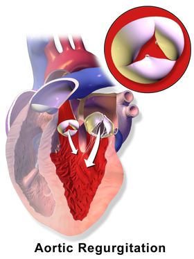
In aortic regurgitation the cusps do not close completely during the filling phase of the heart (diastole), there is backflow of blood into the left ventricle.
Other namesAortic valve reconstruction
SpecialtyCardiology
ICD-9-CM35.9

Aortic valve repair or aortic valve reconstruction is the reconstruction of both form and function of a dysfunctional aortic valve. Most frequently it is used for the treatment of aortic regurgitation. It can also become necessary for the treatment of aortic aneurysm, less frequently for congenital aortic stenosis.

Background

An aortic valve repair will realistically be possible in the absence of calcification or shrinking (retraction) of the aortic valve. Thus, congenital aortic stenosis may be treated by aortic valve repair. In acquired aortic stenosis valve replacement will be the only realistic option. In most instances, aortic valve repair will be performed for aortic regurgitation (insufficiency). Aortic valve repair may also be performed in the treatment of aortic aneurysm or aortic dissection if either aneurysm or dissection involves the aorta close to the valve.

Indications for aortic valve repair:

  • Absence of relevant calcification and
  • Congenital and severe aortic stenosis with symptoms or decreased left ventricular function
  • Severe aortic regurgitation and symptoms, or leftventricular enlargement (>65 to 70 mm), or decreased left ventricular function (EF < 50%)
  • Ascending aortic aneurysm > 55mm
  • Ascending aortic aneurysm > 50mm and risk factors (e.g. high blood pressure)
  • Ascending aortic aneurysm > 50mm and connective tissue disease
  • Ascending aortic aneurysm > 50mm and risk factors and connective tissue disease

Repair versus replacement

The goal of the operation is the improvement of life expectancy and treatment of heart failure as the consequence of dysfunction of the aortic valve. The goal may also be to avert complications of the aorta (rupture or dissection) in the treatment of aneurysm. Repair is a more recent alternative to replacement; in many instances replacement will be the only realistic option because of severe destruction of the valve.

While replacement of the aortic valve is a safe and reproducible procedure it may still be associated with the long-term occurrence of so-called valve-related complications. The probability of these complications depends on the age of the patient and the type of operation. Typical complications are blood clot formation on the valve or dislodgment of thrombus (embolism); bleeding complications are commonly a consequence of "blood-thinning" medication needed to prevent clots (anticoagulation). Biologic/tissue replacement valves have a tendency to degenerate, and there is also an increased risk of infections of valve prosthesis (prosthetic valve endocarditis).

Compared to the results of valve replacement there will be a minimal tendency towards clot formation after aortic valve repair, and anticoagulation is commonly not necessary, thus minimizing the possibility of bleeding complications. The likelihood of infection of the repaired aortic valve is much lower compared to what is seen after aortic valve replacement. A repair procedure may not last forever, but in many instances the durability of an aortic valve repair will markedly exceed that of a biological prosthesis.

Surgical technique

The details of the aortic valve repair procedure depend on the possibility of congenital malformation of the valve, the type and degree of secondary deformation, and the existence of an aortic aneurysm. The goal of the procedure is the restoration of a normal form of the aortic valve, which will then lead to near-normal function and good durability of the repair. A transesophageal echocardiogram during the operation and prior to the repair will be important to define the exact deformation of the aortic valve and thus the mechanism of regurgitation.

In order to best accommodate the complex geometry of the aortic valve, these procedures are generally performed through open-heart surgery. Minimally invasive procedures limit the ability to precisely judge the form of the aortic valve and will lead to a higher uncertainty regarding function and durability of aortic valve repair. As for aortic valve replacement, the heart-lung machine is usually connected to the patient via aorta and right atrium. The heart is arrested through cardioplegia, and the form of the aortic valve is carefully analyzed. Currently documented predicted values for certain aspects of the form of the aortic valve. are available. Using these parameters and a good transesophageal echocardiogram the precise mechanism of regurgitation can be determined in most cases.

Aortic valve stenosis

Congenital aortic valve stenosis can be treated by aortic valve repair if there is no relevant calcification. In this scenario the aortic valve will almost always be unicuspid and the valve configuration must be altered as part of the procedure in order to improve opening of the valve. Because of the unicuspid form of the valve the repair concept will be similar to that of the regurgitant unicuspid valve.

The traditional treatment of congenital aortic stenosis is balloon valvuloplasty or surgical commissurotomy. Both approaches will frequently not eliminate the narrowing of the valve; in addition, they will lead to a variable degree of aortic valve regurgitation which places an additional burden on the heart. In both interventions some of the valve tissue is opened; the peculiar form aspects of unicuspid aortic valves are not taken into consideration. The repair approach differs from commissurotomy mainly in that not only valve tissue is divided to improve opening, but also at least an additional commissure (suspension point of the valve) is created for the aortic valve. Thus, a bicuspid valve is created which results in near-normal function of the aortic valve.

The most reproducible concept is the creation of a bicuspid aortic valve with two normal commissures and two cusps. Tissue of the aortic valve is removed or detached from the aorta in places where it is clearly abnormal. The location of a second commissure of normal height is determined; using a patch or the original cusp tissue the cusps are then sutured to the aortic wall in order to create cusps of sufficient tissue and adequate form reaching the new commissure.

Aortic regurgitation

Tricuspid aortic valve

In tricuspid aortic valves the anatomy is principally normal; if there is an aneurysm of the ascending aorta the principles of aortic aneurysm will have to be applied. Without aneurysm, the cause of regurgitation is frequently stretching of one or two of the valve components (cusps). Such stretching can be combined with the presence of congenital tissue fenestrations. Additionally, enlargement of the aortic annulus can contribute to valve dysfunction. Shrinkage of the cusps is less frequent in industrial countries; this is currently not well treatable by repair.

In surgical treatment, the extent of cusp stretching is exactly determined and then corrected by sutures. Enlargement of the annulus requires its size reduction and stabilization by an annuloplasty. In the case of annular dilatation, the annulus has to be reduced; currently, the largest experience exists with a strong suture that is placed around the annulus and tied to the desired size. Stretching is corrected by plicating sutures to the point that all cusps have a normal configuration. At the end of the operation, the cusp margins should be at an identical height.

Bicuspid aortic valve

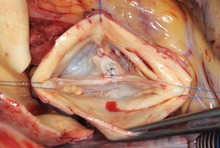
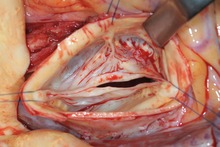
Bicuspid aortic valve which had to be operated on for severe regurgitation. Two of the cusps (upper side right and left) are grown together (fused) since birth. The lack of closure is seen in the central part of the valve, it is caused by stretching of the fused cusp.

In bicuspid aortic valve anatomy, there is congenital fusion of two cusps. This fused cusp is exposed to higher than normal stress and will stretch over time as a consequence. This results in aortic valve regurgitation. Annular enlargement is very frequent in this context, and it increases the tendency to leak. As a result of long-standing dysfunction also the normal cusp may undergo deformation and stretch. In half of the affected individuals there is also an aneurysm of the ascending aorta which has to be treated appropriately.

Repaired bicuspid aortic valve. The stretching of the fused cusp has been corrected by sutures, the correct coaptation of the cusps is easily visible.

Since the bicuspid anatomy commonly has an almost normal valve function (unless deformed) it is left bicuspid; the repair procedure simply corrects the secondary deformations that led to regurgitation. Similar to tricuspid aortic valves, the cusps must be measured to rule out shrinkage. The annulus is commonly enlarged, it must be reduced and stabilized by an annuloplasty. Tissue redundancy through stretching is corrected by sutures.

Unicuspid aortic valve

The unicuspid aortic valve may not only result in relevant stenosis (narrowing), it may also primarily lead to regurgitation. In a proportion of the affected individuals, an aneurysm of the ascending aorta may be present which may need treatment as well. The repair procedure will change the configuration of the valve by creating at least one additional commissure. Commonly the unicuspid valve is changed into a bicuspid configuration; the resulting valve function will be close to normal. Cusp tissue is resected where it is grossly abnormal. Using patch tissue, the cusps are enlarged so they reach the second (new) commissure. If the annulus is enlarged it must be reduced and stabilized.

Quadricuspid aortic valve

Aortic regurgitation in a quadricuspid valve is commonly caused by the additional (4th) commissure, which holds back cusp tissue and keeps it from closing adequately. Currently, the most reliable concept for repair of a quadricuspid valve seems to be its conversion into a tricuspid valve. In some cases a bicuspid configuration may be appropriate. In order to achieve this cusp, tissue is detached from the aorta and the valve is then brought into adequate form.

Aneurysm of the ascending aorta

The enlargement of the ascending aorta may lead to aortic valve regurgitation because the outward tension on the cusps prevents their adequate closure. Regurgitation may also (in part) be due to congenital malformation of the aortic valve or concomitant stretching of a tricuspid aortic valve. Life expectancy may be limited by severe aortic regurgitation. The aneurysm of the ascending aorta may also become so large that it can develop rupture or dissection as life-threatening complications.

The operation must address the aneurysm by replacing the enlarged part of the aorta. Since the aortic valve is very sensitive in its form and function to any changes of the aortic dimensions, the operation will in most cases also have to address the valve, i.e. apply the principles of aortic valve repair. This principle applies to tricuspid valves as well as bicuspid or unicuspid aortic valves.

The goal of the operation is to eliminate the aneurysm and to preserve or repair the aortic valve. The operation may include replacement of the aortic root. Replacement of the root is usually not necessary if its diameter is less than 40 to 45 mm. In those instances replacement of the ascending aorta is sufficient. If root diameter exceeds 45 mm it will have to be replaced in many instances. There are mainly 2 operative techniques currently used, and both lead to similar results. With both techniques the aortic valve must be carefully assessed after replacement of the root; repair of any aortic valve abnormalities is necessary in order to achieve good and durable valve function.

Operative details

There are two options: tubular ascending aortic replacement or replacement of the aortic root.

Tubular ascending aortic replacement

The aorta is divided above the aortic valve and root. The avascular graft is then sutured to the aortic root. The form of the aortic valve may have been changed by this maneuver, it thus has to be carefully checked. Often stretching of a cusp becomes apparent at that point, and this will have to correct by sutures (see 3.3.1, 3.3.2).

Replacement of the aortic root

After the heart has been arrested, the enlarged aorta is removed close to the insertion line of the aortic valve cusps. The origins of the coronary arteries must be detached from the aorta. For the procedure, according to Magdi Yacoub a graft is tailored to create 3 tongues that replace the aneurysmatic aortic wall in the root. The graft is then sutured to the cusp insertion lines. Some surgeons combine this procedure with an annuloplasty. For the procedure according to Tirone David, the aortic valve is mobilized even further from the surrounding tissues. The avascular graft is then positioned around the valve, and the valve is fixed inside the graft with sutures.

With both techniques, the form of the aortic valve must be carefully assessed after completed root replacement. In most instances some cusp stretching will be found which would result in prolapse and relevant regurgitation afterward if uncorrected. Thus an aortic valve repair procedure will frequently be necessary according to the principles of tricuspid or bicuspid aortic valve repair.

Postoperative treatment

Contrary to valve replacement with mechanical prostheses inhibition of the blood clotting system (anticoagulation) is not necessary after aortic valve repair. Blood-thinning may only be necessary if atrial fibrillation occurs or persists in order to prevent blood clot formation in the left atrium.

Following aortic valve replacement, prophylactic administration of antibiotics is recommended for interventions involving mouth and throat (e.g. dental surgery). It is unclear whether this is also necessary after aortic valve repair.

History

First attempts at aortic valve repair were undertaken even before heart valve prostheses were developed. In 1912 the French surgeon Theodore Tuffier widened a stenotic (narrowed) aortic valve. The colleagues of Dwight Harken reported in 1958 on their experience with aortic valve repair for aortic regurgitation by narrowing the annulus of the aortic valve. In those times, both surgeons and cardiologists had minimal information on the exact nature and severity of dysfunction of the aortic valve. This changed with the development of echocardiography by Inge Edler and Carl Hellmuth Hertz in the early 1950s. Nonetheless, the development of heart valve prostheses made replacement the standard approach because of its reproducibility. The first ball-cage valve was implanted in 1961 by the American surgeons Albert Starr and Lowell Edwards, and in the next decades many mechanical and biological prostheses were developed and used. The positive results with the repair of the mitral valve stimulated surgeons in the 1980s and 1990s to develop surgical techniques that could be applied for the different causes of aortic regurgitation. Stepwise improvements were introduced in the subsequent years; today many regurgitant aortic valves can be treated by repair.

Implant (medicine)

From Wikipedia, the free encyclopedia
https://en.wikipedia.org/wiki/Implant_(medicine)
Orthopedic implants to repair fractures to the radius and ulna. Note the visible break in the ulna. (right forearm)
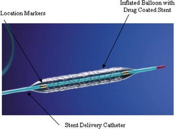
A coronary stent — in this case a drug-eluting stent — is another common item implanted in humans.

An implant is a medical device manufactured to replace a missing biological structure, support a damaged biological structure, or enhance an existing biological structure. For example, an implant may be a rod, used to strengthen weak bones. Medical implants are human-made devices, in contrast to a transplant, which is a transplanted biomedical tissue. The surface of implants that contact the body might be made of a biomedical material such as titanium, silicone, or apatite depending on what is the most functional. In some cases implants contain electronics, e.g. artificial pacemaker and cochlear implants. Some implants are bioactive, such as subcutaneous drug delivery devices in the form of implantable pills or drug-eluting stents.

Applications

Implants can roughly be categorized into groups by application:

Sensory and neurological

Sensory and neurological implants are used for disorders affecting the major senses and the brain, as well as other neurological disorders. They are predominately used in the treatment of conditions such as cataract, glaucoma, keratoconus, and other visual impairments; otosclerosis and other hearing loss issues, as well as middle ear diseases such as otitis media; and neurological diseases such as epilepsy, Parkinson's disease, and treatment-resistant depression. Examples include the intraocular lens, intrastromal corneal ring segment, cochlear implant, tympanostomy tube, and neurostimulator.

Cardiovascular

Cardiovascular medical devices are implanted in cases where the heart, its valves, and the rest of the circulatory system is in disorder. They are used to treat conditions such as heart failure, cardiac arrhythmia, ventricular tachycardia, valvular heart disease, angina pectoris, and atherosclerosis. Examples include the artificial heart, artificial heart valve, implantable cardioverter-defibrillator, artificial cardiac pacemaker, and coronary stent.

Orthopedic

Orthopaedic implants help alleviate issues with the bones and joints of the body. They are used to treat bone fractures, osteoarthritis, scoliosis, spinal stenosis, and chronic pain. Examples include a wide variety of pins, rods, screws, and plates used to anchor fractured bones while they heal.

Metallic glasses based on magnesium with zinc and calcium addition are tested as the potential metallic biomaterials for biodegradable medical implants.

Patients with orthopaedic implants sometimes need to be put under magnetic resonance imaging (MRI) machine for detailed musculoskeletal study. Therefore, concerns have been raised regarding the loosening and migration of implant, heating of the implant metal which could cause thermal damage to surrounding tissues, and distortion of the MRI scan that affects the imaging results. A study of orthopaedic implants in 2005 has shown that majority of the orthopaedic implants does not react with magnetic fields under the 1.0 Tesla MRI scanning machine with the exception of external fixator clamps. However, at 7.0 Tesla, several orthopaedic implants would show significant interaction with the MRI magnetic fields, such as heel and fibular implant.

Electric

Electrical implants are being used to relieve pain from rheumatoid arthritis. The electric implant is embedded in the neck of patients with rheumatoid arthritics, the implant sends electrical signals to electrodes in the vagus nerve. The application of this device is being tested an alternative to medicating people with rheumatoid arthritis for their lifetime.

Contraception

Contraceptive implants are primarily used to prevent unintended pregnancy and treat conditions such as non-pathological forms of menorrhagia. Examples include copper- and hormone-based intrauterine devices.

Cosmetic

Cosmetic implants — often prosthetics — attempt to bring some portion of the body back to an acceptable aesthetic norm. They are used as a follow-up to mastectomy due to breast cancer, for correcting some forms of disfigurement, and modifying aspects of the body (as in buttock augmentation and chin augmentation). Examples include the breast implant, nose prosthesis, ocular prosthesis, and injectable filler.

Other organs and systems

artificial urinary sphincter
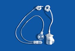
AMS 800 and ZSI 375 artificial urinary sphincters

Other types of organ dysfunction can occur in the systems of the body, including the gastrointestinal, respiratory, and urological systems. Implants are used in those and other locations to treat conditions such as gastroesophageal reflux disease, gastroparesis, respiratory failure, sleep apnea, urinary and fecal incontinence, and erectile dysfunction. Examples include the LINX, implantable gastric stimulator, diaphragmatic/phrenic nerve stimulator, neurostimulator, surgical mesh, artificial urinary sphincter and penile implant.

Classification

United States classification

Medical devices are classified by the US Food and Drug Administration (FDA) under three different classes depending on the risks the medical device may impose on the user. According to 21CFR 860.3, Class I devices are considered to pose the least amount of risk to the user and require the least amount of control. Class I devices include simple devices such as arm slings and hand-held surgical instruments. Class II devices are considered to need more regulation than Class I devices and are required to undergo specific requirements before FDA approval. Class II devices include X-ray systems and physiological monitors. Class III devices require the most regulatory controls since the device supports or sustains human life or may not be well tested. Class III devices include replacement heart valves and implanted cerebellar stimulators. Many implants typically fall under Class II and Class III devices.

Materials

Commonly implanted metals

A variety of minimally bioreactive metals are routinely implanted. The most commonly implanted form of stainless steel is 316L. Cobalt-chromium and titanium-based implant alloys are also permanently implanted. All of these are made passive by a thin layer of oxide on their surface. A consideration, however, is that metal ions diffuse outward through the oxide and end up in the surrounding tissue. Bioreaction to metal implants includes the formation of a small envelope of fibrous tissue. The thickness of this layer is determined by the products being dissolved, and the extent to which the implant moves around within the enclosing tissue. Pure titanium may have only a minimal fibrous encapsulation. Stainless steel, on the other hand, may elicit encapsulation of as much as 2 mm.

List of implantable metal alloys

Stainless Steel

  • ASTM F138/F139 316L
  • ASTM F1314 22Cr-13Ni–5Mn

Titanium Alloy

Cobalt Chrome Alloy

  • ASTM F90 Co-20Cr-15W-10Ni
  • ASTM F562 Co-35Ni-20Cr-10Mo
  • ASTM F1537 Co-28Cr-6Mo

Tantalum

Porosity in Implants

Porous implants are characterized by the presence of voids in the metallic or ceramic matrix. Voids can be regular, such as in additively manufactured (AM) lattices, or stochastic, such as in gas-infiltrated production processes. The reduction in the modulus of the implant follows a complex nonlinear relationship dependent on the volume fraction of base material and morphology of the pores.

Experimental models exist to predict the range of modulus that stochastic porous material may take. Above 10% vol. fraction porosity, models begin to deviate significantly. Different models, such as the rule of mixtures for low porosity, two-material matrices have been developed to describe mechanical properties.

AM lattices have more predictable mechanical properties compared to stochastic porous materials and can be tuned such that they have favorable directional mechanical properties. Variables such as strut diameter, strut shape, and number of cross-beams can have a dramatic effect on loading characteristics of the lattice. AM has the ability to fine-tune the lattice spacing to within a much smaller range than stochastically porous structures, enabling the future cell-development of specific cultures in tissue engineering.

Porosity in implants serves two primary purposes

1) The elastic modulus of the implant is decreased, allowing the implant to better match the elastic modulus of the bone. The elastic modulus of cortical bone (~18 GPa) is significantly lower than typical solid titanium or steel implants (110 GPa and 210 GPa, respectively), causing the implant take up a disproportionate amount of the load applied to the appendage, leading to an effect called stress shielding.

2) Porosity enables osteoblastic cells to grow into the pores of implants. Cells can span gaps of smaller than 75 microns and grow into pores larger than 200 microns. Bone ingrowth is a favorable effect, as it anchors the cells into the implant, increasing the strength of the bone-implant interface. More load is transferred from the implant to the bone, reducing stress shielding effects. The density of the bone around the implant is likely to be higher due to the increased load applied to the bone. Bone ingrowth reduces the likelihood of the implant loosening over time because stress shielding and corresponding bone resorption over extended timescales is avoided. Porosity of greater than 40% is favorable to facilitate sufficient anchoring of the osteoblastic cells.

Complications

Complications can arise from implant failure. Internal rupturing of a breast implant can lead to bacterial infection, for example.

Under ideal conditions, implants should initiate the desired host response. Ideally, the implant should not cause any undesired reaction from neighboring or distant tissues. However, the interaction between the implant and the tissue surrounding the implant can lead to complications. The process of implantation of medical devices is subjected to the same complications that other invasive medical procedures can have during or after surgery. Common complications include infection, inflammation, and pain. Other complications that can occur include risk of rejection from implant-induced coagulation and allergic foreign body response. Depending on the type of implant, the complications may vary.

When the site of an implant becomes infected during or after surgery, the surrounding tissue becomes infected by microorganisms. Three main categories of infection can occur after operation. Superficial immediate infections are caused by organisms that commonly grow near or on skin. The infection usually occurs at the surgical opening. Deep immediate infection, the second type, occurs immediately after surgery at the site of the implant. Skin-dwelling and airborne bacteria cause deep immediate infection. These bacteria enter the body by attaching to the implant's surface prior to implantation. Though not common, deep immediate infections can also occur from dormant bacteria from previous infections of the tissue at the implantation site that have been activated from being disturbed during the surgery. The last type, late infection, occurs months to years after the implantation of the implant. Late infections are caused by dormant blood-borne bacteria attached to the implant prior to implantation. The blood-borne bacteria colonize on the implant and eventually get released from it. Depending on the type of material used to make the implant, it may be infused with antibiotics to lower the risk of infections during surgery. However, only certain types of materials can be infused with antibiotics, the use of antibiotic-infused implants runs the risk of rejection by the patient since the patient may develop a sensitivity to the antibiotic, and the antibiotic may not work on the bacteria.

Inflammation, a common occurrence after any surgical procedure, is the body's response to tissue damage as a result of trauma, infection, intrusion of foreign materials, or local cell death, or as a part of an immune response. Inflammation starts with the rapid dilation of local capillaries to supply the local tissue with blood. The inflow of blood causes the tissue to become swollen and may cause cell death. The excess blood, or edema, can activate pain receptors at the tissue. The site of the inflammation becomes warm from local disturbances of fluid flow and the increased cellular activity to repair the tissue or remove debris from the site.

Implant-induced coagulation is similar to the coagulation process done within the body to prevent blood loss from damaged blood vessels. However, the coagulation process is triggered from proteins that become attached to the implant surface and lose their shapes. When this occurs, the protein changes conformation and different activation sites become exposed, which may trigger an immune system response where the body attempts to attack the implant to remove the foreign material. The trigger of the immune system response can be accompanied by inflammation. The immune system response may lead to chronic inflammation where the implant is rejected and has to be removed from the body. The immune system may encapsulate the implant as an attempt to remove the foreign material from the site of the tissue by encapsulating the implant in fibrinogen and platelets. The encapsulation of the implant can lead to further complications, since the thick layers of fibrous encapsulation may prevent the implant from performing the desired functions. Bacteria may attack the fibrous encapsulation and become embedded into the fibers. Since the layers of fibers are thick, antibiotics may not be able to reach the bacteria and the bacteria may grow and infect the surrounding tissue. In order to remove the bacteria, the implant would have to be removed. Lastly, the immune system may accept the presence of the implant and repair and remodel the surrounding tissue. Similar responses occur when the body initiates an allergic foreign body response. In the case of an allergic foreign body response, the implant would have to be removed.

Failures

The many examples of implant failure include rupture of silicone breast implants, hip replacement joints, and artificial heart valves, such as the Bjork–Shiley valve, all of which have caused FDA intervention. The consequences of implant failure depend on the nature of the implant and its position in the body. Thus, heart valve failure is likely to threaten the life of the individual, while breast implant or hip joint failure is less likely to be life-threatening.

Devices implanted directly in the grey matter of the brain produce the highest quality signals, but are prone to scar-tissue build-up, causing the signal to become weaker, or even non-existent, as the body reacts to a foreign object in the brain.

In 2018, Implant files, an investigation made by ICIJ revealed that medical devices that are unsafe and have not been adequately tested were implanted in patients' bodies. In United Kingdom, Prof Derek Alderson, president of the Royal College of Surgeons, concludes: "All implantable devices should be registered and tracked to monitor efficacy and patient safety in the long-term."

Military logistics

From Wikipedia, the free encyclopedia
U.S. Marines from Combat Logistics Battalion 8 and Navy personnel from Beach Master Unit 2 off-load ISO containers from a Landing Craft Utility with a Logistics Vehicle System Replacement
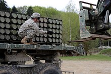
Loading artillery shells (2016)

Military logistics is the discipline of planning and carrying out the movement, supply, and maintenance of military forces. In its most comprehensive sense, it is those aspects or military operations that deal with:

  • Design, development, acquisition, storage, distribution, maintenance, evacuation, and disposition of materiel.
  • Transport of personnel.
  • Acquisition or construction, maintenance, operation and disposition of facilities.
  • Acquisition or furnishing of services.
  • Medical and health service support.

Etymology and definition

The word "logistics" is derived from the Greek adjective logistikos meaning "skilled in calculating", and its corresponding Latin word logisticus. In turn this comes from the Greek logos, which refers to the principles of thought and action. Another Latin root, log-, gave rise around 1380 to logio, meaning to lodge or dwell, and became the French verb loger, meaning "to lodge". Around 1670, the French King Louis XIV created the position of Maréchal des logis, an officer responsible for planning marches, establishing camp sites, and regulating transport and supply. The term logistique soon came to refer to his duties. It was in this sense that Antoine-Henri Jomini referred to the term in his Summary of the Art of War (1838). In the English translation, the word became "logistics".

In 1888, Charles C. Rogers created a course on naval logistics at the Naval War College. In Farrow's Military Encyclopedia (1895), Edward S. Farrow, and instructor in tactics at West Point provided this definition:

Bardin considers the application of this word by some writers as more ambitious than accurate. It is derived from Latin logista, the administrator or intendant of the Roman armies. It is properly that branch of the military art embracing all the details for moving and supplying armies. It includes the operations of the ordnance, quartermaster's, subsistence, medical, and pay departments. It also embraces the preparation and regulation of magazines, for opening a campaign, and all orders of march and other orders from the general-in-chief relative to moving and supplying armies.

The term became popularised during the Second World War. In Logistics in World War II: Final Report of the Army Service Forces, Lieutenant General LeRoy Lutes, the commanding general of the Army Service Forces, gave the term a more expansive definition:

The word "logistics" has been given many different shades of meaning. A common definition is: "That branch of military art which embraces the details of the transport, quartering, and supply of troops in military operations." As the word is used in the following pages, its meaning is even broader. It embraces all military activities not included in the terms "strategy" and "tactics." In this sense, logistics includes procurement, storage, and distribution of equipment and supplies; transport of troops and cargo by land, sea, and air; construction and maintenance of facilities; communication by wire, radio, and the mails; care of the sick and wounded; and the induction, classification, assignment, welfare and separation of personnel.

NATO uses a more restrictive definition:

The science of planning and carrying out the movement and maintenance of forces. In its most comprehensive sense, the aspects of military operations which deal with:

  1. design and development, acquisition, storage, movement, distribution, maintenance, evacuation, and disposal of materiel;
  2. transport of personnel;
  3. acquisition or construction, maintenance, operation, and disposition of facilities;
  4. acquisition or furnishing of services; and
  5. medical and health service support.

In the 1960s, the term "logistics" began to be used in the business world, where it means physical distribution and supply chain management.

Principles

Historian James A. Huston proposed sixteen principles of military logistics:

  1. Equivalence: Strategy, tactics and logistics are inseparable and interdependent facets of military art and science.
  2. Material precedence: Mobilisation of materiel should precede that of personnel, and the provision of logistical units that of combat units.
  3. Forward impetus: The impetus of supply should be from the rear, and combat unit commanders should be spared having to deal with logistical details while still being in control of their logistics.
  4. Mobility: Logistics should facilitate the rapid movement of both combat and logistical units in support of operations.
  5. Dispersion: Multiple sources of supply and lines of communications reduce the enemy interference and congestion of transportation infrastructure.
  6. Economy: Logistical resources are limited, so they must be deployed so as to make the best use of them.
  7. Feasibility: Logistical capabilities are subject to external constraints.
  8. Flexibility: Strategic and operational plans and priorities change and logistical support must change with them.
  9. Relativity: Logistics is relative to time and space.
  10. Continuity: Fundamental changes should not be required to meet an emergency.
  11. Timeliness Logistical tasks must be accomplished so as to take full advantage of opportunities.
  12. Responsibility: Someone must be responsible for logistical performance and outcomes.
  13. Unity of command: Logistics is a function of command and a single authority should be responsible for logistics.
  14. Information: Accurate and timely information is required for effective logistical planning and support.
  15. Quality: Logistics is facilitated by strict quality standards.
  16. Simplicity: Simple solutions are more effective and manageable.

The United States Joint Chiefs of Staff reduced the number of principles to just seven:

  1. Responsiveness: Providing the required support when and where it is needed.
  2. Simplicity: fosters unity and efficiency in planning and execution, and reduces the fog of war and the "friction" caused by combat.
  3. Flexibility: the ability to improvise and adapt to changing situations and requirements.
  4. Economy: using the minimum amount of resources required to bring about an objective.
  5. Attainability: the point at which sufficient supplies, support and distribution capabilities exist to initiate operations at an acceptable level of risk.
  6. Sustainability: the ability to maintain the necessary level and duration of logistics support to achieve objectives.
  7. Survivability: the capacity to prevail in spite of adverse impacts.

Supply options

There are three basic options for the supply of an army in the field, which can be employed individually or in combination.

Obtain supplies in the field

The most basic requirements of an army were food and water. Foraging involved gathering food and fodder for animals in the field. The availability of these tends to be seasonal, with greater abundance around harvest time in agricultural regions. There is also a dependence on geography, for in desert campaigns there may not be food, water or fodder available locally. Looting was another means of obtaining supplies in the field. It is possible to capture supplies from the enemy or enemy population. Another alternative is purchasing, whereby an army takes cash and buys its supplies in the field. Cash can also be obtained in the field through local taxation, backed by the threat of violence. The major drawback of using local sources of supply is that they can be exhausted if an army remains in one place for too long, so a force dependent on it needs to keep moving.

The widespread use of sources in the field gives rise to counter-logistics, whereby resources are denied to the enemy through devastation of the land and removal or destruction of food sources. Pre-emptive purchasing can be used as a form of economic warfare. A besieging force can attempt to starve out a garrison or tempt it to sally through devastation of the surrounding area rather than undertake the more costly operation of assaulting and destroying it, but if it is dependent on local supply then the besieger who might be starved out through their exhaustion.

Carry supplies with the army

A second method was for the army to bring along what was needed, whether by ships, pack animals, wagons or carried on the backs of the soldiers themselves. Since ancient times, troops had carried rations and personal equipment such as weapons, armour, cooking gear and bedrolls. Animals could be driven to accompany the army and consumed for meat. Roads facilitate the movement of wheeled vehicles, and travel by river or sea permits the carriage of large volumes of supplies. This allowed the army some measure of self-sufficiency, and until the development of faster firing weapons in the 19th century most of the ammunition a soldier needed for an entire campaign could be carried on their person or in wagons accompanying the troops. However, this method led to an extensive baggage train which could slow down the army's advance.

Ship supplies from the rear

Obtaining supplies in the field and carrying supplies with the army remained the primary means of supply until the 19th century, but even in the 17th century the much larger armies of the period were highly dependent on food supplies being gathered in magazines and shipped to the front. Starting with the Industrial Revolution, new technological, technical and administrative advances permitted supplies to be transported at speeds and over distances never before possible. At the same time, increased demands for ammunition, and the heavier weight of shells and bombs made it more difficult for armies to carry their requirements, and they soon became dependent on regular replenishment of ammunition from depots. At the same time, mechanisation, with motor vehicles replacing animals, created a demand for fuel and spare parts, neither of which could be obtained locally. This led to a "logistical revolution" which began in the 20th century and drastically improved the capabilities of modern armies while making them highly dependent on this method.

History

The history of military logistics goes back to Neolithic times. The most basic requirements of an army were food and water. Early armies were equipped with weapons used for hunting like spears, knives, axes and bows and arrows, and rarely exceeded 20,000 men due to the practical difficulty of supplying a large number of soldiers. Large armies began to appear in the Iron Age. Animals such as horses, oxen, camels and even elephants were used as beasts of burden to carry supplies. Food, water and fodder for the animals could usually be found or purchased in the field. The Roman Empire and Maurya Empire in India built networks of roads, but it was far less expensive to transport a ton of grain from Egypt to Rome than 80 kilometres (50 mi) by road. After the fall of the Roman Empire in the fifth century there was the shift from a centrally organised army to a combination of military forces made up of local troops. Feudalism was therefore a distributed military logistics system where magnates of the households drew upon their own resources for men and equipment.

From the late sixteenth century, armies in Europe increased in size, to 100,000 or more in some cases. When operating in enemy territory an army was forced to plunder the local countryside for supplies, which allowed war to be conducted at the enemy's expense. However, with the increase in army sizes this reliance on pillage and plunder became problematic, as decisions regarding where and when an army could move or fight became based not on strategic objectives but on whether a given area was capable of supporting the soldiers' needs. Sieges in particular were affected by this, both for an army attempting to lay siege to a town and one coming to its relief. Unless a commander was able to arrange a form of regular resupply, a fortress or town with a devastated countryside could become immune to either operation.

Napoleon made logistics a major part of his strategy. He dispersed his corps along a broad front to maximise the area from which supplies could be drawn. Each day forage parties brought in supplies. This differed from earlier operations living off the land in the size of the forces involved, and because the primary motivation was the emperor's desire for mobility. Ammunition could not as a rule be obtained locally, but it was still possible to carry sufficient ammunition for an entire whole campaign.

The nineteenth century witnessed technological developments that facilitated immense improvements to the storage, handling and transportation of supplies which made it easier to support and army from the rear. Canning simplified storage and distribution of foods, and reduced waste and the incidence of food-related illness. Refrigeration allowed frozen meat and fresh produce to be stored and shipped. Steamships made water transports faster and more reliable. Railways were a more economical form of transport than animal-drawn carts and wagons, although they were limited to tracks, and therefore could not support an advancing army unless its advance was along existing railway lines. At the same time, the advent of industrial warfare in the form of bolt-action rifles, machine guns and quick-firing artillery sent ammunition consumption soaring during the First World War.

In the twentieth century the advent of motor vehicles powered by internal combustion engines offered an alternative to animal transport for moving supplies forward of the railhead, although many armies still used animals during the Second World War. The development of air transport provided an alternative to both land and sea transport, but with limited tonnage and at high cost. An airlift over "the Hump" helped supply the Chinese war effort], and after the war the 1948 Berlin Air Lift was successful in supplying half of the city. With the subsequent development of large jets, aircraft became the preferred method of moving personnel over long distances, although it was still more economical to move cargo by sea and land. In forward areas, the helicopter was well-suited to moving troops and supplies, especially over rugged terrain.

Models

Levels

Akin to the three levels of war, there can be considered to be three levels of logistics. Although modern communications and information technology may have blurred the distinction between them, the three-level hierarchy is deeply embedded in the organisational structure of military forces.

  • Strategic logistics involves logistical activities that are conducted at national and international levels. It includes defining requirements, and arranging for the production and distribution of materiel to operational forces.
  • Operational logistics involves logistical activities within the theatre of operations. It includes the reception, storage, and distribution of supplies and personnel, the hospitalisation of casualties, the maintenance and repair of equipment, and the operation of the intra-theatre transportation system. The operational level of war can be defined by the amount of logistical independence a formation has. For this reason logistics is most often discussed at the operational level.
  • Tactical logistics involves the logistical activities of units engaged in combat.

Unlike business logistics, the objective of military logistics is not cost effectiveness of the supply chain, but maximum sustained combat effectiveness.

Representation of a Lie group

From Wikipedia, the free encyclopedia https://en.wikipedia.org/wiki/Representation_of_a_Lie_group...