Search This Blog

Thursday, October 26, 2023

Analgesic

From Wikipedia, the free encyclopedia
https://en.wikipedia.org/wiki/Analgesic
 
Opium poppies such as this one provide ingredients for the class of analgesics called opiates.
 
Class identifiers

An analgesic drug, also called simply an analgesic, pain reliever, or painkiller, is any member of the group of drugs used to achieve relief from pain (that is, analgesia or pain management). Analgesics are conceptually distinct from anesthetics, which temporarily reduce, and in some instances eliminate, sensation, although analgesia and anesthesia are neurophysiologically overlapping and thus various drugs have both analgesic and anesthetic effects.

Analgesic choice is also determined by the type of pain: For neuropathic pain, recent research has suggested that classes of drugs that are not normally considered analgesics, such as tricyclic antidepressants and anticonvulsants may be considered as an alternative. 

Various analgesics, such as many NSAIDs, are available over the counter in most countries, whereas various others are prescription drugs owing to the substantial risks and high chances of overdose, misuse, and addiction in the absence of medical supervision.

Etymology

The word analgesic derives from Greek an- (ἀν-, "without"), álgos (ἄλγος, "pain"), and -ikos (-ικος, forming adjectives). Such drugs were usually known as "anodynes" before the 20th century.

Classification

Analgesics are typically classified based on their mechanism of action.

A bottle of acetaminophen

Paracetamol (acetaminophen)

Paracetamol, also known as acetaminophen or APAP, is a medication used to treat pain and fever. It is typically used for mild to moderate pain. In combination with opioid pain medication, paracetamol is now used for more severe pain such as cancer pain and after surgery. It is typically used either by mouth or rectally but is also available intravenously. Effects last between two and four hours. Paracetamol is classified as a mild analgesic. Paracetamol is generally safe at recommended doses.

NSAIDs

Nonsteroidal anti-inflammatory drugs (usually abbreviated to NSAIDs), are a drug class that groups together drugs that decrease pain and lower fever, and, in higher doses, decrease inflammation. The most prominent members of this group of drugs, aspirin, ibuprofen and naproxen, are all available over the counter in most countries.

COX-2 inhibitors

These drugs have been derived from NSAIDs. The cyclooxygenase enzyme inhibited by NSAIDs was discovered to have at least two different versions: COX1 and COX2. Research suggested most of the adverse effects of NSAIDs to be mediated by blocking the COX1 (constitutive) enzyme, with the analgesic effects being mediated by the COX2 (inducible) enzyme. Thus, the COX2 inhibitors were developed to inhibit only the COX2 enzyme (traditional NSAIDs block both versions in general). These drugs (such as rofecoxib, celecoxib, and etoricoxib) are equally effective analgesics when compared with NSAIDs, but cause less gastrointestinal hemorrhage in particular.

After widespread adoption of the COX-2 inhibitors, it was discovered that most of the drugs in this class increase the risk of cardiovascular events by 40% on average. This led to the withdrawal of rofecoxib and valdecoxib, and warnings on others. Etoricoxib seems relatively safe, with the risk of thrombotic events similar to that of non-coxib NSAID diclofenac.

Opioids

Morphine, the archetypal opioid, and other opioids (e.g., codeine, oxycodone, hydrocodone, dihydromorphine, pethidine) all exert a similar influence on the cerebral opioid receptor system. Buprenorphine is a partial agonist of the μ-opioid receptor, and tramadol is a serotonin norepinephrine reuptake inhibitor (SNRI) with weak μ-opioid receptor agonist properties. Tramadol is structurally closer to venlafaxine than to codeine and delivers analgesia by not only delivering "opioid-like" effects (through mild agonism of the mu receptor) but also by acting as a weak but fast-acting serotonin releasing agent and norepinephrine reuptake inhibitor. Tapentadol, with some structural similarities to tramadol, presents what is believed to be a novel drug working through two (and possibly three) different modes of action in the fashion of both a traditional opioid and as an SNRI. The effects of serotonin and norepinephrine on pain, while not completely understood, have had causal links established and drugs in the SNRI class are commonly used in conjunction with opioids (especially tapentadol and tramadol) with greater success in pain relief.

Dosing of all opioids may be limited by opioid toxicity (confusion, respiratory depression, myoclonic jerks and pinpoint pupils), seizures (tramadol), but opioid-tolerant individuals usually have higher dose ceilings than patients without tolerance. Opioids, while very effective analgesics, may have some unpleasant side-effects. Patients starting morphine may experience nausea and vomiting (generally relieved by a short course of antiemetics such as phenergan). Pruritus (itching) may require switching to a different opioid. Constipation occurs in almost all patients on opioids, and laxatives (lactulose, macrogol-containing or co-danthramer) are typically co-prescribed.

When used appropriately, opioids and other central analgesics are safe and effective; however, risks such as addiction and the body's becoming used to the drug (tolerance) can occur. The effect of tolerance means that frequent use of the drug may result in its diminished effect. When safe to do so, the dosage may need to be increased to maintain effectiveness against tolerance, which may be of particular concern regarding patients with chronic pain and requiring an analgesic over long periods. Opioid tolerance is often addressed with opioid rotation therapy in which a patient is routinely switched between two or more non-cross-tolerant opioid medications in order to prevent exceeding safe dosages in the attempt to achieve an adequate analgesic effect.

Opioid tolerance should not be confused with opioid-induced hyperalgesia. The symptoms of these two conditions can appear very similar but the mechanism of action is different. Opioid-induced hyperalgesia is when exposure to opioids increases the sensation of pain (hyperalgesia) and can even make non-painful stimuli painful (allodynia).

Alcohol

Alcohol has biological, mental, and social effects which influence the consequences of using alcohol for pain. Moderate use of alcohol can lessen certain types of pain in certain circumstances.

The majority of its analgesic effects come from antagonizing NMDA receptors, similarly to ketamine, thus decreasing the activity of the primary excitatory (signal boosting) neurotransmitter, glutamate. It also functions as an analgesic to a lesser degree by increasing the activity of the primary inhibitory (signal reducing) neurotransmitter, GABA.

Attempting to use alcohol to treat pain has also been observed to lead to negative outcomes including excessive drinking and alcohol use disorder.

Cannabis

Medical cannabis, or medical marijuana, refers to cannabis or its cannabinoids used to treat disease or improve symptoms. There is evidence suggesting that cannabis can be used to treat chronic pain and muscle spasms, with some trials indicating improved relief of neuropathic pain over opioids.

Combinations

Analgesics are frequently used in combination, such as the paracetamol and codeine preparations found in many non-prescription pain relievers. They can also be found in combination with vasoconstrictor drugs such as pseudoephedrine for sinus-related preparations, or with antihistamine drugs for people with allergies.

While the use of paracetamol, aspirin, ibuprofen, naproxen, and other NSAIDS concurrently with weak to mid-range opiates (up to about the hydrocodone level) has been said to show beneficial synergistic effects by combating pain at multiple sites of action, several combination analgesic products have been shown to have few efficacy benefits when compared to similar doses of their individual components. Moreover, these combination analgesics can often result in significant adverse events, including accidental overdoses, most often due to confusion that arises from the multiple (and often non-acting) components of these combinations.

Alternative medicine

There is some evidence that some treatments using alternative medicine can relieve some types of pain more effectively than placebo. The available research concludes that more research would be necessary to better understand the use of alternative medicine.

Other drugs

Nefopam—a monoamine reuptake inhibitor, and calcium and sodium channel modulator—is also approved for the treatment of moderate to severe pain in some countries.

Flupirtine is a centrally acting K+ channel opener with weak NMDA antagonist properties. It was used in Europe for moderate to strong pain, as well as its migraine-treating and muscle-relaxant properties. It has no significant anticholinergic properties, and is believed to be devoid of any activity on dopamine, serotonin, or histamine receptors. It is not addictive, and tolerance usually does not develop. However, tolerance may develop in some cases.

Ziconotide, a blocker of potent N‐type voltage‐gated calcium channels, is administered intrathecally for the relief of severe, usually cancer-related pain.

Adjuvants

Certain drugs that have been introduced for uses other than analgesics are also used in pain management. Both first-generation (such as amitriptyline) and newer antidepressants (such as duloxetine) are used alongside NSAIDs and opioids for pain involving nerve damage and similar problems. Other agents directly potentiate the effects of analgesics, such as using hydroxyzine, promethazine, carisoprodol, or tripelennamine to increase the pain-killing ability of a given dose of opioid analgesic.

Adjuvant analgesics, also called atypical analgesics, include orphenadrine, mexiletine, pregabalin, gabapentin, cyclobenzaprine, hyoscine (scopolamine), and other drugs possessing anticonvulsant, anticholinergic, and/or antispasmodic properties, as well as many other drugs with CNS actions. These drugs are used along with analgesics to modulate and/or modify the action of opioids when used against pain, especially of neuropathic origin.

Dextromethorphan has been noted to slow the development of and reverse tolerance to opioids, as well as to exert additional analgesia by acting upon NMDA receptors, as does ketamine. Some analgesics such as methadone and ketobemidone and perhaps piritramide have intrinsic NMDA action.

High-alcohol liquor, two forms of which were found in the US Pharmacopoeia up until 1916 and in common use by physicians well into the 1930s, has been used in the past as an agent for dulling pain, due to the CNS depressant effects of ethyl alcohol, a notable example being the American Civil War. However, the ability of alcohol to relieve severe pain is likely inferior to many analgesics used today (e.g., morphine, codeine). As such, in general, the idea of alcohol for analgesia is considered a primitive practice in virtually all industrialized countries today.

The anticonvulsant carbamazepine is used to treat neuropathic pain. Similarly, the gabapentinoids gabapentin and pregabalin are prescribed for neuropathic pain, and phenibut is available without prescription. Gabapentinoids work as α2δ-subunit blockers of voltage-gated calcium channels, and tend to have other mechanisms of action as well. Gabapentinoids are all anticonvulsants, which are most commonly used for neuropathic pain, as their mechanism of action tends to inhibit pain sensation originating from the nervous system.

Other uses

Topical analgesia is generally recommended to avoid systemic side-effects. Painful joints, for example, may be treated with an ibuprofen- or diclofenac-containing gel (The labeling for topical diclofenac has been updated to warn about drug-induced hepatotoxicity.); capsaicin also is used topically. Lidocaine, an anesthetic, and steroids may be injected into joints for longer-term pain relief. Lidocaine is also used for painful mouth sores and to numb areas for dental work and minor medical procedures. In February 2007 the FDA notified consumers and healthcare professionals of the potential hazards of topical anesthetics entering the bloodstream when applied in large doses to the skin without medical supervision. These topical anesthetics contain anesthetic drugs such as lidocaine, tetracaine, benzocaine, and prilocaine in a cream, ointment, or gel.

Uses

Topical nonsteroidal anti-inflammatory drugs provide pain relief in common conditions such as muscle sprains and overuse injuries. Since the side effects are also lesser, topical preparations could be preferred over oral medications in these conditions.

List of drugs with comparison

Research

Some novel and investigational analgesics include subtype-selective voltage-gated sodium channel blockers such as funapide and raxatrigine, as well as multimodal agents such as ralfinamide.

Electrosurgery

From Wikipedia, the free encyclopedia
https://en.wikipedia.org/wiki/Electrosurgery
 
Electrosurgery
A surgeon using a monopolar RF electrosurgical instrument to coagulate (and desiccate) tissue in the excision of a lipoma

Electrosurgery is the application of a high-frequency (radio frequency) alternating polarity, electrical current to biological tissue as a means to cut, coagulate, desiccate, or fulgurate tissue. (These terms are used in specific ways for this methodology—see below.) Its benefits include the ability to make precise cuts with limited blood loss. Electrosurgical devices are frequently used during surgical operations helping to prevent blood loss in hospital operating rooms or in outpatient procedures.

In electrosurgical procedures, the tissue is heated by an electric current. Although electrical devices that create a heated probe may be used for the cauterization of tissue in some applications, electrosurgery refers to a different method than electrocautery. Electrocautery uses heat conduction from a probe heated to a high temperature by a direct electrical current (much in the manner of a soldering iron). This may be accomplished by direct current from dry-cells in a penlight-type device.

Electrosurgery, by contrast, uses radio frequency (RF) alternating current to heat the tissue by RF induced intracellular oscillation of ionized molecules that result in an elevation of intracellular temperature. When the intracellular temperature reaches 60 degrees C, instantaneous cell death occurs. If tissue is heated to 60–99 degrees C, the simultaneous processes of tissue desiccation (dehydration) and protein coagulation occur. If the intracellular temperature rapidly reaches 100 degrees C, the intracellular contents undergo a liquid to gas conversion, massive volumetric expansion, and resulting explosive vaporization.

Appropriately applied with electrosurgical forceps, desiccation and coagulation result in the occlusion of blood vessels and halting of bleeding. While the process is technically a process of electrocoagulation, the term "electrocautery" is sometimes loosely, nontechnically and incorrectly used to describe it. The process of vaporization can be used to ablate tissue targets, or, by linear extension, used to transect or cut tissue. While the processes of vaporization/ cutting and desiccation/coagulation are best accomplished with relatively low voltage, continuous or near continuous waveforms, the process of fulguration is performed with relatively high voltage modulated waveforms. Fulguration is a superficial type of coagulation, typically created by arcing modulated high voltage current to tissue that is rapidly desiccated and coagulated. The continued application of current to this high impedance tissue results in resistive heating and the achievement of very high temperatures—enough to cause breakdown of the organic molecules to sugars and even carbon, thus the dark textures from carbonization of tissue.

Diathermy is used by some as a synonym for electrosurgery but in other contexts diathermy means dielectric heating, produced by rotation of molecular dipoles in a high frequency electromagnetic field. This effect is most widely used in microwave ovens or some tissue ablative devices which operate at gigahertz frequencies. Lower frequencies, allowing for deeper penetration, are used in industrial processes.

RF electrosurgery is commonly used in virtually all surgical disciplines including dermatological, gynecological, cardiac, plastic, ocular, spine, ENT, maxillofacial, orthopedic, urological, neuro- and general surgical procedures as well as certain dental procedures.

RF electrosurgery is performed using a RF electrosurgical generator (also referred to as an electrosurgical unit or ESU) and a handpiece including one or two electrodes—a monopolar or bipolar instrument. All RF electrosurgery is bipolar so the difference between monopolar and bipolar instruments is that monopolar instruments comprise only one electrode while bipolar instruments include both electrodes in their design.

The monopolar instrument called an "active electrode" when energized, requires the application of another monopolar instrument called a "dispersive electrode" elsewhere on the patient's body that functions to 'defocus' or disperse the RF current thereby preventing thermal injury to the underlying tissue. This dispersive electrode is frequently and mistakenly called a "ground pad" or "neutral electrode". However virtually all currently available RF electrosurgical systems are designed to function with isolated circuits—the dispersive electrode is directly attached to the ESU, not to "ground". The same electrical current is transmitted across both the dispersive electrode and the active electrode, so it is not "neutral". The term "return electrode" is also technically incorrect since alternating electrical currents refer to alternating polarity, a circumstance that results in bidirectional flow across both electrodes in the circuit.

Bipolar instruments generally are designed with two "active" electrodes, such as a forceps for sealing blood vessels. However, the bipolar instrument can be designed such that one electrode is dispersive. The main advantage of bipolar instruments is that the only part of the patient included in the circuit is that which is between the two electrodes, a circumstance that eliminates the risk of current diversion and related adverse events. However, except for those devices designed to function in fluid, it is difficult to vaporize or cut tissue with bipolar instruments.

Electrical stimulation of neural and muscle cells

Neural and muscle cells are electrically-excitable, i.e. they can be stimulated by electric current. In human patients such stimulation may cause acute pain, muscle spasms, and even cardiac arrest. Sensitivity of the nerve and muscle cells to electric field is due to the voltage-gated ion channels present in their cell membranes. Stimulation threshold does not vary much at low frequencies (so called rheobase-constant level). However, the threshold starts increasing with decreasing duration of a pulse (or a cycle) when it drops below a characteristic minimum (so called chronaxie). Typically, chronaxie of neural cells is in the range of 0.1–10 ms, so the sensitivity to electrical stimulation (inverse of the stimulation threshold) decreases with increasing frequency in the kHz range and above. (Note that frequency of the alternating electric current is an inverse of the duration of a single cycle). To minimize the effects of muscle and neural stimulation, electrosurgical equipment typically operates in the radio frequency (RF) range of 100 kHz to 5 MHz.

Operation at higher frequencies also helps minimizing the amount of hydrogen and oxygen generated by electrolysis of water. This is especially important consideration for applications in liquid medium in closed compartments, where generation of gas bubbles may interfere with the procedure. For example, bubbles produced during an operation inside an eye may obscure a field of view.[citation needed]

Common electrode configurations for devices with isolated circuits

There are several commonly used electrode configurations or circuit topologies:

With "bipolar" instruments the current is applied to the patient using a pair of similarly-sized electrodes. For example, special forceps, with one tine connected to one pole of the RF generator and the other tine connected to the other pole of the generator. When a piece of tissue is held by the forceps, the RF alternating polarity electrical current oscillates between the two forceps tines, heating the intervening tissue by the previously described synchronous oscillation of intracellular ions.

In monopolar configuration the patient is attached to the dispersive electrode, a relatively large metal plate or a flexible metalized plastic pad which is connected to the RF generator or electrosurgical unit (ESU). The surgeon uses a pointed or blade shaped electrode called the "active electrode" to make contact with the tissue and exert a tissue effect - vaporization, and its linear propagation called electrosurgical cutting, or the combination of desiccation and protein coagulation used to seal blood vessels for the purpose of Hemostasis. The electric current oscillates between the active electrode and the dispersive electrode with the entire patient interposed between the two. Since the concentration of the RF current reduces with distance from the active electrode the current density rapidly (quadratically) decreases. Since the rate of tissue heating is proportional to the square of current density, the heating occurs in a very localized region, only near the portion of the electrode, usually the tip, near to or in contact with the target tissue.

On an extremity such as a finger, there is limited cross-sectional area to disperse the current, a circumstance which might result in higher current density and some heating throughout the volume of the extremity.

Another bipolar instrument is characterized with both electrodes on the same design, but the dispersive electrode is much larger than the active one. Since current density is higher in front of the smaller electrode, the heating and associated tissue effects take place only (or primarily) in front of the active electrode, and exact position of the dispersive electrode on tissue is not critical. Sometimes such configuration is called sesquipolar, even though the origin of this term in Latin (sesqui) means a ratio of 1.5.

Dedicated non-grounded machines without a dispersive electrode

Relatively low-powered high frequency electrosurgery can be performed on conscious outpatients with no grounded machines without a dispersive electrode. Operating at low currents with no dispersive electrode is possible because, at the medium RF frequencies (usually 100 – 500 kHz) that the machines generate, the self-capacitance of the patient's body (which is between the patient's body and the machine's ground) is large enough to allow the resulting displacement current to act as a virtual "circuit completion path."

One example of such a machine is called a hyfrecator. This term began in 1940 as a Birtcher Corporation brand name Hyfrecator for "High Frequency Eradicator", but now serves generically to describe a general class of single-electrode, non-isolated (earth-referenced) low-powered electrosurgical machines intended mainly for office use. An accidental circuit completion path through an earth-ground creates the danger of a burn at a site far away from the probe electrode, and for this reason single-electrode devices are used only on conscious patients who would be aware of such complications, and only on carefully insulated tables.

In such a setting, hyfrecators are not used to cut tissue, but to destroy relatively small lesions, and also to stop bleeding in surgical incisions made by blade instruments under local anesthesia.

Electrosurgical modalities

In cutting mode electrode touches the tissue, and sufficiently high power density is applied to vaporize its water content. Since water vapor is not conductive under normal circumstances, electric current cannot flow through the vapor layer. Energy delivery beyond the vaporization threshold can continue if sufficiently high voltage is applied (> +/-200 V) to ionize vapor and convert it into a conductive plasma. Vapor and fragments of the overheated tissue are ejected, forming a crater. Electrode surfaces intended to be used for cutting often feature a finer wire or wire loop, as opposed to a more flat blade with a rounded surface.

Coagulation is performed using waveforms with lower average power, generating heat insufficient for explosive vaporization, but producing a thermal coagulum instead.

Electrosurgical desiccation occurs when the electrode touches the tissue open to air, and the amount of generated heat is lower than that required for cutting. The tissue surface and some of the tissue more deep to the probe dries out and forms a coagulum (a dry patch of dead tissue). This technique may be used for treating nodules under the skin where minimal damage to the skin surface is desired.

In fulguration mode, the electrode is held away from the tissue, so that when the air gap between the electrode and the tissue is ionized, an electric arc discharge develops. In this approach, the burning to the tissue is more superficial, because the current is spread over the tissue area larger than the tip of electrode. Under these conditions, superficial skin charring or carbonization is seen over a wider area than when operating in contact with the probe, and this technique is therefore used for very superficial or protrusive lesions such as skin tags. Ionization of an air gap requires voltage in the kV range.

Besides the thermal effects in tissue, the electric field can produce pores in the cellular membranes – a phenomenon called electroporation. This effect may affect cells beyond the range of thermal damage.

Wet field electrosurgery

There are wet and dry field electrosurgical devices. Wet field devices operate in a saline solution, or in an open wound. Heating is as a result of an alternating current that passes between two electrodes. Heating is usually greatest where the current density is highest. Therefore, it is usually the smallest or sharpest electrode that generates the most heat.

Cut/Coag Most wet field electrosurgical systems operate in two modes: "Cut" causes a small area of tissue to be vaporized, and "Coag" causes the tissue to "dry" (in the sense of bleeding being stopped). "Dried" tissues are killed (and will later slough or be replaced by fibrotic tissue) but they are temporarily physically intact after electrosurgical application. The depth of tissue death is typically a few millimeters near the contact of the electrode.

Cut If the voltage level is high enough, the heat generated can create a vapour pocket. The vapour pocket typically reaches temperatures of approximately 400 degrees Celsius, which vaporizes and explodes a small section of soft tissue, resulting in an incision.

Coag When the system is operating in "coag mode" the voltage output is usually higher than in cut mode. Tissue remains grossly intact, but cells are destroyed at the point of contact, and smaller vessels are destroyed and sealed, stopping capillary and small-arterial bleeding.

Electrosurgical waveforms

Different waveforms can be used for different electrosurgical procedures. For cutting, a continuous single frequency sine wave is often employed. Rapid tissue heating leads to explosive vaporization of interstitial fluid. If the voltage is sufficiently high (> 400 V peak-to-peak) the vapor sheath is ionized, forming conductive plasma. Electric current continues to flow from the metal electrode through the ionized gas into the tissue. Rapid overheating of tissue results in its vaporization, fragmentation and ejection of fragments, allowing for tissue cutting. In applications of a continuous wave the heat diffusion typically leads to formation of a significant thermal damage zone at the edges of the lesion. Open circuit voltage in electrosurgical waveforms is typically in the range of 300–10,000 V peak-to-peak.

Higher precision can be achieved with pulsed waveforms. Using bursts of several tens of microseconds in duration the tissue can be cut, while the size of the heat diffusion zone does not exceed the cellular scale. Heat accumulation during repetitive application of bursts can also be avoided if sufficient delay is provided between the bursts, allowing the tissue to cool down. The proportion of ON time to OFF time can be varied to allow control of the heating rate. A related parameter, duty cycle, is defined as the ratio of the ON time to the period (the time of a single ON-OFF cycle). In the terminology of electrical engineering, the process of altering this ratio to achieve an average amplitude, instead of altering the amplitude directly is called pulse-width modulation.

For coagulation, the average power is typically reduced below the threshold of cutting. Typically, sine wave is turned on and off in rapid succession. The overall effect is a slower heating process, which causes tissue to coagulate. In simple coagulation/cutting mode machines, the lower duty cycle typical of coagulation mode is usually heard by the ear as a lower frequency and a rougher tone than the higher frequency tone typical of cutting mode with the same equipment.

Many modern electrosurgical generators provide sophisticated wave forms with power adjusted in real time, based on changes of the tissue impedance.

Prevention of unintended harm

Burns

For the high power surgical uses during anesthesia the monopolar modality relies on a good electrical contact between a large area of the body (Typically at least the entire back of the patient) and the return electrode or pad (also known as dispersive pad or patient plate). Severe burns (3rd degree) can occur if the contact with the return electrode is insufficient, or when a patient comes into contact with metal objects serving as an unintended (capacitative) leakage path to Earth/Ground.

To prevent unintended burns, the skin is cleaned and a conductive gel is used to enhance the contact with the return electrode. Proper electrical grounding practices must be followed in the electrical wiring of the building. It is also recommended to use a modern ElectroSurgical Unit that includes a return electrode monitoring system that continuously tests for reliable and safe patient contact. These systems interrogate the impedance of a split or dual-pad return electrode and will alarm out, disabling further generator output in case of fault. Prior generators relied on single pad return electrodes and thus had no means of verifying safe patient connection. Return electrodes should always have full contact with the skin and be placed on the same side of the body and close to the body part where the procedure is occurring.

If there is any metal in the body of the patient, the return electrode is placed on the opposite side of the body from the metal and be placed between the metal and the operation site. This prevents current from passing selectively through metal on the way to the return electrode. For example, for a patient who has had a right sided hip replacement who is scheduled for surgery, the return electrode is placed on the left side of the body on the lateral side of the lower abdomen, which places the return electrode between the location of the metal and the surgical site and on the opposite side from the metal. If there is metal on both sides of the body, the return electrode is placed between the metal and the procedure site when possible. Common return electrode locations include lateral portions of the outer thighs, abdomen, back, or shoulder blades.

The use of the bipolar option does not require the placement of a return electrode because the current only passes between tines of the forceps or other bipolar output device.

Electrosurgery should only be performed by a physician who has received specific training in this field and who is familiar with the techniques used to prevent burns.

Smoke toxicity

Concerns have also been raised regarding the toxicity of surgical smoke produced by electrosurgery. This has been shown to contain various volatile organic compounds (VOCs), including formaldehyde, which may cause harm by inhalation by the patients, surgeon or operating theatre staff.

Fire hazard

Electrical knives should not be used around flammable substances, like alcohol-based disinfectants.

History

Development of the first commercial electrosurgical device is credited to William T. Bovie, who developed the first electrosurgical device while employed at Harvard University. The first use of an electrosurgical generator in an operating room occurred on October 1, 1926 at Peter Bent Brigham Hospital in Boston, Massachusetts. The operation—removal of a mass from a patient’s head—was performed by Harvey Cushing. The low powered hyfrecator for office use was introduced in 1940.

Single-photon emission computed tomography

Single-photon emission computed tomography
A SPECT slice of the distribution of technetium exametazime within a patient's brain
ICD-9-CM92.0-92.1
MeSHD01589
OPS-301 code3-72
SPECT image (bone tracer) of a mouse MIP
Collimator used to collimate gamma rays (red arrows) in a gamma camera

Single-photon emission computed tomography (SPECT, or less commonly, SPET) is a nuclear medicine tomographic imaging technique using gamma rays. It is very similar to conventional nuclear medicine planar imaging using a gamma camera (that is, scintigraphy), but is able to provide true 3D information. This information is typically presented as cross-sectional slices through the patient, but can be freely reformatted or manipulated as required.

The technique needs delivery of a gamma-emitting radioisotope (a radionuclide) into the patient, normally through injection into the bloodstream. On occasion, the radioisotope is a simple soluble dissolved ion, such as an isotope of gallium(III). Most of the time, though, a marker radioisotope is attached to a specific ligand to create a radioligand, whose properties bind it to certain types of tissues. This marriage allows the combination of ligand and radiopharmaceutical to be carried and bound to a place of interest in the body, where the ligand concentration is seen by a gamma camera.

Principles

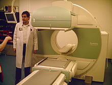
A Siemens brand SPECT scanner, consisting of two gamma cameras.

Instead of just "taking a picture of anatomical structures", a SPECT scan monitors level of biological activity at each place in the 3-D region analyzed. Emissions from the radionuclide indicate amounts of blood flow in the capillaries of the imaged regions. In the same way that a plain X-ray is a 2-dimensional (2-D) view of a 3-dimensional structure, the image obtained by a gamma camera is a 2-D view of 3-D distribution of a radionuclide.

SPECT imaging is performed by using a gamma camera to acquire multiple 2-D images (also called projections), from multiple angles. A computer is then used to apply a tomographic reconstruction algorithm to the multiple projections, yielding a 3-D data set. This data set may then be manipulated to show thin slices along any chosen axis of the body, similar to those obtained from other tomographic techniques, such as magnetic resonance imaging (MRI), X-ray computed tomography (X-ray CT), and positron emission tomography (PET).

SPECT is similar to PET in its use of radioactive tracer material and detection of gamma rays. In contrast with PET, the tracers used in SPECT emit gamma radiation that is measured directly, whereas PET tracers emit positrons that annihilate with electrons up to a few millimeters away, causing two gamma photons to be emitted in opposite directions. A PET scanner detects these emissions "coincident" in time, which provides more radiation event localization information and, thus, higher spatial resolution images than SPECT (which has about 1 cm resolution). SPECT scans are significantly less expensive than PET scans, in part because they are able to use longer-lived and more easily obtained radioisotopes than PET.

Because SPECT acquisition is very similar to planar gamma camera imaging, the same radiopharmaceuticals may be used. If a patient is examined in another type of nuclear medicine scan, but the images are non-diagnostic, it may be possible to proceed straight to SPECT by moving the patient to a SPECT instrument, or even by simply reconfiguring the camera for SPECT image acquisition while the patient remains on the table.

SPECT machine performing a total body bone scan. The patient lies on a table that slides through the machine, while a pair of gamma cameras rotate around her.

To acquire SPECT images, the gamma camera is rotated around the patient. Projections are acquired at defined points during the rotation, typically every 3–6 degrees. In most cases, a full 360-degree rotation is used to obtain an optimal reconstruction. The time taken to obtain each projection is also variable, but 15–20 seconds is typical. This gives a total scan time of 15–20 minutes.

Multi-headed gamma cameras can accelerate acquisition. For example, a dual-headed camera can be used with heads spaced 180 degrees apart, allowing two projections to be acquired simultaneously, with each head requiring 180 degrees of rotation. Triple-head cameras with 120-degree spacing are also used.

Cardiac gated acquisitions are possible with SPECT, just as with planar imaging techniques such as multi gated acquisition scan (MUGA). Triggered by electrocardiogram (EKG) to obtain differential information about the heart in various parts of its cycle, gated myocardial SPECT can be used to obtain quantitative information about myocardial perfusion, thickness, and contractility of the myocardium during various parts of the cardiac cycle, and also to allow calculation of left ventricular ejection fraction, stroke volume, and cardiac output.

Application

SPECT can be used to complement any gamma imaging study, where a true 3D representation can be helpful, such as tumor imaging, infection (leukocyte) imaging, thyroid imaging or bone scintigraphy.

Because SPECT permits accurate localisation in 3D space, it can be used to provide information about localised function in internal organs, such as functional cardiac or brain imaging.

Myocardial perfusion imaging

Myocardial perfusion imaging (MPI) is a form of functional cardiac imaging, used for the diagnosis of ischemic heart disease. The underlying principle is that under conditions of stress, diseased myocardium receives less blood flow than normal myocardium. MPI is one of several types of cardiac stress test.

A cardiac specific radiopharmaceutical is administered, e.g., 99mTc-tetrofosmin (Myoview, GE healthcare), 99mTc-sestamibi (Cardiolite, Bristol-Myers Squibb) or Thallium-201 chloride. Following this, the heart rate is raised to induce myocardial stress, either by exercise on a treadmill or pharmacologically with adenosine, dobutamine, or dipyridamole (aminophylline can be used to reverse the effects of dipyridamole).

SPECT imaging performed after stress reveals the distribution of the radiopharmaceutical, and therefore the relative blood flow to the different regions of the myocardium. Diagnosis is made by comparing stress images to a further set of images obtained at rest which are normally acquired prior to the stress images.

MPI has been demonstrated to have an overall accuracy of about 83% (sensitivity: 85%; specificity: 72%) (in a review, not exclusively of SPECT MPI), and is comparable with (or better than) other non-invasive tests for ischemic heart disease.

Functional brain imaging

Usually, the gamma-emitting tracer used in functional brain imaging is Technetium (99mTc) exametazime. 99mTc is a metastable nuclear isomer that emits gamma rays detectable by a gamma camera. Attaching it to exametazime allows it to be taken up by brain tissue in a manner proportional to brain blood flow, in turn allowing cerebral blood flow to be assessed with the nuclear gamma camera.

Because blood flow in the brain is tightly coupled to local brain metabolism and energy use, the 99mTc-exametazime tracer (as well as the similar 99mTc-EC tracer) is used to assess brain metabolism regionally, in an attempt to diagnose and differentiate the different causal pathologies of dementia. Meta-analysis of many reported studies suggests that SPECT with this tracer is about 74% sensitive at diagnosing Alzheimer's disease vs. 81% sensitivity for clinical exam (cognitive testing, etc.). More recent studies have shown the accuracy of SPECT in Alzheimer's diagnosis may be as high as 88%. In meta analysis, SPECT was superior to clinical exam and clinical criteria (91% vs. 70%) in being able to differentiate Alzheimer's disease from vascular dementias. This latter ability relates to SPECT's imaging of local metabolism of the brain, in which the patchy loss of cortical metabolism seen in multiple strokes differs clearly from the more even or "smooth" loss of non-occipital cortical brain function typical of Alzheimer's disease. Another recent review article showed that multi-headed SPECT cameras with quantitative analysis result in an overall sensitivity of 84-89% and an overall specificity of 83-89% in cross sectional studies and sensitivity of 82-96% and specificity of 83-89% for longitudinal studies of dementia.

99mTc-exametazime SPECT scanning competes with fludeoxyglucose (FDG) PET scanning of the brain, which works to assess regional brain glucose metabolism, to provide very similar information about local brain damage from many processes. SPECT is more widely available, because the radioisotope used is longer-lasting and far less expensive in SPECT, and the gamma scanning equipment is less expensive as well. While 99mTc is extracted from relatively simple technetium-99m generators, which are delivered to hospitals and scanning centers weekly to supply fresh radioisotope, FDG PET relies on FDG, which is made in an expensive medical cyclotron and "hot-lab" (automated chemistry lab for radiopharmaceutical manufacture), and then delivered immediately to scanning sites because of the natural short 110-minute half-life of Fluorine-18.

Applications in nuclear technology

In the nuclear power sector, the SPECT technique can be applied to image radioisotope distributions in irradiated nuclear fuels. Due to the irradiation of nuclear fuel (e.g. uranium) with neutrons in a nuclear reactor, a wide array of gamma-emitting radionuclides are naturally produced in the fuel, such as fission products (cesium-137, barium-140 and europium-154) and activation products (chromium-51 and cobalt-58). These may be imaged using SPECT in order to verify the presence of fuel rods in a stored fuel assembly for IAEA safeguards purposes, to validate predictions of core simulation codes, or to study the behavior of the nuclear fuel in normal operation,  or in accident scenarios.

Reconstruction

SPECT Sinogram

Reconstructed images typically have resolutions of 64×64 or 128×128 pixels, with the pixel sizes ranging from 3–6 mm. The number of projections acquired is chosen to be approximately equal to the width of the resulting images. In general, the resulting reconstructed images will be of lower resolution, have increased noise than planar images, and be susceptible to artifacts.

Scanning is time-consuming, and it is essential that there is no patient movement during the scan time. Movement can cause significant degradation of the reconstructed images, although movement compensation reconstruction techniques can help with this. A highly uneven distribution of radiopharmaceutical also has the potential to cause artifacts. A very intense area of activity (e.g., the bladder) can cause extensive streaking of the images and obscure neighboring areas of activity. This is a limitation of the filtered back projection reconstruction algorithm. Iterative reconstruction is an alternative algorithm that is growing in importance, as it is less sensitive to artifacts and can also correct for attenuation and depth dependent blurring. Furthermore, iterative algorithms can be made more efficacious using the Superiorization methodology.

Attenuation of the gamma rays within the patient can lead to significant underestimation of activity in deep tissues, compared to superficial tissues. Approximate correction is possible, based on relative position of the activity, and optimal correction is obtained with measured attenuation values. Modern SPECT equipment is available with an integrated X-ray CT scanner. As X-ray CT images are an attenuation map of the tissues, this data can be incorporated into the SPECT reconstruction to correct for attenuation. It also provides a precisely registered CT image, which can provide additional anatomical information.

Scatter of the gamma rays as well as the random nature of gamma rays can also lead to the degradation of quality of SPECT images and cause loss of resolution. Scatter correction and resolution recovery are also applied to improve resolution of SPECT images.

Typical SPECT acquisition protocols

Study Radioisotope Emission energy (keV) Half-life Radiopharmaceutical Activity (MBq) Rotation (degrees) Projections Image resolution Time per projection (s)
Bone scan technetium-99m 140 6 hours Phosphonates / Bisphosphonates 800 360 120 128 x 128 30
Myocardial perfusion scan technetium-99m 140 6 hours tetrofosmin; Sestamibi 700 180 60 64 x 64 25
Sestamibi parathyroid scan technetium-99m 140 6 hours Sestamibi


Brain scan technetium-99m 140 6 hours Tc exametazime; ECD 555-1110 360 64 128 x 128 30
Neuroendocrine or neurological tumor scan iodine-123 or iodine-131 159 13 hours or 8 days MIBG 400 360 60 64 x 64 30
White cell scan indium-111 & technetium-99m 171 & 245 67 hours in vitro labelled leucocytes 18 360 60 64 x 64 30

SPECT/CT

In some cases a SPECT gamma scanner may be built to operate with a conventional CT scanner, with coregistration of images. As in PET/CT, this allows location of tumors or tissues which may be seen on SPECT scintigraphy, but are difficult to locate precisely with regard to other anatomical structures. Such scans are most useful for tissues outside the brain, where location of tissues may be far more variable. For example, SPECT/CT may be used in sestamibi parathyroid scan applications, where the technique is useful in locating ectopic parathyroid adenomas which may not be in their usual locations in the thyroid gland.

Quality control

The overall performance of SPECT systems can be performed by quality control tools such as the Jaszczak phantom.

Observable

From Wikipedia, the free encyclopedia

In physics, an observable is a physical property or physical quantity that can be measured. Examples include position and momentum. In systems governed by classical mechanics, it is a real-valued "function" on the set of all possible system states. In quantum physics, it is an operator, or gauge, where the property of the quantum state can be determined by some sequence of operations. For example, these operations might involve submitting the system to various electromagnetic fields and eventually reading a value.

Physically meaningful observables must also satisfy transformation laws that relate observations performed by different observers in different frames of reference. These transformation laws are automorphisms of the state space, that is bijective transformations that preserve certain mathematical properties of the space in question.

Quantum mechanics

In quantum physics, observables manifest as linear operators on a Hilbert space representing the state space of quantum states. The eigenvalues of observables are real numbers that correspond to possible values the dynamical variable represented by the observable can be measured as having. That is, observables in quantum mechanics assign real numbers to outcomes of particular measurements, corresponding to the eigenvalue of the operator with respect to the system's measured quantum state. As a consequence, only certain measurements can determine the value of an observable for some state of a quantum system. In classical mechanics, any measurement can be made to determine the value of an observable.

The relation between the state of a quantum system and the value of an observable requires some linear algebra for its description. In the mathematical formulation of quantum mechanics, up to a phase constant, pure states are given by non-zero vectors in a Hilbert space V. Two vectors v and w are considered to specify the same state if and only if for some non-zero . Observables are given by self-adjoint operators on V. Not every self-adjoint operator corresponds to a physically meaningful observable. Also, not all physical observables are associated with non-trivial self-adjoint operators. For example, in quantum theory, mass appears as a parameter in the Hamiltonian, not as a non-trivial operator. For the case of a system of particles, the space V consists of functions called wave functions or state vectors.

In the case of transformation laws in quantum mechanics, the requisite automorphisms are unitary (or antiunitary) linear transformations of the Hilbert space V. Under Galilean relativity or special relativity, the mathematics of frames of reference is particularly simple, considerably restricting the set of physically meaningful observables.

In quantum mechanics, measurement of observables exhibits some seemingly unintuitive properties. Specifically, if a system is in a state described by a vector in a Hilbert space, the measurement process affects the state in a non-deterministic but statistically predictable way. In particular, after a measurement is applied, the state description by a single vector may be destroyed, being replaced by a statistical ensemble. The irreversible nature of measurement operations in quantum physics is sometimes referred to as the measurement problem and is described mathematically by quantum operations. By the structure of quantum operations, this description is mathematically equivalent to that offered by the relative state interpretation where the original system is regarded as a subsystem of a larger system and the state of the original system is given by the partial trace of the state of the larger system.

In quantum mechanics, dynamical variables such as position, translational (linear) momentum, orbital angular momentum, spin, and total angular momentum are each associated with a Hermitian operator that acts on the state of the quantum system. The eigenvalues of operator correspond to the possible values that the dynamical variable can be observed as having. For example, suppose is an eigenket (eigenvector) of the observable , with eigenvalue , and exists in a Hilbert space. Then

This eigenket equation says that if a measurement of the observable is made while the system of interest is in the state , then the observed value of that particular measurement must return the eigenvalue with certainty. However, if the system of interest is in the general state , then the eigenvalue is returned with probability , by the Born rule.

The above definition is somewhat dependent upon our convention of choosing real numbers to represent real physical quantities. Indeed, just because dynamical variables are "real" and not "unreal" in the metaphysical sense does not mean that they must correspond to real numbers in the mathematical sense.

To be more precise, the dynamical variable/observable is a self-adjoint operator in a Hilbert space.

Operators on finite and infinite dimensional Hilbert spaces

Observables can be represented by a Hermitian matrix if the Hilbert space is finite-dimensional. In an infinite-dimensional Hilbert space, the observable is represented by a symmetric operator, which may not be defined everywhere. The reason for such a change is that in an infinite-dimensional Hilbert space, the observable operator can become unbounded, which means that it no longer has a largest eigenvalue. This is not the case in a finite-dimensional Hilbert space: an operator can have no more eigenvalues than the dimension of the state it acts upon, and by the well-ordering property, any finite set of real numbers has a largest element. For example, the position of a point particle moving along a line can take any real number as its value, and the set of real numbers is uncountably infinite. Since the eigenvalue of an observable represents a possible physical quantity that its corresponding dynamical variable can take, we must conclude that there is no largest eigenvalue for the position observable in this uncountably infinite-dimensional Hilbert space.

Compatible and incompatible observables in quantum mechanics

A crucial difference between classical quantities and quantum mechanical observables is that some pairs of quantum observables may not be simultaneously measurable, a property referred to as complementarity. This is mathematically expressed by non-commutativity of their corresponding operators, to the effect that the commutator

This inequality expresses a dependence of measurement results on the order in which measurements of observables and are performed. A measurement of alters the quantum state in a way that is incompatible with the subsequent measurement of and vice versa.

Observables corresponding to commuting operators are called compatible observables. For example, momentum along say the and axis are compatible. Observables corresponding to non-commuting operators are called incompatible observables or complementary variables. For example, the position and momentum along the same axis are incompatible.

Incompatible observables cannot have a complete set of common eigenfunctions. Note that there can be some simultaneous eigenvectors of and , but not enough in number to constitute a complete basis.

Algorithmic information theory

From Wikipedia, the free encyclopedia https://en.wikipedia.org/wiki/Algorithmic_information_theory ...