Search This Blog

Thursday, October 26, 2023

Nuclear medicine

From Wikipedia, the free encyclopedia
 
Nuclear medicine
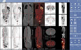
A nuclear medicine PET scan

Nuclear medicine or nucleology is a medical specialty involving the application of radioactive substances in the diagnosis and treatment of disease. Nuclear imaging, in a sense, is "radiology done inside out" because it records radiation emitted from within the body rather than radiation that is transmitted through the body from external sources like X-ray generators. In addition, nuclear medicine scans differ from radiology, as the emphasis is not on imaging anatomy, but on the function. For such reason, it is called a physiological imaging modality. Single photon emission computed tomography (SPECT) and positron emission tomography (PET) scans are the two most common imaging modalities in nuclear medicine.

Diagnostic medical imaging

Diagnostic

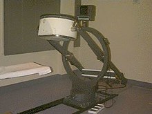
In nuclear medicine imaging, radiopharmaceuticals are taken internally, for example, through inhalation, intravenously, or orally. Then, external detectors (gamma cameras) capture and form images from the radiation emitted by the radiopharmaceuticals. This process is unlike a diagnostic X-ray, where external radiation is passed through the body to form an image.

There are several techniques of diagnostic nuclear medicine.

  • 2D: Scintigraphy ("scint") is the use of internal radionuclides to create two-dimensional images.
  • 3D: SPECT is a 3D tomographic technique that uses gamma camera data from many projections and can be reconstructed in different planes. Positron emission tomography (PET) uses coincidence detection to image functional processes.

Nuclear medicine tests differ from most other imaging modalities in that nuclear medicine scans primarily show the physiological function of the system being investigated as opposed to traditional anatomical imaging such as CT or MRI. Nuclear medicine imaging studies are generally more organ-, tissue- or disease-specific (e.g.: lungs scan, heart scan, bone scan, brain scan, tumor, infection, Parkinson etc.) than those in conventional radiology imaging, which focus on a particular section of the body (e.g.: chest X-ray, abdomen/pelvis CT scan, head CT scan, etc.). In addition, there are nuclear medicine studies that allow imaging of the whole body based on certain cellular receptors or functions. Examples are whole body PET scans or PET/CT scans, gallium scans, indium white blood cell scans, MIBG and octreotide scans.

Iodine-123 whole body scan for thyroid cancer evaluation. The study above was performed after the total thyroidectomy and TSH stimulation with thyroid hormone medication withdrawal. The study shows a small residual thyroid tissue in the neck and a mediastinum lesion, consistent with the thyroid cancer metastatic disease. The observable uptakes in the stomach and bladder are normal physiologic findings.

While the ability of nuclear metabolism to image disease processes from differences in metabolism is unsurpassed, it is not unique. Certain techniques such as fMRI image tissues (particularly cerebral tissues) by blood flow and thus show metabolism. Also, contrast-enhancement techniques in both CT and MRI show regions of tissue that are handling pharmaceuticals differently, due to an inflammatory process.

Diagnostic tests in nuclear medicine exploit the way that the body handles substances differently when there is disease or pathology present. The radionuclide introduced into the body is often chemically bound to a complex that acts characteristically within the body; this is commonly known as a tracer. In the presence of disease, a tracer will often be distributed around the body and/or processed differently. For example, the ligand methylene-diphosphonate (MDP) can be preferentially taken up by bone. By chemically attaching technetium-99m to MDP, radioactivity can be transported and attached to bone via the hydroxyapatite for imaging. Any increased physiological function, such as due to a fracture in the bone, will usually mean increased concentration of the tracer. This often results in the appearance of a "hot spot", which is a focal increase in radio accumulation or a general increase in radio accumulation throughout the physiological system. Some disease processes result in the exclusion of a tracer, resulting in the appearance of a "cold spot". Many tracer complexes have been developed to image or treat many different organs, glands, and physiological processes.

Hybrid scanning techniques

In some centers, the nuclear medicine scans can be superimposed, using software or hybrid cameras, on images from modalities such as CT or MRI to highlight the part of the body in which the radiopharmaceutical is concentrated. This practice is often referred to as image fusion or co-registration, for example SPECT/CT and PET/CT. The fusion imaging technique in nuclear medicine provides information about the anatomy and function, which would otherwise be unavailable or would require a more invasive procedure or surgery.

Practical concerns in nuclear imaging

Although the risks of low-level radiation exposures are not well understood, a cautious approach has been universally adopted that all human radiation exposures should be kept As Low As Reasonably Practicable, "ALARP". (Originally, this was known as "As Low As Reasonably Achievable" (ALARA), but this has changed in modern draftings of the legislation to add more emphasis on the "Reasonably" and less on the "Achievable".)

Working with the ALARP principle, before a patient is exposed for a nuclear medicine examination, the benefit of the examination must be identified. This needs to take into account the particular circumstances of the patient in question, where appropriate. For instance, if a patient is unlikely to be able to tolerate a sufficient amount of the procedure to achieve a diagnosis, then it would be inappropriate to proceed with injecting the patient with the radioactive tracer.

When the benefit does justify the procedure, then the radiation exposure (the amount of radiation given to the patient) should also be kept as low as reasonably practicable. This means that the images produced in nuclear medicine should never be better than required for confident diagnosis. Giving larger radiation exposures can reduce the noise in an image and make it more photographically appealing, but if the clinical question can be answered without this level of detail, then this is inappropriate.

As a result, the radiation dose from nuclear medicine imaging varies greatly depending on the type of study. The effective radiation dose can be lower than or comparable to or can far exceed the general day-to-day environmental annual background radiation dose. Likewise, it can also be less than, in the range of, or higher than the radiation dose from an abdomen/pelvis CT scan.

Some nuclear medicine procedures require special patient preparation before the study to obtain the most accurate result. Pre-imaging preparations may include dietary preparation or the withholding of certain medications. Patients are encouraged to consult with the nuclear medicine department prior to a scan.

Analysis

The result of the nuclear medicine imaging process is a dataset comprising one or more images. In multi-image datasets the array of images may represent a time sequence (i.e. cine or movie) often called a "dynamic" dataset, a cardiac gated time sequence, or a spatial sequence where the gamma-camera is moved relative to the patient. SPECT (single photon emission computed tomography) is the process by which images acquired from a rotating gamma-camera are reconstructed to produce an image of a "slice" through the patient at a particular position. A collection of parallel slices form a slice-stack, a three-dimensional representation of the distribution of radionuclide in the patient.

The nuclear medicine computer may require millions of lines of source code to provide quantitative analysis packages for each of the specific imaging techniques available in nuclear medicine.

Time sequences can be further analysed using kinetic models such as multi-compartment models or a Patlak plot.

Interventional nuclear medicine

Radionuclide therapy can be used to treat conditions such as hyperthyroidism, thyroid cancer, skin cancer and blood disorders.

In nuclear medicine therapy, the radiation treatment dose is administered internally (e.g. intravenous or oral routes) or externally direct above the area to treat in form of a compound (e.g. in case of skin cancer).

The radiopharmaceuticals used in nuclear medicine therapy emit ionizing radiation that travels only a short distance, thereby minimizing unwanted side effects and damage to noninvolved organs or nearby structures. Most nuclear medicine therapies can be performed as outpatient procedures since there are few side effects from the treatment and the radiation exposure to the general public can be kept within a safe limit.

Common nuclear medicine therapies

Substance Condition
Iodine-131-sodium iodide hyperthyroidism and thyroid cancer
Yttrium-90-ibritumomab tiuxetan (Zevalin) and Iodine-131-tositumomab (Bexxar) refractory lymphoma
131I-MIBG (metaiodobenzylguanidine) neuroendocrine tumors
Samarium-153 or Strontium-89 palliative bone pain treatment
Rhenium-188 squamous cell carcinoma or basal cell carcinoma of the skin

In some centers the nuclear medicine department may also use implanted capsules of isotopes (brachytherapy) to treat cancer.

Commonly used radiation sources (radionuclides) for brachytherapy

Radionuclide Type Half-life Energy
Caesium-137 (137Cs) γ-ray 30.17 years 0.662 MeV
Cobalt-60 (60Co) γ-ray 5.26 years 1.17, 1.33 MeV
Iridium-192 (192Ir) β-particles 73.8 days 0.38 MeV (mean)
Iodine-125 (125I) γ-rays 59.6 days 27.4, 31.4 and 35.5 keV
Palladium-103 (103Pd) γ-ray 17.0 days 21 keV (mean)
Ruthenium-106 (106Ru) β-particles 1.02 years 3.54 MeV

History

The history of nuclear medicine contains contributions from scientists across different disciplines in physics, chemistry, engineering, and medicine. The multidisciplinary nature of nuclear medicine makes it difficult for medical historians to determine the birthdate of nuclear medicine. This can probably be best placed between the discovery of artificial radioactivity in 1934 and the production of radionuclides by Oak Ridge National Laboratory for medicine-related use, in 1946.

The origins of this medical idea date back as far as the mid-1920s in Freiburg, Germany, when George de Hevesy made experiments with radionuclides administered to rats, thus displaying metabolic pathways of these substances and establishing the tracer principle. Possibly, the genesis of this medical field took place in 1936, when John Lawrence, known as "the father of nuclear medicine", took a leave of absence from his faculty position at Yale Medical School, to visit his brother Ernest Lawrence at his new radiation laboratory (now known as the Lawrence Berkeley National Laboratory) in Berkeley, California. Later on, John Lawrence made the first application in patients of an artificial radionuclide when he used phosphorus-32 to treat leukemia.

Many historians consider the discovery of artificially produced radionuclides by Frédéric Joliot-Curie and Irène Joliot-Curie in 1934 as the most significant milestone in nuclear medicine. In February 1934, they reported the first artificial production of radioactive material in the journal Nature, after discovering radioactivity in aluminum foil that was irradiated with a polonium preparation. Their work built upon earlier discoveries by Wilhelm Konrad Roentgen for X-ray, Henri Becquerel for radioactive uranium salts, and Marie Curie (mother of Irène Curie) for radioactive thorium, polonium and coining the term "radioactivity." Taro Takemi studied the application of nuclear physics to medicine in the 1930s. The history of nuclear medicine will not be complete without mentioning these early pioneers.

Nuclear medicine gained public recognition as a potential specialty when on May 11, 1946, an article in the Journal of the American Medical Association (JAMA) by Massachusetts General Hospital's Dr. Saul Hertz and Massachusetts Institute of Technology's Dr. Arthur Roberts, described the successful use of treating Graves' Disease with radioactive iodine (RAI) was published. Additionally, Sam Seidlin. brought further development in the field describing a successful treatment of a patient with thyroid cancer metastases using radioiodine (I-131). These articles are considered by many historians as the most important articles ever published in nuclear medicine. Although the earliest use of I-131 was devoted to therapy of thyroid cancer, its use was later expanded to include imaging of the thyroid gland, quantification of the thyroid function, and therapy for hyperthyroidism. Among the many radionuclides that were discovered for medical-use, none were as important as the discovery and development of Technetium-99m. It was first discovered in 1937 by C. Perrier and E. Segre as an artificial element to fill space number 43 in the Periodic Table. The development of a generator system to produce Technetium-99m in the 1960s became a practical method for medical use. Today, Technetium-99m is the most utilized element in nuclear medicine and is employed in a wide variety of nuclear medicine imaging studies.

Widespread clinical use of nuclear medicine began in the early 1950s, as knowledge expanded about radionuclides, detection of radioactivity, and using certain radionuclides to trace biochemical processes. Pioneering works by Benedict Cassen in developing the first rectilinear scanner and Hal O. Anger's scintillation camera (Anger camera) broadened the young discipline of nuclear medicine into a full-fledged medical imaging specialty.

By the early 1960s, in southern Scandinavia, Niels A. Lassen, David H. Ingvar, and Erik Skinhøj developed techniques that provided the first blood flow maps of the brain, which initially involved xenon-133 inhalation; an intra-arterial equivalent was developed soon after, enabling measurement of the local distribution of cerebral activity for patients with neuropsychiatric disorders such as schizophrenia. Later versions would have 254 scintillators so a two-dimensional image could be produced on a color monitor. It allowed them to construct images reflecting brain activation from speaking, reading, visual or auditory perception and voluntary movement. The technique was also used to investigate, e.g., imagined sequential movements, mental calculation and mental spatial navigation.

By the 1970s most organs of the body could be visualized using nuclear medicine procedures. In 1971, American Medical Association officially recognized nuclear medicine as a medical specialty. In 1972, the American Board of Nuclear Medicine was established, and in 1974, the American Osteopathic Board of Nuclear Medicine was established, cementing nuclear medicine as a stand-alone medical specialty.

In the 1980s, radiopharmaceuticals were designed for use in diagnosis of heart disease. The development of single photon emission computed tomography (SPECT), around the same time, led to three-dimensional reconstruction of the heart and establishment of the field of nuclear cardiology.

More recent developments in nuclear medicine include the invention of the first positron emission tomography scanner (PET). The concept of emission and transmission tomography, later developed into single photon emission computed tomography (SPECT), was introduced by David E. Kuhl and Roy Edwards in the late 1950s. Their work led to the design and construction of several tomographic instruments at the University of Pennsylvania. Tomographic imaging techniques were further developed at the Washington University School of Medicine. These innovations led to fusion imaging with SPECT and CT by Bruce Hasegawa from University of California, San Francisco (UCSF), and the first PET/CT prototype by D. W. Townsend from University of Pittsburgh in 1998.

PET and PET/CT imaging experienced slower growth in its early years owing to the cost of the modality and the requirement for an on-site or nearby cyclotron. However, an administrative decision to approve medical reimbursement of limited PET and PET/CT applications in oncology has led to phenomenal growth and widespread acceptance over the last few years, which also was facilitated by establishing 18F-labelled tracers for standard procedures, allowing work at non-cyclotron-equipped sites. PET/CT imaging is now an integral part of oncology for diagnosis, staging and treatment monitoring. A fully integrated MRI/PET scanner is on the market from early 2011.

Sources of radionuclides

99mTc is normally supplied to hospitals through a radionuclide generator containing the parent radionuclide molybdenum-99. 99Mo is typically obtained as a fission product of 235U in nuclear reactors, however global supply shortages have led to the exploration of other methods of production. About a third of the world's supply, and most of Europe's supply, of medical isotopes is produced at the Petten nuclear reactor in the Netherlands. Another third of the world's supply, and most of North America's supply, was produced at the Chalk River Laboratories in Chalk River, Ontario, Canada until its permanent shutdown in 2018.

The most commonly used radioisotope in PET, 18F, is not produced in a nuclear reactor, but rather in a circular accelerator called a cyclotron. The cyclotron is used to accelerate protons to bombard the stable heavy isotope of oxygen 18O. The 18O constitutes about 0.20% of ordinary oxygen (mostly oxygen-16), from which it is extracted. The 18F is then typically used to make FDG.

Common isotopes used in nuclear medicine 
isotope symbol Z T1/2 decay gamma (keV) Maximum β

energy (keV) /

Abundance

Imaging:
fluorine-18 18F 9 109.77 m β+ 511 (193%) 634 (97%)
gallium-67 67Ga 31 3.26 d ec 93 (39%),
185 (21%),
300 (17%)
-
krypton-81m 81mKr 36 13.1 s IT 190 (68%) -
rubidium-82 82Rb 37 1.27 m β+ 511 (191%) 3381 (81.8%)

2605 (13.1%)

1906 (0.14%)

1209 (0.32%)

nitrogen-13 13N 7 9.97 m β+ 511 (200%) 1198 (99.8%)
technetium-99m 99mTc 43 6.01 h IT 140 (89%) -
indium-111 111In 49 2.80 d ec 171 (90%),
245 (94%)
-
iodine-123 123I 53 13.3 h ec 159 (83%) -
xenon-133 133Xe 54 5.24 d β 81 (31%) 346 (99.1%)

267 (0.9%)

thallium-201 201Tl 81 3.04 d ec 69–83* (94%),
167 (10%)
-
Therapy:
yttrium-90 90Y 39 2.67 d β - 2279 (99.98%)
iodine-131 131I 53 8.02 d β 364 (81%) 807 (0.4%)

606 (89.4%)

334 (7.2%)

248 (2.1%)

lutetium-177 177Lu 71 6.65 d β 113 (6.6%),

208 (11%)

498 (79.3%)

385 (9.1%)

177 (11.6%)

Z = atomic number, the number of protons; T1/2 = half-life; decay = mode of decay
photons = principle photon energies in kilo-electron volts, keV, (abundance/decay)
β = beta maximum energy in kilo-electron volts, keV, (abundance/decay)
β+ = β+ decay; β = β decay; IT = isomeric transition; ec = electron capture
* X-rays from progeny, mercury, Hg

A typical nuclear medicine study involves administration of a radionuclide into the body by intravenous injection in liquid or aggregate form, ingestion while combined with food, inhalation as a gas or aerosol, or rarely, injection of a radionuclide that has undergone micro-encapsulation. Some studies require the labeling of a patient's own blood cells with a radionuclide (leukocyte scintigraphy and red blood cell scintigraphy). Most diagnostic radionuclides emit gamma rays either directly from their decay or indirectly through electron–positron annihilation, while the cell-damaging properties of beta particles are used in therapeutic applications. Refined radionuclides for use in nuclear medicine are derived from fission or fusion processes in nuclear reactors, which produce radionuclides with longer half-lives, or cyclotrons, which produce radionuclides with shorter half-lives, or take advantage of natural decay processes in dedicated generators, i.e. molybdenum/technetium or strontium/rubidium.

The most commonly used intravenous radionuclides are technetium-99m, iodine-123, iodine-131, thallium-201, gallium-67, fluorine-18 fluorodeoxyglucose, and indium-111 labeled leukocytes. The most commonly used gaseous/aerosol radionuclides are xenon-133, krypton-81m, (aerosolised) technetium-99m.

Policies and procedures

Radiation dose

A patient undergoing a nuclear medicine procedure will receive a radiation dose. Under present international guidelines it is assumed that any radiation dose, however small, presents a risk. The radiation dose delivered to a patient in a nuclear medicine investigation, though unproven, is generally accepted to present a very small risk of inducing cancer. In this respect it is similar to the risk from X-ray investigations except that the dose is delivered internally rather than from an external source such as an X-ray machine, and dosage amounts are typically significantly higher than those of X-rays.

The radiation dose from a nuclear medicine investigation is expressed as an effective dose with units of sieverts (usually given in millisieverts, mSv). The effective dose resulting from an investigation is influenced by the amount of radioactivity administered in megabecquerels (MBq), the physical properties of the radiopharmaceutical used, its distribution in the body and its rate of clearance from the body.

Effective doses can range from 6 μSv (0.006 mSv) for a 3 MBq chromium-51 EDTA measurement of glomerular filtration rate to 11.2 mSv (11,200 μSv) for a 80 MBq thallium-201 myocardial imaging procedure. The common bone scan with 600 MBq of technetium-99m MDP has an effective dose of approximately 2.9 mSv (2,900 μSv).

Formerly, units of measurement were the curie (Ci), being 3.7E10 Bq, and also 1.0 grams of Radium (Ra-226); the rad (radiation absorbed dose), now replaced by the gray; and the rem (Röntgen equivalent man), now replaced with the sievert. The rad and rem are essentially equivalent for almost all nuclear medicine procedures, and only alpha radiation will produce a higher Rem or Sv value, due to its much higher Relative Biological Effectiveness (RBE). Alpha emitters are nowadays rarely used in nuclear medicine, but were used extensively before the advent of nuclear reactor and accelerator produced radionuclides. The concepts involved in radiation exposure to humans are covered by the field of Health Physics; the development and practice of safe and effective nuclear medicinal techniques is a key focus of Medical Physics.

Regulatory frameworks and guidelines

Different countries around the world maintain regulatory frameworks that are responsible for the management and use of radionuclides in different medical settings. For example, in the US, the Nuclear Regulatory Commission (NRC) and the Food and Drug Administration (FDA) have guidelines in place for hospitals to follow. With the NRC, if radioactive materials aren't involved, like X-rays for example, they are not regulated by the agency and instead are regulated by the individual states. International organizations, such as the International Atomic Energy Agency (IAEA), have regularly published different articles and guidelines for best practices in nuclear medicine as well as reporting on emerging technologies in nuclear medicine. Other factors that are considered in nuclear medicine include a patient's medical history as well as post-treatment management. Groups like International Commission on Radiological Protection have published information on how to manage the release of patients from a hospital with unsealed radionuclides.

Hyperthermia

From Wikipedia, the free encyclopedia
 
Hyperthermia
Other namesOverheating
An analog medical thermometer showing a temperature of 38.7 °C (101.7 °F)
SpecialtyCritical care medicine
SymptomsLack of perspiration, confusion, delirium, decreased blood pressure, increased heart rate and respiration rate, symptoms of dehydration
ComplicationsOrgan failure, unconsciousness
CausesHeat stroke
Risk factorsExposure to hot and/or humid environments, physical exertion, wearing personal protective equipment that covers the body, heatwaves
Diagnostic methodBased on symptoms or body temperature above 37.7 °C (99.9 °F)
Differential diagnosisFever
PreventionMaintaining a moderate temperature, regular hydration, taking regular breaks
TreatmentMild: Staying away from hot environments, rehydrating oneself, mechanical cooling, use of a dehumidifier
Severe: intravenous hydration, gastric lavage with iced saline, hemodialysis, immersing in ice water

Hyperthermia, also known simply as overheating, is a condition in which an individual's body temperature is elevated beyond normal due to failed thermoregulation. The person's body produces or absorbs more heat than it dissipates. When extreme temperature elevation occurs, it becomes a medical emergency requiring immediate treatment to prevent disability or death. Almost half a million deaths are recorded every year from hyperthermia.

The most common causes include heat stroke and adverse reactions to drugs. Heat stroke is an acute temperature elevation caused by exposure to excessive heat, or combination of heat and humidity, that overwhelms the heat-regulating mechanisms of the body. The latter is a relatively rare side effect of many drugs, particularly those that affect the central nervous system. Malignant hyperthermia is a rare complication of some types of general anesthesia. Hyperthermia can also be caused by a traumatic brain injury.

Hyperthermia differs from fever in that the body's temperature set point remains unchanged. The opposite is hypothermia, which occurs when the temperature drops below that required to maintain normal metabolism. The term is from Greek ὑπέρ, hyper, meaning "above", and θέρμος, thermos, meaning "heat".

Classification

In humans, hyperthermia is defined as a temperature greater than 37.5–38.3 °C (99.5–100.9 °F), depending on the reference used, that occurs without a change in the body's temperature set point.

The normal human body temperature can be as high as 37.7 °C (99.9 °F) in the late afternoon. Hyperthermia requires an elevation from the temperature that would otherwise be expected. Such elevations range from mild to extreme; body temperatures above 40 °C (104 °F) can be life-threatening.

Signs and symptoms

An early stage of hyperthermia can be "heat exhaustion" (or "heat prostration" or "heat stress"), whose symptoms can include heavy sweating, rapid breathing and a fast, weak pulse. If the condition progresses to heat stroke, then hot, dry skin is typical as blood vessels dilate in an attempt to increase heat loss. An inability to cool the body through perspiration may cause dry skin. Hyperthermia from neurological disease may include little or no sweating, cardiovascular problems, and confusion or delirium.

Other signs and symptoms vary. Accompanying dehydration can produce nausea, vomiting, headaches, and low blood pressure and the latter can lead to fainting or dizziness, especially if the standing position is assumed quickly.

In severe heat stroke, confusion and aggressive behavior may be observed. Heart rate and respiration rate will increase (tachycardia and tachypnea) as blood pressure drops and the heart attempts to maintain adequate circulation. The decrease in blood pressure can then cause blood vessels to contract reflexively, resulting in a pale or bluish skin color in advanced cases. Young children, in particular, may have seizures. Eventually, organ failure, unconsciousness and death will result.

Causes

Heat stroke occurs when thermoregulation is overwhelmed by a combination of excessive metabolic production of heat (exertion), excessive environmental heat, and insufficient or impaired heat loss, resulting in an abnormally high body temperature. In severe cases, temperatures can exceed 40 °C (104 °F). Heat stroke may be non-exertional (classic) or exertional.

Exertional

Significant physical exertion in hot conditions can generate heat beyond the ability to cool, because, in addition to the heat, humidity of the environment may reduce the efficiency of the body's normal cooling mechanisms. Human heat-loss mechanisms are limited primarily to sweating (which dissipates heat by evaporation, assuming sufficiently low humidity) and vasodilation of skin vessels (which dissipates heat by convection proportional to the temperature difference between the body and its surroundings, according to Newton's law of cooling). Other factors, such as insufficient water intake, consuming alcohol, or lack of air conditioning, can worsen the problem.

The increase in body temperature that results from a breakdown in thermoregulation affects the body biochemically. Enzymes involved in metabolic pathways within the body such as cellular respiration fail to work effectively at higher temperatures, and further increases can lead them to denature, reducing their ability to catalyse essential chemical reactions. This loss of enzymatic control affects the functioning of major organs with high energy demands such as the heart and brain. Loss of fluid and electrolytes cause heat cramps – slow muscular contraction and severe muscular spasm lasting between one and three minutes. Almost all cases of heat cramps involve vigorous physical exertion. Body temperature may remain normal or a little higher than normal and cramps are concentrated in heavily used muscles.

Situational

Situational heat stroke occurs in the absence of exertion. It mostly affects the young and elderly. In the elderly in particular, it can be precipitated by medications that reduce vasodilation and sweating, such as anticholinergic drugs, antihistamines, and diuretics. In this situation, the body's tolerance for high environmental temperature may be insufficient, even at rest.

Heat waves are often followed by a rise in the death rate, and these 'classical hyperthermia' deaths typically involve the elderly and infirm. This is partly because thermoregulation involves cardiovascular, respiratory and renal systems which may be inadequate for the additional stress because of the existing burden of aging and disease, further compromised by medications. During the July 1995 heatwave in Chicago, there were at least 700 heat-related deaths. The strongest risk factors were being confined to bed, and living alone, while the risk was reduced for those with working air conditioners and those with access to transportation. Even then, reported deaths may be underestimated as diagnosis can be mis-classified as stroke or heart attack.

Drugs

Some drugs cause excessive internal heat production. The rate of drug-induced hyperthermia is higher where use of these drugs is higher.

Personal protective equipment

Those working in industry, in the military, or as first responders may be required to wear personal protective equipment (PPE) against hazards such as chemical agents, gases, fire, small arms and improvised explosive devices (IEDs). PPE includes a range of hazmat suits, firefighting turnout gear, body armor and bomb suits, among others. Depending on design, the wearer may be encapsulated in a microclimate, due to an increase in thermal resistance and decrease in vapor permeability. As physical work is performed, the body's natural thermoregulation (i.e. sweating) becomes ineffective. This is compounded by increased work rates, high ambient temperature and humidity levels, and direct exposure to the sun. The net effect is that desired protection from some environmental threats inadvertently increases the threat of heat stress.

The effect of PPE on hyperthermia has been noted in fighting the 2014 Ebola virus epidemic in Western Africa. Doctors and healthcare workers were only able to work for 40 minutes at a time in their protective suits, fearing heat stroke.

Other

Other rare causes of hyperthermia include thyrotoxicosis and an adrenal gland tumor, called pheochromocytoma, both of which can cause increased heat production. Damage to the central nervous system from brain hemorrhage, traumatic brain injury, status epilepticus, and other kinds of injury to the hypothalamus can also cause hyperthermia.

Pathophysiology

A summary of the differences between hyperthermia, hypothermia, and fever.
Hyperthermia: Characterized on the left. Normal body temperature (thermoregulatory set-point) is shown in green, while the hyperthermic temperature is shown in red. As can be seen, hyperthermia can be considered an increase above the thermoregulatory set-point.
Hypothermia: Characterized in the center: Normal body temperature is shown in green, while hypothermic temperature is shown in blue. As can be seen, hypothermia can be conceptualized as a decrease below the thermoregulatory set-point.
Fever: Characterized on the right: Normal body temperature is shown in green. It reads "New Normal" because the thermoregulatory set-point has risen. This has caused what was the normal body temperature (in blue) to be considered hypothermic.

A fever occurs when the core temperature is set higher, through the action of the pre-optic region of the anterior hypothalamus. For example, in response to a bacterial or viral infection, certain white blood cells within the blood will release pyrogens which have a direct effect on the anterior hypothalamus, causing body temperature to rise, much like raising the temperature setting on a thermostat.

In contrast, hyperthermia occurs when the body temperature rises without a change in the heat control centers.

Some of the gastrointestinal symptoms of acute exertional heatstroke, such as vomiting, diarrhea, and gastrointestinal bleeding, may be caused by barrier dysfunction and subsequent endotoxemia. Ultraendurance athletes have been found to have significantly increased plasma endotoxin levels. Endotoxin stimulates many inflammatory cytokines, which in turn may cause multiorgan dysfunction. Experimentally, monkeys treated with oral antibiotics prior to induction of heat stroke do not become endotoxemic.

There is scientific support for the concept of a temperature set point; that is, maintenance of an optimal temperature for the metabolic processes that life depends on. Nervous activity in the preoptic-anterior hypothalamus of the brain triggers heat losing (sweating, etc.) or heat generating (shivering and muscle contraction, etc.) activities through stimulation of the autonomic nervous system. The pre-optic anterior hypothalamus has been shown to contain warm sensitive, cool sensitive, and temperature insensitive neurons, to determine the body's temperature setpoint. As the temperature that these neurons are exposed to rises above 37 °C (99 °F), the rate of electrical discharge of the warm-sensitive neurons increases progressively. Cold-sensitive neurons increase their rate of electrical discharge progressively below 37 °C (99 °F).

Diagnosis

Hyperthermia is generally diagnosed by the combination of unexpectedly high body temperature and a history that supports hyperthermia instead of a fever. Most commonly this means that the elevated temperature has occurred in a hot, humid environment (heat stroke) or in someone taking a drug for which hyperthermia is a known side effect (drug-induced hyperthermia). The presence of signs and symptoms related to hyperthermia syndromes, such as extrapyramidal symptoms characteristic of neuroleptic malignant syndrome, and the absence of signs and symptoms more commonly related to infection-related fevers, are also considered in making the diagnosis.

If fever-reducing drugs lower the body temperature, even if the temperature does not return entirely to normal, then hyperthermia is excluded.

Prevention

When ambient temperature is excessive, humans and many other animals cool themselves below ambient by evaporative cooling of sweat (or other aqueous liquid; saliva in dogs, for example); this helps prevent potentially fatal hyperthermia. The effectiveness of evaporative cooling depends upon humidity. Wet-bulb temperature, which takes humidity into account, or more complex calculated quantities such as wet-bulb globe temperature (WBGT), which also takes solar radiation into account, give useful indications of the degree of heat stress and are used by several agencies as the basis for heat-stress prevention guidelines. (Wet-bulb temperature is essentially the lowest skin temperature attainable by evaporative cooling at a given ambient temperature and humidity.)

A sustained wet-bulb temperature exceeding 35 °C (95 °F) is likely to be fatal even to fit and healthy people unclothed in the shade next to a fan; at this temperature, environmental heat gain instead of loss occurs. As of 2012, wet-bulb temperatures only very rarely exceeded 30 °C (86 °F) anywhere, although significant global warming may change this.

In cases of heat stress caused by physical exertion, hot environments, or protective equipment, prevention or mitigation by frequent rest breaks, careful hydration, and monitoring body temperature should be attempted. However, in situations demanding one is exposed to a hot environment for a prolonged period or must wear protective equipment, a personal cooling system is required as a matter of health and safety. There are a variety of active or passive personal cooling systems; these can be categorized by their power sources and whether they are person- or vehicle-mounted.

Because of the broad variety of operating conditions, these devices must meet specific requirements concerning their rate and duration of cooling, their power source, and their adherence to health and safety regulations. Among other criteria are the user's need for physical mobility and autonomy. For example, active-liquid systems operate by chilling water and circulating it through a garment; the skin surface area is thereby cooled through conduction. This type of system has proven successful in certain military, law enforcement, and industrial applications. Bomb-disposal technicians wearing special suits to protect against improvised explosive devices (IEDs) use a small, ice-based chiller unit that is strapped to one leg; a liquid-circulating garment, usually a vest, is worn over the torso to maintain a safe core body temperature. By contrast, soldiers traveling in combat vehicles can face microclimate temperatures in excess of 65 °C (149 °F) and require a multiple-user, vehicle-powered cooling system with rapid connection capabilities. Requirements for hazmat teams, the medical community, and workers in heavy industry vary further.

Treatment

The underlying cause must be removed. Mild hyperthemia caused by exertion on a hot day may be adequately treated through self-care measures, such as increased water consumption and resting in a cool place. Hyperthermia that results from drug exposure requires prompt cessation of that drug, and occasionally the use of other drugs as counter measures.

Antipyretics (e.g., acetaminophen, aspirin, other nonsteroidal anti-inflammatory drugs) have no role in the treatment of heatstroke because antipyretics interrupt the change in the hypothalamic set point caused by pyrogens; they are not expected to work on a healthy hypothalamus that has been overloaded, as in the case of heatstroke. In this situation, antipyretics actually may be harmful in patients who develop hepatic, hematologic, and renal complications because they may aggravate bleeding tendencies.

When body temperature is significantly elevated, mechanical cooling methods are used to remove heat and to restore the body's ability to regulate its own temperatures. Passive cooling techniques, such as resting in a cool, shady area and removing clothing can be applied immediately. Active cooling methods, such as sponging the head, neck, and trunk with cool water, remove heat from the body and thereby speed the body's return to normal temperatures. When methods such as immersion are impractical, misting the body with water and using a fan have also been shown to be effective.

Sitting in a bathtub of tepid or cool water (immersion method) can remove a significant amount of heat in a relatively short period of time. It was once thought that immersion in very cold water is counterproductive, as it causes vasoconstriction in the skin and thereby prevents heat from escaping the body core. However, a British analysis of various studies stated: "this has never been proven experimentally. Indeed, a recent study using normal volunteers has shown that cooling rates were fastest when the coldest water was used." The analysis concluded that iced water immersion is the most-effective cooling technique for exertional heat stroke. No superior cooling method has been found for non-exertional heat stroke. Thus, aggressive ice-water immersion remains the gold standard for life-threatening heat stroke.

When the body temperature reaches about 40 °C (104 °F), or if the affected person is unconscious or showing signs of confusion, hyperthermia is considered a medical emergency that requires treatment in a proper medical facility. In a hospital, more aggressive cooling measures are available, including intravenous hydration, gastric lavage with iced saline, and even hemodialysis to cool the blood.

Epidemiology

Hyperthermia affects those who are unable to regulate their body heat, mainly due to environmental conditions. The main risk factor for hyperthermia is the lack of ability to sweat. People who are dehydrated or who are older may not produce the sweat they need to regulate their body temperature. High heat conditions can put certain groups at risk for hyperthermia including: physically active individuals, soldiers, construction workers, landscapers and factory workers. Some people that do not have access to cooler living conditions, like people with lower socioeconomic status, may have a difficult time fighting the heat. People are at risk for hyperthermia during high heat and dry conditions, most commonly seen in the summer.

Various cases of different types of hyperthermia have been reported. A research study was published in March 2019 that looked into multiple case reports of drug induced hyperthermia. The study concluded that psychotropic drugs such as anti-psychotics, antidepressants, and anxiolytics were associated with an increased heat-related mortality as opposed to the other drugs researched (anticholinergics, diuretics, cardiovascular agents, etc.). A different study was published in June 2019 that examined the association between hyperthermia in older adults and the temperatures in the United States. Hospitalization records of elderly patients in the US between 1991 and 2006 were analyzed and concluded that cases of hyperthermia were observed to be highest in regions with arid climates. The study discussed finding a disproportionately high number of cases of hyperthermia in early seasonal heat waves indicating that people were not yet practicing proper techniques to stay cool and prevent overheating in the early presence of warm, dry weather.

In urban areas people are at an increased susceptibility to hyperthermia. This is due to a phenomenon called the urban heat island effect. Since the 20th century in the United States, the north-central region (Ohio, Indiana, Illinois, Missouri, Iowa, and Nebraska) was the region with the highest morbidity resulting from hyperthermia. Northeastern states had the next highest. Regions least affected by heat wave-related hyperthermia causing death were Southern and Pacific Coastal states. Northern cities in the United States are at greater risk of hyperthermia during heat waves due to the fact that people tend to have a lower minimum mortality temperature at higher latitudes. In contrast, cities residing in lower latitudes within the continental US typically have higher thresholds for ambient temperatures. In India, hundreds die every year from summer heat waves, including more than 2,500 in the year 2015. Later that same summer, the 2015 Pakistani heat wave killed about 2,000 people. An extreme 2003 European heat wave caused tens of thousands of deaths.

Causes of hyperthermia include dehydration, use of certain medications, using cocaine and amphetamines or excessive alcohol use. Bodily temperatures greater than 37.5–38.3 °C (99.5-101.0 °F) can be diagnosed as a hyperthermic case. As body temperatures increase or excessive body temperatures persist, individuals are at a heightened risk of developing progressive conditions. Greater risk complications of hyperthermia include heat stroke, organ malfunction, organ failure, and death. There are two forms of heat stroke; classical heatstroke and exertional heatstroke. Classical heatstroke occurs from extreme environmental conditions, such as heat waves. Those who are most commonly affected by classical heatstroke are very young, elderly or chronically ill. Exertional heatstroke appears in individuals after vigorous physical activity. Exertional heatstroke is displayed most commonly in healthy 15-50 year old people. Sweating is often present in exertional heatstroke. The associated mortality rate of heatstroke is 40 to 64%.

Research

Hyperthermia can also be deliberately induced using drugs or medical devices, and is being studied and applied in clinical routine as a treatment of some kinds of cancer. Research has shown that medically controlled hyperthermia can shrink tumours. This occurs when a high body temperature damages cancerous cells by destroying proteins and structures within each cell. Hyperthermia has also been researched to investigate whether it causes cancerous tumours to be more prone to radiation as a form of treatment; which as a result has allowed hyperthermia to be used to compliment other forms of cancer therapy. Various techniques of hyperthermia in the treatment of cancer include local or regional hyperthermia, as well as whole body techniques.

Gamma camera

From Wikipedia, the free encyclopedia
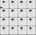
An example of lung scintigraphy examination

A gamma camera (γ-camera), also called a scintillation camera or Anger camera, is a device used to image gamma radiation emitting radioisotopes, a technique known as scintigraphy. The applications of scintigraphy include early drug development and nuclear medical imaging to view and analyse images of the human body or the distribution of medically injected, inhaled, or ingested radionuclides emitting gamma rays.

Imaging techniques

Coded aperture mask for gamma camera (for SPECT)

Scintigraphy ("scint") is the use of gamma cameras to capture emitted radiation from internal radioisotopes to create two-dimensional images.

SPECT (single photon emission computed tomography) imaging, as used in nuclear cardiac stress testing, is performed using gamma cameras. Usually one, two or three detectors or heads, are slowly rotated around the patient's torso.

Multi-headed gamma cameras can also be used for positron emission tomography (PET) scanning, provided that their hardware and software can be configured to detect "coincidences" (near simultaneous events on 2 different heads). Gamma camera PET is markedly inferior to PET imaging with a purpose designed PET scanner, as the scintillator crystal has poor sensitivity for the high-energy annihilation photons, and the detector area is significantly smaller. However, given the low cost of a gamma camera and its additional flexibility compared to a dedicated PET scanner, this technique is useful where the expense and resource implications of a PET scanner cannot be justified.

Construction

Gamma camera
Diagrammatic cross section of a gamma camera detector
Details of the cross section of a gamma camera

A gamma camera consists of one or more flat crystal planes (or detectors) optically coupled to an array of photomultiplier tubes in an assembly known as a "head", mounted on a gantry. The gantry is connected to a computer system that both controls the operation of the camera and acquires and stores images. The construction of a gamma camera is sometimes known as a compartmental radiation construction.

The system accumulates events, or counts, of gamma photons that are absorbed by the crystal in the camera. Usually a large flat crystal of sodium iodide with thallium doping NaI(Tl) in a light-sealed housing is used. The highly efficient capture method of this combination for detecting gamma rays was discovered in 1944 by Sir Samuel Curran whilst he was working on the Manhattan Project at the University of California at Berkeley. Nobel prize-winning physicist Robert Hofstadter also worked on the technique in 1948.

The crystal scintillates in response to incident gamma radiation. When a gamma photon leaves the patient (who has been injected with a radioactive pharmaceutical), it knocks an electron loose from an iodine atom in the crystal, and a faint flash of light is produced when the dislocated electron again finds a minimal energy state. The initial phenomenon of the excited electron is similar to the photoelectric effect and (particularly with gamma rays) the Compton effect. After the flash of light is produced, it is detected. Photomultiplier tubes (PMTs) behind the crystal detect the fluorescent flashes (events) and a computer sums the counts. The computer reconstructs and displays a two dimensional image of the relative spatial count density on a monitor. This reconstructed image reflects the distribution and relative concentration of radioactive tracer elements present in the organs and tissues imaged.

Signal processing

Hal Anger developed the first gamma camera in 1957. His original design, frequently called the Anger camera, is still widely used today. The Anger camera uses sets of vacuum tube photomultipliers (PMT). Generally each tube has an exposed face of about 7.6 cm in diameter and the tubes are arranged in hexagon configurations, behind the absorbing crystal. The electronic circuit connecting the photodetectors is wired so as to reflect the relative coincidence of light fluorescence as sensed by the members of the hexagon detector array. All the PMTs simultaneously detect the (presumed) same flash of light to varying degrees, depending on their position from the actual individual event. Thus the spatial location of each single flash of fluorescence is reflected as a pattern of voltages within the interconnecting circuit array.

The location of the interaction between the gamma ray and the crystal can be determined by processing the voltage signals from the photomultipliers; in simple terms, the location can be found by weighting the position of each photomultiplier tube by the strength of its signal, and then calculating a mean position from the weighted positions. The total sum of the voltages from each photomultiplier, measured by a pulse height analyzer is proportional to the energy of the gamma ray interaction, thus allowing discrimination between different isotopes or between scattered and direct photons.

Spatial resolution

In order to obtain spatial information about the gamma-ray emissions from an imaging subject (e.g. a person's heart muscle cells which have absorbed an intravenous injected radioactive, usually thallium-201 or technetium-99m, medicinal imaging agent) a method of correlating the detected photons with their point of origin is required.

The conventional method is to place a collimator over the detection crystal/PMT array. The collimator consists of a thick sheet of lead, typically 25 to 55 millimetres (1 to 2.2 in) thick, with thousands of adjacent holes through it. There are three types of collimators: low energy, medium energy, and high energy collimators. As the collimators transitioned from low energy to high energy, the hole sizes, thickness, and septations between the holes also increased.  Given a fixed septal thickness, the collimator resolution decreases with increased efficiency and also increasing distance of the source from the collimator. Pulse-height analyser determines the Full width at half maximum that selects certain photons to contribute to the final image, thus determining the collimator resolution.

The individual holes limit photons which can be detected by the crystal to a cone shape; the point of the cone is at the midline center of any given hole and extends from the collimator surface outward. However, the collimator is also one of the sources of blurring within the image; lead does not totally attenuate incident gamma photons, there can be some crosstalk between holes.

Unlike a lens, as used in visible light cameras, the collimator attenuates most (>99%) of incident photons and thus greatly limits the sensitivity of the camera system. Large amounts of radiation must be present so as to provide enough exposure for the camera system to detect sufficient scintillation dots to form a picture.

Other methods of image localization (pinhole, rotating slat collimator with CZT) have been proposed and tested; however, none have entered widespread routine clinical use.

The best current camera system designs can differentiate two separate point sources of gamma photons located at 6 to 12 mm depending on distance from the collimator, the type of collimator and radio-nucleide. Spatial resolution decreases rapidly at increasing distances from the camera face. This limits the spatial accuracy of the computer image: it is a fuzzy image made up of many dots of detected but not precisely located scintillation. This is a major limitation for heart muscle imaging systems; the thickest normal heart muscle in the left ventricle is about 1.2 cm and most of the left ventricle muscle is about 0.8 cm, always moving and much of it beyond 5 cm from the collimator face. To help compensate, better imaging systems limit scintillation counting to a portion of the heart contraction cycle, called gating, however this further limits system sensitivity.

Observable

From Wikipedia, the free encyclopedia

In physics, an observable is a physical property or physical quantity that can be measured. Examples include position and momentum. In systems governed by classical mechanics, it is a real-valued "function" on the set of all possible system states. In quantum physics, it is an operator, or gauge, where the property of the quantum state can be determined by some sequence of operations. For example, these operations might involve submitting the system to various electromagnetic fields and eventually reading a value.

Physically meaningful observables must also satisfy transformation laws that relate observations performed by different observers in different frames of reference. These transformation laws are automorphisms of the state space, that is bijective transformations that preserve certain mathematical properties of the space in question.

Quantum mechanics

In quantum physics, observables manifest as linear operators on a Hilbert space representing the state space of quantum states. The eigenvalues of observables are real numbers that correspond to possible values the dynamical variable represented by the observable can be measured as having. That is, observables in quantum mechanics assign real numbers to outcomes of particular measurements, corresponding to the eigenvalue of the operator with respect to the system's measured quantum state. As a consequence, only certain measurements can determine the value of an observable for some state of a quantum system. In classical mechanics, any measurement can be made to determine the value of an observable.

The relation between the state of a quantum system and the value of an observable requires some linear algebra for its description. In the mathematical formulation of quantum mechanics, up to a phase constant, pure states are given by non-zero vectors in a Hilbert space V. Two vectors v and w are considered to specify the same state if and only if for some non-zero . Observables are given by self-adjoint operators on V. Not every self-adjoint operator corresponds to a physically meaningful observable. Also, not all physical observables are associated with non-trivial self-adjoint operators. For example, in quantum theory, mass appears as a parameter in the Hamiltonian, not as a non-trivial operator. For the case of a system of particles, the space V consists of functions called wave functions or state vectors.

In the case of transformation laws in quantum mechanics, the requisite automorphisms are unitary (or antiunitary) linear transformations of the Hilbert space V. Under Galilean relativity or special relativity, the mathematics of frames of reference is particularly simple, considerably restricting the set of physically meaningful observables.

In quantum mechanics, measurement of observables exhibits some seemingly unintuitive properties. Specifically, if a system is in a state described by a vector in a Hilbert space, the measurement process affects the state in a non-deterministic but statistically predictable way. In particular, after a measurement is applied, the state description by a single vector may be destroyed, being replaced by a statistical ensemble. The irreversible nature of measurement operations in quantum physics is sometimes referred to as the measurement problem and is described mathematically by quantum operations. By the structure of quantum operations, this description is mathematically equivalent to that offered by the relative state interpretation where the original system is regarded as a subsystem of a larger system and the state of the original system is given by the partial trace of the state of the larger system.

In quantum mechanics, dynamical variables such as position, translational (linear) momentum, orbital angular momentum, spin, and total angular momentum are each associated with a Hermitian operator that acts on the state of the quantum system. The eigenvalues of operator correspond to the possible values that the dynamical variable can be observed as having. For example, suppose is an eigenket (eigenvector) of the observable , with eigenvalue , and exists in a Hilbert space. Then

This eigenket equation says that if a measurement of the observable is made while the system of interest is in the state , then the observed value of that particular measurement must return the eigenvalue with certainty. However, if the system of interest is in the general state , then the eigenvalue is returned with probability , by the Born rule.

The above definition is somewhat dependent upon our convention of choosing real numbers to represent real physical quantities. Indeed, just because dynamical variables are "real" and not "unreal" in the metaphysical sense does not mean that they must correspond to real numbers in the mathematical sense.

To be more precise, the dynamical variable/observable is a self-adjoint operator in a Hilbert space.

Operators on finite and infinite dimensional Hilbert spaces

Observables can be represented by a Hermitian matrix if the Hilbert space is finite-dimensional. In an infinite-dimensional Hilbert space, the observable is represented by a symmetric operator, which may not be defined everywhere. The reason for such a change is that in an infinite-dimensional Hilbert space, the observable operator can become unbounded, which means that it no longer has a largest eigenvalue. This is not the case in a finite-dimensional Hilbert space: an operator can have no more eigenvalues than the dimension of the state it acts upon, and by the well-ordering property, any finite set of real numbers has a largest element. For example, the position of a point particle moving along a line can take any real number as its value, and the set of real numbers is uncountably infinite. Since the eigenvalue of an observable represents a possible physical quantity that its corresponding dynamical variable can take, we must conclude that there is no largest eigenvalue for the position observable in this uncountably infinite-dimensional Hilbert space.

Compatible and incompatible observables in quantum mechanics

A crucial difference between classical quantities and quantum mechanical observables is that some pairs of quantum observables may not be simultaneously measurable, a property referred to as complementarity. This is mathematically expressed by non-commutativity of their corresponding operators, to the effect that the commutator

This inequality expresses a dependence of measurement results on the order in which measurements of observables and are performed. A measurement of alters the quantum state in a way that is incompatible with the subsequent measurement of and vice versa.

Observables corresponding to commuting operators are called compatible observables. For example, momentum along say the and axis are compatible. Observables corresponding to non-commuting operators are called incompatible observables or complementary variables. For example, the position and momentum along the same axis are incompatible.

Incompatible observables cannot have a complete set of common eigenfunctions. Note that there can be some simultaneous eigenvectors of and , but not enough in number to constitute a complete basis.

Thermodynamic diagrams

From Wikipedia, the free encyclopedia https://en.wikipedia.org/wiki/Thermodynamic_diagrams Thermodynamic diagrams are diagrams used to repr...