Search This Blog

Tuesday, January 30, 2024

Nanoparticles for drug delivery to the brain

Nanoparticles for drug delivery to the brain is a method for transporting drug molecules across the blood–brain barrier (BBB) using nanoparticles. These drugs cross the BBB and deliver pharmaceuticals to the brain for therapeutic treatment of neurological disorders. These disorders include Parkinson's disease, Alzheimer's disease, schizophrenia, depression, and brain tumors. Part of the difficulty in finding cures for these central nervous system (CNS) disorders is that there is yet no truly efficient delivery method for drugs to cross the BBB. Antibiotics, antineoplastic agents, and a variety of CNS-active drugs, especially neuropeptides, are a few examples of molecules that cannot pass the BBB alone. With the aid of nanoparticle delivery systems, however, studies have shown that some drugs can now cross the BBB, and even exhibit lower toxicity and decrease adverse effects throughout the body. Toxicity is an important concept for pharmacology because high toxicity levels in the body could be detrimental to the patient by affecting other organs and disrupting their function. Further, the BBB is not the only physiological barrier for drug delivery to the brain. Other biological factors influence how drugs are transported throughout the body and how they target specific locations for action. Some of these pathophysiological factors include blood flow alterations, edema and increased intracranial pressure, metabolic perturbations, and altered gene expression and protein synthesis. Though there exist many obstacles that make developing a robust delivery system difficult, nanoparticles provide a promising mechanism for drug transport to the CNS.

Background

The first successful delivery of a drug across the BBB occurred in 1995. The drug used was hexapeptide dalargin, an anti-nociceptive peptide that cannot cross the BBB alone. It was encapsulated in polysorbate 80 coated nanoparticles and intravenously injected. This was a huge breakthrough in the nanoparticle drug delivery field, and it helped advance research and development toward clinical trials of nanoparticle delivery systems. Nanoparticles range in size from 10 - 1000 nm (or 1 µm) and they can be made from natural or artificial polymers, lipids, dendrimers, and micelles. Most polymers used for nanoparticle drug delivery systems are natural, biocompatible, and biodegradable, which helps prevent contamination in the CNS. Several current methods for drug delivery to the brain include the use of liposomes, prodrugs, and carrier-mediated transporters. Many different delivery methods exist to transport these drugs into the body, such as peroral, intranasal, intravenous, and intracranial. For nanoparticles, most studies have shown increasing progression with intravenous delivery. Along with delivery and transport methods, there are several means of functionalizing, or activating, the nanoparticle carriers. These means include dissolving or absorbing a drug throughout the nanoparticle, encapsulating a drug inside the particle, or attaching a drug on the surface of the particle.

Types of nanoparticles for CNS drug delivery

Lipid-based

Diagram of liposome showing a phospholipid bilayer surrounding an aqueous interior.

One type of nanoparticle involves use of liposomes as drug molecule carriers. The diagram on the right shows a standard liposome. It has a phospholipid bilayer separating the interior from the exterior of the cell.

Liposomes are composed of vesicular bilayers, lamellae, made of biocompatible and biodegradable lipids such as sphingomyelin, phosphatidylcholine, and glycerophospholipids. Cholesterol, a type of lipid, is also often incorporated in the lipid-nanoparticle formulation. Cholesterol can increase stability of a liposome and prevent leakage of a bilayer because its hydroxyl group can interact with the polar heads of the bilayer phospholipids. Liposomes have the potential to protect the drug from degradation, target sites for action, and reduce toxicity and adverse effects. Lipid nanoparticles can be manufactured by high pressure homogenization, a current method used to produce parenteral emulsions. This process can ultimately form a uniform dispersion of small droplets in a fluid substance by subdividing particles until the desired consistency is acquired. This manufacturing process is already scaled and in use in the food industry, which therefore makes it more appealing for researchers and for the drug delivery industry.

Liposomes can also be functionalized by attaching various ligands on the surface to enhance brain-targeted delivery.

Cationic liposomes

Another type of lipid-nanoparticle that can be used for drug delivery to the brain is a cationic liposome. These are lipid molecules that are positively charged. One example of cationic liposomes uses bolaamphiphiles, which contain hydrophilic groups surrounding a hydrophobic chain to strengthen the boundary of the nano-vesicle containing the drug. Bolaamphiphile nano-vesicles can cross the BBB, and they allow controlled release of the drug to target sites. Lipoplexes can also be formed from cationic liposomes and DNA solutions, to yield transfection agents. Cationic liposomes cross the BBB through adsorption mediated endocytosis followed by internalization in the endosomes of the endothelial cells. By transfection of endothelial cells through the use of lipoplexes, physical alterations in the cells could be made. These physical changes could potentially improve how some nanoparticle drug-carriers cross the BBB.

Metallic

Metal nanoparticles are promising as carriers for drug delivery to the brain. Common metals used for nanoparticle drug delivery are gold, silver, and platinum, owing to their biocompatibility. These metallic nanoparticles are used due to their large surface area to volume ratio, geometric and chemical tunability, and endogenous antimicrobial properties. Silver cations released from silver nanoparticles can bind to the negatively charged cellular membrane of bacteria and increase membrane permeability, allowing foreign chemicals to enter the intracellular fluid.

Metal nanoparticles are chemically synthesized using reduction reactions. For example, drug-conjugated silver nanoparticles are created by reducing silver nitrate with sodium borohydride in the presence of an ionic drug compound. The drug binds to the surface of the silver, stabilizing the nanoparticles and preventing the nanoparticles from aggregation.

Metallic nanoparticles typically cross the BBB via transcytosis. Nanoparticle delivery through the BBB can be increased by introducing peptide conjugates to improve permeability to the central nervous system. For instance, recent studies have shown an improvement in gold nanoparticle delivery efficiency by conjugating a peptide that binds to the transferrin receptors expressed in brain endothelial cells.

Solid lipid

Diagram displays a solid lipid nanoparticle (SLN). There is only one phospholipid layer because the interior of the particle is solid. Molecules such as antibodies, targeting peptides, and drug molecules can be bound to the surface of the SLN.

Also, solid lipid nanoparticles (SLNs) are lipid nanoparticles with a solid interior as shown in the diagram on the right. SLNs can be made by replacing the liquid lipid oil used in the emulsion process with a solid lipid. In solid lipid nanoparticles, the drug molecules are dissolved in the particle's solid hydrophobic lipid core, this is called the drug payload, and it is surrounded by an aqueous solution. Many SLNs are developed from triglycerides, fatty acids, and waxes. High-pressure homogenization or micro-emulsification can be used for manufacturing. Further, functionalizing the surface of solid lipid nanoparticles with polyethylene glycol (PEG) can result in increased BBB permeability. Different colloidal carriers such as liposomes, polymeric nanoparticles, and emulsions have reduced stability, shelf life and encapsulation efficacy. Solid lipid nanoparticles are designed to overcome these shortcomings and have an excellent drug release and physical stability apart from targeted delivery of drugs.

Nanoemulsions

Another form for nanoparticle delivery systems is oil-in-water emulsions done on a nano-scale. This process uses common biocompatible oils such as triglycerides and fatty acids, and combines them with water and surface-coating surfactants. Oils rich in omega-3 fatty acids especially contain important factors that aid in penetrating the tight junctions of the BBB.

Polymer-based

Other nanoparticles are polymer-based, meaning they are made from a natural polymer such as polylactic acid (PLA), poly D,L-glycolide (PLG),

polylactide-co-glycolide (PLGA), and polycyanoacrylate (PCA). Some studies have found that polymeric nanoparticles may provide better results for drug delivery relative to lipid-based nanoparticles because they may increase the stability of the drugs or proteins being transported. Polymeric nanoparticles may also contain beneficial controlled release mechanisms.

Polymer Branch

Nanoparticles made from natural polymers that are biodegradable have the abilities to target specific organs and tissues in the body, to carry DNA for gene therapy, and to deliver larger molecules such as proteins, peptides, and even genes. To manufacture these polymeric nanoparticles, the drug molecules are first dissolved and then encapsulated or attached to a polymer nanoparticle matrix. Three different structures can then be obtained from this process; nanoparticles, nanocapsules (in which the drug is encapsulated and surrounded by the polymer matrix), and nanospheres (in which the drug is dispersed throughout the polymeric matrix in a spherical form).

One of the most important traits for nanoparticle delivery systems is that they must be biodegradable on the scale of a few days. A few common polymer materials used for drug delivery studies are polybutyl cyanoacrylate (PBCA), poly(isohexyl cyanoacrylate) (PIHCA), polylactic acid (PLA), or polylactide-co-glycolide (PLGA). PBCA undergoes degradation through enzymatic cleavage of its ester bond on the alkyl side chain to produce water-soluble byproducts. PBCA also proves to be the fastest biodegradable material, with studies showing 80% reduction after 24 hours post intravenous therapy injection. PIHCA, however, was recently found to display an even lower degradation rate, which in turn further decreases toxicity. PIHCA, due to this slight advantage, is currently undergoing phase III clinical trials for transporting the drug doxorubicin as a treatment for hepatocellular carcinomas.

Human serum albumin (HSA) and chitosan are also materials of interest for the generation of nanoparticle delivery systems. Using albumin nanoparticles for stroke therapy can overcome numerous limitations. For instance, albumin nanoparticles can enhance BBB permeability, increase solubility, and increase half-life in circulation. Patients who have brain cancer overexpress albumin-binding proteins, such as SPARC and gp60, in their BBB and tumor cells, naturally increasing the uptake of albumin into the brain. Using this relationship, researches have formed albumin nanoparticles that co-encapsulate two anticancer drugs, paclitaxel and fenretinide, modified with low weight molecular protamine (LMWP), a type of cell-penetrating protein, for anti-glioma therapy. Once injected into the patient's body, the albumin nanoparticles can cross the BBB more easily, bind to the proteins and penetrate glioma cells, and then release the contained drugs. This nanoparticle formulation enhances tumor-targeting delivery efficiency and improves the solubility issue of hydrophobic drugs. Specifically, cationic bovine serum albumin-conjugated tanshinone IIA PEGylated nanoparticles injected into a MCAO rat model decreased the volume of infarction and neuronal apoptosis. Chitosan, a naturally abundant polysaccharide, is particularly useful due to its biocompability and lack of toxicity. With its adsorptive and mucoadhesive properties, chitosan can overcome limitations of internasal administration to the brain. It has been shown that cationic chitosan nanoparticles interact with the negatively charged brain endothelium.

Coating these polymeric nanoparticle devices with different surfactants can also aid BBB crossing and uptake in the brain. Surfactants such as polysorbate 80, 20, 40, 60, and poloxamer 188, demonstrated positive drug delivery through the blood–brain barrier, whereas other surfactants did not yield the same results. It has also been shown that functionalizing the surface of nanoparticles with polyethylene glycol (PEG), can induce the "stealth effect", allowing the drug-loaded nanoparticle to circulate throughout the body for prolonged periods of time. Further, the stealth effect, caused in part by the hydrophilic and flexible properties of the PEG chains, facilitates an increase in localizing the drug at target sites in tissues and organs.

Mechanisms for delivery

Liposomes

A mechanism for liposome transport across the BBB is lipid-mediated free diffusion, a type of facilitated diffusion, or lipid-mediated endocytosis. There exist many lipoprotein receptors which bind lipoproteins to form complexes that in turn transport the liposome nano-delivery system across the BBB. Apolipoprotein E (apoE) is a protein that facilitates transport of lipids and cholesterol. ApoE constituents bind to nanoparticles, and then this complex binds to a low-density lipoprotein receptor (LDLR) in the BBB and allows transport to occur.

This diagram shows several ways in which transport across the BBB works. For nanoparticle delivery across the BBB, the most common mechanisms are receptor-mediated transcytosis and adsorptive transcytosis

Polymeric nanoparticles

The mechanism for the transport of polymer-based nanoparticles across the BBB has been characterized as receptor-mediated endocytosis by the brain capillary endothelial cells. Transcytosis then occurs to transport the nanoparticles across the tight junction of endothelial cells and into the brain. Surface coating nanoparticles with surfactants such as polysorbate 80 or poloxamer 188 was shown to increase uptake of the drug into the brain also. This mechanism also relies on certain receptors located on the luminal surface of endothelial cells of the BBB. Ligands coated on the nanoparticle's surface bind to specific receptors to cause a conformational change. Once bound to these receptors, transcytosis can commence, and this involves the formation of vesicles from the plasma membrane pinching off the nanoparticle system after internalization.

Additional receptors identified for receptor-mediated endocytosis of nanoparticle delivery systems are the scavenger receptor class B type I (SR-BI), LDL receptor (LRP1), transferrin receptor, and insulin receptor. As long as a receptor exists on the endothelial surface of the BBB, any ligand can be attached to the nanoparticle's surface to functionalize it so that it can bind and undergo endocytosis.

Another mechanism is adsorption mediated transcytosis, where electrostatic interactions are involved in mediating nanoparticle crossing of the BBB. Cationic nanoparticles (including cationic liposomes) are of interest for this mechanism, because their positive charges assist binding on the brain's endothelial cells. Using TAT-peptides, a cell-penetrating peptide, to functionalize the surface of cationic nanoparticles can further improve drug transport into the brain.

Magnetic and Magnetoelectric nanoparticles

In contrast to the above mechanisms, a delivery with magnetic fields does not strongly depend on the biochemistry of the brain. In this case, nanoparticles are literally pulled across the BBB via application of a magnetic field gradient. The nanoparticles can be pulled in as well as removed from the brain merely by controlling the direction of the gradient. For the approach to work, the nanoparticles must have a non-zero magnetic moment and have a diameter of less than 50 nm. Both magnetic and magnetoelectric nanoparticles (MENs) satisfy the requirements. However, it is only the MENs which display a non-zero magnetoelectric (ME) effect. Due to the ME effect, MENs can provide a direct access to local intrinsic electric fields at the nanoscale to enable a two-way communication with the neural network at the single-neuron level. MENs, proposed by the research group of Professor Sakhrat Khizroev at Florida International University (FIU), have been used for targeted drug delivery and externally controlled release across the BBB to treat HIV and brain tumors, as well as to wirelessly stimulate neurons deep in the brain for treatment of neurodegenerative diseases such as Parkinson's Disease and others.

Focused ultrasound

Studies have shown that focused ultrasound bursts can noninvasively be used to disrupt tight junctions in desired locations of BBB, allowing for the increased passage of particles at that location. This disruption can last up to four hours after burst administration. Focused ultrasound works by generating oscillating microbubbles, which physically interact with the cells of the BBB by oscillating at a frequency which can be tuned by the ultrasound burst. This physical interaction is believed to cause cavitation and ultimately the disintegration of the tight junction complexes which may explain why this effect lasts for several hours. However, the energy applied from ultrasound can result in tissue damage. Fortunately, studies have demonstrated that this risk can be reduced if preformed microbubbles are first injected before focused ultrasound is applied, reducing the energy required from the ultrasound. This technique has applications in the treatment of various diseases. For example, one study has shown that using focused ultrasound with oscillating bubbles loaded with a chemotherapeutic drug, carmustine, facilitates the safe treatment of glioblastoma in an animal model. This drug, like many others, normally requires large dosages to reach the target brain tissue diffusion from the blood, leading to systemic toxicity and the possibilities of multiple harmful side effects manifesting throughout the body. However, focused ultrasound has the potential to increase the safety and efficacy of drug delivery to the brain.

Toxicity

A study was performed to assess the toxicity effects of doxorubicin-loaded polymeric nanoparticle systems. It was found that doses up to 400 mg/kg of PBCA nanoparticles alone did not cause any toxic effects on the organism. These low toxicity effects can most likely be attributed to the controlled release and modified biodistribution of the drug due to the traits of the nanoparticle delivery system. Toxicity is a highly important factor and limit of drug delivery studies, and a major area of interest in research on nanoparticle delivery to the brain.

Metal nanoparticles are associated with risks of neurotoxicity and cytotoxicity. These heavy metals generate reactive oxygen species, which causes oxidative stress and damages the cells' mitochondria and endoplasmic reticulum. This leads to further issues in cellular toxicity, such as damage to DNA and disruption of cellular pathways. Silver nanoparticles in particular have a higher degree of toxicity compared to other metal nanoparticles such as gold or iron. Silver nanoparticles can circulate through the body and accumulate easily in multiple organs, as discovered in a study on the silver nanoparticle distribution in rats. Traces of silver accumulated in the rats' lungs, spleen, kidney, liver, and brain after the nanoparticles were injected subcutaneously. In addition, silver nanoparticles generate more reactive oxygen species compared to other metals, which leads to an overall larger issue of toxicity.
Research

In the early 21st century, extensive research is occurring in the field of nanoparticle drug delivery systems to the brain. One of the common diseases being studied in neuroscience is Alzheimer's disease. Many studies have been done to show how nanoparticles can be used as a platform to deliver therapeutic drugs to these patients with the disease. A few Alzheimer's drugs that have been studied especially are rivastigmine, tacrine, quinoline, piperine, and curcumin. PBCA, chitosan, and PLGA nanoparticles were used as delivery systems for these drugs. Overall, the results from each drug injection with these nanoparticles showed remarkable improvements in the effects of the drug relative to non-nanoparticle delivery systems. This possibly suggests that nanoparticles could provide a promising solution to how these drugs could cross the BBB. One factor that still must be considered and accounted for is nanoparticle accumulation in the body. With long-term and frequent injections that are often required to treat chronic diseases such as Alzheimer's disease, polymeric nanoparticles could potentially build up in the body, causing undesirable effects. This area for concern would have to be further assessed to analyze these possible effects and to improve them.

History of neuroimaging

From Wikipedia, the free encyclopedia
https://en.wikipedia.org/wiki/History_of_neuroimaging

Neuroimaging is a medical technique that allows doctors and researchers to take pictures of the inner workings of the body or brain of a patient. It can show areas with heightened activity, areas with high or low blood flow, the structure of the patients brain/body, as well as certain abnormalities. Neuroimaging is most often used to find the specific location of certain diseases or birth defects such as tumors, cancers, or clogged arteries. Neuroimaging first came about as a medical technique in the 1880's with the invention of the human circulation balance and has since lead to other inventions such as the x-ray, air ventriculography, cerebral angiography, PET/SPECT scans, magnetoencephalography, and xenon CT scanning.

Neuroimaging Techniques

Human Circulation Balance

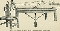
Angelo Mosso's 'human circulation balance.'

The 'human circulation balance' was a non-invasive way to measure blood flow to the brain during mental activities. This technique worked by placing patients on a table that was supported by a fulcrum, allowing the table to sway depending on activity levels. When patients were exposed to more cognitively complex stimuli, the table would sway towards the head. Invented in 1882 by Angelo Mosso, the 'human circulation balance' is said to be the first technique of neuroimaging created and is what Mosso is most known for.

Wilhelm Roentgen, creator of the X-ray.

X-ray

In the year of 1895, Wilhelm Roentgen developed the first radiograph, more commonly known as the X-ray. By 1901, Roentgen had been awarded a Nobel Peace Prize for his discovery. Immediately after its release, X-ray machines were being manufactured and used worldwide in medicine. However, this was only the first step in the development of neuroimaging. The brain is almost entirely composed of soft tissue that is not radio-opaque, meaning it remains essentially invisible to ordinary or plain X-ray examinations. This is also true of most brain abnormalities, though there are exceptions. For example, a calcified tumor (e.g.,meningioma, craniopharyngioma, and some types of glioma) can easily be seen.

Air Ventriculography

To combat this, in 1918, neurosurgeon Walter Dandy developed a technique called air ventriculography. This method injected filtered air directly into the lateral ventricles to better take pictures of the ventricle systems of the brain. Thanks to local anesthetics, this was not a painful procedure, but it was significantly risky. Hemorrhage, severe infection, and extreme changes in intrarenal pressure were all threats to the procedure. Despite this, Dandy did not stop there. In 1919, he proceeded to discover Encephalography, a medical procedure used to record the brain's electrical activity. This method involved attaching sensors to the brain that detect and measure the brain's electrical signals. These signals are then translated into a visual, showing the brain's activity patterns. With these early advances, neuroimaging was beginning to be used to diagnose conditions such as epilepsy, brain injuries, and sleep disorders. Providing invaluable information about brain function that would one day be added upon during the devolvement of modern neuroimaging.

Cerebral Angiography

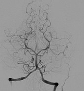
Cerebral angiogram showing a transverse projection of the vertebrobasilar and posterior cerebral circulation.

Introduced in 1927, cerebral angiography enabled doctors to accurately detect and diagnose anomalies in the brain such as tumors and internal carotid artery occlusions. Over the course of a year, Egas Moniz, the inventor of cerebral angiography, ran experiments with various dye solution percentages that were injected into arteries to help better visualize the blood vessels in the brain before discovering that a solution consisting of 25% sodium iodide was the safest for patients, as well as the most effective in the visualization of blood vessels and arteries within the brain.

PET/SPECT Scans

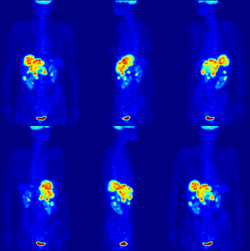
Full body PET scan of an adult female.

A positron emission tomography, or PET scan, is a scan that shows areas of high activity in the body. The way it works is that a patient is first given a radioactive substance (called a tracer) via an injection in the hand or arm. The tracer then circulates through the body and attaches to a specific substance that the organ or tissue produces during metabolism, such as glucose. As a result, positrons are created, and those positrons are scanned by the PET camera. After they are scanned, a computer produces either a 2D or 3D image of the activity occurring within the organ or tissue. The idea for the PET scan was originally proposed by William Sweet in the 1950's, but the first full-body PET scanner wasn't actually developed until 1974 by Michael Phelp.

Similarly, the single-photon emission computed tomography scan, or SPECT scan, also works by scanning a tracer within the patient. The difference, however, is that the SPECT directly scans the gamma rays from where the tracer attaches rather than the positrons that the PET scans. As a result, the images that the SPECT scan creates are not as clear as the images produced by a PET scan, but it's typically a cheaper procedure to undertake. SPECT was developed by David Kuhl in the 1950's. Kuhl also helped set the foundation that would lead to the PET scan.

Magnetoencephalography

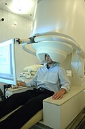
MEG device with patient.

Magnetoencephalography (MEG) is a technique that looks for regions of activity in the brain by detecting large groups of electrically charged ions moving through cells. It was originally developed by physicist David Cohen in the early 1970's as a noninvasive procedure. In order to be noninvasive, the MEG was designed like a giant helmet that the patient would put their head inside of and, once turned on, would read the electromagnetic pulses coming from their brain. Later on, in 1972, Cohen invented the SQUID (superconducting quantum interference device), which gave the MEG the ability to detect extremely small changes in ions and magnetic fields in the brain.   

Xenon CT Scanning

Godfrey Hounsfield, inventor of first CT scanner

Xenon computed tomography is a modern scanning technique that reveals the flow of blood to the areas of the brain. The scan tests for consistent and sufficient blood flow to all areas of the brain by having patients breathe in xenon gas, a contrast agent, to show the areas of high and low blood flow. Although many trial scans and tests were ran during the development process of computed tomography, British biomedical engineer Godfrey Hounsfield is the founder of the technique and invented the first CT scanner in 1967, which he won a Nobel Prize for in 1979. However, the adoption of the scanners in the United States didn't occur until six years later in 1973. Regardless, the CT scanner was already gaining a notable reputation and popularity beforehand.

Magnetic resonance imaging

Shortly after the initial development of CT, magnetic resonance imaging (MRI or MR scanning) was developed. Rather than using ionizing or X-radiation, MRI uses the variation in signals produced by protons in the body when the head is placed in a strong magnetic field. Associated with early application of the basic technique to the human body are the names of Jackson (in 1968), Damadian (in 1972), and Abe and Paul Lauterbur (in 1973). Lauterbur and Sir Peter Mansfield were awarded the 2003 Nobel Prize in Physiology or Medicine for their discoveries concerning MRI. At first, structural imaging benefited more than functional imaging from the introduction of MRI. During the 1980s a veritable explosion of technical refinements and diagnostic MR applications took place, enabling even neurological tyros to diagnose brain pathology that would have been elusive or incapable of demonstration in a living person only a decade or two earlier.

Blood–brain barrier

 
Blood–brain barrier
Solute permeability at the BBB vs. choroid plexus

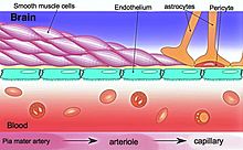
The blood–brain barrier (BBB) is a highly selective semipermeable border of endothelial cells that regulates the transfer of solutes and chemicals between the circulatory system and the central nervous system, thus protecting the brain from harmful or unwanted substances in the blood. The blood–brain barrier is formed by endothelial cells of the capillary wall, astrocyte end-feet ensheathing the capillary, and pericytes embedded in the capillary basement membrane. This system allows the passage of some small molecules by passive diffusion, as well as the selective and active transport of various nutrients, ions, organic anions, and macromolecules such as glucose and amino acids that are crucial to neural function.

The blood–brain barrier restricts the passage of pathogens, the diffusion of solutes in the blood, and large or hydrophilic molecules into the cerebrospinal fluid, while allowing the diffusion of hydrophobic molecules (O2, CO2, hormones) and small non-polar molecules. Cells of the barrier actively transport metabolic products such as glucose across the barrier using specific transport proteins. The barrier also restricts the passage of peripheral immune factors, like signaling molecules, antibodies, and immune cells, into the CNS, thus insulating the brain from damage due to peripheral immune events.

Specialized brain structures participating in sensory and secretory integration within brain neural circuits—the circumventricular organs and choroid plexus—have in contrast highly permeable capillaries.

Structure

Part of a network of capillaries supplying brain cells
The astrocytes type 1 surrounding capillaries in the brain
Sketch showing constitution of blood vessels inside the brain

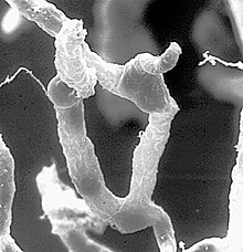
The BBB results from the selectivity of the tight junctions between the endothelial cells of brain capillaries, restricting the passage of solutes. At the interface between blood and the brain, endothelial cells are adjoined continuously by these tight junctions, which are composed of smaller subunits of transmembrane proteins, such as occludin, claudins (such as Claudin-5), junctional adhesion molecule (such as JAM-A). Each of these tight junction proteins is stabilized to the endothelial cell membrane by another protein complex that includes scaffolding proteins such as tight junction protein 1 (ZO1) and associated proteins.

The BBB is composed of endothelial cells restricting passage of substances from the blood more selectively than endothelial cells of capillaries elsewhere in the body. Astrocyte cell projections called astrocytic feet (also known as "glia limitans") surround the endothelial cells of the BBB, providing biochemical support to those cells. The BBB is distinct from the quite similar blood-cerebrospinal fluid barrier, which is a function of the choroidal cells of the choroid plexus, and from the blood-retinal barrier, which can be considered a part of the whole realm of such barriers.

Not all vessels in the human brain exhibit BBB properties. Some examples of this include the circumventricular organs, the roof of the third and fourth ventricles, capillaries in the pineal gland on the roof of the diencephalon and the pineal gland. The pineal gland secretes the hormone melatonin "directly into the systemic circulation", thus melatonin is not affected by the blood–brain barrier.

Development

The BBB appears to be functional by the time of birth. P-glycoprotein, a transporter, exists already in the embryonal endothelium.

Measurement of brain uptake of various blood-borne solutes showed that newborn endothelial cells were functionally similar to those in adults, indicating that a selective BBB is operative at birth.

In mice, Claudin-5 loss during development is lethal and results in size-selective loosening of the BBB.

Function

The blood–brain barrier acts effectively to protect brain tissue from circulating pathogens and other potentially toxic substances. Accordingly, blood-borne infections of the brain are rare. Infections of the brain that do occur are often difficult to treat. Antibodies are too large to cross the blood–brain barrier, and only certain antibiotics are able to pass. In some cases, a drug has to be administered directly into the cerebrospinal fluid where it can enter the brain by crossing the blood-cerebrospinal fluid barrier.

Circumventricular organs

Circumventricular organs (CVOs) are individual structures located adjacent to the fourth ventricle or third ventricle in the brain, and are characterized by dense capillary beds with permeable endothelial cells unlike those of the blood–brain barrier. Included among CVOs having highly permeable capillaries are the area postrema, subfornical organ, vascular organ of the lamina terminalis, median eminence, pineal gland, and three lobes of the pituitary gland.

Permeable capillaries of the sensory CVOs (area postrema, subfornical organ, vascular organ of the lamina terminalis) enable rapid detection of circulating signals in systemic blood, while those of the secretory CVOs (median eminence, pineal gland, pituitary lobes) facilitate transport of brain-derived signals into the circulating blood. Consequently, the CVO permeable capillaries are the point of bidirectional blood–brain communication for neuroendocrine function.

Specialized permeable zones

The border zones between brain tissue "behind" the blood–brain barrier and zones "open" to blood signals in certain CVOs contain specialized hybrid capillaries that are leakier than typical brain capillaries, but not as permeable as CVO capillaries. Such zones exist at the border of the area postrema—nucleus tractus solitarii (NTS), and median eminence—hypothalamic arcuate nucleus. These zones appear to function as rapid transit regions for brain structures involved in diverse neural circuits—like the NTS and arcuate nucleus—to receive blood signals which are then transmitted into neural output. The permeable capillary zone shared between the median eminence and hypothalamic arcuate nucleus is augmented by wide pericapillary spaces, facilitating bidirectional flow of solutes between the two structures, and indicating that the median eminence is not only a secretory organ, but may also be a sensory organ.

Therapeutic research

As a drug target

The blood–brain barrier is formed by the brain capillary endothelium and excludes from the brain 100% of large-molecule neurotherapeutics and more than 98% of all small-molecule drugs. Overcoming the difficulty of delivering therapeutic agents to specific regions of the brain presents a major challenge to treatment of most brain disorders. In its neuroprotective role, the blood–brain barrier functions to hinder the delivery of many potentially important diagnostic and therapeutic agents to the brain. Therapeutic molecules and antibodies that might otherwise be effective in diagnosis and therapy do not cross the BBB in adequate amounts to be clinically effective. The BBB represents an obstacle to some drugs reaching the brain, thus to overcome this barrier some peptides able to naturally cross the BBB have been widely investigated as a drug delivery system.

Mechanisms for drug targeting in the brain involve going either "through" or "behind" the BBB. Modalities for drug delivery to the brain in unit doses through the BBB entail its disruption by osmotic means, or biochemically by the use of vasoactive substances, such as bradykinin, or even by localized exposure to high-intensity focused ultrasound (HIFU).

Other methods used to get through the BBB may entail the use of endogenous transport systems, including carrier-mediated transporters, such as glucose and amino acid carriers, receptor-mediated transcytosis for insulin or transferrin, and the blocking of active efflux transporters such as p-glycoprotein. Some studies have shown that vectors targeting BBB transporters, such as the transferrin receptor, have been found to remain entrapped in brain endothelial cells of capillaries, instead of being ferried across the BBB into the targeted area.

Nanoparticles

Nanotechnology is under preliminary research for its potential to facilitate the transfer of drugs across the BBB. Capillary endothelial cells and associated pericytes may be abnormal in tumors and the blood–brain barrier may not always be intact in brain tumors. Other factors, such as astrocytes, may contribute to the resistance of brain tumors to therapy using nanoparticles. Fat soluble molecules less than 400 daltons in mass can freely diffuse past the BBB through lipid mediated passive diffusion.

Damage in injury and disease

The blood–brain barrier may become damaged in select neurological diseases, as indicated by neuroimaging studies of Alzheimer's disease, amyotrophic lateral sclerosis, epilepsy, ischemic stroke, and brain trauma, and in systemic diseases, such as liver failure. Effects such as impaired glucose transport and endothelial degeneration may lead to metabolic dysfunction within the brain, and an increased permeability of the BBB to proinflammatory factors, potentially allowing antibiotics and phagocytes to move across the BBB.

Prediction

There have been many attempts to correlate the experimental blood–brain barrier permeability with physicochemical properties. In 1988, the first QSAR study of brain–blood distribution conducted reported the in vivo values in rats for a large number of H2 receptor histamine agonists.

The first papers modelling blood-brain barrier permeability identified three properties, i.e., molecular volume, lipophilicity, and hydrogen bonding potential, as contributing to solute transport through the blood-brain barrier. A 2022 dataset selected different classification models based on molecular fingerprints, MACCS166 keys and molecular descriptors.

History

A 1898 study observed that low-concentration "bile salts" failed to affect behavior when injected into the blood of animals. Thus, in theory, the salts failed to enter the brain.

Two years later, Max Lewandowsky may have been the first to coin the term "blood–brain barrier" in 1900, referring to the hypothesized semipermeable membrane. There is some debate over the creation of the term blood–brain barrier as it is often attributed to Lewandowsky, but it does not appear in his papers. The creator of the term may have been Lina Stern. Stern was a Russian scientist who published her work in Russian and French. Due to the language barrier between her publications and English-speaking scientists, this could have made her work a lesser-known origin of the term.

All the while, bacteriologist Paul Ehrlich was studying staining, a procedure that is used in many microscopy studies to make fine biological structures visible using chemical dyes. As Ehrlich injected some of these dyes (notably the aniline dyes that were then widely used), the dye stained all of the organs of some kinds of animals except for their brains. At that time, Ehrlich attributed this lack of staining to the brain simply not picking up as much of the dye.

However, in a later experiment in 1913, Edwin Goldmann (one of Ehrlich's students) injected the dye directly into the cerebrospinal fluid of animal brains. He found then the brains did become dyed, but the rest of the body did not, demonstrating the existence of a compartmentalization between the two. At that time, it was thought that the blood vessels themselves were responsible for the barrier, since no obvious membrane could be found.

Drug delivery to the brain

From Wikipedia, the free encyclopedia

Drug delivery to the brain is the process of passing therapeutically active molecules across the blood–brain barrier into the brain. This is a complex process that must take into account the complex anatomy of the brain as well as the restrictions imposed by the special junctions of the blood–brain barrier.

Anatomy

The blood–brain barrier is formed by special tight junctions between endothelial cells lining brain blood vessels. Blood vessels of all tissues contain this monolayer of endothelial cells, however only brain endothelial cells have tight junctions preventing passive diffusion of most substances into the brain tissue. The structure of these tight junctions was first determined in the 1960s by Tom Reese, Morris Kranovsky, and Milton Brightman. Furthermore, astrocytic "end feet", the terminal regions of the astrocytic processes, surround the outside of brain capillary endothelial cells". The astrocytes are glial cells restricted to the brain and spinal cord and help maintain blood-brain barrier properties in brain endothelial cells.

Physiology

The main function of the blood–brain barrier is to protect the brain and keep it isolated from harmful toxins that are potentially in the blood stream. It accomplishes this because of its structure, as is usual in the body that structure defines its function. The tight junctions between the endothelial cells prevent large molecules as well as many ions from passing between the junction spaces. This forces molecules to go through the endothelial cells in order to enter the brain tissue, meaning that they must pass through the cell membranes of the endothelial cells. Because of this, the only molecules that are easily able to transverse the blood–brain barrier are ones that are very lipid-soluble. These are not the only molecules that can transverse the blood–brain barrier; glucose, oxygen and carbon dioxide are not lipid-soluble but are actively transported across the barrier, to support normal cellular function of the brain. The fact that molecules have to fully transverse the endothelial cells makes them a perfect barricade to unspecified particles from entering the brain, working to protect the brain at all costs. Also, because most molecules are transported across the barrier, it does a very effective job of maintaining homeostasis for the most vital organ of the human body.

Drug delivery to the blood–brain barrier

Because of the difficulty for drugs to pass through the blood–brain barrier, a study was conducted to determine the factors that influence a compound’s ability to transverse the blood–brain barrier. In this study, they examined several different factors to investigate diffusion across the blood–brain barrier. They used lipophilicity, Gibbs Adsorption Isotherm, a Co CMC Plot, and the surface area of the drug to water and air. They began by looking at compounds whose blood–brain permeability was known and labeled them either CNS+ or CNS- for compounds that easily transverse the barrier and those that did not. They then set out to analyze the above factors to determine what is necessary to transverse the blood–brain barrier. What they found was a little surprising; lipophilicity is not the leading characteristic for a drug to pass through the barrier. This is surprising because one would think that the most effective way to make a drug move through a lipophilic barrier is to increase its lipophilicity, it turns out that it is a complex function of all of these characteristics that makes a drug able to pass through the blood–brain barrier. The study found that barrier permittivity is "based on the measurement of the surface activity and as such takes into account the molecular properties of both hydrophobic and charged residues of the molecule of interest." They found that there is not a simple answer to what compounds transverse the blood–brain barrier and what does not. Rather, it is based on the complex analysis of the surface activity of the molecule as well as relative size.

Problems faced in drug delivery

Other problems persist besides just simply getting through the blood–brain barrier. The first of these is that a lot of times, even if a compound transverses the barrier, it does not do it in a way that the drug is in a therapeutically relevant concentration. This can have many causes, the most simple being that the way the drug was produced only allows a small amount to pass through the barrier. Another cause of this would be the binding to other proteins in the body rendering the drug ineffective to either be therapeutically active or able to pass through the barrier with the adhered protein. Another problem that must be accounted for is the presence of enzymes in the brain tissue that could render the drug inactive. The drug may be able to pass through the membrane fine, but will be deconstructed once it is inside the brain tissue rendering it useless. All of these are problems that must be addressed and accounted for in trying to deliver effective drug solutions to the brain tissue.

Possible solutions

Exosomes to deliver treatments across the blood–brain barrier

A group from the University of Oxford led by Prof. Matthew Wood claims that exosomes can cross the blood–brain barrier and deliver siRNAs, antisense oligonucleotides, chemotherapeutic agents and proteins specifically to neurons after inject them systemically (in blood). Because these exosomes are able to cross the blood–brain barrier, this protocol could solve the issue of poor delivery of medications to the central nervous system and cure Alzheimer's, Parkinson's Disease and brain cancer, among other diseases. The laboratory has been recently awarded a major new €30 million project leading experts from 14 academic institutions, two biotechnology companies and seven pharmaceutical companies to translate the concept to the clinic.

Pro-drugs

This is the process of disguising medically active molecules with lipophilic molecules that allow it to better sneak through the blood–brain barrier. Drugs can be disguised using more lipophilic elements or structures. This form of the drug will be inactive because of the lipophilic molecules but then would be activated, by either enzyme degradation or some other mechanism for removal of the lipophilic disguise to release the drug into its active form. There are still some major drawbacks to these pro-drugs. The first of which is that the pro-drug may be able to pass through the barrier and then also re-pass through the barrier without ever releasing the drug in its active form. The second is the sheer size of these types of molecules makes it still difficult to pass through the blood–brain barrier.

Peptide masking

Similar to the idea of pro-drugs, another way of masking the drugs chemical composition is by masking a peptide’s characteristics by combining with other molecular groups that are more likely to pass through the blood–brain barrier. An example of this is using a cholesteryl molecule instead of cholesterol that serves to conceal the water soluble characteristics of the drug. This type of masking as well as aiding in traversing the blood–brain barrier. It also can work to mask the drug peptide from peptide-degrading enzymes in the brain Also a "targetor" molecule could be attached to the drug that helps it pass through the barrier and then once inside the brain, is degraded in such a way that the drug cannot pass back through the brain. Once the drug cannot pass back through the barrier the drug can be concentrated and made effective for therapeutic use. However drawbacks to this exist as well. Once the drug is in the brain there is a point where it needs to be degraded to prevent overdose to the brain tissue. Also if the drug cannot pass back through the blood–brain barrier, it compounds the issues of dosage and intense monitoring would be required. For this to be effective there must be a mechanism for the removal of the active form of the drug from the brain tissue.

Receptor-mediated permabilitizers

These are drug compounds that increase the permeability of the blood–brain barrier. By decreasing the restrictiveness of the barrier, it is much easier to get a molecule to pass through it. These drugs increase the permeability of the blood–brain barrier temporarily by increasing the osmotic pressure in the blood which loosens the tight junctions between the endothelial cells. By loosening the tight junctions normal injection of drugs through an [IV] can take place and be effective to enter the brain. This must be done in a very controlled environment because of the risk associated with these drugs. Firstly, the brain can be flooded with molecules that are floating through the blood stream that are usually blocked by the barrier. Secondly, when the tight junctions loosen, the homeostasis of the brain can also be thrown off which can result in seizures and the compromised function of the brain.

Nanoparticles

The most promising drug delivery system is using nanoparticle delivery systems, these are systems where the drug is bound to a nanoparticle capable of traversing the blood–brain barrier. The most promising compound for the nanoparticles is Human Serum Albumin (HSA). The main benefits of this is that particles made of HSA are well tolerated without serious side effects as well as the albumin functional groups can be utilized for surface modification that allows for specific cell uptake. These nanoparticles have been shown to transverse the blood–brain barrier carrying host drugs. To enhance the effectiveness of nanoparticles, scientists are attempting to coat the nanoparticles to make them more effective to cross the blood–brain barrier. Studies have shown that "the overcoating of the [nanoparticles] with polysorbate 80 yielded doxorubicin concentrations in the brain of up to 6 μg/g after i.v. injection of 5 mg/kg" as compared to no detectable increase in an injection of the drug alone or the uncoated nanoparticle. This is very new science and technology so the real effectiveness of this process has not been fully understood. However young the research is, the results are promising pointing to nanotechnology as the way forward in treating a variety of brain diseases.

Loaded microbubble-enhanced focused ultrasound

Microbubbles are small "bubbles" of mono-lipids that are able to pass through the blood–brain barrier. They form a lipophilic bubble that can easily move through the barrier. One barrier to this however is that these microbubbles are rather large, which prevents their diffusion into the brain. This is counteracted by a focused ultrasound. The ultrasound increases the permeability of the blood–brain barrier by causing interference in the tight junctions in localized areas. This combined with the microbubbles allows for a very specific area of diffusion for the microbubbles, because they can only diffuse where the ultrasound is disrupting the barrier. The hypothesis and usefulness of these is the possibility of loading a microbubble with an active drug to diffuse through the barrier and target a specific area. There are several important factors in making this a viable solution for drug delivery. The first is that the loaded microbubble must not be substantially greater than the unloaded bubble. This ensures that the diffusion will be similar and the ultrasound disruption will be enough to induce diffusion. A second factor that must be determined is the stability of the loaded micro-bubble. This means is the drug fully retained in the bubble or is there leakage. Lastly, it must be determined how the drug is to be released from the microbubble once it passes through the blood–brain barrier. Studies have shown the effectiveness of this method for getting drugs to specific sites in the brain in animal models.

Hippocampal prosthesis

From Wikipedia, the free encyclopedia
https://en.wikipedia.org/wiki/Hippocampal_prosthesis

A hippocampus prosthesis is a type of cognitive prosthesis (a prosthesis implanted into the nervous system in order to improve or replace the function of damaged brain tissue). Prosthetic devices replace normal function of a damaged body part; this can be simply a structural replacement (e.g. reconstructive surgery or glass eye) or a rudimentary, functional replacement (e.g. a pegleg or hook).

However, prosthetics involving the brain have some special categories and requirements. "Input" prosthetics, such as retinal or cochlear implant, supply signals to the brain that the patient eventually learns to interpret as sight or sound. "Output" prosthetics use brain signals to drive a bionic arm, hand or computer device, and require considerable training during which the patient learns to generate the desired action via their thoughts. Both of these types of prosthetics rely on the plasticity of the brain to adapt to the requirement of the prosthesis, thus allowing the user to "learn" the use of his new body part.

A cognitive or "brain-to-brain" prosthesis involves neither learned input nor output signals, but the native signals used normally by the area of the brain to be replaced (or supported). Thus, such a device must be able to fully replace the function of a small section of the nervous system—using that section's normal mode of operation. In order to achieve this, developers require a deep understanding of the functioning of the nervous system. The scope of design must include a reliable mathematical model as well as the technology in order to properly manufacture and install a cognitive prosthesis. The primary goal of an artificial hippocampus is to provide a cure for Alzheimer's disease and other hippocampus—related problems. To do so, the prosthesis has to be able to receive information directly from the brain, analyze the information and give an appropriate output to the cerebral cortex; in other words, it must behave just like a natural hippocampus. At the same time, the artificial organ must be completely autonomous, since any exterior power source will greatly increase the risk of infection.

Hippocampus

Role

The hippocampus is part of the human limbic system, which interacts with the neocortex and other parts of the brain to produce emotions. As a part of the limbic system, the hippocampus plays its part in the formation of emotion in addition to its other roles, such as consolidation of new memories, navigation, and spatial orientation. The hippocampus is responsible for the formation of long term recognition memories. In other words, this is the part of the brain that allows us to associate a face with a name. Because of its close relationship with memory formation, damage to the hippocampus is closely related to Alzheimer's disease.

Anatomy

The hippocampus is a bilateral structure, situated under the neocortex. Each hippocampus is "composed of several different subsystem[s] that form a closed feedback loop, with input from the neocortex entering via the entorhinal cortex, propagating through the intrinsic subregions of the hippocampus and returning to the neocortex." In an electronic sense, the hippocampus is composed of a slice of parallel circuits.

Essential requirements

Biocompatibility

Since the prosthesis will be permanently implanted inside the brain, long term biocompatibility is required. We must also take into account the tendency for supporting braincells like astrocytes to encapsulate the implant. (This is a natural response for braincells, in order to protect neurons), thus impairing its function.

Bio-mimetic

Being biomimetic means that the implant must be able to fulfill the properties a real biological neuron. To do so we must have an in–depth understanding of brain behavior to build a solid mathematical model to be based upon. The field of computational neuroscience has made headway in this endeavor.

First, we must take into account that, like most of biological processes, the behaviors of neurons are highly nonlinear and depend on many factors: input frequency patterns, etc. Also, a good model must take into account the fact that the expression of a single nerve cell is negligible, since the processes are carried by groups of neurons interacting in network. Once installed, the device must assume all (or at least most) of the function of the damaged hippocampus for a prolonged period of time. First, the artificial neurons must be able to work together in network just like real neurons. Then, they must be able, working and effective synaptics connections with the existing neurons of the brain; therefore a model for silicon/neurons interface will be required.

Size

The implant must be small enough to be implantable while minimizing collateral damage during and after the implantation.

Bidirectional communication

In order to fully assume the function of the damaged hippocampus, the prosthesis must be able to communicate with the existing tissue in a bidirectional manner. in other words, the implant must be able to receive information from the brain and give an appropriate and compressible feedback to the surrounding nerve cell.

Personalized

The structural and functional characteristic of the brain varies greatly between individuals; therefore any neural implant has to be specific to each individual, which requires a precise model of the hippocampus and the use of advanced brain imagery to determine individual variance.

Surgical requirement

Since the prosthesis will be installed inside the brain, the operation itself will be much like a tumor removal operation. Although collateral damage will be inevitable, the effect on the patient will be minimal.

Model

"In order to incorporate the nonlinear dynamics of biological neurons into neuron models to develop a prosthesis, it is first necessary to measure them accurately. We have developed and applied methods for quantifying the nonlinear dynamics of hippocampal neurons (Berger et al., 1988a,b, 1991, 1992, 1994; Dalal et al., 1997) using principles of nonlinear systems theory (Lee and Schetzen, 1965; Krausz, 1975; P. Z. Marmarelis and Marmarelis, 1978; Rugh, 1981; Sclabassi et al., 1988). In this approach, properties of neurons are assessed experimentally by applying a random interval train of electrical impulses as an input and electrophysiologically recording the evoked output of the target neuron during stimulation (figure 12.2A). The input train consists of a series of impulses (as many as 4064), with interimpulse intervals varying according to a Poisson process having a mean of 500 ms and a range of 0.2–5000 ms. Thus, the input is "broadband" and stimulates the neuron over most of its operating range; that is, the statistical properties of the random train are highly consistent with the known physiological properties of hippocampal neurons. Nonlinear response properties are expressed in terms of the relation between progressively higher-order temporal properties of a sequence of input events and the probability of neuronal output, and are modeled as the kernels of a functional power series."

Technology involved

Imaging

Technology such as EEG, MEG, fMRI and other type of imaging technology are essential to the installation of the implant, which requires a high precision in order to minimize collateral damage (since the hippocampus is situated inside the cortex), as well as the proper function of the device.

Silicon/neuron interface

A silicon/neuron interface will be needed for the proper interaction of the silicon neurons of the prosthesis and the biological neurons of the brain.

Neuron network processor

In the brain, tasks are carried out by groups of interconnected neuronal network rather than a single cell, which means that any prosthesis must be able to simulate this network behavior. To do so, we will need a high number and density of silicon neurons to produce an effective prosthesis; therefore, a High-density Hippocampal Neuron Network Processor will be required in order for the prosthesis to carry out the task of a biological hippocampus. In addition, a neuron/silicon interface will be essential to the bidirectional communication of the implanted prosthesis. The choice of material and the design must ensure long term viability and bio compatibility while ensuring the density and the specificity of the interconnections.

Power supply

Appropriate power supply is still a major issue for any neural implant. Because the prostheses are implanted inside the brain, long term biocompatibility aside, the power supply will require several specification. First, the power supply must be self recharging. Unlike other prostheses, infection is a much greater issue for neural implant, due to the sensitivity of the brain; therefore an external power source is not envisagable. Because the brain is also highly heat sensitive, the power and the device itself must not generate too much heat to avoid disrupting brain function.

Prosthetic neuronal memory silicon chips

A prosthetic neuronal memory silicon chip is a device that imitates the brain's process of creating long-term memories. A prototype for this device was designed by Theodore Berger, a biomedical engineer and neurologist at University of Southern California. Berger started to work on the design in the early 1990s. He partnered with research colleagues that have been able to implant electrodes into rats and monkeys to test restoration of memory function. Recent work shows that the system can form long-term memories in many different behavioral situations. Berger and colleagues hope to eventually use these chips as electronic implants for humans whose brains that suffer from diseases such as Alzheimer's that disrupt neuronal networks.

Technology and medical application

To begin making a brain prosthesis, Berger and his collaborator Vasilis Marmarelis, a biomedical engineer at USC, worked with the hippocampus slices of rats. Since they knew that neuronal signals travel from one side of the hippocampus to the other, the researchers sent random pulses into the hippocampus, recorded the signals at specific locales to see how they were changed, and then derived equations representing the changes. They then programmed those equations into the computer chips.

Next, they had to determine whether a chip could be used as a prosthesis, or implant, for a damaged region in the hippocampus. To do this, they had to figure out whether they could avoid a central component of the pathway in the brain slices. They put electrodes in the region, which carried electrical pulses to an external chip. The chip then executed the transformations that are normally carried out in the hippocampus, and other electrodes sent the signals back to the slice of brain.

Memory codes

In 1996, Dr. Sam A. Deadwyler of Wake Forest Baptist Medical Center in Winston-Salem, NC, studied the activity patterns of collections of hippocampal neurons while rats performed a task requiring short-term memory.  These 'ensembles' or collections of neurons fired in different patterns in both time and 'space' (in this case, space referred to different neurons distributed throughout the hippocampus) depending on the type of behavior required in the task.  More importantly, Deadwyler and his colleagues could identify patterns that clearly distinguished between the various stimuli in the task including position (similar to place cells), behavioral responses, and what part of the task was occurring.  Analyses based on the neural ensemble activity alone without looking at those variables could identify and even 'predict' some of those variables even before they occurred.  In fact, the patterns would even identify when the rat was about to make an error in the task. Over the following ten years, Deadwyler's laboratory refined the analysis to identify the 'codes' and improved the ability to predict correct and error responses, even to the point of being able to have untrained rats perform the memory task using hippocampal stimulation with codes obtained from fully trained rats. The discovery of the memory codes in hippocampus led Deadwyler to join efforts with Berger for future studies in which Berger's team would develop models of memory function in hippocampus, and Deadwyler's team would test the models in rats and monkeys, and eventually move into human studies.

Trials on rats and monkeys

To transition to awake, behaving animals, Berger partnered with Deadwyler and Dr. Robert E. Hampson of Wake Forest to test a prototype of the memory prosthetic connected to rat and monkey brains via electrodes to analyze information just like the actual hippocampus. The prosthetic model allowed even a damaged hippocampus to generate new memories. In one demonstration, Deadwyler and Hampson impaired the rats' ability to form long-term memories by using pharmacological agents. These disrupted the neural circuitry that transfers messages between two subregions of the hippocampus. These subregions, CA1 and CA3, interact to create long-term memories. The rats were unable to remember which lever they needed to pull to obtain the reward. The researchers then developed an artificial hippocampus that could duplicate the pattern of interaction between CA3-CA1 interactions by analyzing the neural spikes in the cells with an electrode array, and then playing back the same pattern on the same array. After stimulating the rat hippocampi using the mathematical model of the prosthesis, their ability to identify the correct lever to pull improved dramatically. This artificial hippocampus played a significant role in the developmental stage of a memory prosthetic, as it went on to show that if a prosthetic device and its associated electrodes were implanted in the animals with a malfunctioning hippocampus, the device could potentially restore the memory capability to that of normal rats.

Goals for the future

The research teams at USC and Wake Forest are working to possibly make this system applicable to humans whose brains suffer damage from Alzheimer's, stroke, or injury, the disruption of neural networks often stops long-term memories from forming. The system designed by Berger and implemented by Deadwyler and Hampson allows the signal processing to take place that would occur naturally in undamaged neurons. Ultimately, they hope to restore the ability to create long-term memories by implanting chips such as these into the brain.

Recent development

Theodore Berger and his colleagues at the University of Southern California in Los Angeles have developed a working hippocampal prosthesis that passed the live tissue test in slices of brain tissue in 2004,. In 2011, in collaboration with Drs. Sam A. Deadwyler and Robert E. Hampson at Wake Forest Baptist Medical Center successfully tested a proof-of-concept hippocampal prosthesis in awake, behaving rats. The prosthesis was in the form of multisite electrodes positioned to record from both the input and output "sides" of the damaged hippocampus, the input is gathered and analyzed by external computation chips, an appropriate feedback is computed, then used to stimulate the appropriate output pattern in the brain so that the prosthesis functioned like a real hippocampus. In 2012, the team tested a further implementation in macaques prefrontal cortex, further developing the neural prosthesis technology. In 2013, Hampson et al. successfully tested a hippocampal prosthesis on non-human primates. While the device does not yet consist of a fully implantable "chip," these tests, from rat to monkey, demonstrate the effectiveness of the device as a neural prosthetic, and supports application to human trials.

Proof of concept for a human hippocampal prosthetic

In 2018, a team led by Robert E. Hampson at Wake Forest Baptist Medical, and including Berger and Deadwyler, became the first to demonstrate effectiveness of the prosthetic model in human patients. The subjects underwent implantation of electrodes in the brain at Wake Forest as part of a medical diagnostic procedure for epilepsy. While in the hospital, patients with electrodes in hippocampus volunteered to perform a memory task on computer while hippocampal neural activity was recorded in order for Berger and his team at USC team to customize the hippocampal prosthetic model for that patient. With model in hand, the Wake Forest team was able to demonstrate up to 37% improvement in memory function in patients with memory impaired by disease. The improvement was demonstrated for memories up to 75 minutes after stimulation by the hippocampal prosthetic model. As of 2018, studies are planned to test memory codes for additional attributes and features of items to be remembered as well as duration of memory facilitation in excess of 24 hours.

Operator (computer programming)

From Wikipedia, the free encyclopedia https://en.wikipedia.org/wiki/Operator_(computer_programmin...