Search This Blog

Wednesday, March 27, 2024

Major prion protein

From Wikipedia, the free encyclopedia

Major prion protein (PrP) is encoded in the human body by the PRNP gene also known as CD230 (cluster of differentiation 230). Expression of the protein is most predominant in the nervous system but occurs in many other tissues throughout the body.

The protein can exist in multiple isoforms: the normal PrPC form, and the protease-resistant form designated PrPRes such as the disease-causing PrPSc (scrapie) and an isoform located in mitochondria. The misfolded version PrPSc is associated with a variety of cognitive disorders and neurodegenerative diseases such as in animals: ovine scrapie, bovine spongiform encephalopathy (BSE, mad cow disease), feline spongiform encephalopathy, transmissible mink encephalopathy (TME), exotic ungulate encephalopathy, chronic wasting disease (CWD) which affects deer; and in humans: Creutzfeldt–Jakob disease (CJD), fatal familial insomnia (FFI), Gerstmann–Sträussler–Scheinker syndrome (GSS), kuru, and variant Creutzfeldt–Jakob disease (vCJD). Similarities exist between kuru, thought to be due to human ingestion of diseased individuals, and vCJD, thought to be due to human ingestion of BSE-tainted cattle products.

Gene

Chromosome 20

The human PRNP gene is located on the short (p) arm of chromosome 20 between the end (terminus) of the arm and position 13, from base pair 4,615,068 to base pair 4,630,233.

Structure

PrP is highly conserved through mammals, lending credence to application of conclusions from test animals such as mice. Comparison between primates is especially similar, ranging from 92.9-99.6% similarity in amino acid sequences. The human protein structure consists of a globular domain with three α-helices and a two-strand antiparallel β-sheet, an NH2-terminal tail, and a short COOH-terminal tail. A glycophosphatidylinositol (GPI) membrane anchor at the COOH-terminal tethers PrP to cell membranes, and this proves to be integral to the transmission of conformational change; secreted PrP lacking the anchor component is unaffected by the infectious isoform.

The primary sequence of PrP is 253 amino acids long before post-translational modification. Signal sequences in the amino- and carboxy- terminal ends are removed posttranslationally, resulting in a mature length of 208 amino acids. For human and golden hamster PrP, two glycosylated sites exist on helices 2 and 3 at Asn181 and Asn197. Murine PrP has glycosylation sites as Asn180 and Asn196. A disulfide bond exists between Cys179 of the second helix and Cys214 of the third helix (human PrPC numbering).

PrP messenger RNA contains a pseudoknot structure (prion pseudoknot), which is thought to be involved in regulation of PrP protein translation.

Ligand-binding

The mechanism for conformational conversion to the scrapie isoform is speculated to be an elusive ligand-protein, but, so far, no such compound has been identified. However, a large body of research has developed on candidates and their interaction with the PrPC.

Copper, zinc, manganese, and nickel are confirmed PrP ligands that bind to its octarepeat region. Ligand binding causes a conformational change with unknown effect. Heavy metal binding at PrP has been linked to resistance to oxidative stress arising from heavy metal toxicity.

PrPC (normal cellular) isoform

Although the precise function of PrP is not yet known, it is possibly involved in the transport of ionic copper to cells from the surrounding environment. Researchers have also proposed roles for PrP in cell signaling or in the formation of synapses. PrPC attaches to the outer surface of the cell membrane by a glycosylphosphatidylinositol anchor at its C-terminal Ser231.

Prion protein contains five octapeptide repeats with sequence PHGGGWGQ (though the first repeat has the slightly-modified, histidine-deficient sequence PQGGGGWGQ). This is thought to generate a copper-binding domain via nitrogen atoms in the histidine imidazole side-chains and deprotonated amide nitrogens from the 2nd and 3rd glycines in the repeat. The ability to bind copper is, therefore, pH-dependent. NMR shows copper binding results in a conformational change at the N-terminus.

PrPSc (scrapie) isoform

PrPSc is a conformational isoform of PrPC, but this orientation tends to accumulate in compact, protease-resistant aggregates within neural tissue. The abnormal PrPSc isoform has a different secondary and tertiary structure from PrPC, but identical primary sequence. Whereas PrPC has largely alpha helical and disordered domains, PrPSc has no alpha helix and an amyloid fibril core composed of a stack of PrP molecules glued together by parallel in-register intermolecular beta sheets. This refolding renders the PrPSc isoform extremely resistant to proteolysis.

The propagation of PrPSc is a topic of great interest, as its accumulation is a pathological cause of neurodegeneration. Based on the progressive nature of spongiform encephalopathies, the predominant hypothesis posits that the change from normal PrPC is caused by the presence and interaction with PrPSc. Strong support for this is taken from studies in which PRNP-knockout mice are resistant to the introduction of PrPSc. Despite widespread acceptance of the conformation conversion hypothesis, some studies mitigate claims for a direct link between PrPSc and cytotoxicity.

Polymorphisms at sites 136, 154, and 171 are associated with varying susceptibility to ovine scrapie. (These ovine sites correspond to human sites 133, 151, and 168.) Polymorphisms of the PrP-VRQ form and PrP-ARQ form are associated with increased susceptibility, whereas PrP-ARR is associated with resistance. The National Scrapie Plan of the UK aims to breed out these scrapie polymorphisms by increasing the frequency of the resistant allele. However, PrP-ARR polymorphisms are susceptible to atypical scrapie, so this may prove unfruitful.

Function

Nervous system

The strong association to neurodegenerative diseases raises many questions of the function of PrP in the brain. A common approach is using PrP-knockout and transgenic mice to investigate deficiencies and differences. Initial attempts produced two strains of PrP-null mice that show no physiological or developmental differences when subjected to an array of tests. However, more recent strains have shown significant cognitive abnormalities.

As the null mice age, a marked loss of Purkinje cells in the cerebellum results in decreased motor coordination. However, this effect is not a direct result of PrP's absence, and rather arises from increased Doppel gene expression. Other observed differences include reduced stress response and increased exploration of novel environments.

Circadian rhythm is altered in null mice. Fatal familial insomnia is thought to be the result of a point mutation in PRNP at codon 178, which corroborates PrP's involvement in sleep-wake cycles. In addition, circadian regulation has been demonstrated in PrP mRNA, which cycles regularly with day-night.

Memory

While null mice exhibit normal learning ability and short-term memory, long-term memory consolidation deficits have been demonstrated. As with ataxia, this is attributable to Doppel gene expression. However, spatial learning, a predominantly hippocampal-function, is decreased in the null mice and can be recovered with the reinstatement of PrP in neurons; this indicates that loss of PrP function is the cause. The interaction of hippocampal PrP with laminin (LN) is pivotal in memory processing and is likely modulated by the kinases PKA and ERK1/2.

Further support for PrP's role in memory formation is derived from several population studies. A test of healthy young humans showed increased long-term memory ability associated with an MM or MV genotype when compared to VV. Down syndrome patients with a single valine substitution have been linked to earlier cognitive decline. Several polymorphisms in PRNP have been linked with cognitive impairment in the elderly as well as earlier cognitive decline. All of these studies investigated differences in codon 129, indicating its importance in the overall functionality of PrP, in particular with regard to memory.

Neurons and synapses

PrP is present in both the pre- and post-synaptic compartments, with the greatest concentration in the pre-synaptic portion. Considering this and PrP's suite of behavioral influences, the neural cell functions and interactions are of particular interest. Based on the copper ligand, one proposed function casts PrP as a copper buffer for the synaptic cleft. In this role, the protein could serve as either a copper homeostasis mechanism, a calcium modulator, or a sensor for copper or oxidative stress. Loss of PrP function has been linked to long-term potentiation (LTP). This effect can be positive or negative and is due to changes in neuronal excitability and synaptic transmission in the hippocampus.

Some research indicates PrP involvement in neuronal development, differentiation, and neurite outgrowth. The PrP-activated signal transduction pathway is associated with axon and dendritic outgrowth with a series of kinases.

Immune system

Though most attention is focused on PrP's presence in the nervous system, it is also abundant in immune system tissue. PrP immune cells include hematopoietic stem cells, mature lymphoid and myeloid compartments, and certain lymphocytes; also, it has been detected in natural killer cells, platelets, and monocytes. T cell activation is accompanied by a strong up-regulation of PrP, though it is not requisite. The lack of immunoresponse to transmissible spongiform encephalopathies (TSE), neurodegenerative diseases caused by prions, could stem from the tolerance for PrPSc.

Muscles, liver, and pituitary

PrP-null mice provide clues to a role in muscular physiology when subjected to a forced swimming test, which showed reduced locomotor activity. Aging mice with an overexpression of PRNP showed significant degradation of muscle tissue.

Though present, very low levels of PrP exist in the liver and could be associated with liver fibrosis. Presence in the pituitary has been shown to affect neuroendocrine function in amphibians, but little is known concerning mammalian pituitary PrP.

Cellular

Varying expression of PrP through the cell cycle has led to speculation on involvement in development. A wide range of studies has been conducted investigating the role in cell proliferation, differentiation, death, and survival. Engagement of PrP has been linked to activation of signal transduction.

Modulation of signal transduction pathways has been demonstrated in cross-linking with antibodies and ligand-binding (hop/STI1 or copper). Given the diversity of interactions, effects, and distribution, PrP has been proposed as dynamic surface protein functioning in signaling pathways. Specific sites along the protein bind other proteins, biomolecules, and metals. These interfaces allow specific sets of cells to communicate based on level of expression and the surrounding microenvironment. The anchoring on a GPI raft in the lipid bilayer supports claims of an extracellular scaffolding function.

Diseases caused by PrP misfolding

More than 20 mutations in the PRNP gene have been identified in people with inherited prion diseases, which include the following:

The conversion of PrPC to PrPSc conformation is the mechanism of transmission of fatal, neurodegenerative transmissible spongiform encephalopathies (TSE). This can arise from genetic factors, infection from external source, or spontaneously for reasons unknown. Accumulation of PrPSc corresponds with progression of neurodegeneration and is the proposed cause. Some PRNP mutations lead to a change in single amino acids (the building-blocks of proteins) in the prion protein. Others insert additional amino acids into the protein or cause an abnormally short protein to be made. These mutations cause the cell to make prion proteins with an abnormal structure. The abnormal protein PrPSc accumulates in the brain and destroys nerve cells, which leads to the mental and behavioral features of prion diseases.

Several other changes in the PRNP gene (called polymorphisms) do not cause prion diseases but may affect a person's risk of developing these diseases or alter the course of the disorders. An allele that codes for a PRNP variant, G127V, provides resistance to kuru.

In addition, some prion diseases can be transmitted from external sources of PrPSc.

  • Scrapie – fatal neurodegenerative disease in sheep, not transmissible to humans
  • Bovine spongiform encephalopathy (mad-cow disease) – fatal neurodegenerative disease in cows, which can be transmitted to humans by ingestion of brain, spinal, or digestive tract tissue of an infected cow
  • Kuru – TSE in humans, transmitted via funerary cannibalism. Generally, affected family members were given, by tradition, parts of the central nervous system according to ritual when consuming deceased family members.

Alzheimer's disease

PrPC protein is one of several cellular receptors of soluble amyloid beta (Aβ) oligomers, which are canonically implicated in causing Alzheimer's disease. These oligomers are composed of smaller Aβ plaques, and are the most damaging to the integrity of a neuron. The precise mechanism of soluble Aβ oligomers directly inducing neurotoxicity is unknown, and experimental deletion of PRNP in animals has yielded several conflicting findings. When Aβ oligomers were injected into the cerebral ventricles of a mouse model of Alzheimer's, PRNP deletion did not offer protection, only anti-PrPC antibodies prevented long-term memory and spatial learning deficits. This would suggest either an unequal relation between PRNP and Aβ oligomer-mediated neurodegeneration or a site-specific relational significance. In the case of direct injection of Aβ oligomers into the hippocampus, PRNP-knockout mice were found to be indistinguishable from control with respect to both neuronal death rates and measurements of synaptic plasticity. It was further found that Aβ-oligomers bind to PrPC at the postsynaptic density, indirectly overactivating the NMDA receptor via the Fyn enzyme, resulting in excitotoxicity. Soluble Aβ oligomers also bind to PrPC at the dendritic spines, forming a complex with Fyn and excessively activating tau, another protein implicated in Alzheimer's. As the gene FYN codes for the enzyme Fyn, FYN-knockout mice display neither excitotoxic events nor dendritic spine shrinkage when injected with Aβ oligomers. In mammals, the full functional significance of PRNP remains unclear, as PRNP deletion has been prophylactically implemented by the cattle industry without apparent harm. In mice, this same deletion phenotypically varies between Alzheimer's mouse lines, as hAPPJ20 mice and TgCRND8 mice show a slight increase in epileptic activity, contributing to conflicting results when examining Alzheimer's survival rates. Of note, the deletion of PRNP in both APPswe and SEN1dE9, two other transgenic models of Alzheimer's, attenuated the epilepsy-induced death phenotype seen in a subset of these animals. Taken collectively, recent evidence suggests PRNP may be important for conducing the neurotoxic effects of soluble Aβ-oligomers and the emergent disease state of Alzheimer's.

In humans, the methionine/valine polymorphism at codon 129 of PRNP (rs1799990) is most closely associated with Alzheimer's disease. Variant V allele carriers (VV and MV) show a 13% decreased risk with respect to developing Alzheimer's compared to the methionine homozygote (MM). However, the protective effects of variant V carriers have been found exclusively in Caucasians. The decreased risk in V allele carriers is further limited to late-onset Alzheimer's disease only (≥ 65 years). PRNP can also functionally interact with polymorphisms in two other genes implicated in Alzheimer's, PSEN1 and APOE, to compound risk for both Alzheimer's and sporadic Creutzfeldt–Jakob disease. A point mutation on codon 102 of PRNP at least in part contributed to three separate patients' atypical frontotemporal dementia within the same family, suggesting a new phenotype for Gerstmann–Sträussler–Scheinker syndrome. The same study proposed sequencing PRNP in cases of ambiguously diagnosed dementia, as the various forms of dementia can prove challenging to differentially diagnose.

Research

In 2006 the production of cattle lacking PrPC form of the major prion protein (PrP) protein was reported which were resistant to prion propagation with no apparent developmental abnormalities. Besides the study of bovine products free of prion proteins another use could be so that human pharmaceuticals can be made in their blood without the danger that those products might get contaminated with the infectious agent that causes mad cow.

Interactions

A strong interaction exists between PrP and the cochaperone Hop (Hsp70/Hsp90 organizing protein; also called STI1 (Stress-induced protein 1)).

Grey matter

From Wikipedia, the free encyclopedia
https://en.wikipedia.org/wiki/Grey_matter
Grey matter
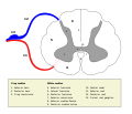
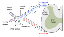
The formation of the spinal nerve from the dorsal and ventral roots (with grey matter labelled at centre right).
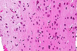
Micrograph showing grey matter, with the characteristic neuronal cell bodies (dark shade of pink), and white matter with its characteristic fine meshwork-like appearance (left of image; lighter shade of pink). HPS stain.

Grey matter, or gray matter in American English, is a major component of the central nervous system, consisting of neuronal cell bodies, neuropil (dendrites and unmyelinated axons), glial cells (astrocytes and oligodendrocytes), synapses, and capillaries. Grey matter is distinguished from white matter in that it contains numerous cell bodies and relatively few myelinated axons, while white matter contains relatively few cell bodies and is composed chiefly of long-range myelinated axons. The colour difference arises mainly from the whiteness of myelin. In living tissue, grey matter actually has a very light grey colour with yellowish or pinkish hues, which come from capillary blood vessels and neuronal cell bodies.

Structure

Grey matter refers to unmyelinated neurons and other cells of the central nervous system. It is present in the brain, brainstem and cerebellum, and present throughout the spinal cord.

Grey matter is distributed at the surface of the cerebral hemispheres (cerebral cortex) and of the cerebellum (cerebellar cortex), as well as in the depths of the cerebrum (the thalamus; hypothalamus; subthalamus, basal gangliaputamen, globus pallidus and nucleus accumbens; as well as the septal nuclei), cerebellum (deep cerebellar nuclei – the dentate nuclei, globose nucleus, emboliform nucleus, and fastigial nucleus), and brainstem (the substantia nigra, red nucleus, olivary nuclei, and cranial nerve nuclei).

Grey matter in the spinal cord is known as the grey column which travels down the spinal cord distributed in three grey columns that are presented in an "H" shape. The forward-facing column is the anterior grey column, the rear-facing one is the posterior grey column and the interlinking one is the lateral grey column. The grey matter on the left and right side is connected by the grey commissure. The grey matter in the spinal cord consists of interneurons, as well as the cell bodies of projection neurons.

Grey matter undergoes development and growth throughout childhood and adolescence. Recent studies using cross-sectional neuroimaging have shown that by around the age of 8 the volume of grey matter begins to decrease. However, the density of grey matter appears to increase as a child develops into early adulthood. Males tend to exhibit grey matter of increased volume but lower density than that of females.

Function

Grey matter contains most of the brain's neuronal cell bodies. The grey matter includes regions of the brain involved in muscle control, and sensory perception such as seeing and hearing, memory, emotions, speech, decision-making, and self-control.

The grey matter in the spinal cord is split into three grey columns:

The grey matter of the spinal cord can be divided into different layers, called Rexed laminae. These describe, in general, the purpose of the cells within the grey matter of the spinal cord at a particular location.

Clinical significance

High alcohol consumption has been correlated with significant reductions in grey matter volume. Short-term cannabis use (30 days) is not correlated with changes in white or grey matter. However, several cross-sectional studies have shown that repeated long-term cannabis use is associated with smaller grey matter volumes in the hippocampus, amygdala, medial temporal cortex, and prefrontal cortex, with increased grey matter volume in the cerebellum. Long-term cannabis use is also associated with alterations in white matter integrity in an age-dependent manner, with heavy cannabis use during adolescence and early adulthood associated with the greatest amount of change.

Meditation has been shown to change grey matter structure.

Habitual playing of action video games has been reported to promote a reduction of grey matter in the hippocampus while 3D platformer games have been reported to increase grey matter in the hippocampus.

Women and men with equivalent IQ scores have differing proportions of grey to white matter in cortical brain regions associated with intelligence.

Pregnancy renders substantial changes in brain structure, primarily reductions in grey matter volume in regions subserving social cognition. The grey matter reductions endured for at least 2 years post-pregnancy. The profile of brain changes is comparable to that taking place during adolescence, a hormonally similar transitional period of life.

History

Etymology

In the current edition of the official Latin nomenclature, Terminologia Anatomica, substantia grisea is used for English grey matter. The adjective grisea for grey is however not attested in classical Latin. The adjective grisea is derived from the French word for grey, gris. Alternative designations like substantia cana  and substantia cinerea are being used alternatively. The adjective cana, attested in classical Latin, can mean grey, or greyish white. The classical Latin cinerea means ash-coloured.

Kuru (disease)

From Wikipedia, the free encyclopedia
Kuru
A Fore child with advanced kuru. He is unable to walk or sit upright without assistance and is severely malnourished.
Pronunciation
  • IPA: kuru
SpecialtyNeuropathology, infectious disease
SymptomsBody tremors, random outbursts of laughter, gradual loss of coordination
ComplicationsInfection and pneumonia during the terminal stage.
Usual onsetOften takes years or even decades for symptoms to appear after exposure
Duration11–14 month life expectancy after onset of symptoms
CausesTransmission of infected prion proteins
Risk factorsCannibalism
Diagnostic methodAutopsy
Differential diagnosisCreutzfeldt–Jakob disease
PreventionAvoiding practices of cannibalism
TreatmentSupportive care
PrognosisFatal
FrequencyRare
DeathsApproximately 2,700 as of 2005

Kuru is a rare, incurable, and fatal neurodegenerative disorder that was formerly common among the Fore people of Papua New Guinea. Kuru is a form of transmissible spongiform encephalopathy (TSE) caused by the transmission of abnormally folded proteins (prions), which leads to symptoms such as tremors and loss of coordination from neurodegeneration.

The term kuru derives from the Fore word kuria or guria ("to shake"), due to the body tremors that are a classic symptom of the disease. Kúru itself means "trembling". It is also known as the "laughing sickness" due to the pathologic bursts of laughter which are a symptom of the disease. It is now widely accepted that kuru was transmitted among members of the Fore tribe of Papua New Guinea via funerary cannibalism. Deceased family members were traditionally cooked and eaten, which was thought to help free the spirit of the dead. Women and children usually consumed the brain, the organ in which infectious prions were most concentrated, thus allowing for transmission of kuru. The disease was therefore more prevalent among women and children.

The epidemic likely started when a villager developed sporadic Creutzfeldt–Jakob disease and died. When villagers ate the brain, they contracted the disease and then spread it to other villagers who ate their infected brains.

While the Fore people stopped consuming human meat in the early 1960s, when it was first speculated to be transmitted via endocannibalism, the disease lingered due to kuru's long incubation period of anywhere from 10 to over 50 years. The epidemic finally declined sharply after half a century, from 200 deaths per year in 1957 to no deaths from at least 2010 onwards, with sources disagreeing on whether the last known kuru victim died in 2005 or 2009.

Signs and symptoms

Kuru, a transmissible spongiform encephalopathy, is a disease of the nervous system that causes physiological and neurological effects which ultimately lead to death. It is characterized by progressive cerebellar ataxia, or loss of coordination and control over muscle movements.

The preclinical or asymptomatic phase, also called the incubation period, averages 10–13 years, but can be as short as five and has been estimated to last as long as 50 years or more after initial exposure.

The clinical stage, which begins at the first onset of symptoms, lasts an average of 12 months. The clinical progression of kuru is divided into three specific stages: the ambulant, sedentary and terminal stages. While there is some variation in these stages from individual to individual, they are highly conserved among the affected population. Before the onset of clinical symptoms, an individual can also present with prodromal symptoms including headache and joint pain in the legs.

Ambulant stage

In the ambulant stage, the infected individual may exhibit unsteady stance and gait, decreased muscle control, difficulty pronouncing words (dysarthria), and tremors (titubation). This stage is named the ambulant because the individual is still able to walk around despite symptoms.

Sedentary stage

In the sedentary stage, the infected individual is incapable of walking without support and experiences ataxia and severe tremors. Furthermore, the individual shows signs of emotional instability and depression, yet exhibits uncontrolled and sporadic laughter. Despite the other neurological symptoms, tendon reflexes are still intact at this stage of the disease.

Terminal stage

In the terminal stage, the infected individual's existing symptoms, like ataxia, progress to the point where it is no longer possible to sit up without support. New symptoms also emerge: the individual develops dysphagia, which can lead to severe malnutrition, and may also become incontinent, lose the ability or will to speak, and become unresponsive to their surroundings despite maintaining consciousness. Towards the end of the terminal stage, patients often develop chronic decubitus ulcerated wounds that can be easily infected. An infected person usually dies within three months to two years after the first terminal stage symptoms, often because of aspiration pneumonia or other secondary infections.

Causes

Kuru is largely localized to the Fore people and people with whom they intermarried. The Fore people ritualistically cooked and consumed body parts of their family members following their death to incorporate "the body of the dead person into the bodies of living relatives, thus helping to free the spirit of the dead". Because the brain is the organ enriched in the infectious prion, women and children, who consumed brain, had a much higher likelihood of being infected than men, who preferentially consumed muscles.

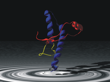
Normally folded prion protein PrPc subdomain-Residues 125–228. Note the presence of alpha helices (blue).

Prion

Cryoelectron microscopy model of the misfolded PrPsc protein, enriched in beta sheets (center)

The infectious agent is a misfolded form of a host-encoded protein called prion (PrP). Prion proteins are encoded by the Prion Protein Gene (PRNP). The two forms of prion are designated as PrPc, which is a normally folded protein, and PrPsc, a misfolded form which gives rise to the disease. The two forms do not differ in their amino acid sequence; however, the pathogenic PrPsc isoform differs from the normal PrPc form in its secondary and tertiary structure. The PrPsc isoform is more enriched in beta sheets, while the normal PrPc form is enriched in alpha helices. The differences in conformation allow PrPsc to aggregate and be extremely resistant to protein degradation by enzymes or by other chemical and physical means. The normal form, on the other hand, is susceptible to complete proteolysis and soluble in non-denaturing detergents.

It has been suggested that pre-existing or acquired PrPsc can promote the conversion of PrPc into PrPsc, which goes on to convert other PrPc. This initiates a chain reaction that allows for its rapid propagation, resulting in the pathogenesis of prion diseases.

Transmission

In 1961, Australian medical researcher Michael Alpers conducted extensive field studies among the Fore accompanied by anthropologist Shirley Lindenbaum. Their historical research suggested the epidemic may have originated around 1900 from a single individual who lived on the edge of Fore territory and who is thought to have spontaneously developed some form of Creutzfeldt–Jakob disease. Alpers and Lindenbaum's research conclusively demonstrated that kuru spread easily and rapidly in the Fore people due to their endocannibalistic funeral practices, in which relatives consumed the bodies of the dead to return the person's "life force" to the hamlet, a Fore social subunit. Corpses of family members were often buried for days, then exhumed once the corpses were colonized by insect larvae, at which point the corpse would be dismembered and served with the larvae as a side dish.

The demographic distribution evident in the infection rates – kuru was eight to nine times more prevalent in women and children than in men at its peak – is because Fore men considered consuming human flesh to weaken them in times of conflict or battle, while the women and children were more likely to eat the bodies of the deceased, including the brain, where the prion particles were particularly concentrated. Also, the strong possibility exists that it was passed on to women and children more easily because they took on the task of cleaning relatives after death and might have had open sores and cuts on their hands.

Although ingestion of the prion particles can lead to the disease, a high degree of transmission occurred if the prion particles could reach the subcutaneous tissue. With elimination of cannibalism because of Australian colonial law enforcement and the local Christian missionaries' efforts, Alpers' research showed that kuru was already declining among the Fore by the mid‑1960s. However, the mean incubation period of the disease is 14 years, and seven cases were reported with latencies of 40 years or more for those who were most genetically resilient, continuing to appear for several more decades. Sources disagree on whether the last person with kuru died in 2005 or 2009.

Diagnosis

Kuru is diagnosed by reviewing the patient's history of cerebellar signs and symptoms, performing neurological exams, and excluding other neurological diseases during exams. The symptoms evaluated are typically coordination issues and involuntary muscle movements, but these markers can be confused with other diseases that affect the nervous and muscle system; physical scans are often required to differentiate Kuru from other disorders. There is no laboratory test to determine the presence of Kuru, except for postmortem evaluation of central nervous system (CNS) tissues, so diagnoses are achieved by eliminating other possible disorders.

Electroencephalogram (EEG) is used to discern kuru from Creutzfeldt–Jakob disease, a similar encephalopathy (any disease that affects the structure of the brain). EEGs search for electrical activity in the patient's brain and measure the frequency of each wave to determine if there is an issue with the brain's activity. Periodic complexes (PC), reoccurring patterns with spike wave-complexes occurring at intervals, are recorded frequently in some diseases but are not presented in the kuru readings. Exams and testing, like EEG, MRIs, blood test, and scans, can be used to determine if the infected person is dealing with Kuru disease or another encephalopathy. However, testing over periods of time can be difficult.

Immunity

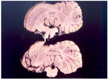
Cerebellum of a kuru patient

In 2009, researchers at the Medical Research Council discovered a naturally occurring variant of a prion protein in a population from Papua New Guinea that confers strong resistance to kuru. In the study, which began in 1996, researchers assessed over 3,000 people from the affected and surrounding Eastern Highland populations, and identified a variation in the prion protein G127. G127 polymorphism is the result of a missense mutation, and is highly geographically restricted to regions where the kuru epidemic was the most widespread. Researchers believe that the PrnP variant occurred very recently, estimating that the most recent common ancestor lived 10 generations ago.

The findings of the study could help researchers better understand and develop treatments for other related prion diseases, such as Creutzfeldt–Jakob disease and other neurodegenerate diseases like Alzheimer's disease.

History

Kuru was first described in official reports by Australian officers patrolling the Eastern Highlands of Papua New Guinea in the early 1950s. Some unofficial accounts place kuru in the region as early as 1910. In 1951, Arthur Carey was the first to use the term kuru in a report to describe a new disease afflicting the Fore tribes of Papua New Guinea (PNG). In his report, Carey noted that kuru mostly affected Fore women, eventually killing them. Kuru was noted in the Fore, Yate and Usurufa people in 1952–1953 by anthropologists Ronald Berndt and Catherine Berndt. In 1953, kuru was observed by patrol officer John McArthur, who provided a description of the disease in his report. McArthur believed that kuru was merely a psychosomatic episode resulting from the sorcery practices of the tribal people in the region. After the disease had progressed into a larger epidemic, the tribal people asked Charles Pfarr, a Lutheran medical officer, to come to the area to report the disease to Australian authorities.

Initially, the Fore people believed the causes of kuru to be sorcery or witchcraft. They also thought that the magic causing kuru was contagious. It was also called negi-nagi, which meant foolish person as the victims laughed at spontaneous intervals. This disease, the Fore people believed, was caused by ghosts, because of the shaking and strange behaviour that comes with kuru. Attempting to cure this, they would feed victims casuarina bark.

When kuru disease had become an epidemic, Daniel Carleton Gajdusek, a virologist, and Vincent Zigas, a medical doctor, started research on the disease. In 1957, Zigas and Gajdusek published a report in the Medical Journal of Australia that suggested that kuru had a genetic origin, and that "any ethnic-environmental variables that are operating in kuru pathogenesis have not yet been determined."

Cannibalism was suspected as a possible cause from the very beginning but was not formally put forth as a hypothesis until 1967 by Glasse and more formally in 1968 by Mathews, Glasse, and Lindenbaum.

Even before anthropophagy had been linked to kuru, cannibalism was banned by the Australian administration of Papua New Guinea, and the practice was nearly eliminated by 1960. While the number of cases of kuru was decreasing, medical researchers were finally able to properly investigate kuru, which eventually led to the modern understanding of prions as its cause.

In an effort to understand the pathology of kuru disease, Gajdusek established the first experimental tests on chimpanzees for kuru at the National Institutes of Health (NIH). Michael Alpers, an Australian doctor, collaborated with Gajdusek by providing samples of brain tissues he had taken from an 11-year-old Fore girl who had died of kuru. In his work, Gajdusek was also the first to compile a bibliography of kuru disease. Joe Gibbs joined Gajdusek to monitor and record the behavior of the apes at the NIH and conduct their autopsies. Within two years, one of the chimps, Daisy, had developed kuru, demonstrating that an unknown disease factor was transmitted through infected biomaterial and that it was capable of crossing the species barrier to other primates. After Elisabeth Beck confirmed that this experiment had brought about the first experimental transmission of kuru, the finding was deemed a very important advance in human medicine, leading to the award of the Nobel Prize in Physiology or Medicine to Gajdusek in 1976.

Subsequently, E. J. Field spent large parts of the late 1960s and early 1970s in New Guinea investigating the disease, connecting it to scrapie and multiple sclerosis. He noted the disease's interactions with glial cells, including the critical observation that the infectious process may depend on the structural rearrangement of the host's molecules. This was an early observation of what was to later become the prion hypothesis.

In popular culture

  • The Czech immunologist-poet Miroslav Holub wrote "Kuru, or the Smiling Death Syndrome" about the disease.
  • The X-Files season 7 episode "Theef" features a character diagnosed with advanced kuru after his sudden death.
  • The video game Dead Island, as well as Dead Island: Riptide, cite kuru as the disease that has swept the fictional islands of Banoi and Palanai.
  • In the film We Are What We Are, the medical examiner is able to identify the family as cannibals after he realises that the family is suffering from kuru.
  • In the post-apocalyptic book and film The Road by Cormac McCarthy, two characters practicing cannibalism demonstrate symptoms of kuru.
  • Tuesday, March 26, 2024

    Glia

    From Wikipedia, the free encyclopedia
     
    Glia
    Illustration of the four different types of glial cells found in the central nervous system: ependymal cells (light pink), astrocytes (green), microglial cells (dark red) and oligodendrocytes (light blue)
    Details
    PrecursorNeuroectoderm for macroglia, and hematopoietic stem cells for microglia
    SystemNervous system
    Identifiers
    MeSHD009457

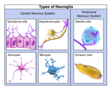
    Glia, also called glial cells (gliocytes) or neuroglia, are non-neuronal cells in the central nervous system (brain and spinal cord) and the peripheral nervous system that do not produce electrical impulses. The neuroglia make up more than one half the volume of neural tissue in our body. They maintain homeostasis, form myelin in the peripheral nervous system, and provide support and protection for neurons. In the central nervous system, glial cells include oligodendrocytes, astrocytes, ependymal cells and microglia, and in the peripheral nervous system they include Schwann cells and satellite cells.

    Function

    They have four main functions:

    • to surround neurons and hold them in place
    • to supply nutrients and oxygen to neurons
    • to insulate one neuron from another
    • to destroy pathogens and remove dead neurons.

    They also play a role in neurotransmission and synaptic connections, and in physiological processes such as breathing. While glia were thought to outnumber neurons by a ratio of 10:1, recent studies using newer methods and reappraisal of historical quantitative evidence suggests an overall ratio of less than 1:1, with substantial variation between different brain tissues.

    Glial cells have far more cellular diversity and functions than neurons, and glial cells can respond to and manipulate neurotransmission in many ways. Additionally, they can affect both the preservation and consolidation of memories. Glia were discovered in 1856, by the pathologist Rudolf Virchow in his search for a "connective tissue" in the brain. The term derives from Greek γλία and γλοία "glue" (English: /ˈɡlə/ or /ˈɡlə/), and suggests the original impression that they were the glue of the nervous system.

    Types

    Neuroglia of the brain shown by Golgi's method
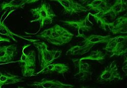
    Astrocytes can be identified in culture because, unlike other mature glia, they express glial fibrillary acidic protein (GFAP)
    Glial cells in a rat brain stained with an antibody against GFAP
    Different types of neuroglia

    Macroglia

    Derived from ectodermal tissue.

    Location Name Description
    CNS Astrocytes

    The most abundant type of macroglial cell in the CNS, astrocytes (also called astroglia) have numerous projections that link neurons to their blood supply while forming the blood–brain barrier. They regulate the external chemical environment of neurons by removing excess potassium ions, and recycling neurotransmitters released during synaptic transmission. Astrocytes may regulate vasoconstriction and vasodilation by producing substances such as arachidonic acid, whose metabolites are vasoactive.

    Astrocytes signal each other using ATP. The gap junctions (also known as electrical synapses) between astrocytes allow the messenger molecule IP3 to diffuse from one astrocyte to another. IP3 activates calcium channels on cellular organelles, releasing calcium into the cytoplasm. This calcium may stimulate the production of more IP3 and cause release of ATP through channels in the membrane made of pannexins. The net effect is a calcium wave that propagates from cell to cell. Extracellular release of ATP, and consequent activation of purinergic receptors on other astrocytes, may also mediate calcium waves in some cases.

    In general, there are two types of astrocytes, protoplasmic and fibrous, similar in function but distinct in morphology and distribution. Protoplasmic astrocytes have short, thick, highly branched processes and are typically found in gray matter. Fibrous astrocytes have long, thin, less-branched processes and are more commonly found in white matter.

    It has recently been shown that astrocyte activity is linked to blood flow in the brain, and that this is what is actually being measured in fMRI. They also have been involved in neuronal circuits playing an inhibitory role after sensing changes in extracellular calcium.

    CNS Oligodendrocytes

    Oligodendrocytes are cells that coat axons in the CNS with their cell membrane, forming a specialized membrane differentiation called myelin, producing the myelin sheath. The myelin sheath provides insulation to the axon that allows electrical signals to propagate more efficiently.

    CNS Ependymal cells

    Ependymal cells, also named ependymocytes, line the spinal cord and the ventricular system of the brain. These cells are involved in the creation and secretion of cerebrospinal fluid (CSF) and beat their cilia to help circulate the CSF and make up the blood-CSF barrier. They are also thought to act as neural stem cells.

    CNS Radial glia

    Radial glia cells arise from neuroepithelial cells after the onset of neurogenesis. Their differentiation abilities are more restricted than those of neuroepithelial cells. In the developing nervous system, radial glia function both as neuronal progenitors and as a scaffold upon which newborn neurons migrate. In the mature brain, the cerebellum and retina retain characteristic radial glial cells. In the cerebellum, these are Bergmann glia, which regulate synaptic plasticity. In the retina, the radial Müller cell is the glial cell that spans the thickness of the retina and, in addition to astroglial cells, participates in a bidirectional communication with neurons.

    PNS Schwann cells

    Similar in function to oligodendrocytes, Schwann cells provide myelination to axons in the peripheral nervous system (PNS). They also have phagocytotic activity and clear cellular debris that allows for regrowth of PNS neurons.

    PNS Satellite cells

    Satellite glial cells are small cells that surround neurons in sensory, sympathetic, and parasympathetic ganglia. These cells help regulate the external chemical environment. Like astrocytes, they are interconnected by gap junctions and respond to ATP by elevating the intracellular concentration of calcium ions. They are highly sensitive to injury and inflammation and appear to contribute to pathological states, such as chronic pain.

    PNS Enteric glial cells

    Are found in the intrinsic ganglia of the digestive system. Glia cells are thought to have many roles in the enteric system, some related to homeostasis and muscular digestive processes.

    Microglia

    Microglia are specialized macrophages capable of phagocytosis that protect neurons of the central nervous system. They are derived from the earliest wave of mononuclear cells that originate in yolk sac blood islands early in development, and colonize the brain shortly after the neural precursors begin to differentiate.

    These cells are found in all regions of the brain and spinal cord. Microglial cells are small relative to macroglial cells, with changing shapes and oblong nuclei. They are mobile within the brain and multiply when the brain is damaged. In the healthy central nervous system, microglia processes constantly sample all aspects of their environment (neurons, macroglia and blood vessels). In a healthy brain, microglia direct the immune response to brain damage and play an important role in the inflammation that accompanies the damage. Many diseases and disorders are associated with deficient microglia, such as Alzheimer's disease, Parkinson's disease and ALS.

    Other

    Pituicytes from the posterior pituitary are glial cells with characteristics in common to astrocytes. Tanycytes in the median eminence of the hypothalamus are a type of ependymal cell that descend from radial glia and line the base of the third ventricle. Drosophila melanogaster, the fruit fly, contains numerous glial types that are functionally similar to mammalian glia but are nonetheless classified differently.

    Total number

    In general, neuroglial cells are smaller than neurons. There are approximately 85 billion glia cells in the human brain, about the same number as neurons. Glial cells make up about half the total volume of the brain and spinal cord. The glia to neuron-ratio varies from one part of the brain to another. The glia to neuron-ratio in the cerebral cortex is 3.72 (60.84 billion glia (72%); 16.34 billion neurons), while that of the cerebellum is only 0.23 (16.04 billion glia; 69.03 billion neurons). The ratio in the cerebral cortex gray matter is 1.48, with 3.76 for the gray and white matter combined. The ratio of the basal ganglia, diencephalon and brainstem combined is 11.35.

    The total number of glia cells in the human brain is distributed into the different types with oligodendrocytes being the most frequent (45–75%), followed by astrocytes (19–40%) and microglia (about 10% or less).

    Development

    23-week fetal brain culture astrocyte

    Most glia are derived from ectodermal tissue of the developing embryo, in particular the neural tube and crest. The exception is microglia, which are derived from hematopoietic stem cells. In the adult, microglia are largely a self-renewing population and are distinct from macrophages and monocytes, which infiltrate an injured and diseased CNS.

    In the central nervous system, glia develop from the ventricular zone of the neural tube. These glia include the oligodendrocytes, ependymal cells, and astrocytes. In the peripheral nervous system, glia derive from the neural crest. These PNS glia include Schwann cells in nerves and satellite glial cells in ganglia.

    Capacity to divide

    Glia retain the ability to undergo cell divisions in adulthood, whereas most neurons cannot. The view is based on the general inability of the mature nervous system to replace neurons after an injury, such as a stroke or trauma, where very often there is a substantial proliferation of glia, or gliosis, near or at the site of damage. However, detailed studies have found no evidence that 'mature' glia, such as astrocytes or oligodendrocytes, retain mitotic capacity. Only the resident oligodendrocyte precursor cells seem to keep this ability once the nervous system matures.

    Glial cells are known to be capable of mitosis. By contrast, scientific understanding of whether neurons are permanently post-mitotic, or capable of mitosis, is still developing. In the past, glia had been considered to lack certain features of neurons. For example, glial cells were not believed to have chemical synapses or to release transmitters. They were considered to be the passive bystanders of neural transmission. However, recent studies have shown this to not be entirely true.

    Functions

    Some glial cells function primarily as the physical support for neurons. Others provide nutrients to neurons and regulate the extracellular fluid of the brain, especially surrounding neurons and their synapses. During early embryogenesis, glial cells direct the migration of neurons and produce molecules that modify the growth of axons and dendrites. Some glial cells display regional diversity in the CNS and their functions may vary between the CNS regions.

    Neuron repair and development

    Glia are crucial in the development of the nervous system and in processes such as synaptic plasticity and synaptogenesis. Glia have a role in the regulation of repair of neurons after injury. In the central nervous system (CNS), glia suppress repair. Glial cells known as astrocytes enlarge and proliferate to form a scar and produce inhibitory molecules that inhibit regrowth of a damaged or severed axon. In the peripheral nervous system (PNS), glial cells known as Schwann cells (or also as neuri-lemmocytes) promote repair. After axonal injury, Schwann cells regress to an earlier developmental state to encourage regrowth of the axon. This difference between the CNS and the PNS, raises hopes for the regeneration of nervous tissue in the CNS. For example, a spinal cord may be able to be repaired following injury or severance.

    Myelin sheath creation

    Oligodendrocytes are found in the CNS and resemble an octopus: they have bulbous cell bodies with up to fifteen arm-like processes. Each process reaches out to an axon and spirals around it, creating a myelin sheath. The myelin sheath insulates the nerve fiber from the extracellular fluid and speeds up signal conduction along the nerve fiber. In the peripheral nervous system, Schwann cells are responsible for myelin production. These cells envelop nerve fibers of the PNS by winding repeatedly around them. This process creates a myelin sheath, which not only aids in conductivity but also assists in the regeneration of damaged fibers.

    Neurotransmission

    Astrocytes are crucial participants in the tripartite synapse. They have several crucial functions, including clearance of neurotransmitters from within the synaptic cleft, which aids in distinguishing between separate action potentials and prevents toxic build-up of certain neurotransmitters such as glutamate, which would otherwise lead to excitotoxicity. Furthermore, astrocytes release gliotransmitters such as glutamate, ATP, and D-serine in response to stimulation.

    Clinical significance

    Neoplastic glial cells stained with an antibody against GFAP (brown), from a brain biopsy

    While glial cells in the PNS frequently assist in regeneration of lost neural functioning, loss of neurons in the CNS does not result in a similar reaction from neuroglia. In the CNS, regrowth will only happen if the trauma was mild, and not severe. When severe trauma presents itself, the survival of the remaining neurons becomes the optimal solution. However, some studies investigating the role of glial cells in Alzheimer's disease are beginning to contradict the usefulness of this feature, and even claim it can "exacerbate" the disease. In addition to affecting the potential repair of neurons in Alzheimer's disease, scarring and inflammation from glial cells have been further implicated in the degeneration of neurons caused by amyotrophic lateral sclerosis.

    In addition to neurodegenerative diseases, a wide range of harmful exposure, such as hypoxia, or physical trauma, can lead to the end result of physical damage to the CNS. Generally, when damage occurs to the CNS, glial cells cause apoptosis among the surrounding cellular bodies. Then, there is a large amount of microglial activity, which results in inflammation, and, finally, there is a heavy release of growth inhibiting molecules.

    History

    Although glial cells and neurons were probably first observed at the same time in the early 19th century, unlike neurons whose morphological and physiological properties were directly observable for the first investigators of the nervous system, glial cells had been considered to be merely "glue" that held neurons together until the mid-20th century.

    Glia were first described in 1856 by the pathologist Rudolf Virchow in a comment to his 1846 publication on connective tissue. A more detailed description of glial cells was provided in the 1858 book 'Cellular Pathology' by the same author.

    When markers for different types of cells were analyzed, Albert Einstein's brain was discovered to contain significantly more glia than normal brains in the left angular gyrus, an area thought to be responsible for mathematical processing and language. However, out of the total of 28 statistical comparisons between Einstein's brain and the control brains, finding one statistically significant result is not surprising, and the claim that Einstein's brain is different is not scientific (c.f. Multiple comparisons problem).

    Not only does the ratio of glia to neurons increase through evolution, but so does the size of the glia. Astroglial cells in human brains have a volume 27 times greater than in mouse brains.

    These important scientific findings may begin to shift the neurocentric perspective into a more holistic view of the brain which encompasses the glial cells as well. For the majority of the twentieth century, scientists had disregarded glial cells as mere physical scaffolds for neurons. Recent publications have proposed that the number of glial cells in the brain is correlated with the intelligence of a species. Moreover, evidences are demonstrating the active role of glia, in particular astroglia, in cognitive processes like learning and memory and, for these reasons, it has been proposed the foundation of a specific field to study these functions because investigations in this area are still limited due to the dominance of the neurocentric perspective.

    Operator (computer programming)

    From Wikipedia, the free encyclopedia https://en.wikipedia.org/wiki/Operator_(computer_programmin...