Search This Blog

Friday, June 14, 2024

Psychoneuroimmunology

From Wikipedia, the free encyclopedia
https://en.wikipedia.org/wiki/Psychoneuroimmunology

Psychoneuroimmunology (PNI), also referred to as psychoendoneuroimmunology (PENI) or psychoneuroendocrinoimmunology (PNEI), is the study of the interaction between psychological processes and the nervous and immune systems of the human body. It is a subfield of psychosomatic medicine. PNI takes an interdisciplinary approach, incorporating psychology, neuroscience, immunology, physiology, genetics, pharmacology, molecular biology, psychiatry, behavioral medicine, infectious diseases, endocrinology, and rheumatology.

The main interests of PNI are the interactions between the nervous and immune systems and the relationships between mental processes and health. PNI studies, among other things, the physiological functioning of the neuroimmune system in health and disease; disorders of the neuroimmune system (autoimmune diseases; hypersensitivities; immune deficiency); and the physical, chemical and physiological characteristics of the components of the neuroimmune system in vitro, in situ, and in vivo.

History

Interest in the relationship between psychiatric syndromes or symptoms and immune function has been a consistent theme since the beginning of modern medicine.

Claude Bernard, the father of modern physiology, with his pupils

Claude Bernard, a French physiologist of the Muséum national d'Histoire naturelle (National Museum of Natural History in English), formulated the concept of the milieu interieur in the mid-1800s. In 1865, Bernard described the perturbation of this internal state: "... there are protective functions of organic elements holding living materials in reserve and maintaining without interruption humidity, heat and other conditions indispensable to vital activity. Sickness and death are only a dislocation or perturbation of that mechanism" (Bernard, 1865). Walter Cannon, a professor of physiology at Harvard University coined the commonly used term, homeostasis, in his book The Wisdom of the Body, 1932, from the Greek word homoios, meaning similar, and stasis, meaning position. In his work with animals, Cannon observed that any change of emotional state in the beast, such as anxiety, distress, or rage, was accompanied by total cessation of movements of the stomach (Bodily Changes in Pain, Hunger, Fear and Rage, 1915). These studies looked into the relationship between the effects of emotions and perceptions on the autonomic nervous system, namely the sympathetic and parasympathetic responses that initiated the recognition of the freeze, fight or flight response. His findings were published from time to time in professional journals, then summed up in book form in The Mechanical Factors of Digestion, published in 1911.
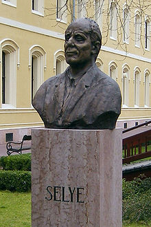
Bust of Hans Selye at Selye János University, Komárno, Slovakia

Hans Selye, a student of Johns Hopkins University and McGill University, and a researcher at Université de Montréal, experimented with animals by putting them under different physical and mental adverse conditions and noted that under these difficult conditions the body consistently adapted to heal and recover. Several years of experimentation that formed the empiric foundation of Selye's concept of the General Adaptation Syndrome. This syndrome consists of an enlargement of the adrenal gland, atrophy of the thymus, spleen, and other lymphoid tissue, and gastric ulcerations.

Selye describes three stages of adaptation, including an initial brief alarm reaction, followed by a prolonged period of resistance, and a terminal stage of exhaustion and death. This foundational work led to a rich line of research on the biological functioning of glucocorticoids.

Mid-20th century studies of psychiatric patients reported immune alterations in psychotic individuals, including lower numbers of lymphocytes and poorer antibody response to pertussis vaccination, compared with nonpsychiatric control subjects. In 1964, George F. Solomon, from the University of California in Los Angeles, and his research team coined the term "psychoimmunology" and published a landmark paper: "Emotions, immunity, and disease: a speculative theoretical integration."

Origins

In 1975, Robert Ader and Nicholas Cohen, at the University of Rochester, advanced PNI with their demonstration of classic conditioning of immune function, and they subsequently coined the term "psychoneuroimmunology". Ader was investigating how long conditioned responses (in the sense of Pavlov's conditioning of dogs to drool when they heard a bell ring) might last in laboratory rats. To condition the rats, he used a combination of saccharin-laced water (the conditioned stimulus) and the drug Cytoxan, which unconditionally induces nausea and taste aversion and suppression of immune function. Ader was surprised to discover that after conditioning, just feeding the rats saccharin-laced water was associated with the death of some animals and he proposed that they had been immunosuppressed after receiving the conditioned stimulus. Ader (a psychologist) and Cohen (an immunologist) directly tested this hypothesis by deliberately immunizing conditioned and unconditioned animals, exposing these and other control groups to the conditioned taste stimulus, and then measuring the amount of antibody produced. The highly reproducible results revealed that conditioned rats exposed to the conditioned stimulus were indeed immunosuppressed. In other words, a signal via the nervous system (taste) was affecting immune function. This was one of the first scientific experiments that demonstrated that the nervous system can affect the immune system.

In the 1970s, Hugo Besedovsky, Adriana del Rey and Ernst Sorkin, working in Switzerland, reported multi-directional immune-neuro-endocrine interactions, since they show that not only the brain can influence immune processes but also the immune response itself can affect the brain and neuroendocrine mechanisms. They found that the immune responses to innocuous antigens triggers an increase in the activity of hypothalamic neurons and hormonal and autonomic nerve responses that are relevant for immunoregulation and are integrated at brain levels (see review). On these bases, they proposed that the immune system acts as a sensorial receptor organ that, besides its peripheral effects, can communicate to the brain and associated neuro-endocrine structures its state of activity. These investigators also identified products from immune cells, later characterized as cytokines, that mediate this immune-brain communication.

In 1981, David L. Felten, then working at the Indiana University School of Medicine, and his colleague JM Williams, discovered a network of nerves leading to blood vessels as well as cells of the immune system. The researchers also found nerves in the thymus and spleen terminating near clusters of lymphocytes, macrophages, and mast cells, all of which help control immune function. This discovery provided one of the first indications of how neuro-immune interaction occurs.

Ader, Cohen, and Felten went on to edit the groundbreaking book Psychoneuroimmunology in 1981, which laid out the underlying premise that the brain and immune system represent a single, integrated system of defense.

In 1985, research by neuropharmacologist Candace Pert, of the National Institutes of Health at Georgetown University, revealed that neuropeptide-specific receptors are present on the cell walls of both the brain and the immune system. The discovery that neuropeptides and neurotransmitters act directly upon the immune system shows their close association with emotions and suggests mechanisms through which emotions, from the limbic system, and immunology are deeply interdependent. Showing that the immune and endocrine systems are modulated not only by the brain but also by the central nervous system itself affected the understanding of emotions, as well as disease.

Contemporary advances in psychiatry, immunology, neurology, and other integrated disciplines of medicine has fostered enormous growth for PNI. The mechanisms underlying behaviorally induced alterations of immune function, and immune alterations inducing behavioral changes, are likely to have clinical and therapeutic implications that will not be fully appreciated until more is known about the extent of these interrelationships in normal and pathophysiological states.

The immune-brain loop

PNI research looks for the exact mechanisms by which specific neuroimmune effects are achieved. Evidence for nervous-immunological interactions exist at multiple biological levels.

The immune system and the brain communicate through signaling pathways. The brain and the immune system are the two major adaptive systems of the body. Two major pathways are involved in this cross-talk: the Hypothalamic-pituitary-adrenal axis (HPA axis), and the sympathetic nervous system (SNS), via the sympathetic-adrenal-medullary axis (SAM axis). The activation of SNS during an immune response might be aimed to localize the inflammatory response.

The body's primary stress management system is the HPA axis. The HPA axis responds to physical and mental challenge to maintain homeostasis in part by controlling the body's cortisol level. Dysregulation of the HPA axis is implicated in numerous stress-related diseases, with evidence from meta-analyses indicating that different types/duration of stressors and unique personal variables can shape the HPA response. HPA axis activity and cytokines are intrinsically intertwined: inflammatory cytokines stimulate adrenocorticotropic hormone (ACTH) and cortisol secretion, while, in turn, glucocorticoids suppress the synthesis of proinflammatory cytokines.

Molecules called pro-inflammatory cytokines, which include interleukin-1 (IL-1), Interleukin-2 (IL-2), interleukin-6 (IL-6), Interleukin-12 (IL-12), Interferon-gamma (IFN-Gamma) and tumor necrosis factor alpha (TNF-alpha) can affect brain growth as well as neuronal function. Circulating immune cells such as macrophages, as well as glial cells (microglia and astrocytes) secrete these molecules. Cytokine regulation of hypothalamic function is an active area of research for the treatment of anxiety-related disorders.

Cytokines mediate and control immune and inflammatory responses. Complex interactions exist between cytokines, inflammation and the adaptive responses in maintaining homeostasis. Like the stress response, the inflammatory reaction is crucial for survival. Systemic inflammatory reaction results in stimulation of four major programs:

These are mediated by the HPA axis and the SNS. Common human diseases such as allergy, autoimmunity, chronic infections and sepsis are characterized by a dysregulation of the pro-inflammatory versus anti-inflammatory and T helper (Th1) versus (Th2) cytokine balance. Recent studies show pro-inflammatory cytokine processes take place during depression, mania and bipolar disease, in addition to autoimmune hypersensitivity and chronic infections.

Chronic secretion of stress hormones, glucocorticoids (GCs) and catecholamines (CAs), as a result of disease, may reduce the effect of neurotransmitters, including serotonin, norepinephrine and dopamine, or other receptors in the brain, thereby leading to the dysregulation of neurohormones. Under stimulation, norepinephrine is released from the sympathetic nerve terminals in organs, and the target immune cells express adrenoreceptors. Through stimulation of these receptors, locally released norepinephrine, or circulating catecholamines such as epinephrine, affect lymphocyte traffic, circulation, and proliferation, and modulate cytokine production and the functional activity of different lymphoid cells.

Glucocorticoids also inhibit the further secretion of corticotropin-releasing hormone from the hypothalamus and ACTH from the pituitary (negative feedback). Under certain conditions stress hormones may facilitate inflammation through induction of signaling pathways and through activation of the corticotropin-releasing hormone.

These abnormalities and the failure of the adaptive systems to resolve inflammation affect the well-being of the individual, including behavioral parameters, quality of life and sleep, as well as indices of metabolic and cardiovascular health, developing into a "systemic anti-inflammatory feedback" and/or "hyperactivity" of the local pro-inflammatory factors which may contribute to the pathogenesis of disease.

This systemic or neuro-inflammation and neuroimmune activation have been shown to play a role in the etiology of a variety of neurodegenerative disorders such as Parkinson's and Alzheimer's disease, multiple sclerosis, pain, and AIDS-associated dementia. However, cytokines and chemokines also modulate central nervous system (CNS) function in the absence of overt immunological, physiological, or psychological challenges.

Psychoneuroimmunological effects

There are now sufficient data to conclude that immune modulation by psychosocial stressors and/or interventions can lead to actual health changes. Although changes related to infectious disease and wound healing have provided the strongest evidence to date, the clinical importance of immunological dysregulation is highlighted by increased risks across diverse conditions and diseases. For example, stressors can produce profound health consequences. In one epidemiological study, all-cause mortality increased in the month following a severe stressor – the death of a spouse. Theorists propose that stressful events trigger cognitive and affective responses which, in turn, induce sympathetic nervous system and endocrine changes, and these ultimately impair immune function. Potential health consequences are broad, but include rates of infection HIV progression cancer incidence and progression, and high rates of infant mortality.

Understanding stress and immune function

Stress is thought to affect immune function through emotional and/or behavioral manifestations such as anxiety, fear, tension, anger and sadness and physiological changes such as heart rate, blood pressure, and sweating. Researchers have suggested that these changes are beneficial if they are of limited duration, but when stress is chronic, the system is unable to maintain equilibrium or homeostasis; the body remains in a state of arousal, where digestion is slower to reactivate or does not reactivate properly, often resulting in indigestion. Furthermore, blood pressure stays at higher levels.

In one of the earlier PNI studies, which was published in 1960, subjects were led to believe that they had accidentally caused serious injury to a companion through misuse of explosives. Since then decades of research resulted in two large meta-analyses, which showed consistent immune dysregulation in healthy people who are experiencing stress.

In the first meta-analysis by Herbert and Cohen in 1993, they examined 38 studies of stressful events and immune function in healthy adults. They included studies of acute laboratory stressors (e.g. a speech task), short-term naturalistic stressors (e.g. medical examinations), and long-term naturalistic stressors (e.g. divorce, bereavement, caregiving, unemployment). They found consistent stress-related increases in numbers of total white blood cells, as well as decreases in the numbers of helper T cells, suppressor T cells, and cytotoxic T cells, B cells, and natural killer cells (NK). They also reported stress-related decreases in NK and T cell function, and T cell proliferative responses to phytohaemagglutinin [PHA] and concanavalin A [Con A]. These effects were consistent for short-term and long-term naturalistic stressors, but not laboratory stressors.

In the second meta-analysis by Zorrilla et al. in 2001, they replicated Herbert and Cohen's meta-analysis. Using the same study selection procedures, they analyzed 75 studies of stressors and human immunity. Naturalistic stressors were associated with increases in number of circulating neutrophils, decreases in number and percentages of total T cells and helper T cells, and decreases in percentages of natural killer cell (NK) cells and cytotoxic T cell lymphocytes. They also replicated Herbert and Cohen's finding of stress-related decreases in NKCC and T cell mitogen proliferation to phytohaemagglutinin (PHA) and concanavalin A (Con A).

A study done by the American Psychological Association did an experiment on rats, where they applied electrical shocks to a rat, and saw how interleukin-1 was released directly into the brain. Interleukin-1 is the same cytokine released when a macrophage chews on a bacterium, which then travels up the vagus nerve, creating a state of heightened immune activity, and behavioral changes.

More recently, there has been increasing interest in the links between interpersonal stressors and immune function. For example, marital conflict, loneliness, caring for a person with a chronic medical condition, and other forms on interpersonal stress dysregulate immune function.

Communication between the brain and immune system

  • Stimulation of brain sites alters immunity (stressed animals have altered immune systems).
  • Damage to brain hemispheres alters immunity (hemispheric lateralization effects).
  • Immune cells produce cytokines that act on the CNS.
  • Immune cells respond to signals from the CNS.

Communication between neuroendocrine and immune system

  • Glucocorticoids and catecholamines influence immune cells.
  • Hypothalamic Pituitary Adrenal axis releases the needed hormones to support the immune system.
  • Activity of the immune system is correlated with neurochemical/neuroendocrine activity of brain cells.

Connections between glucocorticoids and immune system

  • Anti-inflammatory hormones that enhance the organism's response to a stressor.
  • Prevent the overreaction of the body's own defense system.
  • Overactivation of glucocorticoid receptors can lead to health risks.
  • Regulators of the immune system.
  • Affect cell growth, proliferation and differentiation.
  • Cause immunosuppression which can lead to an extended amount of time fighting off infections.
  • High basal levels of cortisol are associated with a higher risk of infection.
  • Suppress cell adhesion, antigen presentation, chemotaxis and cytotoxicity.
  • Increase apoptosis.

Corticotropin-releasing hormone (CRH)

Release of corticotropin-releasing hormone (CRH) from the hypothalamus is influenced by stress.

Furthermore, stressors that enhance the release of CRH suppress the function of the immune system; conversely, stressors that depress CRH release potentiate immunity.

  • Central mediated since peripheral administration of CRH antagonist does not affect immunosuppression.
  • HPA axis/stress axis responds consistently to stressors that are new, unpredictable and that have low-perceived control.
  • As cortisol reaches an appropriate level in response to the stressor, it deregulates the activity of the hippocampus, hypothalamus, and pituitary gland which results in less production of cortisol.

Relationships between prefrontal cortex activation and cellular senescence

  • Psychological stress is regulated by the prefrontal cortex (PFC)
  • The PFC modulates vagal activity
  • Prefrontally modulated and vagally mediated cholinergic input to the spleen reduces inflammatory responses

Pharmaceutical advances

Glutamate agonists, cytokine inhibitors, vanilloid-receptor agonists, catecholamine modulators, ion-channel blockers, anticonvulsants, GABA agonists (including opioids and cannabinoids), COX inhibitors, acetylcholine modulators, melatonin analogs (such as Ramelton), adenosine receptor antagonists and several miscellaneous drugs (including biologics like Passiflora edulis) are being studied for their psychoneuroimmunological effects.

For example, SSRIs, SNRIs and tricyclic antidepressants acting on serotonin, norepinephrine, dopamine and cannabinoid receptors have been shown to be immunomodulatory and anti-inflammatory against pro-inflammatory cytokine processes, specifically on the regulation of IFN-gamma and IL-10, as well as TNF-alpha and IL-6 through a psychoneuroimmunological process. Antidepressants have also been shown to suppress TH1 upregulation.

Tricyclic and dual serotonergic-noradrenergic reuptake inhibition by SNRIs (or SSRI-NRI combinations), have also shown analgesic properties additionally. According to recent evidences antidepressants also seem to exert beneficial effects in experimental autoimmune neuritis in rats by decreasing Interferon-beta (IFN-beta) release or augmenting NK activity in depressed patients.

These studies warrant investigation of antidepressants for use in both psychiatric and non-psychiatric illness and that a psychoneuroimmunological approach may be required for optimal pharmacotherapy in many diseases. Future antidepressants may be made to specifically target the immune system by either blocking the actions of pro-inflammatory cytokines or increasing the production of anti-inflammatory cytokines.

The endocannabinoid system appears to play a significant role in the mechanism of action of clinically effective and potential antidepressants and may serve as a target for drug design and discovery. The endocannabinoid-induced modulation of stress-related behaviors appears to be mediated, at least in part, through the regulation of the serotoninergic system, by which cannabinoid CB1 receptors modulate the excitability of dorsal raphe serotonin neurons. Data suggest that the endocannabinoid system in cortical and subcortical structures is differentially altered in an animal model of depression and that the effects of chronic, unpredictable stress (CUS) on CB1 receptor binding site density are attenuated by antidepressant treatment while those on endocannabinoid content are not.

The increase in amygdalar CB1 receptor binding following imipramine treatment is consistent with prior studies which collectively demonstrate that several treatments which are beneficial to depression, such as electroconvulsive shock and tricyclic antidepressant treatment, increase CB1 receptor activity in subcortical limbic structures, such as the hippocampus, amygdala and hypothalamus. And preclinical studies have demonstrated the CB1 receptor is required for the behavioral effects of noradrenergic based antidepressants but is dispensable for the behavioral effect of serotonergic based antidepressants.

Extrapolating from the observations that positive emotional experiences boost the immune system, Roberts speculates that intensely positive emotional experiences—sometimes brought about during mystical experiences occasioned by psychedelic medicines—may boost the immune system powerfully. Research on salivary IgA supports this hypothesis, but experimental testing has not been done.

Environmental conflict

From Wikipedia, the free encyclopedia
https://en.wikipedia.org/wiki/Environmental_conflict

Hambach Forest protest against coal mine expansion

Environmental conflicts, socio-environmental conflict or ecological distribution conflicts (EDCs) are social conflicts caused by environmental degradation or by unequal distribution of environmental resources. The Environmental Justice Atlas documented 3,100 environmental conflicts worldwide as of April 2020 and emphasised that many more conflicts remained undocumented.

Parties involved in these conflicts include locally affected communities, states, companies and investors, and social or environmental movements; typically environmental defenders are protecting their homelands from resource extraction or hazardous waste disposal. Resource extraction and hazardous waste activities often create resource scarcities (such as by overfishing or deforestation), pollute the environment, and degrade the living space for humans and nature, resulting in conflict. A particular case of environmental conflicts are forestry conflicts, or forest conflicts which "are broadly viewed as struggles of varying intensity between interest groups, over values and issues related to forest policy and the use of forest resources". In the last decades, a growing number of these have been identified globally.

Frequently environmental conflicts focus on environmental justice issues, the rights of indigenous people, the rights of peasants, or threats to communities whose livelihoods are dependent on the ocean. Outcomes of local conflicts are increasingly influenced by trans-national environmental justice networks that comprise the global environmental justice movement.

Environmental conflict can complicate response to natural disaster or exacerbate existing conflicts – especially in the context of geopolitical disputes or where communities have been displaced to create environmental migrants. The study of these conflicts is related to the fields of ecological economics, political ecology, and environmental justice.

Causes

The origin of environmental conflicts can be directly linked to the industrial economy. As less than 10% of materials and energy are recycled, the industrial economy is constantly expanding energy and material extraction at commodity frontiers through two main processes:

  1. Appropriating new natural resources through territorial claims and land grabs.
  2. Making exploitation of existing sites more efficient through investments or social and technical innovation.

EDCs are caused by the unfair distribution of environmental costs and benefits. These conflicts arise from social inequality, contested claims over territory, the proliferation of extractive industries, and the impacts of the economic industrialization over the past centuries. Oil, mining, and agriculture industries are focal points of environmental conflicts.

Types of conflicts

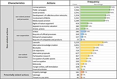
Environmental defenders use a wide range of tactics
Most environmental conflicts are in the mining, energy, and waste disposal sectors.

A 2020 paper mapped the arguments and concerns of environmental defenders in over 2743 conflicts found in the Environmental Justice Atlas (EJAtlas). The analysis found that the industrial sectors most frequently challenged by environmental conflicts were mining (21%), fossil energy (17%), biomass and land uses (15%), and water management (14%). Killings of environmental defenders happened in 13% of the reported cases.

There was also a distinct difference in the types of conflict found in high and low income countries. There were more conflicts around conservation, water management, and biomass and land use in low income countries; while in high income countries almost half of conflicts focused on waste management, tourism, nuclear power, industrial zones, and other infrastructure projects. The study also found that most conflicts start with self-organized local groups defending against infringement, with a focus on non-violent tactics.

Water protectors and land defenders who defend indigenous rights are criminalized at a much higher rate than in other conflicts.

Environmental conflicts can be classified based on the different stages of the commodity chain: during the extraction of energy sources or materials, in the transportation and production of goods, or at the final disposal of waste.

EJAtlas Categories

The EJAtlas was founded and is co-directed by Leah Temper and Joan Martinez-Alier, and it is coordinated by Daniela Del Bene. Its aim is “to document, understand and analyse the political outcomes that emerge or that may emerge” from ecological distribution conflicts. It is housed at the ICTA of the Universitat Autonoma de Barcelona. Since 2012, academics and activists have collaborated to write the entries, reaching 3,500 by July 2021.

The EJ Atlas identifies ten categories of ecological distribution conflicts:

  1. Biodiversity conservation conflicts:
  2. Biomass and land conflicts (Forests, Agriculture, Fisheries and Livestock Management)
  3. Fossil Fuels and Climate Justice/Energy
  4. Industrial and Utilities Conflicts
  5. Infrastructure and Built Environment
  6. Mineral Ores and Building Materials Extraction
  7. Nuclear
  8. Tourism Recreation
  9. Waste Management
  10. Water Management

Ecological distribution conflicts

Ecological Distribution Conflicts (EDCs) were introduced as a concept in 1995 by Joan Martínez-Alier and Martin O'Connor to facilitate more systematic documentation and analysis of environmental conflicts and to produce a more coherent body of academic, activist, and legal work around them. EDCs arise from the unfair access to natural resources, unequally distributed burdens of environmental pollution, and relate to the exercise of power by different social actors when they enter into disputes over access to or impacts on natural resources. For example, a factory may pollute a river thus affecting the community whose livelihood depends on the water of the river. The same can apply to the climate crisis, which may cause sea level rise on some Pacific islands. This type of damage is often not valued by the market, preventing those affected from being compensated.

Ecological conflicts occur at both global and local scales. Often conflicts take place between the global South and the global North, e.g. a Finnish forest company operating in Indonesia, or in econonomic peripheries, although there is a growing emergence of conflicts in Europe, including violent ones. There are also local conflicts that occur within a short commodity chain (e.g. local extraction of sand and gravel for a nearby cement factory).

Intellectual history

Since its conception, the term Ecological Distribution Conflict has been linked to research from the fields of political ecology, ecological economics, and ecofeminism. It has also been adopted into a non-academic setting through the environmental justice movement, where it branches academia and activism to assist social movements in legal struggles.

In his 1874 lecture ‘Wage Labour and Capital’, Karl Marx introduced the idea that economic relations under capitalism are inherently exploitative, meaning economic inequality is an inevitability of the system. He theorised that this is because capitalism expands through capital accumulation, an ever-increasing process which requires the economic subjugation of parts of the population in order to function.

Building on this theory, academics in the field of political economy created the term ‘economic distribution conflicts’ to describe the conflicts that occur from this inherent economic inequality. This type of conflict typically occurs between parties with an economic relationship but unequal power dynamic, such as buyers and sellers, or debtors and creditors.

However, Martinez Allier and Martin O’Connor noticed that this term focuses solely on the economy, omitting the conflicts that do not occur from economic inequality but from the unequal distribution of environmental resources. In response, in 1995, they coined the term ‘ecological distribution conflict’. This type of conflict occurs at commodity frontiers, which are constantly being moved and reframed due to society's unsustainable social metabolism. These conflicts might occur between extractive industries and Indigenous populations, or between polluting actors and those living on marginalised land. Its roots can still be seen in Marxian theory, as it is based on the idea that capitalism's need for expansion drives inequality and conflict.

Unfair ecological distribution can be attributed to capitalism as a system of cost-shifting. Neoclassical economics usually consider these impacts as “market failures” or “externalities” that can be valued in monetary terms and internalized into the price system. Ecological economics and political ecology scholars oppose the idea of economic commensuration that could form the basis of eco-compensation mechanisms for impacted communities. Instead, they advocate for different valuation languages such as sacredness, livelihood, rights of nature, Indigenous territorial rights, archaeological values, and ecological or aesthetic value.

Social movements

Ecological distribution conflicts have given rise to many environmental justice movements around the globe. Environmental justice scholars conclude that these conflicts are a force for sustainability. These scholars study the dynamics that drive these conflicts towards an environmental justice success or a failure.

Globally, around 17% of all environmental conflicts registered in the EJAtlas report environmental justices 'successes', such as stopping an unsustainable project or redistributing resources in a more egalitarian way.

Movements usually shape their repertoires of contention as protest forms and direct actions, which are influenced by national and local backgrounds. In environmental justice struggles, the biophysical characteristics of the conflict can further shape the forms of mobilization and direct action. Resistance strategies can take advantage of ‘biophysical opportunity structures’, where they attempt to identify, change or disrupt the damaging ecological processes they are confronting.

Finally, the ‘collective action frames’ of movements emerging in response to environmental conflicts becomes very powerful when they challenge the mainstream relationship of human societies with the environment. These frames are often expressed through pithy protest slogans, that scholars refer to as the ‘vocabulary of environmental justice’ and which includes concepts and phrases such as ‘environmental racism’, ‘tree plantations are not forests’, ‘keep the oil in the soil’, ‘keep the coal in the hole’ and the like, resonating and empathizing with those communities affected by EDC.

Environmentalism of the poor

Some scholars make a distinction between environmentalist conflicts that have an objective of sustainability or resource conservation and environmental conflicts more broadly (which are any conflict over a natural resource). The former type of conflict gives rise to environmentalism of the poor, in which environmental defenders protect their land from degradation by industrial economic forces. Environmentalist conflicts tend to be intermodal conflicts in which peasant or agricultural land uses are in conflict with industrial uses (such as mining). Intramodal conflicts, in which peasants dispute amongst themselves about land use may not be environmentalist.

In this division movement such as La Via Campesina (LVC), or the International Planning Committee for Food Sovereignty (IPC) can be considered in the halfway between these two approaches. In their defense of peasant agriculture and against large-scale capitalist industrial agriculture, both LVC and the IPC have fundamentally contributed to promoting agroecology as a sustainable agriculture model across the globe, adopting an intermodal approach against industrial agriculture and providing new sources of education to poor communities that could incentive an aware integration in the redistribution of resources. A similar attitude has shaped the action of the Brazilian Landless Farmworkers movement (MST) in the way it has struggled with the idea of productivity and the use of chemical products by several agribusiness realities that destroy resources rich in fertility and biodiversity.

Such movements often question the dominant form of valuation of resource uses (i.e. monetary values and cost-benefit analyses) and renegotiate the values deemed relevant for sustainability. Sometimes, particularly when the resistance weakens, demands for monetary compensation are made (in a framework of ‘weak sustainability’). The same groups, at other times or when feeling stronger, might argue in terms of values which are not commensurate with money, such as indigenous territorial rights, irreversible ecological values, human right to health or the sacredness of redefining the very economic, ecological and social principles behind particular uses of the Mother Earth, implicitly defending a conception of ‘strong sustainability’. In contesting and environment, such intermodal conflicts are those that are most clearly forced towards broader sustainability transitions.

Conflict resolution

A distinct field of conflict resolution called Environmental Conflict Resolution, focuses on developing collaborative methods for deescalating and resolving environmental conflicts. As a field of practice, people working on conflict resolution focus on the collaboration, and consensus building among stakeholders. An analysis of such resolution processes found that the best predictor of successful resolution was sufficient consultation with all parties involved.

A new tool with certain potential in this regard is the development of video games proposing distinct options to the gamers for handling conflicts over environmental resources, for instance in the fishery sector.

Critique

Some scholars critique the focus on natural resources used in descriptions of environmental conflict. Often these approaches focus on the commercialization of the natural environment that doesn't acknowledge the underlying value of a healthy environment.

Apoptosis

From Wikipedia, the free encyclopedia
https://en.wikipedia.org/wiki/Apoptosis

Apoptosis
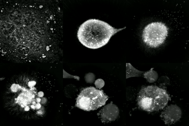
An etoposide-treated DU145 prostate cancer cell exploding into a cascade of apoptotic bodies. The sub images were extracted from a 61-hour time-lapse microscopy video, created using quantitative phase-contrast microscopy. The optical thickness is color-coded. With increasing thickness, color changes from gray to yellow, red, purple and finally black.

Apoptosis (from Ancient Greek: ἀπόπτωσις, romanizedapóptōsis, lit.'falling off') is a form of programmed cell death that occurs in multicellular organisms and in some eukaryotic, single-celled microorganisms such as yeast. Biochemical events lead to characteristic cell changes (morphology) and death. These changes include blebbing, cell shrinkage, nuclear fragmentation, chromatin condensation, DNA fragmentation, and mRNA decay. The average adult human loses 50 to 70 billion cells each day due to apoptosis. For the average human child between 8 and 14 years old, each day the approximate loss is 20 to 30 billion cells.

In contrast to necrosis, which is a form of traumatic cell death that results from acute cellular injury, apoptosis is a highly regulated and controlled process that confers advantages during an organism's life cycle. For example, the separation of fingers and toes in a developing human embryo occurs because cells between the digits undergo apoptosis. Unlike necrosis, apoptosis produces cell fragments called apoptotic bodies that phagocytes are able to engulf and remove before the contents of the cell can spill out onto surrounding cells and cause damage to them.

Because apoptosis cannot stop once it has begun, it is a highly regulated process. Apoptosis can be initiated through one of two pathways. In the intrinsic pathway the cell kills itself because it senses cell stress, while in the extrinsic pathway the cell kills itself because of signals from other cells. Weak external signals may also activate the intrinsic pathway of apoptosis. Both pathways induce cell death by activating caspases, which are proteases, or enzymes that degrade proteins. The two pathways both activate initiator caspases, which then activate executioner caspases, which then kill the cell by degrading proteins indiscriminately.

In addition to its importance as a biological phenomenon, defective apoptotic processes have been implicated in a wide variety of diseases. Excessive apoptosis causes atrophy, whereas an insufficient amount results in uncontrolled cell proliferation, such as cancer. Some factors like Fas receptors and caspases promote apoptosis, while some members of the Bcl-2 family of proteins inhibit apoptosis.

Discovery and etymology

German scientist Carl Vogt was first to describe the principle of apoptosis in 1842. In 1885, anatomist Walther Flemming delivered a more precise description of the process of programmed cell death. However, it was not until 1965 that the topic was resurrected. While studying tissues using electron microscopy, John Kerr at the University of Queensland was able to distinguish apoptosis from traumatic cell death. Following the publication of a paper describing the phenomenon, Kerr was invited to join Alastair Currie, as well as Andrew Wyllie, who was Currie's graduate student, at the University of Aberdeen. In 1972, the trio published a seminal article in the British Journal of Cancer. Kerr had initially used the term programmed cell necrosis, but in the article, the process of natural cell death was called apoptosis. Kerr, Wyllie and Currie credited James Cormack, a professor of Greek language at University of Aberdeen, with suggesting the term apoptosis. Kerr received the Paul Ehrlich and Ludwig Darmstaedter Prize on March 14, 2000, for his description of apoptosis. He shared the prize with Boston biologist H. Robert Horvitz.

For many years, neither "apoptosis" nor "programmed cell death" was a highly cited term. Two discoveries brought cell death from obscurity to a major field of research: identification of the first component of the cell death control and effector mechanisms, and linkage of abnormalities in cell death to human disease, in particular cancer. This occurred in 1988 when it was shown that BCL2, the gene responsible for follicular lymphoma, encoded a protein that inhibited cell death.

The 2002 Nobel Prize in Medicine was awarded to Sydney Brenner, H. Robert Horvitz and John Sulston for their work identifying genes that control apoptosis. The genes were identified by studies in the nematode C. elegans and homologues of these genes function in humans to regulate apoptosis.

John Sulston won the Nobel Prize in Medicine in 2002, for his pioneering research on apoptosis.

In Greek, apoptosis translates to the "falling off" of leaves from a tree. Cormack, professor of Greek language, reintroduced the term for medical use as it had a medical meaning for the Greeks over two thousand years before. Hippocrates used the term to mean "the falling off of the bones". Galen extended its meaning to "the dropping of the scabs". Cormack was no doubt aware of this usage when he suggested the name. Debate continues over the correct pronunciation, with opinion divided between a pronunciation with the second p silent (/æpəˈtsɪs/ ap-ə-TOH-sis) and the second p pronounced (/pəpˈtsɪs/). In English, the p of the Greek -pt- consonant cluster is typically silent at the beginning of a word (e.g. pterodactyl, Ptolemy), but articulated when used in combining forms preceded by a vowel, as in helicopter or the orders of insects: diptera, lepidoptera, etc.

In the original Kerr, Wyllie & Currie paper, there is a footnote regarding the pronunciation:

We are most grateful to Professor James Cormack of the Department of Greek, University of Aberdeen, for suggesting this term. The word "apoptosis" (ἀπόπτωσις) is used in Greek to describe the "dropping off" or "falling off" of petals from flowers, or leaves from trees. To show the derivation clearly, we propose that the stress should be on the penultimate syllable, the second half of the word being pronounced like "ptosis" (with the "p" silent), which comes from the same root "to fall", and is already used to describe the drooping of the upper eyelid.

Activation mechanisms

Control Of The Apoptotic Mechanisms
Control of the apoptotic mechanisms

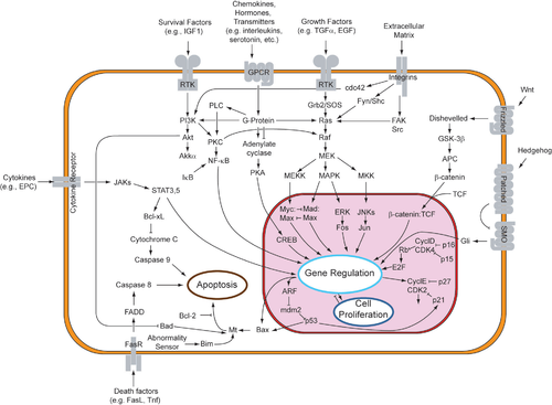
The initiation of apoptosis is tightly regulated by activation mechanisms, because once apoptosis has begun, it inevitably leads to the death of the cell. The two best-understood activation mechanisms are the intrinsic pathway (also called the mitochondrial pathway) and the extrinsic pathway. The intrinsic pathway is activated by intracellular signals generated when cells are stressed and depends on the release of proteins from the intermembrane space of mitochondria. The extrinsic pathway is activated by extracellular ligands binding to cell-surface death receptors, which leads to the formation of the death-inducing signaling complex (DISC).

A cell initiates intracellular apoptotic signaling in response to a stress, which may bring about cell suicide. The binding of nuclear receptors by glucocorticoids, heat, radiation, nutrient deprivation, viral infection, hypoxia, increased intracellular concentration of free fatty acids and increased intracellular calcium concentration, for example, by damage to the membrane, can all trigger the release of intracellular apoptotic signals by a damaged cell. A number of cellular components, such as poly ADP ribose polymerase, may also help regulate apoptosis. Single cell fluctuations have been observed in experimental studies of stress induced apoptosis.

Before the actual process of cell death is precipitated by enzymes, apoptotic signals must cause regulatory proteins to initiate the apoptosis pathway. This step allows those signals to cause cell death, or the process to be stopped, should the cell no longer need to die. Several proteins are involved, but two main methods of regulation have been identified: the targeting of mitochondria functionality, or directly transducing the signal via adaptor proteins to the apoptotic mechanisms. An extrinsic pathway for initiation identified in several toxin studies is an increase in calcium concentration within a cell caused by drug activity, which also can cause apoptosis via a calcium binding protease calpain.

Intrinsic pathway

The intrinsic pathway is also known as the mitochondrial pathway. Mitochondria are essential to multicellular life. Without them, a cell ceases to respire aerobically and quickly dies. This fact forms the basis for some apoptotic pathways. Apoptotic proteins that target mitochondria affect them in different ways. They may cause mitochondrial swelling through the formation of membrane pores, or they may increase the permeability of the mitochondrial membrane and cause apoptotic effectors to leak out. There is also a growing body of evidence indicating that nitric oxide is able to induce apoptosis by helping to dissipate the membrane potential of mitochondria and therefore make it more permeable. Nitric oxide has been implicated in initiating and inhibiting apoptosis through its possible action as a signal molecule of subsequent pathways that activate apoptosis.

During apoptosis, cytochrome c is released from mitochondria through the actions of the proteins Bax and Bak. The mechanism of this release is enigmatic, but appears to stem from a multitude of Bax/Bak homo- and hetero-dimers of Bax/Bak inserted into the outer membrane. Once cytochrome c is released it binds with Apoptotic protease activating factor – 1 (Apaf-1) and ATP, which then bind to pro-caspase-9 to create a protein complex known as an apoptosome. The apoptosome cleaves the pro-caspase to its active form of caspase-9, which in turn cleaves and activates pro-caspase into the effector caspase-3.

Mitochondria also release proteins known as SMACs (second mitochondria-derived activator of caspases) into the cell's cytosol following the increase in permeability of the mitochondria membranes. SMAC binds to proteins that inhibit apoptosis (IAPs) thereby deactivating them, and preventing the IAPs from arresting the process and therefore allowing apoptosis to proceed. IAP also normally suppresses the activity of a group of cysteine proteases called caspases, which carry out the degradation of the cell. Therefore, the actual degradation enzymes can be seen to be indirectly regulated by mitochondrial permeability.

Extrinsic pathway

Overview of signal transduction pathways
Overview of TNF (left) and Fas (right) signalling in apoptosis, an example of direct signal transduction

Two theories of the direct initiation of apoptotic mechanisms in mammals have been suggested: the TNF-induced (tumor necrosis factor) model and the Fas-Fas ligand-mediated model, both involving receptors of the TNF receptor (TNFR) family coupled to extrinsic signals.

TNF pathway

TNF-alpha is a cytokine produced mainly by activated macrophages, and is the major extrinsic mediator of apoptosis. Most cells in the human body have two receptors for TNF-alpha: TNFR1 and TNFR2. The binding of TNF-alpha to TNFR1 has been shown to initiate the pathway that leads to caspase activation via the intermediate membrane proteins TNF receptor-associated death domain (TRADD) and Fas-associated death domain protein (FADD). cIAP1/2 can inhibit TNF-α signaling by binding to TRAF2. FLIP inhibits the activation of caspase-8. Binding of this receptor can also indirectly lead to the activation of transcription factors involved in cell survival and inflammatory responses. However, signalling through TNFR1 might also induce apoptosis in a caspase-independent manner. The link between TNF-alpha and apoptosis shows why an abnormal production of TNF-alpha plays a fundamental role in several human diseases, especially in autoimmune diseases. The TNF-alpha receptor superfamily also includes death receptors (DRs), such as DR4 and DR5. These receptors bind to the protein TRAIL and mediate apoptosis. Apoptosis is known to be one of the primary mechanisms of targeted cancer therapy. Luminescent iridium complex-peptide hybrids (IPHs) have recently been designed, which mimic TRAIL and bind to death receptors on cancer cells, thereby inducing their apoptosis.

Fas pathway

The fas receptor (First apoptosis signal) – (also known as Apo-1 or CD95) is a transmembrane protein of the TNF family which binds the Fas ligand (FasL). The interaction between Fas and FasL results in the formation of the death-inducing signaling complex (DISC), which contains the FADD, caspase-8 and caspase-10. In some types of cells (type I), processed caspase-8 directly activates other members of the caspase family, and triggers the execution of apoptosis of the cell. In other types of cells (type II), the Fas-DISC starts a feedback loop that spirals into increasing release of proapoptotic factors from mitochondria and the amplified activation of caspase-8.[41]

Common components

Following TNF-R1 and Fas activation in mammalian cells a balance between proapoptotic (BAX, BID, BAK, or BAD) and anti-apoptotic (Bcl-Xl and Bcl-2) members of the Bcl-2 family are established. This balance is the proportion of proapoptotic homodimers that form in the outer-membrane of the mitochondrion. The proapoptotic homodimers are required to make the mitochondrial membrane permeable for the release of caspase activators such as cytochrome c and SMAC. Control of proapoptotic proteins under normal cell conditions of nonapoptotic cells is incompletely understood, but in general, Bax or Bak are activated by the activation of BH3-only proteins, part of the Bcl-2 family.

Caspases

Caspases play the central role in the transduction of ER apoptotic signals. Caspases are proteins that are highly conserved, cysteine-dependent aspartate-specific proteases. There are two types of caspases: initiator caspases (caspases 2, 8, 9, 10, 11, and 12) and effector caspases (caspases 3, 6, and 7). The activation of initiator caspases requires binding to specific oligomeric activator protein. Effector caspases are then activated by these active initiator caspases through proteolytic cleavage. The active effector caspases then proteolytically degrade a host of intracellular proteins to carry out the cell death program.

Caspase-independent apoptotic pathway

There also exists a caspase-independent apoptotic pathway that is mediated by AIF (apoptosis-inducing factor).

Apoptosis model in amphibians

The frog Xenopus laevis serves as an ideal model system for the study of the mechanisms of apoptosis. In fact, iodine and thyroxine also stimulate the spectacular apoptosis of the cells of the larval gills, tail and fins in amphibian's metamorphosis, and stimulate the evolution of their nervous system transforming the aquatic, vegetarian tadpole into the terrestrial, carnivorous frog.

Negative regulators of apoptosis

Negative regulation of apoptosis inhibits cell death signaling pathways, helping tumors to evade cell death and developing drug resistance. The ratio between anti-apoptotic (Bcl-2) and pro-apoptotic (Bax) proteins determines whether a cell lives or dies. Many families of proteins act as negative regulators categorized into either antiapoptotic factors, such as IAPs and Bcl-2 proteins or prosurvival factors like cFLIP, BNIP3, FADD, Akt, and NF-κB.

Proteolytic caspase cascade: Killing the cell

Many pathways and signals lead to apoptosis, but these converge on a single mechanism that actually causes the death of the cell. After a cell receives stimulus, it undergoes organized degradation of cellular organelles by activated proteolytic caspases. In addition to the destruction of cellular organelles, mRNA is rapidly and globally degraded by a mechanism that is not yet fully characterized. mRNA decay is triggered very early in apoptosis.

A cell undergoing apoptosis shows a series of characteristic morphological changes. Early alterations include:

  1. Cell shrinkage and rounding occur because of the retraction of lamellipodia and the breakdown of the proteinaceous cytoskeleton by caspases.
  2. The cytoplasm appears dense, and the organelles appear tightly packed.
  3. Chromatin undergoes condensation into compact patches against the nuclear envelope (also known as the perinuclear envelope) in a process known as pyknosis, a hallmark of apoptosis.
  4. The nuclear envelope becomes discontinuous and the DNA inside it is fragmented in a process referred to as karyorrhexis. The nucleus breaks into several discrete chromatin bodies or nucleosomal units due to the degradation of DNA.

Apoptosis progresses quickly and its products are quickly removed, making it difficult to detect or visualize on classical histology sections. During karyorrhexis, endonuclease activation leaves short DNA fragments, regularly spaced in size. These give a characteristic "laddered" appearance on agar gel after electrophoresis. Tests for DNA laddering differentiate apoptosis from ischemic or toxic cell death.

Apoptotic cell disassembly

Different steps in apoptotic cell disassembly

Before the apoptotic cell is disposed of, there is a process of disassembly. There are three recognized steps in apoptotic cell disassembly:

  1. Membrane blebbing: The cell membrane shows irregular buds known as blebs. Initially these are smaller surface blebs. Later these can grow into larger so-called dynamic membrane blebs. An important regulator of apoptotic cell membrane blebbing is ROCK1 (rho associated coiled-coil-containing protein kinase 1).
  2. Formation of membrane protrusions: Some cell types, under specific conditions, may develop different types of long, thin extensions of the cell membrane called membrane protrusions. Three types have been described: microtubule spikes, apoptopodia (feet of death), and beaded apoptopodia (the latter having a beads-on-a-string appearance). Pannexin 1 is an important component of membrane channels involved in the formation of apoptopodia and beaded apoptopodia.
  3. Fragmentation: The cell breaks apart into multiple vesicles called apoptotic bodies, which undergo phagocytosis. The plasma membrane protrusions may help bring apoptotic bodies closer to phagocytes.

Removal of dead cells

The removal of dead cells by neighboring phagocytic cells has been termed efferocytosis. Dying cells that undergo the final stages of apoptosis display phagocytotic molecules, such as phosphatidylserine, on their cell surface. Phosphatidylserine is normally found on the inner leaflet surface of the plasma membrane, but is redistributed during apoptosis to the extracellular surface by a protein known as scramblase. These molecules mark the cell for phagocytosis by cells possessing the appropriate receptors, such as macrophages. The removal of dying cells by phagocytes occurs in an orderly manner without eliciting an inflammatory response. During apoptosis cellular RNA and DNA are separated from each other and sorted to different apoptotic bodies; separation of RNA is initiated as nucleolar segregation.

Pathway knock-outs

Many knock-outs have been made in the apoptosis pathways to test the function of each of the proteins. Several caspases, in addition to APAF1 and FADD, have been mutated to determine the new phenotype. In order to create a tumor necrosis factor (TNF) knockout, an exon containing the nucleotides 3704–5364 was removed from the gene. This exon encodes a portion of the mature TNF domain, as well as the leader sequence, which is a highly conserved region necessary for proper intracellular processing. TNF-/- mice develop normally and have no gross structural or morphological abnormalities. However, upon immunization with SRBC (sheep red blood cells), these mice demonstrated a deficiency in the maturation of an antibody response; they were able to generate normal levels of IgM, but could not develop specific IgG levels. Apaf-1 is the protein that turns on caspase 9 by cleavage to begin the caspase cascade that leads to apoptosis. Since a -/- mutation in the APAF-1 gene is embryonic lethal, a gene trap strategy was used in order to generate an APAF-1 -/- mouse. This assay is used to disrupt gene function by creating an intragenic gene fusion. When an APAF-1 gene trap is introduced into cells, many morphological changes occur, such as spina bifida, the persistence of interdigital webs, and open brain. In addition, after embryonic day 12.5, the brain of the embryos showed several structural changes. APAF-1 cells are protected from apoptosis stimuli such as irradiation. A BAX-1 knock-out mouse exhibits normal forebrain formation and a decreased programmed cell death in some neuronal populations and in the spinal cord, leading to an increase in motor neurons.

The caspase proteins are integral parts of the apoptosis pathway, so it follows that knock-outs made have varying damaging results. A caspase 9 knock-out leads to a severe brain malformation . A caspase 8 knock-out leads to cardiac failure and thus embryonic lethality . However, with the use of cre-lox technology, a caspase 8 knock-out has been created that exhibits an increase in peripheral T cells, an impaired T cell response, and a defect in neural tube closure. These mice were found to be resistant to apoptosis mediated by CD95, TNFR, etc. but not resistant to apoptosis caused by UV irradiation, chemotherapeutic drugs, and other stimuli. Finally, a caspase 3 knock-out was characterized by ectopic cell masses in the brain and abnormal apoptotic features such as membrane blebbing or nuclear fragmentation. A remarkable feature of these KO mice is that they have a very restricted phenotype: Casp3, 9, APAF-1 KO mice have deformations of neural tissue and FADD and Casp 8 KO showed defective heart development, however, in both types of KO other organs developed normally and some cell types were still sensitive to apoptotic stimuli suggesting that unknown proapoptotic pathways exist.

Methods for distinguishing apoptotic from necrotic cells

Long-term live cell imaging (12h) of multinucleated mouse pre-Adipocyte trying to undergo mitosis. Due to the excess of genetic material the cell fails to replicate and dies by apoptosis.

Label-free live cell imaging, time-lapse microscopy, flow fluorocytometry, and transmission electron microscopy can be used to compare apoptotic and necrotic cells. There are also various biochemical techniques for analysis of cell surface markers (phosphatidylserine exposure versus cell permeability by flow cytometry), cellular markers such as DNA fragmentation (flow cytometry), caspase activation, Bid cleavage, and cytochrome c release (Western blotting). Supernatant screening for caspases, HMGB1, and cytokeratin 18 release can identify primary from secondary necrotic cells. However, no distinct surface or biochemical markers of necrotic cell death have been identified yet, and only negative markers are available. These include absence of apoptotic markers (caspase activation, cytochrome c release, and oligonucleosomal DNA fragmentation) and differential kinetics of cell death markers (phosphatidylserine exposure and cell membrane permeabilization). A selection of techniques that can be used to distinguish apoptosis from necroptotic cells could be found in these references.

Implication in disease

A section of mouse liver showing several apoptotic cells, indicated by arrows
A section of mouse liver stained to show cells undergoing apoptosis (orange)
Neonatal cardiomyocytes ultrastructure after anoxia-reoxygenation

Defective pathways

The many different types of apoptotic pathways contain a multitude of different biochemical components, many of them not yet understood. As a pathway is more or less sequential in nature, removing or modifying one component leads to an effect in another. In a living organism, this can have disastrous effects, often in the form of disease or disorder. A discussion of every disease caused by modification of the various apoptotic pathways would be impractical, but the concept overlying each one is the same: The normal functioning of the pathway has been disrupted in such a way as to impair the ability of the cell to undergo normal apoptosis. This results in a cell that lives past its "use-by date" and is able to replicate and pass on any faulty machinery to its progeny, increasing the likelihood of the cell's becoming cancerous or diseased.

A recently described example of this concept in action can be seen in the development of a lung cancer called NCI-H460. The X-linked inhibitor of apoptosis protein (XIAP) is overexpressed in cells of the H460 cell line. XIAPs bind to the processed form of caspase-9 and suppress the activity of apoptotic activator cytochrome c, therefore overexpression leads to a decrease in the number of proapoptotic agonists. As a consequence, the balance of anti-apoptotic and proapoptotic effectors is upset in favour of the former, and the damaged cells continue to replicate despite being directed to die. Defects in regulation of apoptosis in cancer cells occur often at the level of control of transcription factors. As a particular example, defects in molecules that control transcription factor NF-κB in cancer change the mode of transcriptional regulation and the response to apoptotic signals, to curtail dependence on the tissue that the cell belongs. This degree of independence from external survival signals, can enable cancer metastasis.

Dysregulation of p53

The tumor-suppressor protein p53 accumulates when DNA is damaged due to a chain of biochemical factors. Part of this pathway includes alpha-interferon and beta-interferon, which induce transcription of the p53 gene, resulting in the increase of p53 protein level and enhancement of cancer cell-apoptosis. p53 prevents the cell from replicating by stopping the cell cycle at G1, or interphase, to give the cell time to repair; however, it will induce apoptosis if damage is extensive and repair efforts fail. Any disruption to the regulation of the p53 or interferon genes will result in impaired apoptosis and the possible formation of tumors.

Inhibition

Inhibition of apoptosis can result in a number of cancers, inflammatory diseases, and viral infections. It was originally believed that the associated accumulation of cells was due to an increase in cellular proliferation, but it is now known that it is also due to a decrease in cell death. The most common of these diseases is cancer, the disease of excessive cellular proliferation, which is often characterized by an overexpression of IAP family members. As a result, the malignant cells experience an abnormal response to apoptosis induction: Cycle-regulating genes (such as p53, ras or c-myc) are mutated or inactivated in diseased cells, and further genes (such as bcl-2) also modify their expression in tumors. Some apoptotic factors are vital during mitochondrial respiration e.g. cytochrome C. Pathological inactivation of apoptosis in cancer cells is correlated with frequent respiratory metabolic shifts toward glycolysis (an observation known as the "Warburg hypothesis".

HeLa cell

Apoptosis in HeLa cells is inhibited by proteins produced by the cell; these inhibitory proteins target retinoblastoma tumor-suppressing proteins. These tumor-suppressing proteins regulate the cell cycle, but are rendered inactive when bound to an inhibitory protein. HPV E6 and E7 are inhibitory proteins expressed by the human papillomavirus, HPV being responsible for the formation of the cervical tumor from which HeLa cells are derived. HPV E6 causes p53, which regulates the cell cycle, to become inactive. HPV E7 binds to retinoblastoma tumor suppressing proteins and limits its ability to control cell division.hese two inhibitory proteins are partially responsible for HeLa cells' immortality by inhibiting apoptosis to occur.

Treatments

The main method of treatment for potential death from signaling-related diseases involves either increasing or decreasing the susceptibility of apoptosis in diseased cells, depending on whether the disease is caused by either the inhibition of or excess apoptosis. For instance, treatments aim to restore apoptosis to treat diseases with deficient cell death and to increase the apoptotic threshold to treat diseases involved with excessive cell death. To stimulate apoptosis, one can increase the number of death receptor ligands (such as TNF or TRAIL), antagonize the anti-apoptotic Bcl-2 pathway, or introduce Smac mimetics to inhibit the inhibitor (IAPs). The addition of agents such as Herceptin, Iressa, or Gleevec works to stop cells from cycling and causes apoptosis activation by blocking growth and survival signaling further upstream. Finally, adding p53-MDM2 complexes displaces p53 and activates the p53 pathway, leading to cell cycle arrest and apoptosis. Many different methods can be used either to stimulate or to inhibit apoptosis in various places along the death signaling pathway.

Apoptosis is a multi-step, multi-pathway cell-death programme that is inherent in every cell of the body. In cancer, the apoptosis cell-division ratio is altered. Cancer treatment by chemotherapy and irradiation kills target cells primarily by inducing apoptosis.

Hyperactive apoptosis

On the other hand, loss of control of cell death (resulting in excess apoptosis) can lead to neurodegenerative diseases, hematologic diseases, and tissue damage. Neurons that rely on mitochondrial respiration undergo apoptosis in neurodegenerative diseases such as Alzheimer's and Parkinson's. (an observation known as the "Inverse Warburg hypothesis"). Moreover, there is an inverse epidemiological comorbidity between neurodegenerative diseases and cancer. The progression of HIV is directly linked to excess, unregulated apoptosis. In a healthy individual, the number of CD4+ lymphocytes is in balance with the cells generated by the bone marrow; however, in HIV-positive patients, this balance is lost due to an inability of the bone marrow to regenerate CD4+ cells. In the case of HIV, CD4+ lymphocytes die at an accelerated rate through uncontrolled apoptosis, when stimulated. At the molecular level, hyperactive apoptosis can be caused by defects in signaling pathways that regulate the Bcl-2 family proteins. Increased expression of apoptotic proteins such as BIM, or their decreased proteolysis, leads to cell death and can cause a number of pathologies, depending on the cells where excessive activity of BIM occurs. Cancer cells can escape apoptosis through mechanisms that suppress BIM expression or by increased proteolysis of BIM.

Treatments

Treatments aiming to inhibit works to block specific caspases. Finally, the Akt protein kinase promotes cell survival through two pathways. Akt phosphorylates and inhibits Bad (a Bcl-2 family member), causing Bad to interact with the 14-3-3 scaffold, resulting in Bcl dissociation and thus cell survival. Akt also activates IKKα, which leads to NF-κB activation and cell survival. Active NF-κB induces the expression of anti-apoptotic genes such as Bcl-2, resulting in inhibition of apoptosis. NF-κB has been found to play both an antiapoptotic role and a proapoptotic role depending on the stimuli utilized and the cell type.

HIV progression

The progression of the human immunodeficiency virus infection into AIDS is due primarily to the depletion of CD4+ T-helper lymphocytes in a manner that is too rapid for the body's bone marrow to replenish the cells, leading to a compromised immune system. One of the mechanisms by which T-helper cells are depleted is apoptosis, which results from a series of biochemical pathways:

  1. HIV enzymes deactivate anti-apoptotic Bcl-2. This does not directly cause cell death but primes the cell for apoptosis should the appropriate signal be received. In parallel, these enzymes activate proapoptotic procaspase-8, which does directly activate the mitochondrial events of apoptosis.
  2. HIV may increase the level of cellular proteins that prompt Fas-mediated apoptosis.
  3. HIV proteins decrease the amount of CD4 glycoprotein marker present on the cell membrane.
  4. Released viral particles and proteins present in extracellular fluid are able to induce apoptosis in nearby "bystander" T helper cells.
  5. HIV decreases the production of molecules involved in marking the cell for apoptosis, giving the virus time to replicate and continue releasing apoptotic agents and virions into the surrounding tissue.
  6. The infected CD4+ cell may also receive the death signal from a cytotoxic T cell.

Cells may also die as direct consequences of viral infections. HIV-1 expression induces tubular cell G2/M arrest and apoptosis. The progression from HIV to AIDS is not immediate or even necessarily rapid; HIV's cytotoxic activity toward CD4+ lymphocytes is classified as AIDS once a given patient's CD4+ cell count falls below 200.

Researchers from Kumamoto University in Japan have developed a new method to eradicate HIV in viral reservoir cells, named "Lock-in and apoptosis." Using the synthesized compound Heptanoylphosphatidyl L-Inositol Pentakisphophate (or L-Hippo) to bind strongly to the HIV protein PR55Gag, they were able to suppress viral budding. By suppressing viral budding, the researchers were able to trap the HIV virus in the cell and allow for the cell to undergo apoptosis (natural cell death). Associate Professor Mikako Fujita has stated that the approach is not yet available to HIV patients because the research team has to conduct further research on combining the drug therapy that currently exists with this "Lock-in and apoptosis" approach to lead to complete recovery from HIV.

Viral infection

Viral induction of apoptosis occurs when one or several cells of a living organism are infected with a virus, leading to cell death. Cell death in organisms is necessary for the normal development of cells and the cell cycle maturation. It is also important in maintaining the regular functions and activities of cells.

Viruses can trigger apoptosis of infected cells via a range of mechanisms including:

  • Receptor binding
  • Activation of protein kinase R (PKR)
  • Interaction with p53
  • Expression of viral proteins coupled to MHC proteins on the surface of the infected cell, allowing recognition by cells of the immune system (such as natural killer and cytotoxic T cells) that then induce the infected cell to undergo apoptosis.

Canine distemper virus (CDV) is known to cause apoptosis in central nervous system and lymphoid tissue of infected dogs in vivo and in vitro. Apoptosis caused by CDV is typically induced via the extrinsic pathway, which activates caspases that disrupt cellular function and eventually leads to the cells death. In normal cells, CDV activates caspase-8 first, which works as the initiator protein followed by the executioner protein caspase-3. However, apoptosis induced by CDV in HeLa cells does not involve the initiator protein caspase-8. HeLa cell apoptosis caused by CDV follows a different mechanism than that in vero cell lines. This change in the caspase cascade suggests CDV induces apoptosis via the intrinsic pathway, excluding the need for the initiator caspase-8. The executioner protein is instead activated by the internal stimuli caused by viral infection not a caspase cascade.

The Oropouche virus (OROV) is found in the family Bunyaviridae. The study of apoptosis brought on by Bunyaviridae was initiated in 1996, when it was observed that apoptosis was induced by the La Crosse virus into the kidney cells of baby hamsters and into the brains of baby mice.

OROV is a disease that is transmitted between humans by the biting midge (Culicoides paraensis). It is referred to as a zoonotic arbovirus and causes febrile illness, characterized by the onset of a sudden fever known as Oropouche fever.

The Oropouche virus also causes disruption in cultured cells – cells that are cultivated in distinct and specific conditions. An example of this can be seen in HeLa cells, whereby the cells begin to degenerate shortly after they are infected.

With the use of gel electrophoresis, it can be observed that OROV causes DNA fragmentation in HeLa cells. It can be interpreted by counting, measuring, and analyzing the cells of the Sub/G1 cell population. When HeLA cells are infected with OROV, the cytochrome C is released from the membrane of the mitochondria, into the cytosol of the cells. This type of interaction shows that apoptosis is activated via an intrinsic pathway.

In order for apoptosis to occur within OROV, viral uncoating, viral internalization, along with the replication of cells is necessary. Apoptosis in some viruses is activated by extracellular stimuli. However, studies have demonstrated that the OROV infection causes apoptosis to be activated through intracellular stimuli and involves the mitochondria.

Many viruses encode proteins that can inhibit apoptosis. Several viruses encode viral homologs of Bcl-2. These homologs can inhibit proapoptotic proteins such as BAX and BAK, which are essential for the activation of apoptosis. Examples of viral Bcl-2 proteins include the Epstein-Barr virus BHRF1 protein and the adenovirus E1B 19K protein. Some viruses express caspase inhibitors that inhibit caspase activity and an example is the CrmA protein of cowpox viruses. Whilst a number of viruses can block the effects of TNF and Fas. For example, the M-T2 protein of myxoma viruses can bind TNF preventing it from binding the TNF receptor and inducing a response. Furthermore, many viruses express p53 inhibitors that can bind p53 and inhibit its transcriptional transactivation activity. As a consequence, p53 cannot induce apoptosis, since it cannot induce the expression of proapoptotic proteins. The adenovirus E1B-55K protein and the hepatitis B virus HBx protein are examples of viral proteins that can perform such a function.

Viruses can remain intact from apoptosis in particular in the latter stages of infection. They can be exported in the apoptotic bodies that pinch off from the surface of the dying cell, and the fact that they are engulfed by phagocytes prevents the initiation of a host response. This favours the spread of the virus. Prions can cause apoptosis in neurons.

Plants

Programmed cell death in plants has a number of molecular similarities to that of animal apoptosis, but it also has differences, notable ones being the presence of a cell wall and the lack of an immune system that removes the pieces of the dead cell. Instead of an immune response, the dying cell synthesizes substances to break itself down and places them in a vacuole that ruptures as the cell dies. Additionally, plants do not contain phagocytic cells, which are essential in the process of breaking down and removing apoptotic bodies. Whether this whole process resembles animal apoptosis closely enough to warrant using the name apoptosis (as opposed to the more general programmed cell death) is unclear.

Caspase-independent apoptosis

The characterization of the caspases allowed the development of caspase inhibitors, which can be used to determine whether a cellular process involves active caspases. Using these inhibitors it was discovered that cells can die while displaying a morphology similar to apoptosis without caspase activation. Later studies linked this phenomenon to the release of AIF (apoptosis-inducing factor) from the mitochondria and its translocation into the nucleus mediated by its NLS (nuclear localization signal). Inside the mitochondria, AIF is anchored to the inner membrane. In order to be released, the protein is cleaved by a calcium-dependent calpain protease.

Philosophy of self

From Wikipedia, the free encyclopedia https://en.wikipedia.org/wiki/Philosophy_of_self The philosophy of self ...