Search This Blog

Saturday, June 15, 2024

Genetic recombination

From Wikipedia, the free encyclopedia
https://en.wikipedia.org/wiki/Genetic_recombination
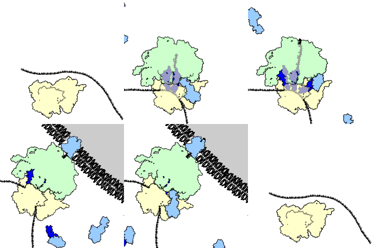
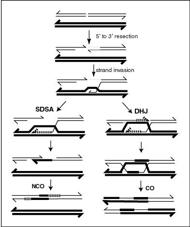
A model of meiotic recombination, initiated by a double-strand break or gap, followed by pairing with an homologous chromosome and strand invasion to initiate the recombinational repair process. Repair of the gap can lead to crossover (CO) or non-crossover (NCO) of the flanking regions. CO recombination is thought to occur by the Double Holliday Junction (DHJ) model, illustrated on the right. NCO recombinants are thought to occur primarily by the Synthesis Dependent Strand Annealing (SDSA) model, illustrated on the left. Most recombination events appear to be the SDSA type.

Genetic recombination (also known as genetic reshuffling) is the exchange of genetic material between different organisms which leads to production of offspring with combinations of traits that differ from those found in either parent. In eukaryotes, genetic recombination during meiosis can lead to a novel set of genetic information that can be further passed on from parents to offspring. Most recombination occurs naturally and can be classified into two types: (1) interchromosomal recombination, occurring through independent assortment of alleles whose loci are on different but homologous chromosomes (random orientation of pairs of homologous chromosomes in meiosis I); & (2) intrachromosomal recombination, occurring through crossing over.

During meiosis in eukaryotes, genetic recombination involves the pairing of homologous chromosomes. This may be followed by information transfer between the chromosomes. The information transfer may occur without physical exchange (a section of genetic material is copied from one chromosome to another, without the donating chromosome being changed) (see SDSA – Synthesis Dependent Strand Annealing pathway in Figure); or by the breaking and rejoining of DNA strands, which forms new molecules of DNA (see DHJ pathway in Figure).

Recombination may also occur during mitosis in eukaryotes where it ordinarily involves the two sister chromosomes formed after chromosomal replication. In this case, new combinations of alleles are not produced since the sister chromosomes are usually identical. In meiosis and mitosis, recombination occurs between similar molecules of DNA (homologous sequences). In meiosis, non-sister homologous chromosomes pair with each other so that recombination characteristically occurs between non-sister homologues. In both meiotic and mitotic cells, recombination between homologous chromosomes is a common mechanism used in DNA repair.

Gene conversion – the process during which homologous sequences are made identical also falls under genetic recombination.

Genetic recombination and recombinational DNA repair also occurs in bacteria and archaea, which use asexual reproduction.

Recombination can be artificially induced in laboratory (in vitro) settings, producing recombinant DNA for purposes including vaccine development.

V(D)J recombination in organisms with an adaptive immune system is a type of site-specific genetic recombination that helps immune cells rapidly diversify to recognize and adapt to new pathogens.

Synapsis

During meiosis, synapsis (the pairing of homologous chromosomes) ordinarily precedes genetic recombination.

Mechanism

Genetic recombination is catalyzed by many different enzymes. Recombinases are key enzymes that catalyse the strand transfer step during recombination. RecA, the chief recombinase found in Escherichia coli, is responsible for the repair of DNA double strand breaks (DSBs). In yeast and other eukaryotic organisms there are two recombinases required for repairing DSBs. The RAD51 protein is required for mitotic and meiotic recombination, whereas the DNA repair protein, DMC1, is specific to meiotic recombination. In the archaea, the ortholog of the bacterial RecA protein is RadA.

Bacterial recombination

In bacteria there is regular genetic recombination, as well as ineffective transfer of genetic material, expressed as unsuccessful transfer or abortive transfer, which is any bacterial DNA transfer of the donor cell to recipients which have set the incoming DNA as part of the genetic material of the recipient. Abortive transfer was registered in the following transduction and conjugation. In all cases, the transmitted fragment is diluted by the culture growth.

Chromosomal crossover
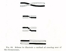
Thomas Hunt Morgan's illustration of crossing over (1916)

In eukaryotes, recombination during meiosis is facilitated by chromosomal crossover. The crossover process leads to offspring having different combinations of genes from those of their parents, and can occasionally produce new chimeric alleles. The shuffling of genes brought about by genetic recombination produces increased genetic variation. It also allows sexually reproducing organisms to avoid Muller's ratchet, in which the genomes of an asexual population tend to accumulate deleterious mutations over time than beneficial or reversing mutations.

Chromosomal crossover involves recombination between the paired chromosomes inherited from each of one's parents, generally occurring during meiosis. During prophase I (pachytene stage) the four available chromatids are in tight formation with one another. While in this formation, homologous sites on two chromatids can closely pair with one another, and may exchange genetic information.

Because there is a small probability of recombination at any location along a chromosome, the frequency of recombination between two locations depends on the distance separating them. Therefore, for genes sufficiently distant on the same chromosome, the amount of crossover is high enough to destroy the correlation between alleles.

Tracking the movement of genes resulting from crossovers has proven quite useful to geneticists. Because two genes that are close together are less likely to become separated than genes that are farther apart, geneticists can deduce roughly how far apart two genes are on a chromosome if they know the frequency of the crossovers. Geneticists can also use this method to infer the presence of certain genes. Genes that typically stay together during recombination are said to be linked. One gene in a linked pair can sometimes be used as a marker to deduce the presence of the other gene. This is typically used to detect the presence of a disease-causing gene.

The recombination frequency between two loci observed is the crossing-over value. It is the frequency of crossing over between two linked gene loci (markers), and depends on the distance between the genetic loci observed. For any fixed set of genetic and environmental conditions, recombination in a particular region of a linkage structure (chromosome) tends to be constant, and the same is then true for the crossing-over value which is used in the production of genetic maps.

Gene conversion

In gene conversion, a section of genetic material is copied from one chromosome to another, without the donating chromosome being changed. Gene conversion occurs at high frequency at the actual site of the recombination event during meiosis. It is a process by which a DNA sequence is copied from one DNA helix (which remains unchanged) to another DNA helix, whose sequence is altered. Gene conversion has often been studied in fungal crosses where the 4 products of individual meioses can be conveniently observed. Gene conversion events can be distinguished as deviations in an individual meiosis from the normal 2:2 segregation pattern (e.g. a 3:1 pattern).

Nonhomologous recombination

Recombination can occur between DNA sequences that contain no sequence homology. This can cause chromosomal translocations, sometimes leading to cancer.

In B cells

B cells of the immune system perform genetic recombination, called immunoglobulin class switching. It is a biological mechanism that changes an antibody from one class to another, for example, from an isotype called IgM to an isotype called IgG.

Genetic engineering

In genetic engineering, recombination can also refer to artificial and deliberate recombination of disparate pieces of DNA, often from different organisms, creating what is called recombinant DNA. A prime example of such a use of genetic recombination is gene targeting, which can be used to add, delete or otherwise change an organism's genes. This technique is important to biomedical researchers as it allows them to study the effects of specific genes. Techniques based on genetic recombination are also applied in protein engineering to develop new proteins of biological interest.

Examples include Restriction enzyme mediated integration, Gibson assembly and Golden Gate Cloning.

Recombinational repair

DNA damages caused by a variety of exogenous agents (e.g. UV light, X-rays, chemical cross-linking agents) can be repaired by homologous recombinational repair (HRR). These findings suggest that DNA damages arising from natural processes, such as exposure to reactive oxygen species that are byproducts of normal metabolism, are also repaired by HRR. In humans, deficiencies in the gene products necessary for HRR during meiosis likely cause infertility In humans, deficiencies in gene products necessary for HRR, such as BRCA1 and BRCA2, increase the risk of cancer (see DNA repair-deficiency disorder).

In bacteria, transformation is a process of gene transfer that ordinarily occurs between individual cells of the same bacterial species. Transformation involves integration of donor DNA into the recipient chromosome by recombination. This process appears to be an adaptation for repairing DNA damages in the recipient chromosome by HRR. Transformation may provide a benefit to pathogenic bacteria by allowing repair of DNA damage, particularly damages that occur in the inflammatory, oxidizing environment associated with infection of a host.

When two or more viruses, each containing lethal genomic damages, infect the same host cell, the virus genomes can often pair with each other and undergo HRR to produce viable progeny. This process, referred to as multiplicity reactivation, has been studied in lambda and T4 bacteriophages, as well as in several pathogenic viruses. In the case of pathogenic viruses, multiplicity reactivation may be an adaptive benefit to the virus since it allows the repair of DNA damages caused by exposure to the oxidizing environment produced during host infection. See also reassortment.

Meiotic recombination

A molecular model for the mechanism of meiotic recombination presented by Anderson and Sekelsky is outlined in the first figure in this article. Two of the four chromatids present early in meiosis (prophase I) are paired with each other and able to interact. Recombination, in this model, is initiated by a double-strand break (or gap) shown in the DNA molecule (chromatid) at the top of the figure. Other types of DNA damage may also initiate recombination. For instance, an inter-strand cross-link (caused by exposure to a cross-linking agent such as mitomycin C) can be repaired by HRR.

Two types of recombinant product are produced. Indicated on the right side is a "crossover" (CO) type, where the flanking regions of the chromosomes are exchanged, and on the left side, a "non-crossover" (NCO) type where the flanking regions are not exchanged. The CO type of recombination involves the intermediate formation of two "Holliday junctions" indicated in the lower right of the figure by two X-shaped structures in each of which there is an exchange of single strands between the two participating chromatids. This pathway is labeled in the figure as the DHJ (double-Holliday junction) pathway.

The NCO recombinants (illustrated on the left in the figure) are produced by a process referred to as "synthesis dependent strand annealing" (SDSA). Recombination events of the NCO/SDSA type appear to be more common than the CO/DHJ type. The NCO/SDSA pathway contributes little to genetic variation, since the arms of the chromosomes flanking the recombination event remain in the parental configuration. Thus, explanations for the adaptive function of meiosis that focus exclusively on crossing-over are inadequate to explain the majority of recombination events.

Achiasmy and heterochiasmy

Achiasmy is the phenomenon where autosomal recombination is completely absent in one sex of a species. Achiasmatic chromosomal segregation is well documented in male Drosophila melanogaster. Heterochiasmy occurs when recombination rates differ between the sexes of a species. This sexual dimorphic pattern in recombination rate has been observed in many species. In mammals, females most often have higher rates of recombination. The "Haldane-Huxley rule" states that achiasmy usually occurs in the heterogametic sex.

RNA virus recombination

Numerous RNA viruses are capable of genetic recombination when at least two viral genomes are present in the same host cell. Recombination is largely responsible for RNA virus diversity and immune evasion. RNA recombination appears to be a major driving force in determining genome architecture and the course of viral evolution among picornaviridae ((+)ssRNA) (e.g. poliovirus). In the retroviridae ((+)ssRNA)(e.g. HIV), damage in the RNA genome appears to be avoided during reverse transcription by strand switching, a form of recombination.

Recombination also occurs in the reoviridae (dsRNA)(e.g. reovirus), orthomyxoviridae ((-)ssRNA)(e.g. influenza virus) and coronaviridae ((+)ssRNA) (e.g. SARS).

Recombination in RNA viruses appears to be an adaptation for coping with genome damage. Switching between template strands during genome replication, referred to as copy-choice recombination, was originally proposed to explain the positive correlation of recombination events over short distances in organisms with a DNA genome (see first Figure, SDSA pathway).

Recombination can occur infrequently between animal viruses of the same species but of divergent lineages. The resulting recombinant viruses may sometimes cause an outbreak of infection in humans.

Especially in coronaviruses, recombination may also occur even among distantly related evolutionary groups (subgenera), due to their characteristic transcription mechanism, that involves subgenomic mRNAs that are formed by template switching.

When replicating its (+)ssRNA genome, the poliovirus RNA-dependent RNA polymerase (RdRp) is able to carry out recombination. Recombination appears to occur by a copy choice mechanism in which the RdRp switches (+)ssRNA templates during negative strand synthesis. Recombination by RdRp strand switching also occurs in the (+)ssRNA plant carmoviruses and tombusviruses.

Recombination appears to be a major driving force in determining genetic variability within coronaviruses, as well as the ability of coronavirus species to jump from one host to another and, infrequently, for the emergence of novel species, although the mechanism of recombination in is unclear.

In early 2020, many genomic sequences of Australian SARS‐CoV‐2 isolates have deletions or mutations (29742G>A or 29742G>U; "G19A" or "G19U") in the s2m, suggesting that RNA recombination may have occurred in this RNA element. 29742G("G19"), 29744G("G21"), and 29751G("G28") were predicted as recombination hotspots. During the first months of the COVID-19 pandemic, such a recombination event was suggested to have been a critical step in the evolution of SARS-CoV-2's ability to infect humans. Linkage disequilibrium analysis confirmed that RNA recombination with the 11083G > T mutation also contributed to the increase of mutations among the viral progeny. The findings indicate that the 11083G > T mutation of SARS-CoV-2 spread during Diamond Princess shipboard quarantine and arose through de novo RNA recombination under positive selection pressure. In three patients on the Diamond Princess cruise, two mutations, 29736G > T and 29751G > T (G13 and G28) were located in Coronavirus 3′ stem-loop II-like motif (s2m) of SARS-CoV-2. Although s2m is considered an RNA motif highly conserved in 3' untranslated region among many coronavirus species, this result also suggests that s2m of SARS-CoV-2 is RNA recombination/mutation hotspot.
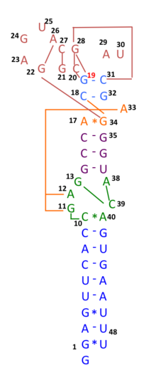
Schematic representation of the s2m RNA secondary structure, with tertiary structural interactions indicated as long range contacts.

SARS-CoV-2's entire receptor binding motif appeared, based on preliminary observations, to have been introduced through recombination from coronaviruses of pangolins. However, more comprehensive analyses later refuted this suggestion and showed that SARS-CoV-2 likely evolved solely within bats and with little or no recombination.

Role of recombination in the origin of life

Nowak and Ohtsuki noted that the origin of life (abiogenesis) is also the origin of biological evolution. They pointed out that all known life on earth is based on biopolymers and proposed that any theory for the origin of life must involve biological polymers that act as information carriers and catalysts. Lehman argued that recombination was an evolutionary development as ancient as the origins of life. Smail et al. proposed that in the primordial Earth, recombination played a key role in the expansion of the initially short informational polymers (presumed to be RNA) that were the precursors to life.

First universal common ancestor

From Wikipedia, the free encyclopedia
https://en.wikipedia.org/wiki/First_universal_common_ancestor

The first universal common ancestor (FUCA) is a proposed non-cellular entity that was the earliest organism with a genetic code capable of biological translation of RNA molecules into peptides to produce proteins. Its descendents include the last universal common ancestor (LUCA) and every modern cell. FUCA would also be the ancestor of ancient sister lineages of LUCA, none of which have modern descendants, but which are thought to have horizontally transferred some of their genes into the genome of early descendants of LUCA.

FUCA is thought to have been composed of progenotes, proposed ancient biological systems that would have used RNA for their genome and self-replication. By comparison, LUCA would have had a complex metabolism and a DNA genome with hundreds of genes and gene families.

Origins

Long before the appearance of compartmentalized biological entities like FUCA, life had already began to organize itself and emerge in a pre-cellular era known as the RNA world. The universal presence of both biological translation mechanism and genetic code in every biological systems indicates monophyly, a unique origin for all biological systems including viruses and cells.

FUCA would have been the first organism capable of biological translation, using RNA molecules to convert information into peptides and produce proteins. This first translation system would have been assembled together with primeval, possibly error-prone genetic code. That is, FUCA would be the first biological system to have genetic code for proteins.

The development of FUCA likely took a long time. FUCA was generated without genetic code, from the ribosome, itself a system evolved from the maturation of a ribonucleoprotein machinery. FUCA appeared when a proto-peptidyl transferase center started to first emerge, when RNA world replicators started to be capable to catalyze the bonding of amino acids into oligopeptides.

The first genes of FUCA were most likely encoding ribosomal, primitive tRNA-aminoacyl transferases and other proteins that helped to stabilize and maintain biological translation. These random peptides produced possibly bound back to the single strand nucleic acid polymers and allowed a higher stabilization of the system that got more robust and was further bound to other stabilizing molecules. When FUCA had matured, its genetic code was completely established.

FUCA was composed by a population of open-systems, self-replicating ribonucleoproteins. With the arrival of these systems, began the progenote era. These systems evolved into maturity when self-organization processes resulted in the creation of a genetic code. This genetic code was for the first time capable to organize an ordered interaction between nucleic acids and proteins through the formation of a biological language. This caused pre-cellular open systems to then start to accumulate information and self-organize, producing the first genomes by the assembly of biochemical pathways, which probably appeared in different progenote populations evolving independently.

In the reduction hypothesis, where giant viruses evolved from primordial cells that became parasitic, viruses might have evolved after FUCA and before LUCA.

Progenotes

Progenotes (also called ribocytes or ribocells) are semi-open or open biological systems capable of performing an intense exchange of genetic information, before the existence of cells and LUCA. The term progenote was coined by Carl Woese in 1977, around the time he introduced the concept of the three domains of life (bacteria, archaea, and eukaryotes) and proposed that each domain originated from a different progenote. The meaning of the term changed with time. In the 1980s, Doolittle and Darnell used the word progenote to designate the ancestor of all three domains of life, now referred to as the last universal common ancestor (LUCA).

The terms ribocyte and ribocell refer to progenotes as protoribosomes, primeval ribosomes that were hypothetical cellular organisms with self-replicating RNA but without DNA, and thus with a RNA genome instead of the usual DNA genome. In Carl Woese's Darwinian threshold period of cellular evolution, the progenotes are also thought to have had RNA as informational molecule instead of DNA.

The evolution of the ribosome from ancient ribocytes, self-replicating machines, into its current form as a translational machine may have been the selective pressure to incorporate proteins into the ribosome's self-replicating mechanisms, so as to increase its capacity for self-replication. Ribosomal RNA is thought to have emerged before cells or viruses, at the time of progenotes.

Progenotes composed and were the descendants of FUCA, and FUCA is thought to have organized the process between the initial organization of biological systems and the maturation of progenotes. Progenotes were dominants in the Progenote age, the time where biological systems originated and initially assembled. The Progenote age would have happened after the pre-biotic age of the RNA-world and Peptide-world but before the age of organisms and mature biological systems like viruses, bacteria and archaea.

The most successful progenotes populations were probably the ones capable to bind and process carbohydrates, amino acids and other intermediated metabolites and co-factors. In progenotes, compartmentalization with membranes was not yet completed and translation of proteins was not precise. Not every progenote had its own metabolism; different metabolic steps were present in different progenotes. Therefore, it is assumed that there existed a community of sub-systems that started to cooperate collectively and culminated in the LUCA.

Ribocytes and viruses

In the eocyte hypothesis, the organism at the root of all eocytes may have been a ribocyte of the RNA-world. For cellular DNA and DNA handling, an "out of virus" scenario has been proposed: storing genetic information in DNA may have been an innovation performed by viruses and later handed over to ribocytes twice, once transforming them into bacteria and once transforming them into archaea.

Similarly in viral eukaryogenesis, a hypothesis theorizing that eukaryotes evolved from a DNA Virus, ribocytes may have been an ancient host for the DNA virus. As ribocytes used RNA to store their genetic info, viruses may initially have adopted DNA as a way to resist RNA-degrading enzymes in the host ribocells. Hence, the contribution from such a new component may have been as significant as the contribution from chloroplasts or mitochondria. Following this hypothesis, archaea, bacteria, and eukaryotes each obtained their DNA informational system from a different virus.

Intestinal gland

From Wikipedia, the free encyclopedia
https://en.wikipedia.org/wiki/Intestinal_gland

Intestinal gland
Micrograph of the small intestine mucosa showing the intestinal glands - bottom 1/3 of image. H&E stain.

In histology, an intestinal gland (also crypt of Lieberkühn and intestinal crypt) is a gland found in between villi in the intestinal epithelium lining of the small intestine and large intestine (or colon). The glands and intestinal villi are covered by epithelium, which contains multiple types of cells: enterocytes (absorbing water and electrolytes), goblet cells (secreting mucus), enteroendocrine cells (secreting hormones), cup cells, tuft cells, and at the base of the gland, Paneth cells (secreting anti-microbial peptides) and stem cells.

Structure

Intestinal glands are found in the epithelia of the small intestine, namely the duodenum, jejunum, and ileum, and in the large intestine (colon), where they are sometimes called colonic crypts. Intestinal glands of the small intestine contain a base of replicating stem cells, Paneth cells of the innate immune system, and goblet cells, which produce mucus. In the colon, crypts do not have Paneth cells.

Function

The enterocytes in the small intestinal mucosa contain digestive enzymes that digest specific foods while they are being absorbed through the epithelium. These enzymes include peptidase, sucrase, maltase, lactase and intestinal lipase. This is in contrast to the gastric glands of the stomach where chief cells secrete pepsinogen.

Also, new epithelium is formed here, which is important because the cells at this site are continuously worn away by the passing food. The basal (further from the intestinal lumen) portion of the crypt contains multipotent stem cells. During each mitosis, one of the two daughter cells remains in the crypt as a stem cell, while the other differentiates and migrates up the side of the crypt and eventually into the villus. These stem cells can differentiate into either an absorptive (enterocytes) or secretory (Goblet cells, Paneth cells, enteroendocrine cells) lineages. Both Wnt and Notch signaling pathways play a large role in regulating cell proliferation and in intestinal morphogenesis and homeostasis.

Loss of proliferation control in the crypts is thought to lead to colorectal cancer.

Intestinal juice

Intestinal juice (also called succus entericus) refers to the clear to pale yellow watery secretions from the glands lining the small intestine walls. The Brunner's glands secrete large amounts of alkaline mucus in response to (1) tactile or irritating stimuli on the duodenal mucosa; (2) vagal stimulation, which increases Brunner's glands secretion concurrently with increase in stomach secretion; and (3) gastrointestinal hormones, especially secretin.

Its function is to complete the process begun by pancreatic juice; the enzyme trypsin exists in pancreatic juice in the inactive form trypsinogen, it is activated by the intestinal enterokinase in intestinal juice. Trypsin can then activate other protease enzymes and catalyze the reaction pro-colipase → colipase. Colipase is necessary, along with bile salts, to enable lipase function. 

Intestinal juice also contains hormones, digestive enzymes, mucus, substances to neutralize hydrochloric acid coming from the stomach. Various exopeptidase which further digests polypeptides into amino acids complete the digestion of proteins.

Colonic crypts

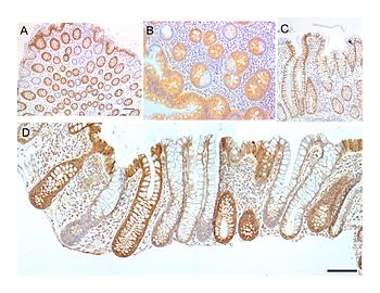
Colonic crypts (intestinal glands) within four tissue sections. In panel A, the bar shows 100 μm and allows an estimate of the frequency of crypts in the colonic epithelium. Panel B includes three crypts in cross-section, each with one segment deficient for CCOI expression and at least one crypt, on the right side, undergoing fission into two crypts. Panel C shows, on the left side, a crypt fissioning into two crypts. Panel D shows typical small clusters of two and three CCOI deficient crypts (the bar shows 50 μm). The images were made from original photomicrographs, but panels A, B and D were also included in an article

The intestinal glands in the colon are often referred to as colonic crypts. The epithelial inner surface of the colon is punctuated by invaginations, the colonic crypts. The colon crypts are shaped like microscopic thick-walled test tubes with a central hole down the length of the tube (the crypt lumen). Four tissue sections are shown here, two (A and B) cut across the long axes of the crypts and two (C and D) cut parallel to the long axes.

In these images the cells have been stained to show a brown-orange color if the cells produce a mitochondrial protein called cytochrome c oxidase subunit I (CCOI or COX-1). The nuclei of the cells (located at the outer edges of the cells lining the walls of the crypts) are stained blue-gray with haematoxylin. As seen in panels C and D, crypts are about 75 to about 110 cells long. The average crypt circumference is 23 cells. From the images, an average is shown to be about 1,725 to 2530 cells per colonic crypt. Another measure was attained giving a range of 1500 to 4900 cells per colonic crypt. Cells are produced at the crypt base and migrate upward along the crypt axis before being shed into the colonic lumen days later. There are 5 to 6 stem cells at the bases of the crypts.

As estimated from the image in panel A, there are about 100 colonic crypts per square millimeter of the colonic epithelium. The length of the human colon is, on average 160.5 cm (measured from the bottom of the cecum to the colorectal junction) with a range of 80 cm to 313 cm. The average inner circumference of the colon is 6.2 cm. Thus, the inner surface epithelial area of the human colon has an area, on average, of about 995 cm2, which includes 9,950,000 (close to 10 million) crypts.

In the four tissue sections shown here, many of the intestinal glands have cells with a mitochondrial DNA mutation in the CCOI gene and appear mostly white, with their main color being the blue-gray staining of the nuclei. As seen in panel B, a portion of the stem cells of three crypts appear to have a mutation in CCOI, so that 40% to 50% of the cells arising from those stem cells form a white segment in the cross cut area.

Overall, the percentage of crypts deficient for CCOI is less than 1% before age 40, but then increases linearly with age. Colonic crypts deficient for CCOI reaches, on average, 18% in women and 23% in men, by 80–84 years of age.

Crypts of the colon can reproduce by fission, as seen in panel C, where a crypt is dividing to form two crypts, and in panel B where at least one crypt appears to be fissioning. Most crypts deficient in CCOI are in clusters of crypts (clones of crypts) with two or more CCOI-deficient crypts adjacent to each other (see panel D).

Clinical significance

Crypt inflammation is known as cryptitis and characterized by the presence of neutrophils between the enterocytes. A severe cryptitis may lead to a crypt abscess.

Pathologic processes that lead to Crohn's disease, i.e. progressive intestinal crypt destruction, are associated with branching of the crypts.

Causes of crypt branching include:

Research

Intestinal glands contain adult stem cells referred to as intestinal stem cells. These cells have been used in the field of stem biology to further understand stem cell niches, and to generate intestinal organoids.

History

The crypts of Lieberkühn are named after the eighteenth-century German anatomist Johann Nathanael Lieberkühn.

Digestive enzyme

From Wikipedia, the free encyclopedia
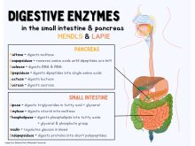
Diagram of the digestive enzymes in the small intestine and pancreas

Digestive enzymes take part in the chemical process of digestion, which follows the mechanical process of digestion. Food consists of macromolecules of proteins, carbohydrates, and fats that need to be broken down chemically by digestive enzymes in the mouth, stomach, pancreas, and duodenum, before being able to be absorbed into the bloodstream. Initial breakdown is achieved by chewing (mastication) and the use of digestive enzymes of saliva. Once in the stomach further mechanical churning takes place mixing the food with secreted gastric acid. Digestive gastric enzymes take part in some of the chemical process needed for absorption. Most of the enzymatic activity, and hence absorption takes place in the duodenum.

Digestive enzymes are found in the digestive tracts of animals (including humans) and in the tracts of carnivorous plants, where they aid in the digestion of food, as well as inside cells, especially in their lysosomes, where they function to maintain cellular survival.

Digestive enzymes are classified based on their target substrates: lipases split fatty acids into fats and oils; proteases and peptidases split proteins into small peptides and amino acids; amylases split carbohydrates such as starch and sugars into simple sugars such as glucose, and nucleases split nucleic acids into nucleotides.

Types

Table of the different major digestive enzymes

Digestive enzymes are found throughout much of the gastrointestinal tract. In the human digestive system, the main sites of digestion are the mouth, stomach, and small intestine. Digestive enzymes are secreted by different exocrine glands including salivary glands, gastric glands, secretory cells in the pancreas, and secretory glands in the small intestine. In some carnivorous plants plant-specific digestive enzymes are used to break down their captured organisms.

Mouth

Complex food substances that are eaten must be broken down into simple, soluble, and diffusible substances before they can be absorbed. In the oral cavity, salivary glands secrete an array of enzymes and substances that aid in digestion and also disinfection. They include the following:

  • Lingual lipase: Lipid digestion initiates in the mouth. Lingual lipase starts the digestion of the lipids/fats.
  • Salivary amylase: Carbohydrate digestion also initiates in the mouth. Amylase, produced by the salivary glands, breaks complex carbohydrates, mainly cooked starch, to smaller chains, or even simple sugars. It is sometimes referred to as ptyalin.
  • Lysozyme: Considering that food contains more than just essential nutrients, e.g. bacteria or viruses, the lysozyme offers a limited and non-specific, yet beneficial antiseptic function in digestion.

Of note is the diversity of the salivary glands. There are two types of salivary glands:

  • Serous glands: These glands produce a secretion rich in water, electrolytes, and enzymes. A great example of a serous oral gland is the parotid gland.
  • Mixed glands: These glands have both serous cells and mucous cells, and include sublingual and submandibular glands. Their secretion is mucinous and high in viscosity.

Stomach

The enzymes that are secreted in the stomach are gastric enzymes. The stomach plays a major role in digestion, both in a mechanical sense by mixing and crushing the food, and also in an enzymatic sense, by digesting it. The following are enzymes produced by the stomach and their respective function:

  • Pepsin is the main gastric enzyme. It is produced by the stomach cells called "chief cells" in its inactive form pepsinogen, which is a zymogen. Pepsinogen is then activated by the stomach acid into its active form, pepsin. Pepsin breaks down the protein in the food into smaller particles, such as peptide fragments and amino acids. Protein digestion, therefore, primarily starts in the stomach, unlike carbohydrate and lipids, which start their digestion in the mouth (however, trace amounts of the enzyme kallikrein, which catabolises certain protein, is found in saliva in the mouth).
  • Gastric lipase: Gastric lipase is an acidic lipase secreted by the gastric chief cells in the fundic mucosa of the stomach. It has a pH level of 3–6. Gastric lipase, together with lingual lipase, comprise the two acidic lipases. These lipases, unlike alkaline lipases (such as pancreatic lipase), do not require bile acid or colipase for optimal enzymatic activity. Acidic lipases make up 30% of lipid hydrolysis occurring during digestion in the human adult, with gastric lipase contributing the most of the two acidic lipases. In neonates, acidic lipases are much more important, providing up to 50% of total lipolytic activity.
  • Cathepsin F: is a cysteine protease.

Pancreas

Pancreas is both an endocrine and an exocrine gland, in that it functions to produce endocrinic hormones released into the circulatory system (such as insulin, and glucagon), to control glucose metabolism, and also to secrete digestive/exocrinic pancreatic juice, which is secreted eventually via the pancreatic duct into the duodenum. Digestive or exocrine function of pancreas is as significant to the maintenance of health as its endocrine function.

Two of the population of cells in the pancreatic parenchyma make up its digestive enzymes:

  • Ductal cells: Mainly responsible for production of bicarbonate (HCO3), which acts to neutralize the acidity of the stomach chyme entering duodenum through the pylorus. Ductal cells of the pancreas are stimulated by the hormone secretin to produce their bicarbonate-rich secretions, in what is in essence a bio-feedback mechanism; highly acidic stomach chyme entering the duodenum stimulates duodenal cells called "S cells" to produce the hormone secretin and release to the bloodstream. Secretin having entered the blood eventually comes into contact with the pancreatic ductal cells, stimulating them to produce their bicarbonate-rich juice. Secretin also inhibits production of gastrin by "G cells", and also stimulates acinar cells of the pancreas to produce their pancreatic enzyme.
  • Acinar cells: Mainly responsible for production of the inactive pancreatic enzymes (zymogens) that, once present in the small bowel, become activated and perform their major digestive functions by breaking down proteins, fat, and DNA/RNA. Acinar cells are stimulated by cholecystokinin (CCK), which is a hormone/neurotransmitter produced by the intestinal cells (I cells) in the duodenum. CCK stimulates production of the pancreatic zymogens.

Pancreatic juice, composed of the secretions of both ductal and acinar cells, contains the following digestive enzymes:

Some of the preceding endogenous enzymes have pharmaceutical counterparts (pancreatic enzymes) that are administered to people with exocrine pancreatic insufficiency.

The pancreas's exocrine function owes part of its notable reliability to biofeedback mechanisms controlling secretion of the juice. The following significant pancreatic biofeedback mechanisms are essential to the maintenance of pancreatic juice balance/production:

  • Secretin, a hormone produced by the duodenal "S cells" in response to the stomach chyme containing high hydrogen atom concentration (high acidicity), is released into the blood stream; upon return to the digestive tract, secretion decreases gastric emptying, increases secretion of the pancreatic ductal cells, as well as stimulating pancreatic acinar cells to release their zymogenic juice.
  • Cholecystokinin (CCK) is a unique peptide released by the duodenal "I cells" in response to chyme containing high fat or protein content. Unlike secretin, which is an endocrine hormone, CCK actually works via stimulation of a neuronal circuit, the end-result of which is stimulation of the acinar cells to release their content. CCK also increases gallbladder contraction, resulting in bile squeezed into the cystic duct, common bile duct and eventually the duodenum. Bile of course helps absorption of the fat by emulsifying it, increasing its absorptive surface. Bile is made by the liver, but is stored in the gallbladder.
  • Gastric inhibitory peptide (GIP) is produced by the mucosal duodenal cells in response to chyme containing high amounts of carbohydrate, proteins, and fatty acids. Main function of GIP is to decrease gastric emptying.
  • Somatostatin is a hormone produced by the mucosal cells of the duodenum and also the "delta cells" of the pancreas. Somatostatin has a major inhibitory effect, including on pancreatic production.

Duodenum

The following enzymes/hormones are produced in the duodenum:

  • secretin: This is an endocrine hormone produced by the duodenal "S cells" in response to the acidity of the gastric chyme.
  • Cholecystokinin (CCK) is a unique peptide released by the duodenal "I cells" in response to chyme containing high fat or protein content. Unlike secretin, which is an endocrine hormone, CCK actually works via stimulation of a neuronal circuit, the end-result of which is stimulation of the acinar cells to release their content. CCK also increases gallbladder contraction, causing release of pre-stored bile into the cystic duct, and eventually into the common bile duct and via the ampulla of Vater into the second anatomic position of the duodenum. CCK also decreases the tone of the sphincter of Oddi, which is the sphincter that regulates flow through the ampulla of Vater. CCK also decreases gastric activity and decreases gastric emptying, thereby giving more time to the pancreatic juices to neutralize the acidity of the gastric chyme.
  • Gastric inhibitory peptide (GIP): This peptide decreases gastric motility and is produced by duodenal mucosal cells.
  • motilin: This substance increases gastro-intestinal motility via specialized receptors called "motilin receptors".
  • somatostatin: This hormone is produced by duodenal mucosa and also by the delta cells of the pancreas. Its main function is to inhibit a variety of secretory mechanisms.

Throughout the lining of the small intestine there are numerous brush border enzymes whose function is to further break down the chyme released from the stomach into absorbable particles. These enzymes are absorbed whilst peristalsis occurs. Some of these enzymes include:

  • Various exopeptidases and endopeptidases including dipeptidase and aminopeptidases that convert peptones and polypeptides into amino acids.
  • Maltase: converts maltose into glucose.
  • Lactase: This is a significant enzyme that converts lactose into glucose and galactose. A majority of Middle-Eastern and Asian populations lack this enzyme. This enzyme also decreases with age. As such lactose intolerance is often a common abdominal complaint in the Middle-Eastern, Asian, and older populations, manifesting with bloating, abdominal pain, and osmotic diarrhea.
  • Sucrase: converts sucrose into glucose and fructose.
  • Other disaccharidases

Plants

In carnivorous plants, digestive enzymes and acids break down insects and in some plants small animals. In some plants, the leaf collapses on the prey to increase contact, others have a small vessel of digestive liquid. Then digestion fluids are used to digest the prey to get at the needed nitrates and phosphorus. The absorption of the needed nutrients are usually more efficient than in other plants. Digestive enzymes independently came about in carnivorous plants and animals.

Some carnivorous plants like the Heliamphora do not use digestive enzymes, but use bacteria to break down the food. These plants do not have digestive juices, but use the rot of the prey.

Some carnivorous plants digestive enzymes:

Endoplasmic reticulum

From Wikipedia, the free encyclopedia
https://en.wikipedia.org/wiki/Endoplasmic_reticulum

Cell biology
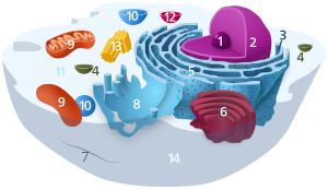
Animal cell diagram
Micrograph of rough endoplasmic reticulum network around the nucleus (shown in the lower right-hand area of the picture). Dark small circles in the network are mitochondria.

The endoplasmic reticulum (ER) is a part of a transportation system of the eukaryotic cell, and has many other important functions such as protein folding. It is a type of organelle made up of two subunits – rough endoplasmic reticulum (RER), and smooth endoplasmic reticulum (SER). The endoplasmic reticulum is found in most eukaryotic cells and forms an interconnected network of flattened, membrane-enclosed sacs known as cisternae (in the RER), and tubular structures in the SER. The membranes of the ER are continuous with the outer nuclear membrane. The endoplasmic reticulum is not found in red blood cells, or spermatozoa.

The two types of ER share many of the same proteins and engage in certain common activities such as the synthesis of certain lipids and cholesterol. Different types of cells contain different ratios of the two types of ER depending on the activities of the cell. RER is found mainly toward the nucleus of cell and SER towards the cell membrane or plasma membrane of cell.

The outer (cytosolic) face of the RER is studded with ribosomes that are the sites of protein synthesis. The RER is especially prominent in cells such as hepatocytes. The SER lacks ribosomes and functions in lipid synthesis but not metabolism, the production of steroid hormones, and detoxification. The SER is especially abundant in mammalian liver and gonad cells.

The ER was observed by light microscopy by Garnier in 1897, who coined the term ergastoplasm. The lacy membranes of the endoplasmic reticulum were first seen by electron microscopy in 1945 by Keith R. Porter, Albert Claude, and Ernest F. Fullam. Later, the word reticulum, which means "network", was applied by Porter in 1953 to describe this fabric of membranes.

Structure

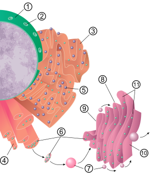
1 Nucleus   2 Nuclear pore   3 Rough endoplasmic reticulum (RER)   4 Smooth endoplasmic reticulum (SER)   5 Ribosome on the rough ER   6 Proteins that are transported   7 Transport vesicle   8 Golgi apparatus   9 Cis face of the Golgi apparatus   10 Trans face of the Golgi apparatus   11 Cisternae of the Golgi apparatus
3D rendering of endoplasmic reticulum

The general structure of the endoplasmic reticulum is a network of membranes called cisternae. These sac-like structures are held together by the cytoskeleton. The phospholipid membrane encloses the cisternal space (or lumen), which is continuous with the perinuclear space but separate from the cytosol. The functions of the endoplasmic reticulum can be summarized as the synthesis and export of proteins and membrane lipids, but varies between ER and cell type and cell function. The quantity of both rough and smooth endoplasmic reticulum in a cell can slowly interchange from one type to the other, depending on the changing metabolic activities of the cell. Transformation can include embedding of new proteins in membrane as well as structural changes. Changes in protein content may occur without noticeable structural changes.

Rough endoplasmic reticulum

A 2-minute animation showing how a protein destined for the secretory pathway is synthesized and secreted into the rough endoplasmic reticulum, which appears at the upper right approximately halfway through the animation

The surface of the rough endoplasmic reticulum (often abbreviated RER or rough ER; also called granular endoplasmic reticulum) is studded with protein-manufacturing ribosomes giving it a "rough" appearance (hence its name). The binding site of the ribosome on the rough endoplasmic reticulum is the translocon. However, the ribosomes are not a stable part of this organelle's structure as they are constantly being bound and released from the membrane. A ribosome only binds to the RER once a specific protein-nucleic acid complex forms in the cytosol. This special complex forms when a free ribosome begins translating the mRNA of a protein destined for the secretory pathway. The first 5–30 amino acids polymerized encode a signal peptide, a molecular message that is recognized and bound by a signal recognition particle (SRP). Translation pauses and the ribosome complex binds to the RER translocon where translation continues with the nascent (new) protein forming into the RER lumen and/or membrane. The protein is processed in the ER lumen by an enzyme (a signal peptidase), which removes the signal peptide. Ribosomes at this point may be released back into the cytosol; however, non-translating ribosomes are also known to stay associated with translocons.

The membrane of the rough endoplasmic reticulum is in the form of large double-membrane sheets that are located near, and continuous with, the outer layer of the nuclear envelope. The double membrane sheets are stacked and connected through several right- or left-handed helical ramps, the "Terasaki ramps", giving rise to a structure resembling a parking garage. Although there is no continuous membrane between the endoplasmic reticulum and the Golgi apparatus, membrane-bound transport vesicles shuttle proteins between these two compartments. Vesicles are surrounded by coating proteins called COPI and COPII. COPII targets vesicles to the Golgi apparatus and COPI marks them to be brought back to the rough endoplasmic reticulum. The rough endoplasmic reticulum works in concert with the Golgi complex to target new proteins to their proper destinations. The second method of transport out of the endoplasmic reticulum involves areas called membrane contact sites, where the membranes of the endoplasmic reticulum and other organelles are held closely together, allowing the transfer of lipids and other small molecules.

The rough endoplasmic reticulum is key in multiple functions:

  • Manufacture of lysosomal enzymes with a mannose-6-phosphate marker added in the cis-Golgi network.
  • Manufacture of secreted proteins, either secreted constitutively with no tag or secreted in a regulatory manner involving clathrin and paired basic amino acids in the signal peptide.
  • Integral membrane proteins that stay embedded in the membrane as vesicles exit and bind to new membranes. Rab proteins are key in targeting the membrane; SNAP and SNARE proteins are key in the fusion event.
  • Initial glycosylation as assembly continues. This is N-linked (O-linking occurs in the Golgi).
    • N-linked glycosylation: If the protein is properly folded, oligosaccharyltransferase recognizes the AA sequence NXS or NXT (with the S/T residue phosphorylated) and adds a 14-sugar backbone (2-N-acetylglucosamine, 9-branching mannose, and 3-glucose at the end) to the side-chain nitrogen of Asn.

Smooth endoplasmic reticulum

Electron micrograph showing smooth ER (arrow) in mouse tissue, at 110,510× magnification

In most cells the smooth endoplasmic reticulum (abbreviated SER) is scarce. Instead there are areas where the ER is partly smooth and partly rough, this area is called the transitional ER. The transitional ER gets its name because it contains ER exit sites. These are areas where the transport vesicles which contain lipids and proteins made in the ER, detach from the ER and start moving to the Golgi apparatus. Specialized cells can have a lot of smooth endoplasmic reticulum and in these cells the smooth ER has many functions. It synthesizes lipids, phospholipids, and steroids. Cells which secrete these products, such as those in the testes, ovaries, and sebaceous glands have an abundance of smooth endoplasmic reticulum. It also carries out the metabolism of carbohydrates, detoxification of natural metabolism products and of alcohol and drugs, attachment of receptors on cell membrane proteins, and steroid metabolism. In muscle cells, it regulates calcium ion concentration. Smooth endoplasmic reticulum is found in a variety of cell types (both animal and plant), and it serves different functions in each. The smooth endoplasmic reticulum also contains the enzyme glucose-6-phosphatase, which converts glucose-6-phosphate to glucose, a step in gluconeogenesis. It is connected to the nuclear envelope and consists of tubules that are located near the cell periphery. These tubes sometimes branch forming a network that is reticular in appearance. In some cells, there are dilated areas like the sacs of rough endoplasmic reticulum. The network of smooth endoplasmic reticulum allows for an increased surface area to be devoted to the action or storage of key enzymes and the products of these enzymes.

Sarcoplasmic reticulum

Skeletal muscle fiber, with sarcoplasmic reticulum colored in blue

The sarcoplasmic reticulum (SR), from the Greek σάρξ sarx ("flesh"), is smooth ER found in muscle cells. The only structural difference between this organelle and the smooth endoplasmic reticulum is the composition of proteins they have, both bound to their membranes and drifting within the confines of their lumens. This fundamental difference is indicative of their functions: The endoplasmic reticulum synthesizes molecules, while the sarcoplasmic reticulum stores calcium ions and pumps them out into the sarcoplasm when the muscle fiber is stimulated. After their release from the sarcoplasmic reticulum, calcium ions interact with contractile proteins that utilize ATP to shorten the muscle fiber. The sarcoplasmic reticulum plays a major role in excitation-contraction coupling.

Functions

The endoplasmic reticulum serves many general functions, including the folding of protein molecules in sacs called cisternae and the transport of synthesized proteins in vesicles to the Golgi apparatus. Rough endoplasmic reticulum is also involved in protein synthesis. Correct folding of newly made proteins is made possible by several endoplasmic reticulum chaperone proteins, including protein disulfide isomerase (PDI), ERp29, the Hsp70 family member BiP/Grp78, calnexin, calreticulin, and the peptidylprolyl isomerase family. Only properly folded proteins are transported from the rough ER to the Golgi apparatus – unfolded proteins cause an unfolded protein response as a stress response in the ER. Disturbances in redox regulation, calcium regulation, glucose deprivation, and viral infection or the over-expression of proteins can lead to endoplasmic reticulum stress response (ER stress), a state in which the folding of proteins slows, leading to an increase in unfolded proteins. This stress is emerging as a potential cause of damage in hypoxia/ischemia, insulin resistance, and other disorders.

Protein transport

Secretory proteins, mostly glycoproteins, are moved across the endoplasmic reticulum membrane. Proteins that are transported by the endoplasmic reticulum throughout the cell are marked with an address tag called a signal sequence. The N-terminus (one end) of a polypeptide chain (i.e., a protein) contains a few amino acids that work as an address tag, which are removed when the polypeptide reaches its destination. Nascent peptides reach the ER via the translocon, a membrane-embedded multiprotein complex. Proteins that are destined for places outside the endoplasmic reticulum are packed into transport vesicles and moved along the cytoskeleton toward their destination. In human fibroblasts, the ER is always co-distributed with microtubules and the depolymerisation of the latter cause its co-aggregation with mitochondria, which are also associated with the ER.

The endoplasmic reticulum is also part of a protein sorting pathway. It is, in essence, the transportation system of the eukaryotic cell. The majority of its resident proteins are retained within it through a retention motif. This motif is composed of four amino acids at the end of the protein sequence. The most common retention sequences are KDEL for lumen-located proteins and KKXX for transmembrane proteins. However, variations of KDEL and KKXX do occur, and other sequences can also give rise to endoplasmic reticulum retention. It is not known whether such variation can lead to sub-ER localizations. There are three KDEL (1, 2 and 3) receptors in mammalian cells, and they have a very high degree of sequence identity. The functional differences between these receptors remain to be established.

Bioenergetics regulation of ER ATP supply by a CaATiER mechanism

Ca2+-antagonized transport into the endoplasmic reticulum (CaATiER) model

The endoplasmic reticulum does not harbor an ATP-regeneration machinery, and therefore requires ATP import from mitochondria. The imported ATP is vital for the ER to carry out its house keeping cellular functions, such as for protein folding and trafficking.

The ER ATP transporter, SLC35B1/AXER, was recently cloned and characterized, and the mitochondria supply ATP to the ER through a Ca2+-antagonized transport into the ER (CaATiER) mechanism. The CaATiER mechanism shows sensitivity to cytosolic Ca2+ ranging from high nM to low μM range, with the Ca2+-sensing element yet to be identified and validated.

Clinical significance

Increased and supraphysiological ER stress in pancreatic β cells disrupts normal insulin secretion, leading to hyperinsulinemia and consequently peripheral insulin resistance associated with obesity in humans. Human clinical trials also suggested a causal link between obesity-induced increase in insulin secretion and peripheral insulin resistance.

Abnormalities in XBP1 lead to a heightened endoplasmic reticulum stress response and subsequently causes a higher susceptibility for inflammatory processes that may even contribute to Alzheimer's disease. In the colon, XBP1 anomalies have been linked to the inflammatory bowel diseases including Crohn's disease.

The unfolded protein response (UPR) is a cellular stress response related to the endoplasmic reticulum. The UPR is activated in response to an accumulation of unfolded or misfolded proteins in the lumen of the endoplasmic reticulum. The UPR functions to restore normal function of the cell by halting protein translation, degrading misfolded proteins, and activating the signaling pathways that lead to increasing the production of molecular chaperones involved in protein folding. Sustained overactivation of the UPR has been implicated in prion diseases as well as several other neurodegenerative diseases and the inhibition of the UPR could become a treatment for those diseases.

Child abandonment

From Wikipedia, the free encyclopedia https://en.wikipedia.org/wiki/Child_abandonment ...