Search This Blog

Thursday, August 8, 2024

Chromatography

From Wikipedia, the free encyclopedia
https://en.wikipedia.org/wiki/Chromatography

In chemical analysis, chromatography is a laboratory technique for the separation of a mixture into its components. The mixture is dissolved in a fluid solvent (gas or liquid) called the mobile phase, which carries it through a system (a column, a capillary tube, a plate, or a sheet) on which a material called the stationary phase is fixed. Because the different constituents of the mixture tend to have different affinities for the stationary phase and are retained for different lengths of time depending on their interactions with its surface sites, the constituents travel at different apparent velocities in the mobile fluid, causing them to separate. The separation is based on the differential partitioning between the mobile and the stationary phases. Subtle differences in a compound's partition coefficient result in differential retention on the stationary phase and thus affect the separation.

Chromatography may be preparative or analytical. The purpose of preparative chromatography is to separate the components of a mixture for later use, and is thus a form of purification. This process is associated with higher costs due to its mode of production. Analytical chromatography is done normally with smaller amounts of material and is for establishing the presence or measuring the relative proportions of analytes in a mixture. The two types are not mutually exclusive.

Etymology and pronunciation

Chromatography, pronounced /ˌkrməˈtɒɡrəfi/, is derived from Greek χρῶμα chroma, which means "color", and γράφειν graphein, which means "to write". The combination of these two terms was directly inherited from the invention of the technique first used to separate biological pigments.

History

Chromatography was first devised at the University of Kazan by the Italian-born Russian scientist Mikhail Tsvet in 1900. He developed the technique and coined the term chromatography in the first decade of the 20th century, primarily for the separation of plant pigments such as chlorophyll, carotenes, and xanthophylls. Since these components separate in bands of different colors (green, orange, and yellow, respectively) they directly inspired the name of the technique. New types of chromatography developed during the 1930s and 1940s made the technique useful for many separation processes.

Chromatography technique developed substantially as a result of the work of Archer John Porter Martin and Richard Laurence Millington Synge during the 1940s and 1950s, for which they won the 1952 Nobel Prize in Chemistry. They established the principles and basic techniques of partition chromatography, and their work encouraged the rapid development of several chromatographic methods: paper chromatography, gas chromatography, and what would become known as high-performance liquid chromatography. Since then, the technology has advanced rapidly. Researchers found that the main principles of Tsvet's chromatography could be applied in many different ways, resulting in the different varieties of chromatography described below. Advances are continually improving the technical performance of chromatography, allowing the separation of increasingly similar molecules.

Terms

  • Analyte – the substance to be separated during chromatography. It is also normally what is needed from the mixture.
  • Analytical chromatography – the use of chromatography to determine the existence and possibly also the concentration of analyte(s) in a sample.
  • Bonded phase – a stationary phase that is covalently bonded to the support particles or to the inside wall of the column tubing.
  • Chromatogram – the visual output of the chromatograph. In the case of an optimal separation, different peaks or patterns on the chromatogram correspond to different components of the separated mixture.
    Chromatogram with unresolved peaks Chromatogram with two resolved peaks
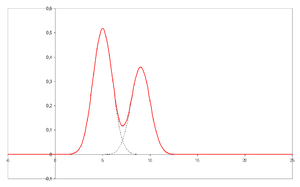
    Plotted on the x-axis is the retention time and plotted on the y-axis a signal (for example obtained by a spectrophotometer, mass spectrometer or a variety of other detectors) corresponding to the response created by the analytes exiting the system. In the case of an optimal system the signal is proportional to the concentration of the specific analyte separated.
  • Chromatograph – an instrument that enables a sophisticated separation, e.g. gas chromatographic or liquid chromatographic separation.
  • Chromatography – a physical method of separation that distributes components to separate between two phases, one stationary (stationary phase), the other (the mobile phase) moving in a definite direction.
  • Eluent (sometimes spelled eluant) – the solvent or solvent fixure used in elution chromatography and is synonymous with mobile phase.
  • Eluate – the mixture of solute (see Eluite) and solvent (see Eluent) exiting the column.
  • Effluent – the stream flowing out of a chromatographic column. In practise, it is used synonymously with eluate, but the term more precisely refers to the stream independent of separation taking place.
  • Eluite – a more precise term for solute or analyte. It is a sample component leaving the chromatographic column.
  • Eluotropic series – a list of solvents ranked according to their eluting power.
  • Immobilized phase – a stationary phase that is immobilized on the support particles, or on the inner wall of the column tubing.
  • Mobile phase – the phase that moves in a definite direction. It may be a liquid (LC and capillary electrochromatography, CEC), a gas (GC), or a supercritical fluid (supercritical-fluid chromatography, SFC). The mobile phase consists of the sample being separated/analyzed and the solvent that moves the sample through the column. In the case of HPLC the mobile phase consists of a non-polar solvent(s) such as hexane in normal phase or a polar solvent such as methanol in reverse phase chromatography and the sample being separated. The mobile phase moves through the chromatography column (the stationary phase) where the sample interacts with the stationary phase and is separated.
  • Preparative chromatography – the use of chromatography to purify sufficient quantities of a substance for further use, rather than analysis.
  • Retention time – the characteristic time it takes for a particular analyte to pass through the system (from the column inlet to the detector) under set conditions. See also: Kovats' retention index
  • Sample – the matter analyzed in chromatography. It may consist of a single component or it may be a mixture of components. When the sample is treated in the course of an analysis, the phase or the phases containing the analytes of interest is/are referred to as the sample whereas everything out of interest separated from the sample before or in the course of the analysis is referred to as waste.
  • Solute – the sample components in partition chromatography.
  • Solvent – any substance capable of solubilizing another substance, and especially the liquid mobile phase in liquid chromatography.
  • Stationary phase – the substance fixed in place for the chromatography procedure. Examples include the silica layer in thin-layer chromatography
  • Detector – the instrument used for qualitative and quantitative detection of analytes after separation.

Chromatography is based on the concept of partition coefficient. Any solute partitions between two immiscible solvents. When one make one solvent immobile (by adsorption on a solid support matrix) and another mobile it results in most common applications of chromatography. If the matrix support, or stationary phase, is polar (e.g, cellulose, silica etc.) it is forward phase chromatography. Otherwise this technique is known as reversed phase, where a non-polar stationary phase (e.g, non-polar derivative of C-18) is used.

Techniques by chromatographic bed shape

Column chromatography

Column chromatography is a separation technique in which the stationary bed is within a tube. The particles of the solid stationary phase or the support coated with a liquid stationary phase may fill the whole inside volume of the tube (packed column) or be concentrated on or along the inside tube wall leaving an open, unrestricted path for the mobile phase in the middle part of the tube (open tubular column). Differences in rates of movement through the medium are calculated to different retention times of the sample. In 1978, W. Clark Still introduced a modified version of column chromatography called flash column chromatography (flash). The technique is very similar to the traditional column chromatography, except that the solvent is driven through the column by applying positive pressure. This allowed most separations to be performed in less than 20 minutes, with improved separations compared to the old method. Modern flash chromatography systems are sold as pre-packed plastic cartridges, and the solvent is pumped through the cartridge. Systems may also be linked with detectors and fraction collectors providing automation. The introduction of gradient pumps resulted in quicker separations and less solvent usage.

In expanded bed adsorption, a fluidized bed is used, rather than a solid phase made by a packed bed. This allows omission of initial clearing steps such as centrifugation and filtration, for culture broths or slurries of broken cells.

Phosphocellulose chromatography utilizes the binding affinity of many DNA-binding proteins for phosphocellulose. The stronger a protein's interaction with DNA, the higher the salt concentration needed to elute that protein.

Planar chromatography

Planar chromatography is a separation technique in which the stationary phase is present as or on a plane. The plane can be a paper, serving as such or impregnated by a substance as the stationary bed (paper chromatography) or a layer of solid particles spread on a support such as a glass plate (thin-layer chromatography). Different compounds in the sample mixture travel different distances according to how strongly they interact with the stationary phase as compared to the mobile phase. The specific Retention factor (Rf) of each chemical can be used to aid in the identification of an unknown substance.

Paper chromatography

Paper chromatography in progress
Paper chromatography

Paper chromatography is a technique that involves placing a small dot or line of sample solution onto a strip of chromatography paper. The paper is placed in a container with a shallow layer of solvent and sealed. As the solvent rises through the paper, it meets the sample mixture, which starts to travel up the paper with the solvent. This paper is made of cellulose, a polar substance, and the compounds within the mixture travel further if they are less polar. More polar substances bond with the cellulose paper more quickly, and therefore do not travel as far.

Thin-layer chromatography (TLC)

Thin layer chromatography

Thin-layer chromatography (TLC) is a widely employed laboratory technique used to separate different biochemicals on the basis of their relative attractions to the stationary and mobile phases. It is similar to paper chromatography. However, instead of using a stationary phase of paper, it involves a stationary phase of a thin layer of adsorbent like silica gel, alumina, or cellulose on a flat, inert substrate. TLC is very versatile; multiple samples can be separated simultaneously on the same layer, making it very useful for screening applications such as testing drug levels and water purity.

Possibility of cross-contamination is low since each separation is performed on a new layer. Compared to paper, it has the advantage of faster runs, better separations, better quantitative analysis, and the choice between different adsorbents. For even better resolution and faster separation that utilizes less solvent, high-performance TLC can be used. An older popular use had been to differentiate chromosomes by observing distance in gel (separation of was a separate step).

Displacement chromatography

The basic principle of displacement chromatography is: A molecule with a high affinity for the chromatography matrix (the displacer) competes effectively for binding sites, and thus displaces all molecules with lesser affinities. There are distinct differences between displacement and elution chromatography. In elution mode, substances typically emerge from a column in narrow, Gaussian peaks. Wide separation of peaks, preferably to baseline, is desired for maximum purification. The speed at which any component of a mixture travels down the column in elution mode depends on many factors. But for two substances to travel at different speeds, and thereby be resolved, there must be substantial differences in some interaction between the biomolecules and the chromatography matrix. Operating parameters are adjusted to maximize the effect of this difference. In many cases, baseline separation of the peaks can be achieved only with gradient elution and low column loadings. Thus, two drawbacks to elution mode chromatography, especially at the preparative scale, are operational complexity, due to gradient solvent pumping, and low throughput, due to low column loadings. Displacement chromatography has advantages over elution chromatography in that components are resolved into consecutive zones of pure substances rather than "peaks". Because the process takes advantage of the nonlinearity of the isotherms, a larger column feed can be separated on a given column with the purified components recovered at significantly higher concentrations.

Techniques by physical state of mobile phase

Gas chromatography

Gas chromatography (GC), also sometimes known as gas-liquid chromatography, (GLC), is a separation technique in which the mobile phase is a gas. Gas chromatographic separation is always carried out in a column, which is typically "packed" or "capillary". Packed columns are the routine work horses of gas chromatography, being cheaper and easier to use and often giving adequate performance. Capillary columns generally give far superior resolution and although more expensive are becoming widely used, especially for complex mixtures. Further, capillary columns can be split into three classes: porous layer open tubular (PLOT), wall-coated open tubular (WCOT) and support-coated open tubular (SCOT) columns. PLOT columns are unique in a way that the stationary phase is adsorbed to the column walls, while WCOT columns have a stationary phase that is chemically bonded to the walls. SCOT columns are in a way the combination of the two types mentioned in a way that they have support particles adhered to column walls, but those particles have liquid phase chemically bonded onto them. Both types of column are made from non-adsorbent and chemically inert materials. Stainless steel and glass are the usual materials for packed columns and quartz or fused silica for capillary columns.

Gas chromatography is based on a partition equilibrium of analyte between a solid or viscous liquid stationary phase (often a liquid silicone-based material) and a mobile gas (most often helium). The stationary phase is adhered to the inside of a small-diameter (commonly 0.53 – 0.18mm inside diameter) glass or fused-silica tube (a capillary column) or a solid matrix inside a larger metal tube (a packed column). It is widely used in analytical chemistry; though the high temperatures used in GC make it unsuitable for high molecular weight biopolymers or proteins (heat denatures them), frequently encountered in biochemistry, it is well suited for use in the petrochemical, environmental monitoring and remediation, and industrial chemical fields. It is also used extensively in chemistry research.

Liquid chromatography

Preparative HPLC apparatus

Liquid chromatography (LC) is a separation technique in which the mobile phase is a liquid. It can be carried out either in a column or a plane. Present day liquid chromatography that generally utilizes very small packing particles and a relatively high pressure is referred to as high-performance liquid chromatography.

In HPLC the sample is forced by a liquid at high pressure (the mobile phase) through a column that is packed with a stationary phase composed of irregularly or spherically shaped particles, a porous monolithic layer, or a porous membrane. Monoliths are "sponge-like chromatographic media" and are made up of an unending block of organic or inorganic parts. HPLC is historically divided into two different sub-classes based on the polarity of the mobile and stationary phases. Methods in which the stationary phase is more polar than the mobile phase (e.g., toluene as the mobile phase, silica as the stationary phase) are termed normal phase liquid chromatography (NPLC) and the opposite (e.g., water-methanol mixture as the mobile phase and C18 (octadecylsilyl) as the stationary phase) is termed reversed phase liquid chromatography (RPLC).

Supercritical fluid chromatography

Supercritical fluid chromatography is a separation technique in which the mobile phase is a fluid above and relatively close to its critical temperature and pressure.

Specific techniques under this broad heading are listed below.

Affinity chromatography

Affinity chromatography is based on selective non-covalent interaction between an analyte and specific molecules. It is very specific, but not very robust. It is often used in biochemistry in the purification of proteins bound to tags. These fusion proteins are labeled with compounds such as His-tags, biotin or antigens, which bind to the stationary phase specifically. After purification, these tags are usually removed and the pure protein is obtained.

Affinity chromatography often utilizes a biomolecule's affinity for the cations of a metal (Zn, Cu, Fe, etc.). Columns are often manually prepared and could be designed specifically for the proteins of interest. Traditional affinity columns are used as a preparative step to flush out unwanted biomolecules, or as a primary step in analyzing a protein with unknown physical properties.

However, liquid chromatography techniques exist that do utilize affinity chromatography properties. Immobilized metal affinity chromatography (IMAC) is useful to separate the aforementioned molecules based on the relative affinity for the metal. Often these columns can be loaded with different metals to create a column with a targeted affinity. 

Techniques by separation mechanism

Ion exchange chromatography

Ion exchange chromatography (usually referred to as ion chromatography) uses an ion exchange mechanism to separate analytes based on their respective charges. It is usually performed in columns but can also be useful in planar mode. Ion exchange chromatography uses a charged stationary phase to separate charged compounds including anions, cations, amino acids, peptides, and proteins. In conventional methods the stationary phase is an ion-exchange resin that carries charged functional groups that interact with oppositely charged groups of the compound to retain. There are two types of ion exchange chromatography: Cation-Exchange and Anion-Exchange. In the Cation-Exchange Chromatography the stationary phase has negative charge and the exchangeable ion is a cation, whereas, in the Anion-Exchange Chromatography the stationary phase has positive charge and the exchangeable ion is an anion. Ion exchange chromatography is commonly used to purify proteins using FPLC.

Size-exclusion chromatography

Size-exclusion chromatography (SEC) is also known as gel permeation chromatography (GPC) or gel filtration chromatography and separates molecules according to their size (or more accurately according to their hydrodynamic diameter or hydrodynamic volume). Smaller molecules are able to enter the pores of the media and, therefore, molecules are trapped and removed from the flow of the mobile phase. The average residence time in the pores depends upon the effective size of the analyte molecules. However, molecules that are larger than the average pore size of the packing are excluded and thus suffer essentially no retention; such species are the first to be eluted. It is generally a low-resolution chromatography technique and thus it is often reserved for the final, "polishing" step of a purification. It is also useful for determining the tertiary structure and quaternary structure of purified proteins, especially since it can be carried out under native solution conditions.

Expanded bed adsorption chromatographic separation

An expanded bed chromatographic adsorption (EBA) column for a biochemical separation process comprises a pressure equalization liquid distributor having a self-cleaning function below a porous blocking sieve plate at the bottom of the expanded bed, an upper part nozzle assembly having a backflush cleaning function at the top of the expanded bed, a better distribution of the feedstock liquor added into the expanded bed ensuring that the fluid passed through the expanded bed layer displays a state of piston flow. The expanded bed layer displays a state of piston flow. The expanded bed chromatographic separation column has advantages of increasing the separation efficiency of the expanded bed.

Expanded-bed adsorption (EBA) chromatography is a convenient and effective technique for the capture of proteins directly from unclarified crude sample. In EBA chromatography, the settled bed is first expanded by upward flow of equilibration buffer. The crude feed, which is a mixture of soluble proteins, contaminants, cells, and cell debris, is then passed upward through the expanded bed. Target proteins are captured on the adsorbent, while particulates and contaminants pass through. A change to elution buffer while maintaining upward flow results in desorption of the target protein in expanded-bed mode. Alternatively, if the flow is reversed, the adsorbed particles will quickly settle and the proteins can be desorbed by an elution buffer. The mode used for elution (expanded-bed versus settled-bed) depends on the characteristics of the feed. After elution, the adsorbent is cleaned with a predefined cleaning-in-place (CIP) solution, with cleaning followed by either column regeneration (for further use) or storage.

Special techniques

Reversed-phase chromatography

Reversed-phase chromatography (RPC) is any liquid chromatography procedure in which the mobile phase is significantly more polar than the stationary phase. It is so named because in normal-phase liquid chromatography, the mobile phase is significantly less polar than the stationary phase. Hydrophobic molecules in the mobile phase tend to adsorb to the relatively hydrophobic stationary phase. Hydrophilic molecules in the mobile phase will tend to elute first. Separating columns typically comprise a C8 or C18 carbon-chain bonded to a silica particle substrate.

Hydrophobic interaction chromatography

Hydrophobic Interaction Chromatography (HIC) is a purification and analytical technique that separates analytes, such as proteins, based on hydrophobic interactions between that analyte and the chromatographic matrix. It can provide a non-denaturing orthogonal approach to reversed phase separation, preserving native structures and potentially protein activity. In hydrophobic interaction chromatography, the matrix material is lightly substituted with hydrophobic groups. These groups can range from methyl, ethyl, propyl, butyl, octyl, or phenyl groups. At high salt concentrations, non-polar sidechains on the surface on proteins "interact" with the hydrophobic groups; that is, both types of groups are excluded by the polar solvent (hydrophobic effects are augmented by increased ionic strength). Thus, the sample is applied to the column in a buffer which is highly polar, which drives an association of hydrophobic patches on the analyte with the stationary phase. The eluent is typically an aqueous buffer with decreasing salt concentrations, increasing concentrations of detergent (which disrupts hydrophobic interactions), or changes in pH. Of critical importance is the type of salt used, with more kosmotropic salts as defined by the Hofmeister series providing the most water structuring around the molecule and resulting hydrophobic pressure. Ammonium sulfate is frequently used for this purpose. The addition of organic solvents or other less polar constituents may assist in improving resolution.

In general, Hydrophobic Interaction Chromatography (HIC) is advantageous if the sample is sensitive to pH change or harsh solvents typically used in other types of chromatography but not high salt concentrations. Commonly, it is the amount of salt in the buffer which is varied. In 2012, Müller and Franzreb described the effects of temperature on HIC using Bovine Serum Albumin (BSA) with four different types of hydrophobic resin. The study altered temperature as to effect the binding affinity of BSA onto the matrix. It was concluded that cycling temperature from 40 to 10 degrees Celsius would not be adequate to effectively wash all BSA from the matrix but could be very effective if the column would only be used a few times. Using temperature to effect change allows labs to cut costs on buying salt and saves money.

If high salt concentrations along with temperature fluctuations want to be avoided one can use a more hydrophobic to compete with one's sample to elute it. This so-called salt independent method of HIC showed a direct isolation of Human Immunoglobulin G (IgG) from serum with satisfactory yield and used β-cyclodextrin as a competitor to displace IgG from the matrix. This largely opens up the possibility of using HIC with samples which are salt sensitive as we know high salt concentrations precipitate proteins.

Hydrodynamic chromatography

Hydrodynamic chromatography (HDC) is derived from the observed phenomenon that large droplets move faster than small ones. In a column, this happens because the center of mass of larger droplets is prevented from being as close to the sides of the column as smaller droplets because of their larger overall size. Larger droplets will elute first from the middle of the column while smaller droplets stick to the sides of the column and elute last. This form of chromatography is useful for separating analytes by molar mass (or molecular mass), size, shape, and structure when used in conjunction with light scattering detectors, viscometers, and refractometers. The two main types of HDC are open tube and packed column. Open tube offers rapid separation times for small particles, whereas packed column HDC can increase resolution and is better suited for particles with an average molecular mass larger than daltons. HDC differs from other types of chromatography because the separation only takes place in the interstitial volume, which is the volume surrounding and in between particles in a packed column.

HDC shares the same order of elution as Size Exclusion Chromatography (SEC) but the two processes still vary in many ways. In a study comparing the two types of separation, Isenberg, Brewer, Côté, and Striegel use both methods for polysaccharide characterization and conclude that HDC coupled with multiangle light scattering (MALS) achieves more accurate molar mass distribution when compared to off-line MALS than SEC in significantly less time. This is largely due to SEC being a more destructive technique because of the pores in the column degrading the analyte during separation, which tends to impact the mass distribution. However, the main disadvantage of HDC is low resolution of analyte peaks, which makes SEC a more viable option when used with chemicals that are not easily degradable and where rapid elution is not important.

HDC plays an especially important role in the field of microfluidics. The first successful apparatus for HDC-on-a-chip system was proposed by Chmela, et al. in 2002. Their design was able to achieve separations using an 80 mm long channel on the timescale of 3 minutes for particles with diameters ranging from 26 to 110 nm, but the authors expressed a need to improve the retention and dispersion parameters. In a 2010 publication by Jellema, Markesteijn, Westerweel, and Verpoorte, implementing HDC with a recirculating bidirectional flow resulted in high resolution, size based separation with only a 3 mm long channel. Having such a short channel and high resolution was viewed as especially impressive considering that previous studies used channels that were 80 mm in length. For a biological application, in 2007, Huh, et al. proposed a microfluidic sorting device based on HDC and gravity, which was useful for preventing potentially dangerous particles with diameter larger than 6 microns from entering the bloodstream when injecting contrast agents in ultrasounds. This study also made advances for environmental sustainability in microfluidics due to the lack of outside electronics driving the flow, which came as an advantage of using a gravity based device.

Two-dimensional chromatograph GCxGC-TOFMS at Chemical Faculty of GUT Gdańsk, Poland, 2016

Two-dimensional chromatography

In some cases, the selectivity provided by the use of one column can be insufficient to provide resolution of analytes in complex samples. Two-dimensional chromatography aims to increase the resolution of these peaks by using a second column with different physico-chemical (chemical classification) properties. Since the mechanism of retention on this new solid support is different from the first dimensional separation, it can be possible to separate compounds by two-dimensional chromatography that are indistinguishable by one-dimensional chromatography. Furthermore, the separation on the second dimension occurs faster than the first dimension. An example of a TDC separation is where the sample is spotted at one corner of a square plate, developed, air-dried, then rotated by 90° and usually redeveloped in a second solvent system.

Two-dimensional chromatography can be applied to GC or LC separations. The heart-cutting approach selects a specific region of interest on the first dimension for separation, and the comprehensive approach uses all analytes in the second-dimension separation.

Simulated moving-bed chromatography

The simulated moving bed (SMB) technique is a variant of high performance liquid chromatography; it is used to separate particles and/or chemical compounds that would be difficult or impossible to resolve otherwise. This increased separation is brought about by a valve-and-column arrangement that is used to lengthen the stationary phase indefinitely. In the moving bed technique of preparative chromatography the feed entry and the analyte recovery are simultaneous and continuous, but because of practical difficulties with a continuously moving bed, simulated moving bed technique was proposed. In the simulated moving bed technique instead of moving the bed, the sample inlet and the analyte exit positions are moved continuously, giving the impression of a moving bed. True moving bed chromatography (TMBC) is only a theoretical concept. Its simulation, SMBC is achieved by the use of a multiplicity of columns in series and a complex valve arrangement. This valve arrangement provides for sample and solvent feed and analyte and waste takeoff at appropriate locations of any column, whereby it allows switching at regular intervals the sample entry in one direction, the solvent entry in the opposite direction, whilst changing the analyte and waste takeoff positions appropriately as well.

Pyrolysis gas chromatography

Pyrolysis–gas chromatography–mass spectrometry is a method of chemical analysis in which the sample is heated to decomposition to produce smaller molecules that are separated by gas chromatography and detected using mass spectrometry.

Pyrolysis is the thermal decomposition of materials in an inert atmosphere or a vacuum. The sample is put into direct contact with a platinum wire, or placed in a quartz sample tube, and rapidly heated to 600–1000 °C. Depending on the application even higher temperatures are used. Three different heating techniques are used in actual pyrolyzers: Isothermal furnace, inductive heating (Curie point filament), and resistive heating using platinum filaments. Large molecules cleave at their weakest points and produce smaller, more volatile fragments. These fragments can be separated by gas chromatography. Pyrolysis GC chromatograms are typically complex because a wide range of different decomposition products is formed. The data can either be used as fingerprints to prove material identity or the GC/MS data is used to identify individual fragments to obtain structural information. To increase the volatility of polar fragments, various methylating reagents can be added to a sample before pyrolysis.

Besides the usage of dedicated pyrolyzers, pyrolysis GC of solid and liquid samples can be performed directly inside Programmable Temperature Vaporizer (PTV) injectors that provide quick heating (up to 30 °C/s) and high maximum temperatures of 600–650 °C. This is sufficient for some pyrolysis applications. The main advantage is that no dedicated instrument has to be purchased and pyrolysis can be performed as part of routine GC analysis. In this case, quartz GC inlet liners have to be used. Quantitative data can be acquired, and good results of derivatization inside the PTV injector are published as well.

Fast protein liquid chromatography

Fast protein liquid chromatography (FPLC), is a form of liquid chromatography that is often used to analyze or purify mixtures of proteins. As in other forms of chromatography, separation is possible because the different components of a mixture have different affinities for two materials, a moving fluid (the "mobile phase") and a porous solid (the stationary phase). In FPLC the mobile phase is an aqueous solution, or "buffer". The buffer flow rate is controlled by a positive-displacement pump and is normally kept constant, while the composition of the buffer can be varied by drawing fluids in different proportions from two or more external reservoirs. The stationary phase is a resin composed of beads, usually of cross-linked agarose, packed into a cylindrical glass or plastic column. FPLC resins are available in a wide range of bead sizes and surface ligands depending on the application.

Countercurrent chromatography

Countercurrent chromatography (CCC) is a type of liquid-liquid chromatography, where both the stationary and mobile phases are liquids and the liquid stationary phase is held stagnant by a strong centrifugal force.

Hydrodynamic countercurrent chromatography (CCC)

The operating principle of CCC instrument requires a column consisting of an open tube coiled around a bobbin. The bobbin is rotated in a double-axis gyratory motion (a cardioid), which causes a variable gravity (G) field to act on the column during each rotation. This motion causes the column to see one partitioning step per revolution and components of the sample separate in the column due to their partitioning coefficient between the two immiscible liquid phases used. There are many types of CCC available today. These include HSCCC (High Speed CCC) and HPCCC (High Performance CCC). HPCCC is the latest and best-performing version of the instrumentation available currently.

Centrifugal partition chromatography (CPC)

In the CPC (centrifugal partition chromatography or hydrostatic countercurrent chromatography) instrument, the column consists of a series of cells interconnected by ducts attached to a rotor. This rotor rotates on its central axis creating the centrifugal field necessary to hold the stationary phase in place. The separation process in CPC is governed solely by the partitioning of solutes between the stationary and mobile phases, which mechanism can be easily described using the partition coefficients (KD) of solutes. CPC instruments are commercially available for laboratory, pilot, and industrial-scale separations with different sizes of columns ranging from some 10 milliliters to 10 liters in volume.

Periodic counter-current chromatography

In contrast to Counter current chromatography (see above), periodic counter-current chromatography (PCC) uses a solid stationary phase and only a liquid mobile phase. It thus is much more similar to conventional affinity chromatography than to counter current chromatography. PCC uses multiple columns, which during the loading phase are connected in line. This mode allows for overloading the first column in this series without losing product, which already breaks through the column before the resin is fully saturated. The breakthrough product is captured on the subsequent column(s). In a next step the columns are disconnected from one another. The first column is washed and eluted, while the other column(s) are still being loaded. Once the (initially) first column is re-equilibrated, it is re-introduced to the loading stream, but as last column. The process then continues in a cyclic fashion.

Chiral chromatography

Chiral chromatography involves the separation of stereoisomers. In the case of enantiomers, these have no chemical or physical differences apart from being three-dimensional mirror images. To enable chiral separations to take place, either the mobile phase or the stationary phase must themselves be made chiral, giving differing affinities between the analytes. Chiral chromatography HPLC columns (with a chiral stationary phase) in both normal and reversed phase are commercially available.

Conventional chromatography are incapable of separating racemic mixtures of enantiomers. However, in some cases nonracemic mixtures of enantiomers may be separated unexpectedly by conventional liquid chromatography (e.g. HPLC without chiral mobile phase or stationary phase ).

Aqueous normal-phase chromatography

Aqueous normal-phase (ANP) chromatography is characterized by the elution behavior of classical normal phase mode (i.e. where the mobile phase is significantly less polar than the stationary phase) in which water is one of the mobile phase solvent system components. It is distinguished from hydrophilic interaction liquid chromatography (HILIC) in that the retention mechanism is due to adsorption rather than partitioning.

Applications

Chromatography is used in many fields including the pharmaceutical industry, the food and beverage industry, the chemical industry, forensic science, environment analysis, and hospitals.

Chiral column chromatography

Chiral column chromatography is a variant of column chromatography that is employed for the separation of chiral compounds, i.e. enantiomers, in mixtures such as racemates or related compounds. The chiral stationary phase (CSP) is made of a support, usually silica based, on which a chiral reagent or a macromolecule with numerous chiral centers is bonded or immobilized.

The chiral stationary phase can be prepared by attaching a chiral compound to the surface of an achiral support such as silica gel. For example, one class of the most commonly used chiral stationary phases both in liquid chromatography and supercritical fluid chromatography is based on oligosaccharides such as Amylose Cellulose or Cyclodextrin (in particular with β-cyclodextrin, a seven sugar ring molecule) immobilized on silica gel.

The principle can be also applied to the fabrication of Monolithic HPLC columns or Gas Chromatography columns. or Supercritical Fluid Chromatography columns. Principle of Chiral Column Chromatography

The chiral stationary phase, CSP, can interact differently with two enantiomers, by a process known as chiral recognition. Chiral recognition depends on various interactions such as hydrogen bonding, π-π interaction, dipole stacking, inclusion complexation, steric, hydrophobic and electrostatic interaction, charge-transfer interactions, ionic interactions etc, between the analyte and the CSP, to form in-situ transient-diastereomeric complexes.

Most of the types of stationary phases can be classified as Pirkle type (Brush type), Protein-based, Cyclodextrins based, Polymer-based carbohydrates (polysaccharide-based CSPs), Macrocyclic antibiotic, Chiral crown ethers, imprinted polymers, etc.

Brush type columns (Pirkle Type)

The brush type, or Pirkle type chiral stationary phases are also called π-π Donnor-Acceptor columns. According to some theoretical models separation on these CSPs is based on a three-point attachment between the solute and the bonded chiral ligand on the surface of the stationary phase. These interactions may be attractive or repulsive in nature, depending on the mutual properties. Pirkle columns discriminate enantiomers by binding of one enantiomer with the chiral stationary phase, thereby forming a diastereomeric complex through π-π bonding, hydrogen bonding, steric interactions, and/or dipole stacking. Pirkle CSP can be categorized into three classes:

(i)                 π-electron acceptor

(ii)               π-electron donor

(iii)             π-electron donor-π-electron acceptor.

Protein-based chiral stationary phases

A protein-based chiral stationary phase is based on silica-gel, on which a protein is immobilized or bonded. The protein is based on many chiral centers, therefore the mechanism of chiral interaction between the protein and the analytes involves many interactions, such as hydrophobic and electrostatic interactions, hydrogen bonding and charge-transfer interactions, which may contribute to chiral recognition. Hydrophobic interactions between the protein and the analyte are affected by percent organic in the mobile phase. As the organic content increases, retention on protein-based columns decreases.

Polysaccharide chiral stationary phases

The naturally occurring polysaccharide form the basis for an important group of columns designed for chiral separation. The main polysaccharides are cellulose, amylose, chitosan, dextran, xylan, curdlan, and inulin. Polysaccharide-based stationary phase have a high loading capacity, many chiral centers and complicated stereochemistry, and can be used for the separation of a wide range of compounds.

Polysaccharide-based chiral stationary phases have a wide application due to their high separation efficiency, selectivity, sensitivity and reproducibility under normal and reversed-phase conditions, as well as their broad applicability for structurally diversified compounds. The mechanism of chiral interaction on the polysaccharide-based chiral stationary phase has not yet been elucidated. However, the following interactions are believed to play a role in the retention:

(i) Hydrogen bonding interactions of the polar chiral analyte with carbamate groups on the CSP;

(ii)  π-π interactions between phenyl groups on the CSP and aromatic groups of the solute;

(i) Dipole-dipole interactions

(ii) Steric interactions due to the helical structure of the CSP.

These effects on the retention process originate also from the functionality of the derivatives of the polysaccharide, its average molecular weight, and size distribution, the solvent used to immobilize it on the macroporous silica support, and the nature of the macroporous silica support itself.

Cyclodextrin (CD) chiral stationary phases

Cyclodextrin (CD) chiral stationary phase is produced by partial degradation of starch by the enzyme cyclodextrin glycosyltransferase, followed by enzymatic coupling of the glucose units, forming a toroidal structure. CDs are cyclic oligosaccharides consisting of six (α CDs), seven (β CDs) and eight (γ CDs) glucopyranose units. The chiral recognition mechanism is based on a sort of inclusion complexation. Complexation involves the interaction of the hydrophobic portion of an analyte enantiomer with the non-polar interior of the cavity, while the polar functional groups can form a hydrogen bond with the polar hydroxyl chiral cavity space. The most important factor that determines whether the analyte molecule will fit into the cyclodextrin cavity is its size. The α-CD consists of 30 stereo-selective centers, β-CD consists of 35 stereo-selective centers and γ-CD consists of 40 stereo-selective centers. When the hydrophobic portion of the analyte is larger or smaller than the toroid's cavity size, inclusion will not occur.

Macrocyclic chiral stationary phases

Macrocyclic chiral stationary phases consist of a silica support, on which macrocyclic antibiotic molecules are bonded. The commonly used macrocyclic antibiotics include rifamycin, glycopeptides (for example, avoparcin, teicoplanin, ristocetin A, vancomycin, and their analogs), polypeptide antibiotic thiostrepton, and aminoglycosides (for example, fradiomycin, kanamycin, and streptomycin). The macrocyclic antibiotics interact with the analyte through hydrogen bonds, dipole-dipole interactions with the polar groups of the analyte, ionic interactions and π-π interactions.

Chiral crown ether

Chiral crown stationary phases consist Crown ethers, immobilized or bonded to the support particles, are polyethers with a macrocyclic structure that can create host-guest complexes with alkali, earth-alkali metal ions, and ammonium cations. The skeleton of the cyclic structure is composed of oxygen and methylene groups arranged alternately. The electron-donating ether oxygens are positioned within the inner wall of the crown cavity, and are encircled by methylene groups in a collar-like arrangement. The chiral recognition is based on two distinct diastereomeric inclusion complexes that can be generated. The primary interactions facilitating complexation involve hydrogen bonds, formed between the three amine hydrogens and the oxygens of the macrocyclic ether, arranged in a tripod configuration. Additionally, ionic interactions, dipole-dipole interactions, or hydrogen bonds can occur between the carbocyclic groups and polar groups of the analytes, providing further support for the complexes.

Method Development

Method development of chiral chromatography is still done by screening of columns from the various classes of chiral columns. While chiral separation mechanisms are understandable in certain scenarios, and the retention characteristics of analytes within the chromatographic columns can occasionally be elucidated, the precise combination of chiral stationary phases (CSPs) and mobile-phase compositions that required to effectively resolve a specific enantiomeric pair often remains elusive.

The chemistry of CSP ligands significantly influences the creation of in-situ diastereomeric complexes upon the stationary phase surface. However, other method's conditions, such as mobile-phase solvents, their composition, mobile phase additives and column temperature can play equally critical roles. The final resolution of the enantiomers is the outcome of combination of intermolecular forces, and even a subtle change in them can determine the success or failure of separation. This complexity prevents from establishing routine method-development protocols that are universally applicable to a diverse range of enantiomers. In fact, sometimes the outcome of previous unsuccessful experiments do not provide any clue for the subsequent steps. Therefore, in practice, a chiral method development laboratory settings, acts like a high-throughput screening protocol, of conducting a systematic screening of various CSP's by advanced column switching devices, trying automatically and systematically various mobile-phase combinations, effectively employing a trial-and-error strategy.

Because of the highly complex retention mechanism of a chiral stationary-phase due to chiral recognition, whose principles have not been deciphered, it is often difficult, if not impossible to predict in advance the steps that can be successfully applied to the enantiomers at hand as part of method development. That's why the standard approach in the method development is high throughput screening, to evaluate or examine a series of stationary phases, using various mobile-phase combinations, to increase the chance of finding a suitable separation condition.

Chiral resolution

From Wikipedia, the free encyclopedia
https://en.wikipedia.org/wiki/Chiral_resolution

Chiral resolution, or enantiomeric resolution, is a process in stereochemistry for the separation of racemic mixture into their enantiomers. It is an important tool in the production of optically active compounds, including drugs. Another term with the same meaning is optical resolution.

The use of chiral resolution to obtain enantiomerically pure compounds has the disadvantage of necessarily discarding at least half of the starting racemic mixture. Asymmetric synthesis of one of the enantiomers is one means of avoiding this waste.

Crystallization of diastereomeric salts

The most common method for chiral resolution involves conversion of the racemic mixture to a pair of diastereomeric derivatives by reacting them with chiral derivatizing agents, also known as chiral resolving agents. The derivatives which are then separated by conventional crystallization, and converted back to the enantiomers by removal of the resolving agent. The process can be laborious and depends on the divergent solubilities of the diastereomers, which is difficult to predict. Often the less soluble diastereomer is targeted and the other is discarded or racemized for reuse. It is common to test several resolving agents. Typical derivatization involves salt formation between an amine and a carboxylic acid. Simple deprotonation then yields back the pure enantiomer. Examples of chiral derivatizing agents are tartaric acid and the amine brucine. The method was introduced (again) by Louis Pasteur in 1853 by resolving racemic tartaric acid with optically active (+)-cinchotoxine.

Case study

One modern-day method of chiral resolution is used in the organic synthesis of the drug duloxetine:

RRR synthesis

In one of its steps the racemic alcohol 1 is dissolved in a mixture of toluene and methanol to which solution is added optically active (S)-mandelic acid 3. The alcohol (S)-enantiomer forms an insoluble diastereomeric salt with the mandelic acid and can be filtered from the solution. Simple deprotonation with sodium hydroxide liberates free (S)-alcohol. In the meanwhile the (R)-alcohol remains in solution unaffected and is recycled back to the racemic mixture by epimerization with hydrochloric acid in toluene. This process is known as RRR synthesis in which the R's stand for Resolution-Racemization-Recycle.

Common resolving agents

The chiral pool consists of many widely available resolving agents.

Via the process known as spontaneous resolution, 5-10% of all racemates crystallize as mixtures of enantiopure crystals. This phenomenon allowed Louis Pasteur to separate left-handed and right-handed sodium ammonium tartrate crystals. These experiments underpinned his discovery of optical activity. In 1882 he went on to demonstrate that by seeding a supersaturated solution of sodium ammonium tartrate with a d-crystal on one side of the reactor and a l-crystal on the opposite side, crystals of opposite handedness will form on the opposite sides of the reactor.

Spontaneous resolution has also been demonstrated with racemic methadone. In a typical setup 50 grams dl-methadone is dissolved in petroleum ether and concentrated. Two millimeter-sized d- and l-crystals are added and after stirring for 125 hours at 40 °C two large d- and l-crystals are recovered in 50% yield.

Another form of direct crystallization is preferential crystallization also called resolution by entrainment of one of the enantiomers. For example, seed crystals of (−)-hydrobenzoin induce crystallization of this enantiomer from an ethanol solution of (±)-hydrobenzoin.

Chiral column chromatography

In chiral column chromatography the stationary phase is made chiral with similar resolving agents as described above.

Protein crystallization

From Wikipedia, the free encyclopedia
https://en.wikipedia.org/wiki/Protein_crystallization
Crystals of proteins grown on the U.S. Space Shuttle or Russian Space Station, Mir.

Protein crystallization is the process of formation of a regular array of individual protein molecules stabilized by crystal contacts. If the crystal is sufficiently ordered, it will diffract. Some proteins naturally form crystalline arrays, like aquaporin in the lens of the eye.

In the process of protein crystallization, proteins are dissolved in an aqueous environment and sample solution until they reach the supersaturated state. Different methods are used to reach that state such as vapor diffusion, microbatch, microdialysis, and free-interface diffusion. Developing protein crystals is a difficult process influenced by many factors, including pH, temperature, ionic strength in the crystallization solution, and even gravity. Once formed, these crystals can be used in structural biology to study the molecular structure of the protein, particularly for various industrial or medical purposes.

Development of protein crystallization

For over 150 years, scientists from all around the world have known about the crystallization of protein molecules.

In 1840, Friedrich Ludwig Hünefeld accidentally discovered the formation of crystalline material in samples of earthworm blood held under two glass slides and occasionally observed small plate-like crystals in desiccated swine or human blood samples. These crystals were named as 'haemoglobin', by Felix Hoppe-Seyler in 1864. The seminal findings of Hünefeld inspired many scientists in the future.

In 1851, Otto Funke described the process of producing human haemoglobin crystals by diluting red blood cells with solvents, such as pure water, alcohol or ether, followed by slow evaporation of the solvent from the protein solution. In 1871, William T. Preyer, Professor at University of Jena, published a book entitled Die Blutkrystalle (The Crystals of Blood), reviewing the features of haemoglobin crystals from around 50 species of mammals, birds, reptiles and fishes.

In 1909, the physiologist Edward T. Reichert, together with the mineralogist Amos P. Brown, published a treatise on the preparation, physiology and geometrical characterization of haemoglobin crystals from several hundreds animals, including extinct species such as the Tasmanian wolf. Increasing protein crystals were found.

In 1934, John Desmond Bernal and his student Dorothy Hodgkin discovered that protein crystals surrounded by their mother liquor gave better diffraction patterns than dried crystals. Using pepsin, they were the first to discern the diffraction pattern of a wet, globular protein. Prior to Bernal and Hodgkin, protein crystallography had only been performed in dry conditions with inconsistent and unreliable results. This is the first X‐ray diffraction pattern of a protein crystal.

In 1958, the structure of myoglobin (a red protein containing heme), determined by X-ray crystallography, was first reported by John Kendrew. Kendrew shared the 1962 Nobel Prize in Chemistry with Max Perutz for this discovery.

Now, based on the protein crystals, the structures of them play a significant role in biochemistry and translational medicine.

The basics of protein crystallization

Lysozyme crystals observed through polarizing filter.

The theory of protein crystallization

Protein crystallization is governed by the same physics that governs the formation of inorganic crystals. For crystallization to occur spontaneously, the crystal state must be favored thermodynamically. This is described by Gibb's free energy (∆G), defined as ∆G = ∆H- T∆S, which captures how the energetics of a process, ∆H, trades off with the corresponding change in entropy, ∆S. Entropy, roughly, describes the disorder of a system. Highly ordered states, such as protein crystals, are disfavored thermodynamically compared to more disordered states, such as solutions of proteins in solvent, because the transition to a more ordered state would decrease the total entropy of the system (positive ∆S). For crystals to form spontaneously, the ∆G of crystal formation must be negative. In other words, the entropic penalty must be paid by a corresponding decrease in the total energy of the system (∆H). Familiar inorganic crystals such as sodium chloride spontaneously form at ambient conditions because the crystal state decreases the total energy of the system. However, crystallization of some proteins under ambient conditions would both decrease the entropy (positive ∆S) and increase the total energy (positive ∆H) of the system, and thus does not occur spontaneously. To achieve crystallization of such proteins conditions are modified to make crystal formation energetically favorable. This is often accomplished by creation of a supersaturated solution of the sample.

A molecular view going from solution to crystal

Crystal formation requires two steps: nucleation and growth. Nucleation is the initiation step for crystallization. At the nucleation phase, protein molecules in solution come together as aggregates to form a stable solid nucleus. As the nucleus forms, the crystal grows bigger and bigger by molecules attaching to this stable nucleus. The nucleation step is critical for crystal formation since it is the first-order phase transition of samples moving from having a high degree of freedom to obtaining an ordered state (aqueous to solid). For the nucleation step to succeed, the manipulation of crystallization parameters is essential. The approach behind getting a protein to crystallize is to yield a lower solubility of the targeted protein in solution. Once the solubility limit is exceeded and crystals are present, crystallization is accomplished.

Methods of protein crystallization

Vapor diffusion

Three methods of preparing crystals, A: Hanging drop. B: Sitting drop. C: Microdialysis

Vapor diffusion is the most commonly employed method of protein crystallization. In this method, droplets containing purified protein, buffer, and precipitant are allowed to equilibrate with a larger reservoir containing similar buffers and precipitants in higher concentrations. Initially, the droplet of protein solution contains comparatively low precipitant and protein concentrations, but as the drop and reservoir equilibrate, the precipitant and protein concentrations increase in the drop. If the appropriate crystallization solutions are used for a given protein, crystal growth occurs in the drop. This method is used because it allows for gentle and gradual changes in concentration of protein and precipitant concentration, which aid in the growth of large and well-ordered crystals.

Vapor diffusion can be performed in either hanging-drop or sitting-drop format. Hanging-drop apparatus involve a drop of protein solution placed on an inverted cover slip, which is then suspended above the reservoir. Sitting-drop crystallization apparatus place the drop on a pedestal that is separated from the reservoir. Both of these methods require sealing of the environment so that equilibration between the drop and reservoir can occur.

Microbatch

A microbatch usually involves immersing a very small volume of protein droplets in oil (as little as 1 μL). The reason that oil is required is because such low volume of protein solution is used and therefore evaporation must be inhibited to carry out the experiment aqueously. Although there are various oils that can be used, the two most common sealing agent are paraffin oils (described by Chayen et al.) and silicon oils (described by D’Arcy). There are also other methods for microbatching that don't use a liquid sealing agent and instead require a scientist to quickly place a film or some tape on a welled plate after placing the drop in the well.

Besides the very limited amounts of sample needed, this method also has as a further advantage that the samples are protected from airborne contamination, as they are never exposed to the air during the experiment.

Microdialysis

Microdialysis takes advantage of a semi-permeable membrane, across which small molecules and ions can pass, while proteins and large polymers cannot cross. By establishing a gradient of solute concentration across the membrane and allowing the system to progress toward equilibrium, the system can slowly move toward supersaturation, at which point protein crystals may form.

Microdialysis can produce crystals by salting out, employing high concentrations of salt or other small membrane-permeable compounds that decrease the solubility of the protein. Very occasionally, some proteins can be crystallized by dialysis salting in, by dialyzing against pure water, removing solutes, driving self-association and crystallization.

Free-interface diffusion

This technique brings together protein and precipitation solutions without premixing them, but instead, injecting them through either sides of a channel, allowing equilibrium through diffusion. The two solutions come into contact in a reagent chamber, both at their maximum concentrations, initiating spontaneous nucleation. As the system comes into equilibrium, the level of supersaturation decreases, favouring crystal growth.

Factors influencing protein crystallization

pH

The basic driving force for protein crystallization is to optimize the number of bonds one can form with another protein through intermolecular interactions. These interactions depend on electron densities of molecules and the protein side chains that change as a function of pH. The tertiary and quaternary structure of proteins are determined by intermolecular interactions between the amino acids’ side groups, in which the hydrophilic groups are usually facing outwards to the solution to form a hydration shell to the solvent (water). As the pH changes, the charge on these polar side group also change with respect to the solution pH and the protein's pKa. Hence, the choice of pH is essential either to promote the formation of crystals where the bonding between molecules to each other is more favorable than with water molecules. pH is one of the most powerful manipulations that one can assign for the optimal crystallization condition.

Temperature

Temperature is another interesting parameter to discuss since protein solubility is a function of temperature. In protein crystallization, manipulation of temperature to yield successful crystals is one common strategy. Unlike pH, temperature of different components of the crystallography experiments could impact the final results such as temperature of buffer preparation, temperature of the actual crystallization experiment, etc.

Chemical Additives

Chemical additives are small chemical compounds that are added to the crystallization process to increase the yield of crystals. The role of small molecules in protein crystallization had not been well thought of in the early days since they were thought of as contaminants in most case. Smaller molecules crystallize better than macromolecules such as proteins, therefore, the use of chemical additives had been limited prior to the study by McPherson. However, this is a powerful aspect of the experimental parameters for crystallization that is important for biochemists and crystallographers to further investigate and apply.

Technologies assisting protein crystallization

High throughput crystallization screening 

High through-put methods exist to help streamline the large number of experiments required to explore the various conditions that are necessary for successful crystal growth. There are numerous commercial kits available for order which apply preassembled ingredients in systems guaranteed to produce successful crystallization. Using such a kit, a scientist avoids the hassle of purifying a protein and determining the appropriate crystallization conditions.

Liquid-handling robots can be used to set up and automate large number of crystallization experiments simultaneously. What would otherwise be slow and potentially error-prone process carried out by a human can be accomplished efficiently and accurately with an automated system. Robotic crystallization systems use the same components described above, but carry out each step of the procedure quickly and with a large number of replicates. Each experiment utilizes tiny amounts of solution, and the advantage of the smaller size is two-fold: the smaller sample sizes not only cut-down on expenditure of purified protein, but smaller amounts of solution lead to quicker crystallizations. Each experiment is monitored by a camera which detects crystal growth.

Protein engineering

Proteins can be engineered to improve the chance of successful protein crystallization by using techniques like Surface Entropy Reduction or engineering in crystal contacts. Frequently, problematic cysteine residues can be replaced by alanine to avoid disulfide-mediated aggregation, and residues such as lysine, glutamate, and glutamine can be changed to alanine to reduce intrinsic protein flexibility, which can hinder crystallization..

Applications of protein crystallography

Macromolecular structures can be determined from protein crystal using a variety of methods, including X-Ray Diffraction/X-ray crystallography, Cryogenic Electron Microscopy (CryoEM) (including Electron Crystallography and Microcrystal Electron Diffraction (MicroED)), Small-angle X-ray scattering, and Neutron diffraction. See also Structural biology.

Crystallization of proteins can also be useful in the formulation of proteins for pharmaceutical purposes.

Recrystallization (chemistry)

From Wikipedia, the free encyclopedia
https://en.wikipedia.org/wiki/Recrystallization_(chemistry)

In chemistry, recrystallization is a technique used to purify chemicals. By dissolving a mixture of a compound and impurities in an appropriate solvent, either the desired compound or impurities can be removed from the solution, leaving the other behind. It is named for the crystals often formed when the compound precipitates out. Alternatively, recrystallization can refer to the natural growth of larger ice crystals at the expense of smaller ones.

Chemistry

In chemistry, recrystallization is a procedure for purifying compounds. The most typical situation is that a desired "compound A" is contaminated by a small amount of "impurity B". There are various methods of purification that may be attempted (see Separation process), recrystallization being one of them. There are also different recrystallization techniques that can be used such as:

Single-solvent recrystallization

Typically, the mixture of "compound A" and "impurity B" is dissolved in the smallest amount of hot solvent to fully dissolve the mixture, thus making a saturated solution. The solution is then allowed to cool. As the solution cools the solubility of compounds in the solution drops. This results in the desired compound dropping (recrystallizing) from the solution. The slower the rate of cooling, the bigger the crystals form.

→ Solvent added (clear) to compound (orange) → Solvent heated to give saturated compound solution (orange) → Saturated compound solution (orange) allowed to cool over time to give crystals (orange) and a saturated solution (pale-orange).
Crystallization of Ibuprofen in HCl(aq)

In an ideal situation the solubility product of the impurity, B, is not exceeded at any temperature. In that case, the solid crystals will consist of pure A and all the impurities will remain in the solution. The solid crystals are collected by filtration and the filtrate is discarded. If the solubility product of the impurity is exceeded, some of the impurities will co-precipitate. However, because of the relatively low concentration of the impurity, its concentration in the precipitated crystals will be less than its concentration in the original solid. Repeated recrystallization will result in an even purer crystalline precipitate. The purity is checked after each recrystallization by measuring the melting point, since impurities lower the melting point. NMR spectroscopy can also be used to check the level of impurity. Repeated recrystallization results in some loss of material because of the non-zero solubility of compound A.

The crystallization process requires an initiation step, such as the addition of a "seed" crystal. In the laboratory, a minuscule fragment of glass, produced by scratching the side of the glass recrystallization vessel, may provide the nucleus on which crystals may grow. Successful recrystallization depends on finding the right solvent. This is usually a combination of prediction/experience and trial/error. The compounds must be more soluble at higher temperatures than at lower temperatures. Any insoluble impurity is removed by the technique of hot filtration.

Multi-solvent recrystallization

This method is the same as the above but where two (or more) solvents are used. This relies on both "compound A" and "impurity B" being soluble in a first solvent. A second solvent is slowly added. Either "compound A" or "impurity B" will be insoluble in this solvent and precipitate, whilst the other of "compound A"/"impurity B" will remain in solution. Thus the proportion of first and second solvents is critical. Typically the second solvent is added slowly until one of the compounds begins to crystallize from the solution and then the solution is cooled. Heating is not required for this technique but can be used.

→ Solvent added (clear) to compound (orange) → Solvent heated to give saturated compound solution (orange) → Second solvent (blue) added to the compound solution (orange) to give a mixed solvent system (green) → Mixed solvent system (green) allowed to cool over time to give crystals (orange) and a saturated mixed solvent system (green-blue).

The reverse of this method can be used where a mixture of solvents dissolves both A and B. One of the solvents is then removed by distillation or by an applied vacuum. This results in a change in the proportions of the solvent causing either "compound A" or "impurity B" to precipitate.

→ First solvent added (clear) to compound (orange) → Solvent heated to give saturated compound solution (orange) → Second solvent (blue) added to the compound solution (orange) to give the first mixed solvent system (green) → Volatile first solvent (clear) is removed (e.g. evaporation) from the first mixed solvent system (green) to give a second mixed solvent system (dark green) → Second mixed solvent system (dark-green) allowed to cool over time to give crystals (orange) and a saturated second mixed solvent system (green-blue).

Hot filtration-recrystallization

Hot filtration can be used to separate "compound A" from both "impurity B" and some "insoluble matter C". This technique normally uses a single-solvent system as described above. When both "compound A" and "impurity B" are dissolved in the minimum amount of hot solvent, the solution is filtered to remove "insoluble matter C". This matter may be anything from a third impurity compound to fragments of broken glass. For a successful procedure, one must ensure that the filtration apparatus is hot in order to stop the dissolved compounds from crystallizing from the solution during filtration, thus forming crystals on the filter paper or funnel.

One way to achieve this is to heat a conical flask containing a small amount of clean solvent on a hot plate. A filter funnel is rested on the mouth, and hot solvent vapors keep the stem warm. Jacketed filter funnels may also be used. The filter paper is preferably fluted, rather than folded into a quarter; this allows quicker filtration, thus less opportunity for the desired compound to cool and crystallize from the solution.

Often it is simpler to do the filtration and recrystallization as two independent and separate steps. That is dissolve "compound A" and "impurity B" in a suitable solvent at room temperature, filter (to remove insoluble compound/glass), remove the solvent and then recrystallize using any of the methods listed above.

→ Solvent added (clear) to a mixture of compound (orange) + insoluble substance (purple) → Solvent heated to give saturated compound solution (orange) + insoluble substance (purple) → Saturated compound solution (orange) filtered to remove insoluble substance (purple) → Saturated compound solution (orange) allowed to cool over time to give crystals (orange) and a saturated solution (pale-orange).

Seeding

Crystallization requires an initiation step. This can be spontaneous or can be done by adding a small amount of the pure compound (a seed crystal) to the saturated solution, or can be done by simply scratching the glass surface to create a seeding surface for crystal growth. It is thought that even dust particles can act as simple seeds.

Single perfect crystals (for X-ray analysis)

Growing crystals for X-ray crystallography can be quite difficult. For X-ray analysis, single perfect crystals are required. Typically a small amount (5–100 mg) of a pure compound is used, and crystals are allowed to grow very slowly. Several techniques can be used to grow these perfect crystals:

  • Slow evaporation of a single solvent - typically the compound is dissolved in a suitable solvent and the solvent is allowed to slowly evaporate. Once the solution is saturated crystals can be formed.
→ Solvent added (clear) to compound (orange) to give compound solution (orange) → Vessel sealed but a small hole allows solvent vapour (clear) to slowly evaporate from compound solution (orange) over time to give crystals (orange) and a saturated solution (pale-orange).
  • Slow evaporation of a multi-solvent system - the same as above, however as the solvent composition changes due to evaporation of the more volatile solvent. The compound is more soluble in the volatile solvent, and so the compound becomes increasingly insoluble in solution and crystallizes.
→ Solvent added (clear) to compound (orange) to give compound solution (orange) → The second solvent added (blue) to the compound solution (orange) to give mixed solvent system (green) → Vessel sealed but a small hole allows solvent vapour (clear) to slowly evaporate over time to give crystals (orange) and a saturated mixed solvent solution (blue-green).
  • Slow diffusion - similar to the above. However, a second solvent is allowed to evaporate from one container into a container holding the compound solution (gas diffusion). As the solvent composition changes due to an increase in the solvent that has gas diffused into the solution, the compound becomes increasingly insoluble in the solution and crystallizes.
→ Solvent added (clear) to compound (orange) in the first vessel to give compound solution (orange) → The first vessel is placed in a second vessel contain second solvent (blue). The second vessel is sealed, and the first vessel is also sealed, although a small hole in the first vessel is present. This hole allows volatile solvent vapour (blue) to slowly evaporate from the second vessel and condensate (that is infuse) into the first vessel, to give a mixed solvent system (green) → Over time this gives crystals (orange) and a saturated mixed solvent system (green-blue).
  • Interface/slow mixing (often performed in an NMR tube). Similar to the above, but instead of one solvent gas-diffusing into another, the two solvents mix (diffuse) by liquid-liquid diffusion. Typically a second solvent is "layered" carefully on top of the solution containing the compound. Over time the two solution mix. As the solvent composition changes due to diffusion, the compound becomes increasingly insoluble in solution and crystallizes, usually at the interface. Additionally, it is better to use a denser solvent as the lower layer, and/or a hotter solvent as the upper layer because this results in the slower mixing of the solvents.
→ Solvent added (clear) to compound (orange) to give compound solution (orange) → The second solvent added (blue) carefully so that the two solvents do not mix. → The two solvents mix (diffuse) slowly over time to give crystals (orange) at the solvent interface (green)
  • Specialized equipment can be used in the shape of an "H" to perform the above, where one of the vertical lines of the "H" is a tube containing a solution of the compound, and the other vertical line of the "H" is a tube containing a solvent which the compound is not soluble in, and the horizontal line of the "H" is a tube which joins the two vertical tubes, which also has a fine glass sinter that restricts the mixing of the two solvents.
→ Solvent added (clear) to compound (orange) to give a compound solution (orange) → The second solvent added (blue) to the second tube chamber → The two solvents mix slowly over time, the mixing is slowed by a fine sinter separating the two solvent chambers, to give crystals (orange) at the solvent interface (green) over time
  • Once single perfect crystals have been obtained, it is recommended that the crystals are kept in a sealed vessel with some of the liquid of crystallization to prevent the crystal from 'drying out'. Single perfect crystals may contain solvent of crystallization in the crystal lattice. Loss of this internal solvent from the crystals can result in the crystal lattice breaking down, and the crystals turning to powder.

Ice

For ice, recrystallization refers to the growth of larger crystals at the expense of smaller ones. Some biological antifreeze proteins have been shown to inhibit this process, and the effect may be relevant in freezing-tolerant organisms.

Nanoelectronics

From Wikipedia, the free encyclopedia
https://en.wikipedia.org/wiki/Nanoelectronics

Nanoelectronics
refers to the use of nanotechnology in electronic components. The term covers a diverse set of devices and materials, with the common characteristic that they are so small that inter-atomic interactions and quantum mechanical properties need to be studied extensively. Some of these candidates include: hybrid molecular/semiconductor electronics, one-dimensional nanotubes/nanowires (e.g. silicon nanowires or carbon nanotubes) or advanced molecular electronics.

Nanoelectronic devices have critical dimensions with a size range between 1 nm and 100 nm. Recent silicon MOSFET (metal–oxide–semiconductor field-effect transistor, or MOS transistor) technology generations are already within this regime, including 22 nanometers CMOS (complementary MOS) nodes and succeeding 14 nm, 10 nm and 7 nm FinFET (fin field-effect transistor) generations. Nanoelectronics is sometimes considered as disruptive technology because present candidates are significantly different from traditional transistors.

Fundamental concepts

In 1965, Gordon Moore observed that silicon transistors were undergoing a continual process of scaling downward, an observation which was later codified as Moore's law. Since his observation, transistor minimum feature sizes have decreased from 10 micrometers to the 10 nm range as of 2019. Note that the technology node doesn't directly represent the minimum feature size. The field of nanoelectronics aims to enable the continued realization of this law by using new methods and materials to build electronic devices with feature sizes on the nanoscale.

Mechanical issues

The volume of an object decreases as the third power of its linear dimensions, but the surface area only decreases as its second power. This somewhat subtle and unavoidable principle has significant ramifications. For example, the power of a drill (or any other machine) is proportional to the volume, while the friction of the drill's bearings and gears is proportional to their surface area. For a normal-sized drill, the power of the device is enough to handily overcome any friction. However, scaling its length down by a factor of 1000, for example, decreases its power by 10003 (a factor of a billion) while reducing the friction by only 10002 (a factor of only a million). Proportionally it has 1000 times less power per unit friction than the original drill. If the original friction-to-power ratio was, say, 1%, that implies the smaller drill will have 10 times as much friction as power; the drill is useless.

For this reason, while super-miniature electronic integrated circuits are fully functional, the same technology cannot be used to make working mechanical devices beyond the scales where frictional forces start to exceed the available power. So even though you may see microphotographs of delicately etched silicon gears, such devices are currently little more than curiosities with limited real world applications, for example, in moving mirrors and shutters. Surface tension increases in much the same way, thus magnifying the tendency for very small objects to stick together. This could possibly make any kind of "micro factory" impractical: even if robotic arms and hands could be scaled down, anything they pick up will tend to be impossible to put down. The above being said, molecular evolution has resulted in working cilia, flagella, muscle fibers and rotary motors in aqueous environments, all on the nanoscale. These machines exploit the increased frictional forces found at the micro or nanoscale. Unlike a paddle or a propeller which depends on normal frictional forces (the frictional forces perpendicular to the surface) to achieve propulsion, cilia develop motion from the exaggerated drag or laminar forces (frictional forces parallel to the surface) present at micro and nano dimensions. To build meaningful "machines" at the nanoscale, the relevant forces need to be considered. We are faced with the development and design of intrinsically pertinent machines rather than the simple reproductions of macroscopic ones.

All scaling issues therefore need to be assessed thoroughly when evaluating nanotechnology for practical applications.

Approaches

Nanofabrication

For example, electron transistors, which involve transistor operation based on a single electron. Nanoelectromechanical systems also fall under this category. Nanofabrication can be used to construct ultradense parallel arrays of nanowires, as an alternative to synthesizing nanowires individually. Of particular prominence in this field, Silicon nanowires are being increasingly studied towards diverse applications in nanoelectronics, energy conversion and storage. Such SiNWs can be fabricated by thermal oxidation in large quantities to yield nanowires with controllable thickness.

Nanomaterials electronics

Besides being small and allowing more transistors to be packed into a single chip, the uniform and symmetrical structure of nanowires and/or nanotubes allows a higher electron mobility (faster electron movement in the material), a higher dielectric constant (faster frequency), and a symmetrical electron/hole characteristic.

Also, nanoparticles can be used as quantum dots.

Molecular electronics

Single-molecule electronic devices are extensively researched. These schemes would make heavy use of molecular self-assembly, designing the device components to construct a larger structure or even a complete system on their own. This can be very useful for reconfigurable computing, and may even completely replace present FPGA technology.

Molecular electronics is a technology under development brings hope for future atomic-scale electronic systems. A promising application of molecular electronics was proposed by the IBM researcher Ari Aviram and the theoretical chemist Mark Ratner in their 1974 and 1988 papers Molecules for Memory, Logic and Amplification (see unimolecular rectifier).

Many nanowire structures have been studied as candidates for interconnecting nanoelectronic devices: nanotubes of carbon and other materials, metal atom chaines, cumulene or polyyne carbon atom chains, and many polymers such as polythiophenes.

Other approaches

Nanoionics studies the transport of ions rather than electrons in nanoscale systems.

Nanophotonics studies the behavior of light on the nanoscale, and has the goal of developing devices that take advantage of this behavior.

Nanoelectronic devices

Current high-technology production processes are based on traditional top down strategies, where nanotechnology has already been introduced silently. The critical length scale of integrated circuits is already at the nanoscale (50 nm and below) regarding the gate length of transistors in CPUs or DRAM devices.

Computers

Simulation result for formation of inversion channel (electron density) and attainment of threshold voltage (IV) in a nanowire MOSFET. Note that the threshold voltage for this device lies around 0.45V.

Nanoelectronics holds the promise of making computer processors more powerful than are possible with conventional semiconductor fabrication techniques. A number of approaches are currently being researched, including new forms of nanolithography, as well as the use of nanomaterials such as nanowires or small molecules in place of traditional CMOS components. Field effect transistors have been made using both semiconducting carbon nanotubes and with heterostructured semiconductor nanowires (SiNWs).

Memory storage

Electronic memory designs in the past have largely relied on the formation of transistors. However, research into crossbar switch based electronics have offered an alternative using reconfigurable interconnections between vertical and horizontal wiring arrays to create ultra high density memories. Two leaders in this area are Nantero which has developed a carbon nanotube based crossbar memory called Nano-RAM and Hewlett-Packard which has proposed the use of memristor material as a future replacement of Flash memory.

An example of such novel devices is based on spintronics. The dependence of the resistance of a material (due to the spin of the electrons) on an external field is called magnetoresistance. This effect can be significantly amplified (GMR - Giant Magneto-Resistance) for nanosized objects, for example when two ferromagnetic layers are separated by a nonmagnetic layer, which is several nanometers thick (e.g. Co-Cu-Co). The GMR effect has led to a strong increase in the data storage density of hard disks and made the gigabyte range possible. The so-called tunneling magnetoresistance (TMR) is very similar to GMR and based on the spin dependent tunneling of electrons through adjacent ferromagnetic layers. Both GMR and TMR effects can be used to create a non-volatile main memory for computers, such as the so-called magnetic random access memory or MRAM.

Novel optoelectronic devices

In the modern communication technology traditional analog electrical devices are increasingly replaced by optical or optoelectronic devices due to their enormous bandwidth and capacity, respectively. Two promising examples are photonic crystals and quantum dots. Photonic crystals are materials with a periodic variation in the refractive index with a lattice constant that is half the wavelength of the light used. They offer a selectable band gap for the propagation of a certain wavelength, thus they resemble a semiconductor, but for light or photons instead of electrons. Quantum dots are nanoscaled objects, which can be used, among many other things, for the construction of lasers. The advantage of a quantum dot laser over the traditional semiconductor laser is that their emitted wavelength depends on the diameter of the dot. Quantum dot lasers are cheaper and offer a higher beam quality than conventional laser diodes.

Displays

The production of displays with low energy consumption might be accomplished using carbon nanotubes (CNT) and/or Silicon nanowires. Such nanostructures are electrically conductive and due to their small diameter of several nanometers, they can be used as field emitters with extremely high efficiency for field emission displays (FED). The principle of operation resembles that of the cathode ray tube, but on a much smaller length scale.

Quantum computers

Entirely new approaches for computing exploit the laws of quantum mechanics for novel quantum computers, which enable the use of fast quantum algorithms. The Quantum computer has quantum bit memory space termed "Qubit" for several computations at the same time. In nanoelectronic devices, the qubit is encoded by the quantum state of one or more electrons spin. The spin are confined by either a semiconductor quantum dot or a dopant.

Radios

Nanoradios have been developed structured around carbon nanotubes.

Energy production

Research is ongoing to use nanowires and other nanostructured materials with the hope to create cheaper and more efficient solar cells than are possible with conventional planar silicon solar cells. It is believed that the invention of more efficient solar energy would have a great effect on satisfying global energy needs.

There is also research into energy production for devices that would operate in vivo, called bio-nano generators. A bio-nano generator is a nanoscale electrochemical device, like a fuel cell or galvanic cell, but drawing power from blood glucose in a living body, much the same as how the body generates energy from food. To achieve the effect, an enzyme is used that is capable of stripping glucose of its electrons, freeing them for use in electrical devices. The average person's body could, theoretically, generate 100 watts of electricity (about 2000 food calories per day) using a bio-nano generator. However, this estimate is only true if all food was converted to electricity, and the human body needs some energy consistently, so possible power generated is likely much lower. The electricity generated by such a device could power devices embedded in the body (such as pacemakers), or sugar-fed nanorobots. Much of the research done on bio-nano generators is still experimental, with Panasonic's Nanotechnology Research Laboratory among those at the forefront.

Medical diagnostics

There is great interest in constructing nanoelectronic devices that could detect the concentrations of biomolecules in real time for use as medical diagnostics, thus falling into the category of nanomedicine. A parallel line of research seeks to create nanoelectronic devices which could interact with single cells for use in basic biological research. These devices are called nanosensors. Such miniaturization on nanoelectronics towards in vivo proteomic sensing should enable new approaches for health monitoring, surveillance, and defense technology.

Historical materialism

From Wikipedia, the free encyclopedia https://en.wikipedia.org/wiki/Dialectic Historical materialism is Karl M...