Search This Blog

Monday, August 26, 2024

Hypothalamus

From Wikipedia, the free encyclopedia
Hypothalamus
 
Location of the human hypothalamus
Location of the hypothalamus (cyan) in relation to the pituitary and to the rest of the brain
Details
Part ofBrain
Identifiers
Latinhypothalamus
MeSHD007031
NeuroLex IDbirnlex_734
TA98A14.1.08.401
A14.1.08.901
TA25714
FMA62008

The hypothalamus (pl.: hypothalami; from Ancient Greek ὑπό (hupó) 'under' and θάλαμος (thálamos) 'chamber') is a small part of the vertebrate brain that contains a number of nuclei with a variety of functions. One of the most important functions is to link the nervous system to the endocrine system via the pituitary gland. The hypothalamus is located below the thalamus and is part of the limbic system. It forms the basal part of the diencephalon. All vertebrate brains contain a hypothalamus. In humans, it is about the size of an almond.

The hypothalamus has the function of regulating certain metabolic processes and other activities of the autonomic nervous system. It synthesizes and secretes certain neurohormones, called releasing hormones or hypothalamic hormones, and these in turn stimulate or inhibit the secretion of hormones from the pituitary gland. The hypothalamus controls body temperature, hunger, important aspects of parenting and maternal attachment behaviours, thirst, fatigue, sleep, circadian rhythms, and is important in certain social behaviors, such as sexual and aggressive behaviors.

Structure

The hypothalamus is divided into four regions (preoptic, supraoptic, tuberal, mammillary) in a parasagittal plane, indicating location anterior-posterior; and three zones (periventricular, intermediate, lateral) in the coronal plane, indicating location medial-lateral. Hypothalamic nuclei are located within these specific regions and zones. It is found in all vertebrate nervous systems. In mammals, magnocellular neurosecretory cells in the paraventricular nucleus and the supraoptic nucleus of the hypothalamus produce neurohypophysial hormones, oxytocin and vasopressin. These hormones are released into the blood in the posterior pituitary. Much smaller parvocellular neurosecretory cells, neurons of the paraventricular nucleus, release corticotropin-releasing hormone and other hormones into the hypophyseal portal system, where these hormones diffuse to the anterior pituitary.

Nuclei

The hypothalamic nuclei include the following:

List of nuclei, their functions, and the neurotransmitters, neuropeptides, or hormones that they utilize
Region Area Nucleus Function
Anterior (supraoptic) Preoptic Preoptic nucleus
Ventrolateral preoptic nucleus Sleep
Medial Medial preoptic nucleus
  • Regulates the release of gonadotropic hormones from the adenohypophysis
  • Contains the sexually dimorphic nucleus, which releases GnRH, differential development between sexes is based upon in utero testosterone levels
  • Thermoregulation
Supraoptic nucleus
Paraventricular nucleus
Anterior hypothalamic nucleus
Suprachiasmatic nucleus
Lateral Lateral nucleus See Lateral hypothalamus § Function – primary source of orexin neurons that project throughout the brain and spinal cord
Middle (tuberal) Medial Dorsomedial hypothalamic nucleus
Ventromedial nucleus
Arcuate nucleus
Lateral Lateral nucleus See Lateral hypothalamus § Function – primary source of orexin neurons that project throughout the brain and spinal cord
Lateral tuberal nuclei
Posterior (mammillary) Medial Mammillary nuclei (part of mammillary bodies)
Posterior nucleus
Lateral Lateral nucleus See Lateral hypothalamus § Function – primary source of orexin neurons that project throughout the brain and spinal cord
Tuberomammillary nucleus

Connections

The hypothalamus is highly interconnected with other parts of the central nervous system, in particular the brainstem and its reticular formation. As part of the limbic system, it has connections to other limbic structures including the amygdala and septum, and is also connected with areas of the autonomous nervous system.

The hypothalamus receives many inputs from the brainstem, the most notable from the nucleus of the solitary tract, the locus coeruleus, and the ventrolateral medulla.

Most nerve fibres within the hypothalamus run in two ways (bidirectional).

Sexual dimorphism

Several hypothalamic nuclei are sexually dimorphic; i.e., there are clear differences in both structure and function between males and females. Some differences are apparent even in gross neuroanatomy: most notable is the sexually dimorphic nucleus within the preoptic area, in which the differences are subtle changes in the connectivity and chemical sensitivity of particular sets of neurons. The importance of these changes can be recognized by functional differences between males and females. For instance, males of most species prefer the odor and appearance of females over males, which is instrumental in stimulating male sexual behavior. If the sexually dimorphic nucleus is lesioned, this preference for females by males diminishes. Also, the pattern of secretion of growth hormone is sexually dimorphic; this is why in many species, adult males are visibly distinct sizes from females.

Responsiveness to ovarian steroids

Other striking functional dimorphisms are in the behavioral responses to ovarian steroids of the adult. Males and females respond to ovarian steroids in different ways, partly because the expression of estrogen-sensitive neurons in the hypothalamus is sexually dimorphic; i.e., estrogen receptors are expressed in different sets of neurons.

Estrogen and progesterone can influence gene expression in particular neurons or induce changes in cell membrane potential and kinase activation, leading to diverse non-genomic cellular functions. Estrogen and progesterone bind to their cognate nuclear hormone receptors, which translocate to the cell nucleus and interact with regions of DNA known as hormone response elements (HREs) or get tethered to another transcription factor's binding site. Estrogen receptor (ER) has been shown to transactivate other transcription factors in this manner, despite the absence of an estrogen response element (ERE) in the proximal promoter region of the gene. In general, ERs and progesterone receptors (PRs) are gene activators, with increased mRNA and subsequent protein synthesis following hormone exposure.

Male and female brains differ in the distribution of estrogen receptors, and this difference is an irreversible consequence of neonatal steroid exposure.[citation needed] Estrogen receptors (and progesterone receptors) are found mainly in neurons in the anterior and mediobasal hypothalamus, notably:

Development

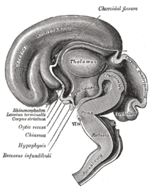
Median sagittal section of brain of human embryo of three months

In neonatal life, gonadal steroids influence the development of the neuroendocrine hypothalamus. For instance, they determine the ability of females to exhibit a normal reproductive cycle, and of males and females to display appropriate reproductive behaviors in adult life.

  • If a female rat is injected once with testosterone in the first few days of postnatal life (during the "critical period" of sex-steroid influence), the hypothalamus is irreversibly masculinized; the adult rat will be incapable of generating an LH surge in response to estrogen (a characteristic of females), but will be capable of exhibiting male sexual behaviors (mounting a sexually receptive female).
  • By contrast, a male rat castrated just after birth will be feminized, and the adult will show female sexual behavior in response to estrogen (sexual receptivity, lordosis behavior).

In primates, the developmental influence of androgens is less clear, and the consequences are less understood. Within the brain, testosterone is aromatized (to estradiol), which is the principal active hormone for developmental influences. The human testis secretes high levels of testosterone from about week 8 of fetal life until 5–6 months after birth (a similar perinatal surge in testosterone is observed in many species), a process that appears to underlie the male phenotype. Estrogen from the maternal circulation is relatively ineffective, partly because of the high circulating levels of steroid-binding proteins in pregnancy.

Sex steroids are not the only important influences upon hypothalamic development; in particular, pre-pubertal stress in early life (of rats) determines the capacity of the adult hypothalamus to respond to an acute stressor. Unlike gonadal steroid receptors, glucocorticoid receptors are very widespread throughout the brain; in the paraventricular nucleus, they mediate negative feedback control of CRF synthesis and secretion, but elsewhere their role is not well understood.

Function

Hormone release

Endocrine glands in the human head and neck and their hormones

The hypothalamus has a central neuroendocrine function, most notably by its control of the anterior pituitary, which in turn regulates various endocrine glands and organs. Releasing hormones (also called releasing factors) are produced in hypothalamic nuclei then transported along axons to either the median eminence or the posterior pituitary, where they are stored and released as needed.

Anterior pituitary

In the hypothalamic–adenohypophyseal axis, releasing hormones, also known as hypophysiotropic or hypothalamic hormones, are released from the median eminence, a prolongation of the hypothalamus, into the hypophyseal portal system, which carries them to the anterior pituitary where they exert their regulatory functions on the secretion of adenohypophyseal hormones. These hypophysiotropic hormones are stimulated by parvocellular neurosecretory cells located in the periventricular area of the hypothalamus. After their release into the capillaries of the third ventricle, the hypophysiotropic hormones travel through what is known as the hypothalamo-pituitary portal circulation. Once they reach their destination in the anterior pituitary, these hormones bind to specific receptors located on the surface of pituitary cells. Depending on which cells are activated through this binding, the pituitary will either begin secreting or stop secreting hormones into the rest of the bloodstream.

Secreted hormone Abbreviation Produced by Effect
Thyrotropin-releasing hormone
(Prolactin-releasing hormone)
TRH, TRF, or PRH Parvocellular neurosecretory cells of the paraventricular nucleus Stimulate thyroid-stimulating hormone (TSH) release from anterior pituitary (primarily)
Stimulate prolactin release from anterior pituitary
Corticotropin-releasing hormone CRH or CRF Parvocellular neurosecretory cells of the paraventricular nucleus Stimulate adrenocorticotropic hormone (ACTH) release from anterior pituitary
Dopamine
(Prolactin-inhibiting hormone)
DA or PIH Dopamine neurons of the arcuate nucleus Inhibit prolactin release from anterior pituitary
Growth-hormone-releasing hormone GHRH Neuroendocrine neurons of the Arcuate nucleus Stimulate growth-hormone (GH) release from anterior pituitary
Gonadotropin-releasing hormone GnRH or LHRH Neuroendocrine cells of the Preoptic area Stimulate follicle-stimulating hormone (FSH) release from anterior pituitary
Stimulate luteinizing hormone (LH) release from anterior pituitary
Somatostatin
(growth-hormone-inhibiting hormone)
SS, GHIH, or SRIF Neuroendocrine cells of the Periventricular nucleus Inhibit growth-hormone (GH) release from anterior pituitary
Inhibit (moderately) thyroid-stimulating hormone (TSH) release from anterior pituitary

Other hormones secreted from the median eminence include vasopressin, oxytocin, and neurotensin.

Posterior pituitary

In the hypothalamic–pituitary–adrenal axis, neurohypophysial hormones are released from the posterior pituitary, which is actually a prolongation of the hypothalamus, into the circulation.

Secreted hormone Abbreviation Produced by Effect
Oxytocin OXY or OXT Magnocellular neurosecretory cells of the paraventricular nucleus and supraoptic nucleus Uterine contraction
Lactation (letdown reflex)
Vasopressin
(antidiuretic hormone)
ADH or AVP Magnocellular and parvocellular neurosecretory cells of the paraventricular nucleus, magnocellular cells in supraoptic nucleus Increase in the permeability to water of the cells of distal tubule and collecting duct in the kidney and thus allows water reabsorption and excretion of concentrated urine

It is also known that hypothalamic–pituitary–adrenal axis (HPA) hormones are related to certain skin diseases and skin homeostasis. There is evidence linking hyperactivity of HPA hormones to stress-related skin diseases and skin tumors.

Stimulation

The hypothalamus coordinates many hormonal and behavioural circadian rhythms, complex patterns of neuroendocrine outputs, complex homeostatic mechanisms, and important behaviours. The hypothalamus must, therefore, respond to many different signals, some of which are generated externally and some internally. Delta wave signalling arising either in the thalamus or in the cortex influences the secretion of releasing hormones; GHRH and prolactin are stimulated whilst TRH is inhibited.

The hypothalamus is responsive to:

Olfactory stimuli

Olfactory stimuli are important for sexual reproduction and neuroendocrine function in many species. For instance if a pregnant mouse is exposed to the urine of a 'strange' male during a critical period after coitus then the pregnancy fails (the Bruce effect). Thus, during coitus, a female mouse forms a precise 'olfactory memory' of her partner that persists for several days. Pheromonal cues aid synchronization of oestrus in many species; in women, synchronized menstruation may also arise from pheromonal cues, although the role of pheromones in humans is disputed.

Blood-borne stimuli

Peptide hormones have important influences upon the hypothalamus, and to do so they must pass through the blood–brain barrier. The hypothalamus is bounded in part by specialized brain regions that lack an effective blood–brain barrier; the capillary endothelium at these sites is fenestrated to allow free passage of even large proteins and other molecules. Some of these sites are the sites of neurosecretion - the neurohypophysis and the median eminence. However, others are sites at which the brain samples the composition of the blood. Two of these sites, the SFO (subfornical organ) and the OVLT (organum vasculosum of the lamina terminalis) are so-called circumventricular organs, where neurons are in intimate contact with both blood and CSF. These structures are densely vascularized, and contain osmoreceptive and sodium-receptive neurons that control drinking, vasopressin release, sodium excretion, and sodium appetite. They also contain neurons with receptors for angiotensin, atrial natriuretic factor, endothelin and relaxin, each of which important in the regulation of fluid and electrolyte balance. Neurons in the OVLT and SFO project to the supraoptic nucleus and paraventricular nucleus, and also to preoptic hypothalamic areas. The circumventricular organs may also be the site of action of interleukins to elicit both fever and ACTH secretion, via effects on paraventricular neurons.

It is not clear how all peptides that influence hypothalamic activity gain the necessary access. In the case of prolactin and leptin, there is evidence of active uptake at the choroid plexus from the blood into the cerebrospinal fluid (CSF). Some pituitary hormones have a negative feedback influence upon hypothalamic secretion; for example, growth hormone feeds back on the hypothalamus, but how it enters the brain is not clear. There is also evidence for central actions of prolactin.

Findings have suggested that thyroid hormone (T4) is taken up by the hypothalamic glial cells in the infundibular nucleus/ median eminence, and that it is here converted into T3 by the type 2 deiodinase (D2). Subsequent to this, T3 is transported into the thyrotropin-releasing hormone (TRH)-producing neurons in the paraventricular nucleus. Thyroid hormone receptors have been found in these neurons, indicating that they are indeed sensitive to T3 stimuli. In addition, these neurons expressed MCT8, a thyroid hormone transporter, supporting the theory that T3 is transported into them. T3 could then bind to the thyroid hormone receptor in these neurons and affect the production of thyrotropin-releasing hormone, thereby regulating thyroid hormone production.

The hypothalamus functions as a type of thermostat for the body. It sets a desired body temperature, and stimulates either heat production and retention to raise the blood temperature to a higher setting or sweating and vasodilation to cool the blood to a lower temperature. All fevers result from a raised setting in the hypothalamus; elevated body temperatures due to any other cause are classified as hyperthermia. Rarely, direct damage to the hypothalamus, such as from a stroke, will cause a fever; this is sometimes called a hypothalamic fever. However, it is more common for such damage to cause abnormally low body temperatures.

Steroids

The hypothalamus contains neurons that react strongly to steroids and glucocorticoids (the steroid hormones of the adrenal gland, released in response to ACTH). It also contains specialized glucose-sensitive neurons (in the arcuate nucleus and ventromedial hypothalamus), which are important for appetite. The preoptic area contains thermosensitive neurons; these are important for TRH secretion.

Neural

Oxytocin secretion in response to suckling or vagino-cervical stimulation is mediated by some of these pathways; vasopressin secretion in response to cardiovascular stimuli arising from chemoreceptors in the carotid body and aortic arch, and from low-pressure atrial volume receptors, is mediated by others. In the rat, stimulation of the vagina also causes prolactin secretion, and this results in pseudo-pregnancy following an infertile mating. In the rabbit, coitus elicits reflex ovulation. In the sheep, cervical stimulation in the presence of high levels of estrogen can induce maternal behavior in a virgin ewe. These effects are all mediated by the hypothalamus, and the information is carried mainly by spinal pathways that relay in the brainstem. Stimulation of the nipples stimulates release of oxytocin and prolactin and suppresses the release of LH and FSH.

Cardiovascular stimuli are carried by the vagus nerve. The vagus also conveys a variety of visceral information, including for instance signals arising from gastric distension or emptying, to suppress or promote feeding, by signalling the release of leptin or gastrin, respectively. Again this information reaches the hypothalamus via relays in the brainstem.

In addition hypothalamic function is responsive to—and regulated by—levels of all three classical monoamine neurotransmitters, noradrenaline, dopamine, and serotonin (5-hydroxytryptamine), in those tracts from which it receives innervation. For example, noradrenergic inputs arising from the locus coeruleus have important regulatory effects upon corticotropin-releasing hormone (CRH) levels.

Control of food intake

Peptide hormones and neuropeptides that regulate feeding
Peptides that increase
feeding behavior
Peptides that decrease
feeding behavior
Ghrelin Leptin
Neuropeptide Y (α,β,γ)-Melanocyte-stimulating hormones
Agouti-related peptide Cocaine- and amphetamine-regulated transcript peptides
Orexins (A,B) Corticotropin-releasing hormone
Melanin-concentrating hormone Cholecystokinin
Galanin Insulin

Glucagon-like peptide 1

The extreme lateral part of the ventromedial nucleus of the hypothalamus is responsible for the control of food intake. Stimulation of this area causes increased food intake. Bilateral lesion of this area causes complete cessation of food intake. Medial parts of the nucleus have a controlling effect on the lateral part. Bilateral lesion of the medial part of the ventromedial nucleus causes hyperphagia and obesity of the animal. Further lesion of the lateral part of the ventromedial nucleus in the same animal produces complete cessation of food intake.

There are different hypotheses related to this regulation:

  1. Lipostatic hypothesis: This hypothesis holds that adipose tissue produces a humoral signal that is proportionate to the amount of fat and acts on the hypothalamus to decrease food intake and increase energy output. It has been evident that a hormone leptin acts on the hypothalamus to decrease food intake and increase energy output.
  2. Gutpeptide hypothesis: gastrointestinal hormones like Grp, glucagons, CCK and others claimed to inhibit food intake. The food entering the gastrointestinal tract triggers the release of these hormones, which act on the brain to produce satiety. The brain contains both CCK-A and CCK-B receptors.
  3. Glucostatic hypothesis: The activity of the satiety center in the ventromedial nuclei is probably governed by the glucose utilization in the neurons. It has been postulated that when their glucose utilization is low and consequently when the arteriovenous blood glucose difference across them is low, the activity across the neurons decrease. Under these conditions, the activity of the feeding center is unchecked and the individual feels hungry. Food intake is rapidly increased by intraventricular administration of 2-deoxyglucose therefore decreasing glucose utilization in cells.
  4. Thermostatic hypothesis: According to this hypothesis, a decrease in body temperature below a given set-point stimulates appetite, whereas an increase above the set-point inhibits appetite.

Fear processing

The medial zone of hypothalamus is part of a circuitry that controls motivated behaviors, like defensive behaviors. Analyses of Fos-labeling showed that a series of nuclei in the "behavioral control column" is important in regulating the expression of innate and conditioned defensive behaviors.

Antipredatory defensive behavior

Exposure to a predator (such as a cat) elicits defensive behaviors in laboratory rodents, even when the animal has never been exposed to a cat. In the hypothalamus, this exposure causes an increase in Fos-labeled cells in the anterior hypothalamic nucleus, the dorsomedial part of the ventromedial nucleus, and in the ventrolateral part of the premammillary nucleus (PMDvl). The premammillary nucleus has an important role in expression of defensive behaviors towards a predator, since lesions in this nucleus abolish defensive behaviors, like freezing and flight.  The PMD does not modulate defensive behavior in other situations, as lesions of this nucleus had minimal effects on post-shock freezing scores. The PMD has important connections to the dorsal periaqueductal gray, an important structure in fear expression. In addition, animals display risk assessment behaviors to the environment previously associated with the cat. Fos-labeled cell analysis showed that the PMDvl is the most activated structure in the hypothalamus, and inactivation with muscimol prior to exposure to the context abolishes the defensive behavior. Therefore, the hypothalamus, mainly the PMDvl, has an important role in expression of innate and conditioned defensive behaviors to a predator.

Social defeat

Likewise, the hypothalamus has a role in social defeat: Nuclei in medial zone are also mobilized during an encounter with an aggressive conspecific. The defeated animal has an increase in Fos levels in sexually dimorphic structures, such as the medial pre-optic nucleus, the ventrolateral part of ventromedial nucleus, and the ventral premammilary nucleus. Such structures are important in other social behaviors, such as sexual and aggressive behaviors. Moreover, the premammillary nucleus also is mobilized, the dorsomedial part but not the ventrolateral part. Lesions in this nucleus abolish passive defensive behavior, like freezing and the "on-the-back" posture.

Learning arbitrator

Recent research has questioned whether the lateral hypothalamus's role is only restricted to initiating and stopping innate behaviors and argued it learns about food-related cues. Specifically that it opposes learning about information what is neutral or distant to food. According this view, the lateral hypothalamus is "a unique arbitrator of learning capable of shifting behavior toward or away from important events".

Septal area

From Wikipedia, the free encyclopedia
Septal area
Lateral and medial septal nuclei in the septal area of the mouse brain
 
Details
Identifiers
Latinnuclei septales
MeSHD012686
NeuroNames259
TA98A14.1.09.266
TA25548
FMA61845

The septal area (medial olfactory area), consisting of the lateral septum and medial septum, is an area in the lower, posterior part of the medial surface of the frontal lobe, and refers to the nearby septum pellucidum.

The septal nuclei are located in this area. The septal nuclei are composed of medium-size neurons which are classified into dorsal, ventral, medial, and caudal groups. The septal nuclei receive reciprocal connections from the olfactory bulb, hippocampus, amygdala, hypothalamus, midbrain, habenula, cingulate gyrus, and thalamus. The septal nuclei are essential in generating the theta rhythm of the hippocampus.

The septal area (medial olfactory area) has no relation to the sense of smell, but it is considered a pleasure zone in animals. The septal nuclei play a role in reward and reinforcement along with the nucleus accumbens. In the 1950s, Olds & Milner showed that rats with electrodes implanted in this area will self-stimulate repeatedly (e.g., press a bar to receive electric current that stimulate the neurons). Experiments on the septal area of humans have taken place since the 1960s.

Connections

Detail of drawing showing components of septal area below corpus callosum.

The septal area is located on the lower posterior part of the frontal lobe. The septal area refers to the nearby septum pellucidum. It is located underneath the corpus callosum and in front of the lamina terminalis. The lamina terminalis is a layer of gray matter that connects the optic chiasma and the anterior commissure. The septal nuclei in the septal area are essential in generating the theta rhythm of the hippocampus.

The dorsal septum projects to the lateral preoptic area, lateral hypothalamus, periventricular hypothalamus and midline thalamus.

Fibers from the ventral half of the septum project topographically to the hippocampal formation, thalamus, hypothalamus and midbrain. Specifically, neurons located along the midline in the vertical limb of the diagonal band of Broca project through the dorsal fornix to all CA fields of the dorsal hippocampus and adjacent subicular cortex. Other fibers from this region project through the stria medullaris to the medial and lateral habenular nuclei, the paratenial and anteromedial nucleus of the thalamus, and through the medial forebrain bundle to the pars posterior of the medial mammillary nucleus.

Cells located in the intermediolateral septum also project through the lateral part of the fimbria to all CA fields of the ventral hippocampus and adjacent subicular and entorhinal cortices. These cells also send fibers through the stria medullaris to the lateral habenular nucleus and mediodorsal thalamic nucleus. Other axons arising from these cells descend through the medial forebrain bundle to terminate in a region dorsal to the interpeduncular nucleus.

The lateral septum is a relay center for connections from the CA3 of the hippocampus to the ventral tegmental area. These connections help link reward signals with the context in which they occur.

Fibers from the most lateral part of the ventral septum (i.e., bed nucleus of the anterior commissure) project through the stria terminalis to the ventral subiculum. In addition, cells located in the horizontal limb of the diagonal band project massively to the pars posterior of the medial mammillary nucleus, the ventral tegmental area, and amygdala.

Functions of the lateral septum

The lateral septum and movement and reward

The lateral septum is involved in a variety of functions, including emotional, motivational, and spatial behavior. It has been suggested that the LS may regulate interactions between the hippocampus and other regions that mediate goal directed behavior, such as the ventral tegmental area. Firing of LS neurons is modulated by both speed and acceleration  and spatial location, and that firing is also related to reward and context. It has thus been suggested that the lateral septum may incorporate movement into the evaluation of environmental context with respect to motivation and reward.

Lateral septum and social behavior

Inhibitory GABA, and excitatory glutamate, which regulate lateral septum (LS) activity, have been found to be increased during social play in juvenile rats. No sex differences were found in extracellular GABA concentrations during social playing, however, glutamate plays a major role in female social playing. When glutamate receptors are blocked in the LS pharmacologically, there is a significant decrease in female social playing, while males had no decrease in playing. This suggests that in the lateral septum, GABA neurotransmission is involved in social play behavior regulation in both sexes, while glutamate neurotransmission is sex-specific, involved in regulation of social play only in female juvenile rats.

Lateral septum and reproduction

Experiments have shown that both testosterone and dihydrotestosterone, when implanted directly into the lateral septum of male rats, caused a significant rise in serum LH and FSH levels, while not significantly increasing serum testosterone and dihydrotestosterone levels respectively. This indicates that the effect is occurring through testosterone receptors and independently of conversion to estrogen via aromatization. As of now, it is unclear, what exact pathway is mediating this phenomenon. Septal lesions significantly decreased serum LH and testosterone levels in male rats, while FSH and prolactin production were unaffected by the surgery. Electrical stimulation of the septum induced the elevation of serum LH and FSH levels. These data suggest that the lateral and/or the medial septum may play a role in the control of GnRH/gonadotropin secretion, and thus, the reproductive axis.

Lateral septum and memory

Despite the diverse direct projection system between the hippocampus and the lateral septum, the first pieces of evidence regarding the role of latter brain area in memory formation and retention have only started to emerge as of 2022. However, recent research has started to shed light on the potentially diverse roles of the lateral septum.

Social memory

The roles of the lateral septum in formation of social memories remain unclear and controversial. It is clear, that some role is being played, as oxytocin and vasopressin, when administered into the lateral septum after social training were able to enhance memory formation, while their respective receptor blockers had the opposite effect. However, data on effects of pre-training administration of these compounds is mixed, hence the lack of consensus.

Fear memory

Pieces of evidence suggesting the role the lateral septum could be playing in fear memory formation and consolidation have started emerging at the end of the XX. century. Inhibitory avoidance tasks using footshock chambers on mice were deployed to study the effects of the disruption of the lateral septum and hippocampal inputs on fear memory formation and retrieval respectively. In both cases, fear memory formation/retrieval was impaired, supporting the hypothesis that hippocampal projections to the lateral septum are at least in part responsible for these mechanisms. The lateral septum could also prove essential in fear memory consolidation. Post-shock adminitration of CRF into the lateral septum enhanced fear memory in the relelvant context; however, this finding still needs to be supported by further studies.

Anxiety

From Wikipedia, the free encyclopedia
Anxiety, an 1894 portrait by Edvard Munch

Anxiety is an emotion which is characterised by an unpleasant state of inner turmoil and includes feelings of dread over anticipated events. Anxiety is different from fear in that fear is defined as the emotional response to a present threat, whereas anxiety is the anticipation of a future one. It is often accompanied by nervous behavior such as pacing back and forth, somatic complaints, and rumination.

Anxiety is a feeling of uneasiness and worry, usually generalized and unfocused as an overreaction to a situation that is only subjectively seen as menacing. It is often accompanied by muscular tension, restlessness, fatigue, inability to catch one's breath, tightness in the abdominal region, nausea, and problems in concentration. Anxiety is closely related to fear, which is a response to a real or perceived immediate threat (fight-or-flight response); anxiety involves the expectation of a future threat including dread. People facing anxiety may withdraw from situations which have provoked anxiety in the past.

The emotion of anxiety can persist beyond the developmentally appropriate time-periods in response to specific events, and thus turning into one of the multiple anxiety disorders (e.g. generalized anxiety disorder, panic disorder). The difference between anxiety disorder (as mental disorder) and anxiety (as normal emotion), is that people with an anxiety disorder experience anxiety excessively or persistently during approximately 6 months, or even during shorter time-periods in children. Anxiety disorders are among the most persistent mental problems and often last decades. Anxiety can also be experienced within other mental disorders, e.g., obsessive-compulsive disorder, post-traumatic stress disorder.

Anxiety vs. fear

Anxiety is distinguished from fear, which is an appropriate cognitive and emotional response to a perceived threat. Anxiety is related to the specific behaviors of fight-or-flight responses, defensive behavior or escape. There is a false presumption that often circulates that anxiety only occurs in situations perceived as uncontrollable or unavoidable, but this is not always so. David Barlow defines anxiety as "a future-oriented mood state in which one is not ready or prepared to attempt to cope with upcoming negative events," and that it is a distinction between future and present dangers which divides anxiety and fear. Another description of anxiety is agony, dread, terror, or even apprehension. In positive psychology, anxiety is described as the mental state that results from a difficult challenge for which the subject has insufficient coping skills.

Fear and anxiety can be differentiated into four domains: (1) duration of emotional experience, (2) temporal focus, (3) specificity of the threat, and (4) motivated direction. Fear is short-lived, present-focused, geared towards a specific threat, and facilitating escape from threat. On the other hand, anxiety is long-acting, future-focused, broadly focused towards a diffuse threat, and promoting excessive caution while approaching a potential threat and interferes with constructive coping.

Joseph E. LeDoux and Lisa Feldman Barrett have both sought to separate automatic threat responses from additional associated cognitive activity within anxiety.

Symptoms

Anxiety can be experienced with long, drawn-out daily symptoms that reduce quality of life, known as chronic (or generalized) anxiety, or it can be experienced in short spurts with sporadic, stressful panic attacks, known as acute anxiety. Symptoms of anxiety can range in number, intensity, and frequency, depending on the person. However, most people do not suffer from chronic anxiety.

Anxiety can induce several psychological pains (e.g. depression) or mental disorders, and may lead to self-harm or suicide.

The behavioral effects of anxiety may include withdrawal from situations which have provoked anxiety or negative feelings in the past. Other effects may include changes in sleeping patterns, changes in habits, increase or decrease in food intake, and increased motor tension (such as foot tapping).

The emotional effects of anxiety may include feelings of apprehension or dread, trouble concentrating, feeling tense or jumpy, anticipating the worst, irritability, restlessness, watching for signs of danger, and a feeling of empty mindedness. as well as "nightmares/bad dreams, obsessions about sensations, déjà vu, a trapped-in-your-mind feeling, and feeling like everything is scary." It may include a vague experience and feeling of helplessness.

The cognitive effects of anxiety may include thoughts about suspected dangers, such as an irrational fear of dying or having a heart attack, when in reality all one is experiencing is mild chest pain, for example.

The physiological symptoms of anxiety may include:

Types

There are various types of anxiety. Existential anxiety can occur when a person faces angst, an existential crisis, or nihilistic feelings. People can also face mathematical anxiety, somatic anxiety, stage fright, or test anxiety. Social anxiety refers to a fear of rejection and negative evaluation (being judged) by other people.

Existential

The philosopher Søren Kierkegaard, in The Concept of Anxiety (1844), described anxiety or dread associated with the "dizziness of freedom" and suggested the possibility for positive resolution of anxiety through the self-conscious exercise of responsibility and choosing. In Art and Artist (1932), the psychologist Otto Rank wrote that the psychological trauma of birth was the pre-eminent human symbol of existential anxiety and encompasses the creative person's simultaneous fear of – and desire for – separation, individuation, and differentiation.

The theologian Paul Tillich characterized existential anxiety as "the state in which a being is aware of its possible nonbeing" and he listed three categories for the nonbeing and resulting anxiety: ontic (fate and death), moral (guilt and condemnation), and spiritual (emptiness and meaninglessness). According to Tillich, the last of these three types of existential anxiety, i.e. spiritual anxiety, is predominant in modern times while the others were predominant in earlier periods. Tillich argues that this anxiety can be accepted as part of the human condition or it can be resisted but with negative consequences. In its pathological form, spiritual anxiety may tend to "drive the person toward the creation of certitude in systems of meaning which are supported by tradition and authority" even though such "undoubted certitude is not built on the rock of reality".

According to Viktor Frankl, the author of Man's Search for Meaning, when a person is faced with extreme mortal dangers, the most basic of all human wishes is to find a meaning of life to combat the "trauma of nonbeing" as death is near.

Depending on the source of the threat, psychoanalytic theory distinguishes three types of anxiety: realistic, neurotic and moral.

Test, performance, and competitive

Test

According to Yerkes-Dodson law, an optimal level of arousal is necessary to best complete a task such as an exam, performance, or competitive event. However, when the anxiety or level of arousal exceeds that optimum, the result is a decline in performance.

Test anxiety is the uneasiness, apprehension, or nervousness felt by students who have a fear of failing an exam. Students who have test anxiety may experience any of the following: the association of grades with personal worth; fear of embarrassment by a teacher; fear of alienation from parents or friends; time pressures; or feeling a loss of control. Sweating, dizziness, headaches, racing heartbeats, nausea, fidgeting, uncontrollable crying or laughing and drumming on a desk are all common. Because test anxiety hinges on fear of negative evaluation, debate exists as to whether test anxiety is itself a unique anxiety disorder or whether it is a specific type of social phobia. The DSM-IV classifies test anxiety as a type of social phobia.

Research indicates that test anxiety among U.S. high-school and college students has been rising since the late 1950s. Test anxiety remains a challenge for students, regardless of age, and has considerable physiological and psychological impacts. Management of test anxiety focuses on achieving relaxation and developing mechanisms to manage anxiety. The routine practice of slow, Device-Guided Breathing (DGB) is a major component of behavioral treatments for anxiety conditions.

Performance and competitive

Performance anxiety and competitive anxiety (competitive trait anxiety, competitive state anxiety) happen when an individual's performance is measured against others. An important distinction between competitive and non-competitive anxiety is that competitive anxiety makes people view their performance as a threat. As a result, they experience a drop in their ordinary ability, whether physical or mental, due to that perceived stress.

Competitive anxiety is caused by a range of internal factors including high expectations, outside pressure, lack of experience, and external factors like the location of a competition. It commonly occurs in those participating in high pressure activities like sports and debates. Some common symptoms of competitive anxiety include muscle tension, fatigue, weakness, sense of panic, apprehensiveness, and panic attacks.

There are 4 major theories of how anxiety affects performance: Drive theory, Inverted U theory, Reversal theory, and The Zone of Optimal Functioning theory.

Drive theory believes that anxiety is positive and performance improves proportionally to the level of anxiety. This theory is not well accepted.

The Inverted U theory is based on the idea that performance peaks at a moderate stress level. It is called Inverted U theory because the graph that plots performance against anxiety looks like an inverted "U".

Reversal theory suggests that performance increases in relation to the individual's interpretation of their arousal levels. If they believed their physical arousal level would help them, their performance would increase, if they didn't, their performance would decrease. For example: Athletes were shown to worry more when focusing on results and perfection rather than the effort and growth involved.

The Zone of Optimal Functioning theory proposes that there is a zone where positive and negative emotions are in a balance which lead to feelings of dissociation and intense concentration, optimizing the individual's performance levels.

Stranger, social, and intergroup anxiety

Humans generally require social acceptance and thus sometimes dread the disapproval of others. Apprehension of being judged by others may cause anxiety in social environments.

Anxiety during social interactions, particularly between strangers, is common among young people. It may persist into adulthood and become social anxiety or social phobia. "Stranger anxiety" in small children is not considered a phobia. In adults, an excessive fear of other people is not a developmentally common stage; it is called social anxiety. According to Cutting, social phobics do not fear the crowd but the fact that they may be judged negatively.

Social anxiety varies in degree and severity. For some people, it is characterized by experiencing discomfort or awkwardness during physical social contact (e.g. embracing, shaking hands, etc.), while in other cases it can lead to a fear of interacting with unfamiliar people altogether. Those with this condition may restrict their lifestyles to accommodate the anxiety, minimizing social interaction whenever possible. Social anxiety also forms a core aspect of certain personality disorders, including avoidant personality disorder.

To the extent that a person is fearful of social encounters with unfamiliar others, some people may experience anxiety particularly during interactions with outgroup members, or people who share different group memberships (i.e., by race, ethnicity, class, gender, etc.). Depending on the nature of the antecedent relations, cognitions, and situational factors, intergroup contact may be stressful and lead to feelings of anxiety. This apprehension or fear of contact with outgroup members is often called interracial or intergroup anxiety.

As is the case with the more generalized forms of social anxiety, intergroup anxiety has behavioral, cognitive, and affective effects. For instance, increases in schematic processing and simplified information processing can occur when anxiety is high. Indeed, such is consistent with related work on attentional bias in implicit memory. Additionally recent research has found that implicit racial evaluations (i.e. automatic prejudiced attitudes) can be amplified during intergroup interaction. Negative experiences have been illustrated in producing not only negative expectations, but also avoidant, or antagonistic, behavior such as hostility. Furthermore, when compared to anxiety levels and cognitive effort (e.g., impression management and self-presentation) in intragroup contexts, levels and depletion of resources may be exacerbated in the intergroup situation.

Trait

Anxiety can be either a short-term "state" or a long-term "personality trait". Trait anxiety reflects a stable tendency across the lifespan of responding with acute, state anxiety in the anticipation of threatening situations (whether they are actually deemed threatening or not). A meta-analysis showed that a high level of neuroticism is a risk factor for development of anxiety symptoms and disorders. Such anxiety may be conscious or unconscious.

Personality can also be a trait leading to anxiety and depression and their persistence. Through experience, many find it difficult to collect themselves due to their own personal nature.

Choice or decision

Anxiety induced by the need to choose between similar options is recognized as a problem for some individuals and for organizations. In 2004, Capgemini wrote: "Today we're all faced with greater choice, more competition and less time to consider our options or seek out the right advice." Overthinking a choice is called analysis paralysis.

In a decision context, unpredictability or uncertainty may trigger emotional responses in anxious individuals that systematically alter decision-making. There are primarily two forms of this anxiety type. The first form refers to a choice in which there are multiple potential outcomes with known or calculable probabilities. The second form refers to the uncertainty and ambiguity related to a decision context in which there are multiple possible outcomes with unknown probabilities.

Panic disorder

Panic disorder may share symptoms of stress and anxiety, but it is actually very different. Panic disorder is an anxiety disorder that occurs without any triggers. According to the U.S. Department of Health and Human Services, this disorder can be distinguished by unexpected and repeated episodes of intense fear. Someone with panic disorder will eventually develop constant fear of another attack and as this progresses it will begin to affect daily functioning and an individual's general quality of life. It is reported by the Cleveland Clinic that panic disorder affects 2 to 3 percent of adult Americans and can begin around the time of the teenage and early adult years. Some symptoms include: difficulty breathing, chest pain, dizziness, trembling or shaking, feeling faint, nausea, fear that you are losing control or are about to die. Even though they have these symptoms during an attack, the main symptom is the persistent fear of having future panic attacks.

Anxiety disorders

Anxiety disorders are a group of mental disorders characterized by exaggerated feelings of anxiety and fear responses. Anxiety is a worry about future events and fear is a reaction to current events. These feelings may cause physical symptoms, such as a fast heart rate and shakiness. There are a number of anxiety disorders: including generalized anxiety disorder, specific phobia, social anxiety disorder, separation anxiety disorder, agoraphobia, panic disorder, and selective mutism. The disorder differs by what results in the symptoms. People often have more than one anxiety disorder.

Anxiety disorders are caused by a complex combination of genetic and environmental factors. To be diagnosed, symptoms typically need to be present for at least six months, be more than would be expected for the situation, and decrease a person's ability to function in their daily lives. Other problems that may result in similar symptoms include hyperthyroidism, heart disease, caffeine, alcohol, or cannabis use, and withdrawal from certain drugs, among others.

Without treatment, anxiety disorders tend to remain. Treatment may include lifestyle changes, counselling, and medications. Counselling is typically with a type of cognitive behavioral therapy. Medications, such as antidepressants or beta blockers, may improve symptoms. A 2023 review found that regular physical activity is effective for reducing anxiety.

About 12% of people are affected by an anxiety disorder in a given year and between 12% and 30% are affected at some point in their life. They occur about twice as often in women than they do in men, and generally begin before the age of 25. The most common anxiety disorders are specific phobias, which affect nearly 12% of people, and social anxiety disorder, which affects 10% of people at some point in their life. They affect those between the ages of 15 and 35 the most and become less common after the age of 55. Rates appear to be higher in the United States and Europe.

Short- and long-term anxiety

Anxiety can be either a short-term "state" or a long-term "trait". Whereas trait anxiety represents worrying about future events, anxiety disorders are a group of mental disorders characterized by feelings of anxiety and fears.

Four ways to be anxious

In his book Anxious: The Modern Mind in the Age of Anxiety Joseph LeDoux examines four experiences of anxiety through a brain-based lens:

  1. In the presence of an existing or imminent external threat, you worry about the event and its implications for your physical and/or psychological well-being. When a threat signal occurs, it signifies either that danger is present or near in space and time or that it might be coming in the future. Nonconscious threats processing by the brain activates defensive survival circuits, resulting in changes in information processing in the brain, controlled in part by increases in arousal and behavioral and physiological responses in the body that then produce signals that feed back to the brain and complement the physiological changes there, intensifying them and extending their duration.
  2. When you notice body sensations, you worry about what they might mean for your physical and/or psychological well-being. The trigger stimulus does not have to be an external stimulus but can be an internal one, as some people are particularly sensitive to body signals.
  3. Thoughts and memories may lead to you to worry about your physical and/or psychological well-being. We do not need to be in the presence of an external or internal stimulus to be anxious. An episodic memory of a past trauma or of a panic attack in the past is sufficient to activate the defence circuits.
  4. Thoughts and memories may result in existential dread, such as worry about leading a meaningful life or the eventuality of death. Examples are contemplations of whether one's life has been meaningful, the inevitability of death, or the difficulty of making decisions that have a moral value. These do not necessarily activate defensive systems; they are more or less pure forms of cognitive anxiety.

Co-morbidity

Anxiety disorders often occur with other mental health disorders, particularly major depressive disorder, bipolar disorder, eating disorders, or certain personality disorders. It also commonly occurs with personality traits such as neuroticism. This observed co-occurrence is partly due to genetic and environmental influences shared between these traits and anxiety.

It is common for those with obsessive–compulsive disorder to experience anxiety. Anxiety is also commonly found in those who experience panic disorders, phobic anxiety disorders, severe stress, dissociative disorders, somatoform disorders, and some neurotic disorders. Anxiety has also been linked to the experience of intrusive thoughts. Studies have revealed that individuals who experience high levels of anxiety (also known as clinical anxiety) are highly vulnerable to the experience of intense intrusive thoughts or psychological disorders that are characterised by intrusive thoughts.

Risk factors

A marble bust of the Roman Emperor Decius from the Capitoline Museum, conveying "an impression of anxiety and weariness, as of a man shouldering heavy [state] responsibilities"

Anxiety disorders are partly genetic, with twin studies suggesting 30-40% genetic influence on individual differences in anxiety. Environmental factors are also important. Twin studies show that individual-specific environments have a large influence on anxiety, whereas shared environmental influences (environments that affect twins in the same way) operate during childhood but decline through adolescence. Specific measured 'environments' that have been associated with anxiety include child abuse, family history of mental health disorders, and poverty. Anxiety is also associated with drug use, including alcohol, caffeine, and benzodiazepines, which are often prescribed to treat anxiety.

Neuroanatomy

Neural circuitry involving the amygdala, which regulates emotions like anxiety and fear, stimulating the HPA axis and sympathetic nervous system, and hippocampus, which is implicated in emotional memory along with the amygdala, is thought to underlie anxiety. People who have anxiety tend to show high activity in response to emotional stimuli in the amygdala. Some writers believe that excessive anxiety can lead to an overpotentiation of the limbic system (which includes the amygdala and nucleus accumbens), giving increased future anxiety, but this does not appear to have been proven.

Research upon adolescents who as infants had been highly apprehensive, vigilant, and fearful finds that their nucleus accumbens is more sensitive than that in other people when deciding to make an action that determined whether they received a reward. This suggests a link between circuits responsible for fear and also reward in anxious people. As researchers note, "a sense of 'responsibility', or self-agency, in a context of uncertainty (probabilistic outcomes) drives the neural system underlying appetitive motivation (i.e., nucleus accumbens) more strongly in temperamentally inhibited than noninhibited adolescents".

The microbes of the gut can connect with the brain to affect anxiety. There are various pathways along which this communication can take place. One is through the major neurotransmitters. The gut microbes such as Bifidobacterium and Bacillus produce the neurotransmitters GABA and dopamine, respectively. The neurotransmitters signal to the nervous system of the gastrointestinal tract, and those signals will be carried to the brain through the vagus nerve or the spinal system. This is demonstrated by the fact that altering the microbiome has shown anxiety- and depression-reducing effects in mice, but not in subjects without vagus nerves.

Another key pathway is the HPA axis, as mentioned above. The microbes can control the levels of cytokines in the body, and altering cytokine levels creates direct effects on areas of the brain such as the hypothalamus, the area that triggers HPA axis activity. The HPA axis regulates production of cortisol, a hormone that takes part in the body's stress response. When HPA activity spikes, cortisol levels increase, processing and reducing anxiety in stressful situations. These pathways, as well as the specific effects of individual taxa of microbes, are not yet completely clear, but the communication between the gut microbiome and the brain is undeniable, as is the ability of these pathways to alter anxiety levels.

With this communication comes the potential to treat. Prebiotics and probiotics have been shown to reduce anxiety. For example, experiments in which mice were given fructo- and galacto-oligosaccharide prebiotics and Lactobacillus probiotics have both demonstrated a capability to reduce anxiety. In humans, results are not as concrete, but promising.

Genetics

Genetics and family history (e.g. parental anxiety) may put an individual at increased risk of an anxiety disorder, but generally external stimuli will trigger its onset or exacerbation. Estimates of genetic influence on anxiety, based on studies of twins, range from 25 to 40% depending on the specific type and age-group under study. For example, genetic differences account for about 43% of variance in panic disorder and 28% in generalized anxiety disorder. Longitudinal twin studies have shown the moderate stability of anxiety from childhood through to adulthood is mainly influenced by stability in genetic influence. When investigating how anxiety is passed on from parents to children, it is important to account for sharing of genes as well as environments, for example using the intergenerational children-of-twins design.

Many studies in the past used a candidate gene approach to test whether single genes were associated with anxiety. These investigations were based on hypotheses about how certain known genes influence neurotransmitters (such as serotonin and norepinephrine) and hormones (such as cortisol) that are implicated in anxiety. None of these findings are well replicated, with the possible exception of TMEM132D, COMT and MAO-A. The epigenetic signature of BDNF, a gene that codes for a protein called brain derived neurotrophic factor that is found in the brain, has also been associated with anxiety and specific patterns of neural activity. and a receptor gene for BDNF called NTRK2 was associated with anxiety in a large genome-wide investigation. The reason that most candidate gene findings have not replicated is that anxiety is a complex trait that is influenced by many genomic variants, each of which has a small effect on its own. Increasingly, studies of anxiety are using a hypothesis-free approach to look for parts of the genome that are implicated in anxiety using big enough samples to find associations with variants that have small effects. The largest explorations of the common genetic architecture of anxiety have been facilitated by the UK Biobank, the ANGST consortium and the CRC Fear, Anxiety and Anxiety Disorders Archived 2019-04-29 at the Wayback Machine.

Epigenetics

Epigenetics of anxiety and stress–related disorders is the field studying the relationship between epigenetic modifications of genes and anxiety and stress-related disorders, including mental health disorders such as generalized anxiety disorder (GAD), post-traumatic stress disorder, obsessive-compulsive disorder (OCD), and more. These changes can lead to transgenerational stress inheritance.

Epigenetic modifications play a role in the development and heritability of these disorders and related symptoms. For example, regulation of the hypothalamus-pituitary-adrenal axis by glucocorticoids plays a major role in stress response and is known to be epigenetically regulated.

As of 2015 most work has been done in animal models in laboratories, and little work has been done in humans; the work is not yet applicable to clinical psychiatry. Stress-induced epigenetic changes, particularly to genes that effect the hypothalamic–pituitary–adrenal (HPA) axis, persist into future generations, negatively impacting the capacity of offspring to adapt to stress. Early life experiences, even when generations removed, can cause permanent epigenetic modifications of DNA resulting in changes in gene expression, endocrine function and metabolism. These heritable epigenetic modifications include DNA methylation of the promoter regions of genes that affect sensitivity to stress.

Medical conditions

Many medical conditions can cause anxiety. This includes conditions that affect the ability to breathe, like COPD and asthma, and the difficulty in breathing that often occurs near death. Conditions that cause abdominal pain or chest pain can cause anxiety and may in some cases be a somatization of anxiety; the same is true for some sexual dysfunctions. Conditions that affect the face or the skin can cause social anxiety especially among adolescents, and developmental disabilities often lead to social anxiety for children as well. Life-threatening conditions like cancer also cause anxiety.

Furthermore, certain organic diseases may present with anxiety or symptoms that mimic anxiety. These disorders include certain endocrine diseases (hypo- and hyperthyroidism, hyperprolactinemia), metabolic disorders (diabetes), deficiency states (low levels of vitamin D, B2, B12, folic acid), gastrointestinal diseases (celiac disease, non-celiac gluten sensitivity, inflammatory bowel disease), heart diseases, blood diseases (anemia), cerebral vascular accidents (transient ischemic attack, stroke), and brain degenerative diseases (Parkinson's disease, dementia, multiple sclerosis, Huntington's disease), among others.

Substance-induced

Several drugs can cause or worsen anxiety, whether in intoxication, withdrawal or as side effect. These include alcohol, tobacco, sedatives (including prescription benzodiazepines), opioids (including prescription pain killers and illicit drugs like heroin), stimulants (such as caffeine, cocaine and amphetamines), hallucinogens, and inhalants.

While many often report self-medicating anxiety with these substances, improvements in anxiety from drugs are usually short-lived (with worsening of anxiety in the long term, sometimes with acute anxiety as soon as the drug effects wear off) and tend to be exaggerated. Acute exposure to toxic levels of benzene may cause euphoria, anxiety, and irritability lasting up to 2 weeks after the exposure.

Psychological

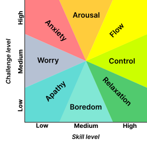
AnxietyArousalFlow (psychology)WorryControl (psychology)ApathyBoredomRelaxation (psychology)
Mental state in terms of challenge level and skill level, according to Csikszentmihalyi's flow model. (Click on a fragment of the image to go to the appropriate article)

Poor coping skills (e.g., rigidity/inflexible problem solving, denial, avoidance, impulsivity, extreme self-expectation, negative thoughts, affective instability, and inability to focus on problems) are associated with anxiety. Anxiety is also linked and perpetuated by the person's own pessimistic outcome expectancy and how they cope with feedback negativity. Temperament (e.g., neuroticism) and attitudes (e.g. pessimism) have been found to be risk factors for anxiety.

Cognitive distortions such as overgeneralizing, catastrophizing, mind reading, emotional reasoning, binocular trick, and mental filter can result in anxiety. For example, an overgeneralized belief that something bad "always" happens may lead someone to have excessive fears of even minimally risky situations and to avoid benign social situations due to anticipatory anxiety of embarrassment. In addition, those who have high anxiety can also create future stressful life events. Together, these findings suggest that anxious thoughts can lead to anticipatory anxiety as well as stressful events, which in turn cause more anxiety. Such unhealthy thoughts can be targets for successful treatment with cognitive therapy.

Psychodynamic theory posits that anxiety is often the result of opposing unconscious wishes or fears that manifest via maladaptive defense mechanisms (such as suppression, repression, anticipation, regression, somatization, passive aggression, dissociation) that develop to adapt to problems with early objects (e.g., caregivers) and empathic failures in childhood. For example, persistent parental discouragement of anger may result in repression/suppression of angry feelings which manifests as gastrointestinal distress (somatization) when provoked by another while the anger remains unconscious and outside the individual's awareness. Such conflicts can be targets for successful treatment with psychodynamic therapy. While psychodynamic therapy tends to explore the underlying roots of anxiety, cognitive behavioral therapy has also been shown to be a successful treatment for anxiety by altering irrational thoughts and unwanted behaviors.

Evolutionary psychology

An evolutionary psychology explanation is that increased anxiety serves the purpose of increased vigilance regarding potential threats in the environment as well as increased tendency to take proactive actions regarding such possible threats. This may cause false positive reactions but an individual with anxiety may also avoid real threats. This may explain why anxious people are less likely to die due to accidents. There is ample empirical evidence that anxiety can have adaptive value. Within a school, timid fish are more likely than bold fish to survive a predator.

When people are confronted with unpleasant and potentially harmful stimuli such as foul odors or tastes, PET-scans show increased blood flow in the amygdala. In these studies, the participants also reported moderate anxiety. This might indicate that anxiety is a protective mechanism designed to prevent the organism from engaging in potentially harmful behaviors.

Social

Social risk factors for anxiety include a history of trauma (e.g., physical, sexual or emotional abuse or assault), bullying, early life experiences and parenting factors (e.g., rejection, lack of warmth, high hostility, harsh discipline, high parental negative affect, anxious childrearing, modelling of dysfunctional and drug-abusing behaviour, discouragement of emotions, poor socialization, poor attachment, and child abuse and neglect), cultural factors (e.g., stoic families/cultures, persecuted minorities including those with disabilities), and socioeconomics (e.g., uneducated, unemployed, impoverished although developed countries have higher rates of anxiety disorders than developing countries). A 2019 comprehensive systematic review of over 50 studies showed that food insecurity in the United States is strongly associated with depression, anxiety, and sleep disorders. Food-insecure individuals had an almost 3 fold risk increase of testing positive for anxiety when compared to food-secure individuals.

Gender socialization

Contextual factors that are thought to contribute to anxiety include gender socialization and learning experiences. In particular, learning mastery (the degree to which people perceive their lives to be under their own control) and instrumentality, which includes such traits as self-confidence, self-efficacy, independence, and competitiveness fully mediate the relation between gender and anxiety. That is, though gender differences in anxiety exist, with higher levels of anxiety in women compared to men, gender socialization and learning mastery explain these gender differences.

Treatment

The first step in the management of a person with anxiety symptoms involves evaluating the possible presence of an underlying medical cause, the recognition of which is essential in order to decide the correct treatment. Anxiety symptoms may mask an organic disease, or appear associated with or as a result of a medical disorder.

Cognitive behavioral therapy (CBT) is effective for anxiety disorders and is a first line treatment. CBT appears to be equally effective when carried out via the internet. While evidence for mental health apps is promising, it is preliminary.

Anxiety often affects relationships, and interpersonal psychotherapy addresses these issues by improving communication and relationship skills.

Psychopharmacological treatment can be used in parallel to CBT or can be used alone. As a general rule, most anxiety disorders respond well to first-line agents. Such drugs, also used as anti-depressants, are the selective serotonin reuptake inhibitors and serotonin-norepinephrine reuptake inhibitors, that work by blocking the reuptake of specific neurotransmitters and resulting in the increase in availability of these neurotransmitters. Additionally, benzodiazepines are often prescribed to individuals with anxiety disorder. Benzodiazepines produce an anxiolytic response by modulating GABA and increasing its receptor binding. A third common treatment involves a category of drug known as serotonin agonists. This category of drug works by initiating a physiological response at 5-HT1A receptor by increasing the action of serotonin at this receptor. Other treatment options include pregabalin, tricyclic antidepressants, and moclobemide, among others.

Anxiety is considered to be a serious psychiatric illness that has an unknown true pervasiveness due to affected individuals not asking for proper treatment or aid, and due to professionals missing the diagnosis.

Prevention

The above risk factors give natural avenues for prevention. A 2017 review found that psychological or educational interventions have a small yet statistically significant benefit for the prevention of anxiety in varied population types.

Pathophysiology

Anxiety disorder appears to be a genetically inherited neurochemical dysfunction that may involve autonomic imbalance; decreased GABA-ergic tone; allelic polymorphism of the catechol-O-methyltransferase (COMT) gene; increased adenosine receptor function; increased cortisol.

In the central nervous system (CNS), the major mediators of the symptoms of anxiety disorders appear to be norepinephrine, serotonin, dopamine, and gamma-aminobutyric acid (GABA). Other neurotransmitters and peptides, such as corticotropin-releasing factor, may be involved. Peripherally, the autonomic nervous system, especially the sympathetic nervous system, mediates many of the symptoms. Increased flow in the right parahippocampal region and reduced serotonin type 1A receptor binding in the anterior and posterior cingulate and raphe of patients are the diagnostic factors for prevalence of anxiety disorder.

The amygdala is central to the processing of fear and anxiety, and its function may be disrupted in anxiety disorders. Anxiety processing in the basolateral amygdala has been implicated with expansion of dendritic arborization of the amygdaloid neurons. SK2 potassium channels mediate inhibitory influence on action potentials and reduce arborization.

Software testing

From Wikipedia, the free encyclopedia https://en.wikipedia.org/wiki/Software_testing TestingCup – Polish Championship in Software Tes...