Search This Blog

Thursday, November 7, 2024

Temporoparietal junction

From Wikipedia, the free encyclopedia
 
Temporoparietal junction
Side view of the human brain. TPJ is indicated by red circle.
 
Side view of the human brain. TPJ is indicated by red circle.

The temporoparietal junction (TPJ) is an area of the brain where the temporal and parietal lobes meet, at the posterior end of the lateral sulcus (Sylvian fissure). The TPJ incorporates information from the thalamus and the limbic system as well as from the visual, auditory, and somatosensory systems. The TPJ also integrates information from both the external environment as well as from within the body. The TPJ is responsible for collecting all of this information and then processing it.

This area is also known to play a crucial role in self–other distinctions processes and theory of mind (ToM). Furthermore, damage to the TPJ has been implicated in having adverse effects on an individual's ability to make moral decisions and has been known to produce out-of-body experiences (OBEs). Electromagnetic stimulation of the TPJ can also cause these effects. Apart from these diverse roles that the TPJ plays, it is also known for its involvement in a variety of widespread disorders including anxiety disorders, amnesia, Alzheimer's disease, autism spectrum disorder, and schizophrenia.

Anatomy and function
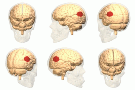
Animation. Both left and right temporoparietal junctions are shown in red.

The brain contains four main lobes: temporal lobe, parietal lobe, frontal lobe, and the occipital lobe. The temporoparietal junction lies in the region between the temporal and parietal lobes, near the lateral sulcus (Sylvian fissure). Specifically, it is composed of the inferior parietal lobule and the caudal parts of the superior temporal sulcus. There are two halves to the temporoparietal junction, with each component in their respective hemispheres of the brain. Each half of the TPJ pertains to various aspects of cognitive function. Often, however, the separate halves of the TPJ will work in coordination. The TPJ is mainly involved in information processing and perception.

Right temporoparietal junction

The right temporoparietal junction (rTPJ) is involved in the processing of information in terms of the ability of an individual to orient attention to new stimuli. Evidence from neuroimaging studies as well as lesion studies revealed that the rTPJ plays a pivotal role in analyzing signals from self-produced actions as well as with signals from the external environment. For example, an individual with lesions in their rTPJ would more than likely exhibit a sense of hemi-neglect, wherein they would no longer be able to pay attention to anything they observe on the left. So, if someone were to have a lesion in their rTPJ, then over time the awareness of the left limbs may fade without treatment. Visual signals provide the sensory information necessary for the brain to process spatial recognition of the world. When vision is limited, knowledge of existence begins to fade away since as far as the brain is concerned the object does not exist. Furthermore, the rTPJ plays a role in the way individuals observe and process information, thus impacting social interaction. Empathy and sympathy require an individual to simultaneously distinguish between different possible perspectives on the same situation. Imaging studies show that this ability depends upon the coordinated interaction of the rTPJ to identify and process the social cues presented to it. This rapid process allows for an individual to quickly react to situations.

Left temporoparietal junction

The left temporoparietal junction (lTPJ) contains both Wernicke's area and the angular gyrus, both prominent anatomical structures of the brain that are involved in language cognition, processing, and comprehension of both written and spoken language. Steven Pinker discusses this brain region, theorising that it underlies an amodal 'language of thought' or Mentalese. The lTPJ, in this account, takes in observations from external environments, such as conversations, makes connections in the brain regarding memories or incidents and then converts those thoughts and connections to written and spoken language. Pinker's full account of this is explained in The Language Instinct: How the Mind Creates Language. The lTPJ also plays an important role in reasoning of other's beliefs, intentions, and desires. Activation of the lTPJ was observed in patients processing mental states such as beliefs when an fMRI was used on patients as they were asked to make inferences regarding the mental states of others such as lying. This study was further supplemented by a study which identified that lesions to the left TPJ can impair cognitive processes specifically involved in the inference of someone else's belief, intention, or desire. Individuals with lesions in the lTPJ were no longer able to correctly identify when someone was lying or insinuating a false sense of belief or desire. The lTPJ is also involved in the processing of associating and remembering the names of individuals and objects.

Disorders

The dopaminergic-serotonergic system mediates our ability to distinguish and understand others’ beliefs as well as predict their behavior in light of that understanding. In certain disorders involving the dopaminergic-serotonergic system, this mentalizing process is disrupted and part or all of the process is impaired; this includes amnesia, Alzheimer's disease, and schizophrenia.

Amnesia

Amnesia is a deficit in memory caused by brain damage, disease, or physiological trauma. Amnesia is best understood via Henry Molaison, or patient H.M., who suffered from severe epilepsy and eventually had a temporal lobectomy. After surgery, his epilepsy improved but then he had anterograde amnesia, wherein long-term memory formation is inhibited. Short-term memory remained normal except that he could never remember anything that had happened after his surgery for very long. Based on general known roles of the TPJ, it is known that the TPJ is involved in the memory processing system of the body. Studies have also revealed that certain types of epileptic amnesia could be attributed to TPJ. fMRI studies indicated that there was lower activation of the rTPJ in patients with epileptic amnesia. Furthermore, it was noticed the autobiographical memories were affected in these patients. As such, the rTPJ along with the right cerebellum were identified as core components of autobiographical memory.

In terms of treatment, most forms of amnesia fix themselves without actually undergoing treatment. However, options such as cognitive therapy or occupational therapy have proved to help. Therapy will focus on various methods to improve a patient's memory and with repetition over time, a patient's memory as a whole will improve and eventually become close to normal.

Alzheimer's disease

Alzheimer's disease is the most common form of dementia and is also the sixth leading cause of death in the United States. This disease has no known cure and is a disease that worsens as it progresses and eventually leads to death. Reduced metabolism in the TPJ, along with the superior frontal sulcus, correlates with Alzheimer's patients’ inability to perceive themselves as others do (with a third-person point of view); the discrepancy between a patient's understanding of their own cognitive impairment and the actual extent of their cognitive impairment increases as metabolism in the TPJ decreases. Additionally, the TPJ contains the praxicon, a dictionary of representations of different human actions, which is necessary to distinguishing between actions of the self and other people. Because dementia (including Alzheimer's) patients with anosognosia are unable to distinguish between the normal actions of other people and their own diminished abilities, it is expected that damage to the TPJ is arresting this cognitive function.

Autism spectrum disorder

There may be a connection between the temporoparietal junction and how individuals with autism spectrum disorder's recognition of socially awkward situations may differ from neurotypicals’. Research reported in 2015 from an experiment in which participants, high-functioning adults with autism spectrum disorder (ASD) and neurotypical (NT) controls, were asked to watch socially awkward situations (a complete episode of the sitcom The Office) under an fMRI, which measured their brain activity. Several brain regions implicated in social perceptual and cognitive processes were of interest: "the dorsal, middle and ventral parts of medial prefrontal cortex (DMPFC, MMPFC and VMPFC), right and left temporo-parietal junctions (RTPJ and LTPJ), right superior temporal sulcus (RSTS) and temporal pole, and posterior medial cortices [posterior cingulate, precuneus (PC)]." In general, participants’ activity in several of those brain regions tracked the episode's socially awkward moments to similar extents—the results were evidence of a lack of group difference except in one region: their activity near the RTPJ, spanning into the posterior end of the RSTS, showed notable quantitative differences between the ASD and NT groups (with ASD group showing lower activity).

Research reported in 2016 on ASC-related structural or physiological differences found using neuroimaging noted that results are often inconsistent across the literature, which could be caused by a variety of variance sources. (Re-)analysis using a technique they developed to reduce one common external source of variance showed group differences in TPJ. However, although statistically significant, results did not display the discriminative power sufficient to classify diagnostic groups, instead yielding accuracy results close to random. They concluded that ASD is a highly heterogeneous syndrome/diagnostic category whose differences from NT controls are difficult to characterize globally using neuroimaging.

Schizophrenia

The decreased ability for schizophrenia patients to function in social situations has been related to a deficit within the theory of mind process. There have been relatively few studies that have examined the role of theory of mind in schizophrenia patients; the findings of these studies as they relate to the activation of the TPJ are varied. Some studies have found decreased activation of the TPJ in schizophrenia patients who were asked to make inferences about other peoples' social intentions based on cartoons; other studies, however, performed similar assessments of schizophrenia patients and found that the TPJ actually became hyperactive, compared to control individuals without schizophrenia, in the TPJ. This indicates that there is abnormal activation of the TPJ in these patients while performing tasks that involving understanding social intention of others, but the directionality of this abnormal activity is not clear, or possibly not universal throughout schizophrenia patients. It was found that the changes in activation in the TPJ were lateralized; they found that there was reduced activity in only the right TPJ and proposed that based on previous research about the different roles of the right and left TPJ the findings indicated that there was a more general deficit in the overall mentalizing process for these patients, but their ability to understand other individuals' basic social intentions through observing interaction is not impaired.

A study found that there was a connection between the auditory hallucinations in schizophrenia and the TPJ; the TPJ has been determined as a critical node in the auditory-verbal hallucination system. This study found that there was a significant decrease in the connectivity between the left TPJ and the right hemispheric homotope of the Broca's area, which is related to the production of language that is also characteristic of AVH events. This aspect of impairment seen in schizophrenia patients may also be related to the involvement of the TPJ with producing out of body experiences.

Anxiety disorders

A recent study showed reduced activity in the TPJ of adolescents compared to adults during an extinction task, suggesting a role for the TPJ in anxiety disorders.

Future of possible treatments

Vasopressin is a neuropeptide that is involved in regulating social behaviors, including social memory and recognition. One study examined the connection between vasopressin and cortical areas that are involved in processing social interactions including the TPJ. This study looked specifically at the brain regions that were active in men who were given vasopressin and tested based on familiarity related tasks. They found that the introduction of vasopressin caused a localized specific change in social recognition-related activity in the left TPJ/Brodmann area 39; the presence of vasopressin diminishes the heightened activity in the left TPJ that is present upon exposure to an unfamiliar social stimulus indicating that the presence of vasopressin leads individuals to associate an unfamiliar face with a familiar category more readily. While recognizing that this is the first study that has looked into this connection, the authors propose that it has potential to lead into further research about regulating the TPJ with vasopressin or a similar compound, which could allow pharmacologists to target this area of the brain and help with certain disorders including autism, social anxiety disorder. Perhaps such an approach could also be used to treat certain symptoms of schizophrenia or other disorders with know social cognitive impairments.

Current research

Current research involving the TPJ is extensive, ranging from issues of physiology to issues of mental state. A wide range of cognitive processes rely on the TPJ and as such gaining information about it is crucial. Research is conducted by studying the role TPJ plays both with and without lesions when stimulated, and with task-based fMRI. Research concerns various issues such as theory of mind, out-of-body experiences, temporal order judgments, morality, etc. This is a growing field due to the prevalence of ailments that involve TPJ as well as because of the importance of perception in everyday life.

Theory of mind

Theory of mind requires the collaboration of functionally related regions of the brain to form the distinction between self and other mental states and to create a comprehensive understanding of those mental states so that we may recognize, understand, and predict behavior. In general the theory of mind process is mediated by the dopaminergic-serotonergic system, which involves the TPJ as well as other associative regions necessary for mentalizing. Recent studies suggest that both the left TPJ, working in conjunction with the frontal cortex, and the right TPJ are involved in the representation of mental states; furthermore they suggest that the TPJ is particularly active in making the distinction between the mental states of self and others. A study in Nature Neuroscience from 2004 describes how the TPJ is involved in processing socially relevant cues including gaze direction and goal-directed action and also explains that results from the study show that lesions to this area of the brain result in an impaired ability to detect another persons belief. Moreover, studies have reported an increase in activity in the TPJ when patients are absorbing information through reading or images regarding other peoples' beliefs but not while observing information about physical control stimuli. Some studies, however, have shown that the TPJ, along with the cingulate cortex, is more specifically involved with attributing beliefs, but the process of mentalizing more generally is associated more with the medial prefrontal cortex. Another study in Current Biology from 2012 identifies the importance of the TPJ in both low-level, such as simple discrimination, and high-level, such as the ability to empathize, sociocognitive operations. In July 2011, a review from Neuropsychologia presented a model of the mentalizing network that established that mental states are first detected in the TPJ. The TPJ is composed of two discrete anatomical regions, the inferior parietal lobule (IPL) and the caudal parts of the superior temporal sulcus (pSTS), and both are active in the process of distinction between mental states of different individuals; thus, it is probable that this detection is the outcome of the combination and coordination of these two parts. Additionally, the right TPJ is involved in the ventral attention stream and contributes to the ability to focus attention on a particular stimuli or objective. It has also been observed that the interaction and communication between the dorsal and ventral streams involves the TPJ.

Out-of-body experiences

The TPJ is also a crucial structure for self-processing. Several neuro-imaging studies have shown an activation of the TPJ during different aspects of self-processing such as visuo-spatial perspective, self-other distinction, mental own body imagery, and vestibular and multi sensory integration. Damage in the TPJ has been linked to out-of-body experiences (OBEs), the feeling that one's self is located outside one's physical body.

An OBE is defined by the presence of three characteristics: disembodiment, the impression of seeing the world from a distant and elevated visuo-spatial perspective, and the impression of seeing one's own body from this elevated perspective. OBEs mostly occur to people with epilepsy or migraines, but approximately 10% of the healthy population also experience OBEs once or twice in a lifetime. They usually occur spontaneously and are of short duration, making OBEs hard to study. Here is an example of a patient describing what he or she experienced during an OBE:

“I was in bed and about to fall asleep when I had the distinct impression that 'I' was at the ceiling level looking down at my body in the bed. I was very startled and frightened; immediately [afterward] I felt that, I was consciously back in the bed again.”

It is suggested that OBEs are caused by multi-sensory disintegration in the TPJ disrupting different aspects of self-processing such as illusory reduplication, illusory self-location, and illusory perspective. The brain integrates different sensory inputs to create a representation of one's body and its location in its surrounding. Some inhibition of discrepant inputs is required to have coherency, but in some cases, those discrepant inputs are so strong and come from more than one sensory source that it leads to two different representations of one's own body. This multi-sensory disintegration at the TPJ leads to OBEs. An electromagnetic stimulation to the right TPJ of a patient with epilepsy induced an OBE. The author also states that these experiences are closely related to schizophrenia and phantom limb.

Temporal order judgement

Temporal order is the arrangement of events in time. By judging this, one can understand how we process things. Temporal order judgments require an individual to determine the relative timing between two spatially separate events. One study revealed that subjects had to determine the order of appearance of two objects as well as which object fit a certain property better. What was learned from this study was that when identifying the order or appearance, fMRI studies showed that there was bilateral activation of the TPJ. Meanwhile, when it comes to object characterization based on a property, it was noticed that there was only activation of the lTPJ. As such, it is evident that TPJ is involved in the “when” pathway of the brain.

Morality

Part of judging how virtuous an action was, whether someone is an ethical person or what one ought to do, morality usually (among other considerations) differentiates by actor intention. This applies to self-assessment as well as of others.

Connections made at the TPJ help an individual understand their emotions: the TPJ allows association of emotions with events or individuals, aiding in any related decision-making process. Studies also show a relation between theory of mind and moral judgment, which further implicates the rTPJ in morality cognition.

However, errors in this emotional processing can arise when patients have lesions in the TPJ or when the brain is electrically stimulated. Transcranial magnetic stimulation (TMS) to the rTPJ seems to affect the ability of an individual, when they make moral decisions, to consider actors’ mental states. Patients’ general ability to judge moral scenarios was not obviously impaired, but it did seem to specifically affect how much they integrated a protagonist's belief into the judgement—only affecting the judgement of a scenario in which the protagonist explicitly intends and so deliberately acts to cause significant harm but completely fails solely due to an incorrect belief (about tool/weapon used). TMS can be used to disrupt neural activity in the rTPJ just before a patient was to make a moral decision or during that decision-making process—constituting two different testing environments, but experimental results were unaffected.

Wernicke's area

From Wikipedia, the free encyclopedia

Wernicke's area (/ˈvɛərnɪkə/; German: [ˈvɛɐ̯nɪkə]), also called Wernicke's speech area, is one of the two parts of the cerebral cortex that are linked to speech, the other being Broca's area. It is involved in the comprehension of written and spoken language, in contrast to Broca's area, which is primarily involved in the production of language. It is traditionally thought to reside in Brodmann area 22, which is located in the superior temporal gyrus in the dominant cerebral hemisphere, which is the left hemisphere in about 95% of right-handed individuals and 70% of left-handed individuals.

Damage caused to Wernicke's area results in receptive, fluent aphasia. This means that the person with aphasia will be able to fluently connect words, but the phrases will lack meaning. This is unlike non-fluent aphasia, in which the person will use meaningful words, but in a non-fluent, telegraphic manner.

Emerging research on the developmental trajectory of Wernicke's area highlights its evolving role in language acquisition and processing during childhood. This includes studies on the maturation of neural pathways associated with this region, which contribute to the progressive complexity of language comprehension and production abilities in developing individuals.

Structure

Wernicke's area, more precisely defined, spans the posterior part of the superior temporal gyrus (STG) and extends to involve adjacent areas like the angular gyrus and parts of the parietal lobe reflecting a more intricate neuroanatomical network than previously understood. This area shows considerable variability in its exact location and extent among individuals, challenging the traditional view of a uniformly located language center.

However, there is an absence of consistent definitions as to the location. Some identify it with the unimodal auditory association in the superior temporal gyrus anterior to the primary auditory cortex (the anterior part of BA 22). This is the site most consistently implicated in auditory word recognition by functional brain imaging experiments. Others include also adjacent parts of the heteromodal cortex in BA 39 and BA40 in the parietal lobe. Despite the overwhelming notion of a specifically defined "Wernicke's Area", the most careful current research suggests that it is not a unified concept.

While previously thought to connect Wernicke's area and Broca's area, new research demonstrates that the arcuate fasciculus instead connects to posterior receptive areas with premotor/motor areas, and not to Broca's area. Consistent with the word recognition site identified in brain imaging, the uncinate fasciculus connects anterior superior temporal regions with Broca's area.

Function

Right homologous area

Research using Transcranial magnetic stimulation suggests that the area corresponding to the Wernicke's area in the non-dominant cerebral hemisphere has a role in processing and resolution of subordinate meanings of ambiguous words—such as "river" when given the ambiguous word "bank". In contrast, the Wernicke's area in the dominant hemisphere processes dominant word meanings ("teller" given "bank").

Modern views

Emerging research, including advanced neuroimaging studies, underscores a more distributed network of brain regions involved in language processing, challenging the traditional dichotomy of Wernicke's and Broca's areas. This includes findings on how Wernicke's area collaborates with other brain regions in processing both verbal and non-verbal auditory information, reshaping our understanding of its functional significance.

There are some suggestions that middle and inferior temporal gyri and basal temporal cortex reflect lexical processing ... there is consensus that the STG from rostral to caudal fields and the STS constitute the neural tissue in which many of the critical computations for speech recognition are executed ... aspects of Broca’s area (Brodmann areas 44 and 45) are also regularly implicated in speech processing. ... the range of areas implicated in speech processing go well beyond the classical language areas typically mentioned for speech; the vast majority of textbooks still state that this aspect of perception and language processing occurs in Wernicke’s area (the posterior third of the STG).

Support for a broad range of speech processing areas was furthered by a recent study carried out at the University of Rochester in which American Sign Language native speakers were subject to MRI while interpreting sentences that identified a relationship using either syntax (relationship is determined by the word order) or inflection (relationship is determined by physical motion of "moving hands through space or signing on one side of the body"). Distinct areas of the brain were activated with the frontal cortex (associated with ability to put information into sequences) being more active in the syntax condition and the temporal lobes (associated with dividing information into its constituent parts) being more active in the inflection condition. However, these areas are not mutually exclusive and show a large amount of overlap. These findings imply that while speech processing is a very complex process, the brain may be using fairly basic, preexisting computational methods.

Recent neuroimaging studies suggest that Wernicke's area plays a pivotal role in the nuanced aspects of language processing, including the interpretation of ambiguous words and the integration of linguistic context. Its functions extend beyond mere speech comprehension, encompassing complex cognitive tasks like semantic processing, discerning metaphorical language, and even contributing to the understanding of non-verbal elements in communication.

Comparative neurology studies have shed light on the evolutionary aspects of Wernicke's area. Similar regions have been identified in non-human primates, suggesting an evolutionary trajectory for language and communication skills. This comparative approach helps in understanding the fundamental neurobiological underpinnings of language and its evolutionary significance.

Clinical significance

Human brain with Wernicke's area highlighted in red

Aphasia

Wernicke's area is named after Carl Wernicke, a German neurologist and psychiatrist who, in 1874, hypothesized a link between the left posterior section of the superior temporal gyrus and the reflexive mimicking of words and their syllables that associated the sensory and motor images of spoken words. He did this on the basis of the location of brain injuries that caused aphasia. Receptive aphasia in which such abilities are preserved is also known as Wernicke's aphasia. In this condition there is a major impairment of language comprehension, while speech retains a natural-sounding rhythm and a relatively normal syntax. Language as a result is largely meaningless (a condition sometimes called fluent or jargon aphasia).

Wernicke's area receives information from the auditory cortex, and functions to assign word meanings. This is why damage to this area results in meaningless speech, often with paraphasic errors and newly created words or expressions. Paraphasia can involve substituting one word for another, known as semantic paraphasia, or substituting one sound or syllable for another, defined as phonemic paraphasia. This speech is often referred to as "word salad", as speech sounds fluent but does not have sensible meaning. Normal sentence structure and prosody are preserved, with normal intonation, inflection, rate, and rhythm. This differs from Broca's aphasia, which is characterized by nonfluency. Patients are typically not aware that their speech is impaired in this way, as they have altered comprehension of their speech. Written language, reading, and repetition are affected as well.

Damage to the posterior temporal lobe of the dominant hemisphere is the cause of Wernicke's aphasia. The etiology of this damage can vary greatly, with the most common cause being a cerebrovascular event such as an ischemic stroke. Ischemic stroke is the result of a thrombus occluding a blood vessel, restricting blood supply to a particular area of the brain. Other causes of focal damage potentially leading to Wernicke's aphasia include head trauma, infections affecting the central nervous system, neurodegenerative disease, and neoplasms. A cerebrovascular event is more likely the cause in an acute-onset presentation of aphasia, whereas a degenerative disease should be suspected in aphasia with gradual progression over time. Imaging is often useful in identifying a lesion, with most common initial imaging consisting of computed tomography (CT) scan or magnetic resonance imaging (MRI). Electroencephalography (EEG) can also be useful in patients with transient aphasia, where findings may be due to seizures, although this is a less common cause.

Diagnosis of aphasia, as well as characterization of type of aphasia, is done with language testing by the provider. Testing should evaluate fluency of speech, comprehension, repetition, ability to name objects, and writing skills.[20] Fluency is assessed by observing the patient's spontaneous speech. Abnormalities in fluency would include shortened phrases, decreased number of words per minute, increased effort with speech, and agrammatism.[19] Patients with Wernicke's aphasia should have fluent speech, so abnormalities in fluency may indicate a different type of aphasia. Comprehension is assessed by giving the patient commands to follow, beginning with simple commands and progressing to more complex commands. Repetition is evaluated by having the patient repeat phrases, progressing from simple to more complex phrases. Both comprehension and repetition would be abnormal in Wernicke's aphasia. Content should also be assessed, by listening to a patient's spontaneous or instructed speech. Content abnormalities include paraphasic errors and neologisms, both indicative of a diagnosis of Wernicke's aphasia. Neologisms are novel words that may resemble existing words. Patients with severe Wernicke's aphasia may also produce strings of such neologisms with a few connecting words, known as jargon. Errors in the selection of phonemes of patients with Wernicke's aphasia include addition, omission, or change in position. Another symptom of Wernicke's aphasia is use of semantic paraphasias or "empty speech" which is the use of generic terms like "stuff" or "things" to stand in for the specific words that the patient cannot think of. Some Wernicke's aphasia patients also talk around missing words, which is called "circumlocution". Patients with Wernicke's aphasia can tend to run on when they talk, due to circumlocution combined with deficient self-monitoring. This overabundance of words or press of speech can be described as logorrhea. If symptoms are present, a full neurologic exam should also be done, which will help differentiate aphasia from other neurologic diagnoses potentially causing altered mental status with abnormal speech and comprehension.

As an example, a patient with Wernicke's aphasia was asked what brought him to the hospital. His response was,

Is this some of the work that we work as we did before? ... All right ... From when wine [why] I'm here. What's wrong with me because I ... was myself until the taenz took something about the time between me and my regular time in that time and they took the time in that time here and that's when the time took around here and saw me around in it's started with me no time and I bekan [began] work of nothing else that's the way the doctor find me that way...

In diagnosing Wernicke's aphasia, clinicians employ a range of assessments focusing on speech fluency, comprehension, and repetition abilities. Treatment strategies extend beyond traditional speech therapy, incorporating multimodal approaches like music therapy and assistive communication technologies. Understanding the variability in the clinical presentation of aphasia is critical for tailoring individualized therapeutic interventions.

While neuroimaging and lesion evidence generally support the idea that malfunction of or damage to Wernicke's area is common in people with receptive aphasia, this is not always so. Some people may use the right hemisphere for language, and isolated damage of Wernicke's area cortex (sparing white matter and other areas) may not cause severe receptive aphasia. Even when patients with Wernicke's area lesions have comprehension deficits, these are usually not restricted to language processing alone. For example, one study found that patients with posterior lesions also had trouble understanding nonverbal sounds like animal and machine noises. In fact, for Wernicke's area, the impairments in nonverbal sounds were statistically stronger than for verbal sounds.

Arcuate fasciculus

From Wikipedia, the free encyclopedia
 
Arcuate fasciculus
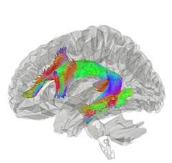
The arcuate fasciculus connects two important areas for language use, Broca's area and Wernicke's area.
Tractography showing arcuate fasciculus
Details
FromBroca's area of the frontal lobe
ToWernicke's area of the temporal lobe
Identifiers
Latinfasciculus arcuatus
TA98A14.1.09.557
TA25599
FMA260714

In neuroanatomy, the arcuate fasciculus (AF; from Latin 'curved bundle') is a bundle of axons that generally connects Broca's area and Wernicke's area in the brain. It is an association fiber tract connecting caudal temporal lobe and inferior frontal lobe.

Structure

The arcuate fasciculus is a white matter tract that runs parallel to the superior longitudinal fasciculus. Due to their proximity, they are sometimes referred to interchangeably. They can be distinguished by the location and function of their endpoints in the frontal cortex. The arcuate fasciculus terminates in Broca's area (specifically BA 44) which is linked to processing complex syntax. However, the superior longitudinal fasciculus ends in the premotor cortex which is implicated in acoustic-motor mapping.

Connection

Historically, the arcuate fasciculus has been understood to connect two important areas for language use: Broca's area in the inferior frontal gyrus and Wernicke's area in the posterior superior temporal gyrus. It is mostly considered to be an oversimplification, but this model is still utilized because a satisfactory replacement has not been developed. The topographical relationships between independent measures of white matter and gray matter integrity suggest that rich developmental or environmental interactions influence brain structure and function. The presence and strength of such associations may elucidate pathophysiological processes influencing systems such as language and motor planning.

As the technique of diffusion MRI has improved, this has become a testable hypothesis. Research indicates more diffuse termination of the fibers of the arcuate than previously thought. While the main caudal source of the fiber tract appears to be posterior superior temporal cortex, the rostral terminations are mostly in premotor cortex, part of Brodmann area 44.

Developmental differences

Myelination is a process by which axons are covered with a protective substance called myelin that drastically increases the signaling efficiency of the neuron. The arcuate fasciculus is heavily myelinated in healthy adult brains. The density of this myelination has been found to predict the accuracy and speed to which one can comprehend sentences. However, the arcuate fasciculus of newborns is unmyelinated. The myelination process occurs gradually during childhood; myelin density has been shown to increase between the age of 3 and 10. A study comparing a group of 6-year-olds to a group of 3-year-olds found that the 6-year-olds had stronger functional connectivity of the arcuate fasciculus. The arcuate fasciculus is similarly undeveloped in non-human primates such as chimpanzees and macaques. This supports the theory that the arcuate fasciculus is a critical component in language.

Dorsal stream

The two-streams hypothesis of language proposes that there are two streams by which the brain processes language information: the dorsal and ventral streams. The basis of this model is generally accepted, however the details of it are highly contentious. The dorsal pathway consists of multiple fiber tracts, one of which is the arcuate fasciculus. The dorsal pathway as a whole is implicated in sensory-to-motor mapping and processing complex syntax.

Role in language

Syntax

Syntax refers to a set of rules by which we order words within a language. Some researchers argue that syntax is what distinguishes language as a uniquely human capacity. Though the exact function of the arcuate fasciculus is still debated, the predominant theory is that it is involved with processing complex sequences of syntax. Studies indicate that as the arcuate fasciculus matures and undergoes myelination, there is a corresponding increase in the ability to process syntax. Furthermore, lesions in the arcuate fasciculus often result in difficulties with syntax. Researchers have found that when subjects are confronted with difficult syntactic structures, there is high synchronicity between the left frontal and parietal regions due to their connection by the arcuate fasciculus. This research further supports the arcuate fasciculus as the key component of human language.

Lateralization

The arcuate fasciculus is a bilateral structure; this means that it is present in both the right and left hemispheres of the brain. These fiber tracts are asymmetrical; the left arcuate fasciculus is stronger than the right. While the left arcuate fasciculus is thought to be the one involved with syntax processing, the right arcuate fasciculus has been implicated in prosody processing. Studies further suggest that the right arcuate fasciculus is involved with the ability to read emotion from human facial expression.

Clinical significance

Conduction aphasia

Historically the arcuate fasciculus has been linked to conduction aphasia, which is usually the result of damage to the inferior parietal lobule that extends into the subcortical white matter and compromises the arcuate fasciculus. This type of aphasia is characterized by difficulty with repetition and prevalent phonemic paraphasias. Patients otherwise exhibit a relatively normal control of language. The symptoms of conduction aphasia suggest that the connection between the posterior temporal cortex and frontal cortex plays a vital role in short-term memory of words and speech sounds that are new or have just been heard. The arcuate fasciculus is the main connection between these two regions. Studies that challenge the claim that the arcuate fasciculus is responsible for repetition cite that in some cases lesions to the arcuate fasciculus nor total agenesis produce conduction aphasia.

Progressive aphasia

Progressive aphasia is a type of aphasia that slowly worsens over time. It can affect both the production and comprehension of language. Progressive aphasic patients that have lesions in their arcuate fasciculus were especially deficient in their syntax processing abilities. Worsened syntax processing correlated with the degree of degradation in the arcuate fasciculus.

Tone deafness

In nine out of ten people with tone deafness, the superior arcuate fasciculus in the right hemisphere could not be detected, suggesting a disconnection between the posterior superior temporal gyrus and the posterior inferior frontal gyrus. Researchers suggested the posterior superior temporal gyrus was the origin of the disorder.

Stuttering

In stutterers, the arcuate fasciculus appears to have bilateral deficits that reduce it by one-third or more relative to non-stutterers. However, there is ongoing debate concerning the contribution of each hemisphere. Diffusion-based evidence of differences between stutterers and controls is not isolated to the arcuate fasciculus.

Specific language impairment

Specific language impairment is a disorder that prevents children from developing language normally. These children particularly have difficulty with the syntactic and hierarchal structures of language. Damage to the arcuate fasciculus is implicated as a possible cause of specific language impairment, however further data is required to validate this claim.

Dyslexia

Dyslexia is a disorder that is primarily characterized by reading deficits. Research has shown that decreases in the integrity of the arcuate fasciculus coincide with worsened reading ability in dyslexic subjects.

Functional disconnection

From Wikipedia, the free encyclopedia
https://en.wikipedia.org/wiki/Functional_disconnection

Functional disconnection
is the disintegrated function in the brain in the absence of anatomical damage, in distinction to physical disconnection of the cerebral hemispheres by surgical resection, trauma or lesion. Applications have included alexia without agraphia dyslexia, persistent vegetative state and minimally conscious state as well as autistic spectrum disorders. Functional disconnection itself is not a medically recognized condition. It is a theoretical concept used to facilitate research into the causes and symptoms within recognized conditions. 

History

In 1977, Witleson reported that developmental dyslexia may be associated with (i) bi-hemisphere representation of spatial functions, in contrast to the unitary right hemisphere control of these functions observed in normal individuals. The bilateral neural involvement in spatial processing may interfere with the left hemisphere's processing of its own specialized functions and result in deficient linguistic, sequential cognitive processing and in overuse of the spatial, holistic cognitive mode, reflecting a functional disconnection syndrome in these individuals confirmed by Leisman in the 1980s and in the 2000s.

The concept of functional disconnection developed further with Stachowiak and Poeck in 1976. who reported on a case in 1976 of a 67-yr-old male with hemianopia resulting from a cerebrovascular accident resulting in pure alexia and a color naming deficit that he suggested was due to a functional disconnection mechanism. He noted that the underlying disconnection mechanism is improved by the facilitating effect of unblocking methods (in the tactile, somesthetic, auditory, and visual systems), so that pathways other than the one impaired by the brain lesion are used.

In 1998, Fritson presented a mechanistic account of how dysfunctional integration among neuronal systems arises, based on the central role played by synaptic plasticity in shaping the connections. He hypothesized that the pathophysiology of schizophrenia is expressed at the level of modulation of associative changes in synaptic efficacy; specifically the modulation of plasticity in those brain systems responsible for emotional learning and emotional memory in the postnatal period. This modulation is mediated by ascending neurotransmitter systems that: (i) have been implicated in schizophrenia; and (ii) are known to be involved in consolidating synaptic connections during learning. The pathophysiology results in a disruption of the reinforcement of adaptive behavior consistent with the disintegrative aspects of the disorder. Kim and colleagues in 2003 further described the disconnection hypothesis in schizophrenia as the result of a prefrontal-parietal lobe functional disconnection, particularly prefrontal dissociation and abnormal prefrontal-parietal interaction during working memory processing.

The concept of functional disconnection developed still further when it was applied to the understanding of the nature of autistic spectrum disorder. Geschwind and Levitt in 2007 suggested a model of the symptoms of autism in which higher-order association areas of the brain (that normally connect to the frontal lobe) are partially disconnected during development, thereby explaining the heterogeneity of autism etiology. The autism group at Cambridge University provided evidence that the functional connectivity of medial temporal lobe structures specifically is abnormal in people with Asperger’s syndrome, at least during fearful face processing. Melillo and Leisman have similarly concluded that a functional disconnection syndrome is a basis for explaining the symptoms of autistic spectrum disorder.

Disconnection syndrome

From Wikipedia, the free encyclopedia
https://en.wikipedia.org/wiki/Disconnection_syndrome
Diffusion tensor imaging of the brain showing the right and left arcuate fasciculus (Raf & Laf), the right and left superior longitudinal fasciculus (Rslf & Lslf), and tapetum of corpus callosum (Ta).

Disconnection syndrome is a general term for a collection of neurological symptoms caused – via lesions to associational or commissural nerve fibres – by damage to the white matter axons of communication pathways in the cerebrum (not to be confused with the cerebellum), independent of any lesions to the cortex. The behavioral effects of such disconnections are relatively predictable in adults. Disconnection syndromes usually reflect circumstances where regions A and B still have their functional specializations except in domains that depend on the interconnections between the two regions.

Callosal syndrome, or split-brain, is an example of a disconnection syndrome from damage to the corpus callosum between the two hemispheres of the brain. Disconnection syndrome can also lead to aphasia, left-sided apraxia, and tactile aphasia, among other symptoms. Other types of disconnection syndrome include conduction aphasia (lesion of the association tract connecting Broca’s area and Wernicke’s), agnosia, apraxia, pure alexia, etc.

Anatomy of cerebral connections

Theodore Meynert, a neuroanatomist of the late 1800s, developed a detailed anatomy of white matter pathways. He classified the white matter fibers that connect the neocortex into three important categories – projection fibers, commissural fibers and association fibers. Projection fibers are the ascending and descending pathways to and from the neocortex. Commissural fibers are responsible for connecting the two hemispheres while the association fibers connect cortical regions within a hemisphere. These fibers make up the interhemispheric connections in the cortex.

Callosal disconnection syndrome is characterized by left ideomotor apraxia and left-hand agraphia and/or tactile anomia, and is relatively rare.

Hemispheric disconnection

Many studies have shown that disconnection syndromes such as aphasia, agnosia, apraxia, pure alexia and many others are not caused by direct damage to functional neocortical regions. They can also be present on only one side of the body which is why these are categorized as hemispheric disconnections. The cause for hemispheric disconnection is if the interhemispheric fibers, as mentioned earlier, are cut or reduced.

An example is commissural disconnect in adults which usually results from surgical intervention, tumor, or interruption of the blood supply to the corpus callosum or the immediately adjacent structures. Callosal disconnection syndrome is characterized by left ideomotor apraxia and left-hand agraphia and/or tactile anomia, and is relatively rare.

Other examples include commissurotomy, the surgical cutting of cerebral commissures to treat epilepsy and callosal agenesis which is when individuals are born without a corpus callosum. Those with callosal agenesis can still perform interhemispheric comparisons of visual and tactile information but with deficits in processing complex information when performing the respective tasks.

Sensorimotor disconnection

Hemispheric disconnection has impacted behaviors relating to the sensory and motor systems. The different systems affected are listed below:

  • Olfaction – The olfactory system is not crossed across hemispheres like the other senses, which means that left input goes to the left hemisphere and right input goes to the right hemisphere. Fibers in the anterior commissure control the olfactory regions in each hemisphere. A patient who lacks an anterior commissure cannot name odors entering the right nostril or use the right hand to pick up the object corresponding to the odor because the left hemisphere, responsible for language and controlling the right hand, is disconnected from the sensory information.
  • Vision – Information from one visual field travels to the contralateral hemisphere. Therefore, with a commissurotomy patient, visual information presented in the left visual field travelling to the right hemisphere would be disconnected from verbal output since the left hemisphere is responsible for speech.
  • Somatosensory – If the two hemispheres are disconnected, the somatosensory functions of the left and right parts of the body become independent. For example, when something is placed on the left hand of a blindfolded patient with the two hemispheres disconnected, the left hand can pick the correct object within a set of objects but the right hand cannot.
  • Audition – Though most of the input from one ear would go through the same ear, the opposite ear also receives some input. Therefore, the disconnection effects seems to be reduced in audition compared to the other systems. However, studies have shown that when the hemispheres are disconnected, the individual does not hear anything from the left and only hears from the right.
  • MovementApraxia and agraphia may occur where responding to any verbal instructions by movement or writing in the left hand is inhibited because the left hand cannot receive these instructions from the right hemisphere,

History

The concept of disconnection syndrome emerged in the late nineteenth century when scientists became aware that certain neurological disorders result from communication problems among brain areas. In 1874, Carl Wernicke introduced this concept in his dissertation when he suggested that conduction aphasia could result from the disconnection of the sensory speech zone from the motor speech area by a single lesion in the left hemisphere to the arcuate fasciculus. As the father of the disconnection theory, Wernicke believed that instead of being localized in specific regions of the brain, higher functions resulted from associative connections between the motor and sensory memory areas.

Lissauer, a pupil of Wernicke, described a case of visual agnosia as a disconnection between the visual and language areas.

Dejerine in 1892 described specific symptoms resulting from a lesion to the corpus callosum that caused alexia without agraphia. The patient had a lesion in the left occipital lobe, blocking sight in the right visual field (hemianopia), and in the splenium of the corpus callosum. Dejerine interpreted this case as a disconnection of the speech area in the left hemisphere from the right visual cortex.

In 1965, Norman Geschwind, an American neurologist, wrote ‘Disconnexion syndromes in animals and man’ where he described a disconnectionist framework that revolutionized neurosciences and clinical neurology. Studies of the monkey brain led to his theory that disconnection syndromes were higher function deficits. Building on Wernicke and previously mentioned psychologists’ idea that disconnection syndromes involved white matter lesion to association tracts connecting two regions of the brain, Geschwind was more detailed in explaining some disconnection syndromes as lesions of the association cortex itself, specifically in the parietal lobe. He described the callosal syndrome, an example of a disconnection syndrome, which is a lesion in the corpus callosum that leads to tactile anomia in just the patient’s left hand.

Though Geschwind made significant advances in describing disconnection syndromes, he was not completely accurate. He didn’t think the association cortex had any specialized role of its own besides acting as a relay station between the primary sensory and motor areas. However, in the 1960s and 1970s, Mesulam and Damasio incorporated specific functional roles for the association cortex. With Mesulam and Damasio’s contributions, Geschwind’s model has evolved over the past 50 years to include connections between brain regions as well as specializations of association cortices.

More recently, neurologists have been using imaging techniques such as diffusion tensor imaging (DTI) and functional magnetic resonance imaging (fMRI) to visualize association pathways in the human brain to advance the future of this disconnection theme.

Hemispherectomy

From Wikipedia, the free encyclopedia
https://en.wikipedia.org/wiki/Hemispherectomy

Hemispherectomy is a surgery that is performed by a neurosurgeon where an unhealthy hemisphere of the brain is disconnected or removed. There are two types of hemispherectomy. Functional hemispherectomy refers to when the diseased brain is simply disconnected so that it can no longer send signals to the rest of the brain and body. Anatomical hemispherectomy refers to when not only is there disconnection, but also the diseased brain is physically removed from the skull. This surgery is mostly used as a treatment for medically intractable epilepsy, which is the term used when anti-seizure medications are unable to control seizures.

History

The first anatomical hemispherectomy was performed and described in 1928 by Walter Dandy. This was done as an attempt to treat glioma, a brain tumor. The first known anatomical hemispherectomy performed as a treatment for intractable epilepsy was in 1938 by Kenneth McKenzie, a Canadian neurosurgeon. Krynaw, a neurosurgeon from South Africa, was one of the first to perform and report a case series on hemispherectomies in 1950. He performed the surgery on pediatric patients with infantile hemiplegia, specifically as a treatment for their seizures and cognitive impairment. His hemispherectomy technique removed the damaged hemisphere except the thalamus and caudate structures. Krynaw reported good outcomes overall, although there was one post-operative death. Specifically, there was an overall theme of improvement in weakness, spasticity and cognition. Amazingly, ten out of the twelve patients had seizures prior to the operation and none of the patients had seizures afterwards. Other neurosurgeons began performing hemispherectomies as well, primarily for the treatment of seizures. For the most part, the surgeries would go well initially, but there was a general theme of subsequent deterioration and even death years after the surgery. As a result of the complication risk and the introduction of new anti-seizure medications, the popularity of the procedure began to decline in the 1950s. Oppenheimer and Griffith were one of the first to describe the potential complications, and they reported their findings in 1966, describing superficial hemosiderosis, granular ependymitis and obstructive hydrocephalus. They posited a theoretical solution to this problem, a surgery that is now known as a functional hemispherectomy. Rasmussen was one of the first neurosurgeons to develop and apply a functional hemispherectomy in practice. He initially made modifications to the original hemispherectomy by preserving the least epileptogenic quarter or third of the hemisphere, hoping this would ameliorate the known complications of the original anatomic hemispherectomy. Although this modification seemed to solve this issue, patients undergoing the modified hemispherectomy continued to have seizures, which was problematic. Therefore, he further modified his surgery to functionally sever residual portions of the frontal and parieto-occipital lobes. This surgery, the functional hemispherectomy, has been further modified over the years by several different neurosurgeons, and to this day there is not a consensus as to which exact technique should be used. Hemispherotomy refers to some of the more recently developed approaches to disconnect the epileptic hemisphere while minimizing brain removal and the risk for complications.

Nomenclature

There are two main types of hemispherectomy: Anatomical and Functional.

Anatomical hemispherectomy refers to the resection and removal of an entire hemisphere of the brain, which includes all four lobes, with or without the removal of basal ganglia and thalamus.

Functional hemispherectomy refers to surgeries that disable the function of one hemisphere, while maintaining its blood supply and without physically removing the entire hemisphere from the skull. Functional hemispherectomies are performed more frequently than anatomical hemispherectomies due to their lower complication rates. However, they do carry a risk of incomplete disconnection, which refers to when the surgeon inadvertently leaves remnants of fibers that continue to connect the hemisphere to the brain and body. These remaining fibers can be problematic, as they may lead to seizure recurrence.

Another term that falls under the hemispherectomy umbrella includes hemidecortication, which is the removal of the cortex from one half of the cerebrum, while attempting to preserve the ventricular system by maintaining the surrounding white matter. Hemidecortication was originally developed as a possible strategy to mitigate some of the complications seen with complete anatomical hemispherectomy.

The term hemispherotomy refers to a surgery that is akin to a functional hemispherectomy in that it functionally severs the damaged hemisphere from the other and leaves some of the severed hemisphere within the skull, but the difference is that it removes even less tissue from the skull. The term hemispherotomy is now used as an umbrella term to describe the group of modern techniques and procedures that predominate at most contemporary epilepsy centers.

There is no statistically significant difference in seizure-free rates between the four different types of surgeries:  Hemispherotomy, functional hemispherectomy, anatomical hemispherectomy and hemidecortication. The overall rate of seizure freedom is estimated to be 73.4%.  However, hemispherotomy procedures may be associated with a more favorable complication profile.

Candidates

The typical candidates for hemispherectomy are pediatric patients who have intractable epilepsy due to extensive cerebral unilateral hemispheric injuries. In addition, the seizures should ideally be emanating from that same hemisphere. In some situations, a hemispherectomy may still be performed if there are seizures from both hemispheres, as long as the majority come from one side. In order to assess the patient’s epilepsy completely, patients undergo extensive testing, including EEG and MRI. Most patients also undergo other studies including functional MRI (fMRI), positron emission tomography (PET) or magnetoencephalography (MEG).

Today, hemispherectomy is performed as a treatment for severe and intractable epilepsy, including for young children whose epilepsy has been found to be drug-resistant. The most common underlying etiologies include malformations of cortical development (MCD), perinatal stroke and Rasmussen’s encephalitis. MCD is an umbrella term for a wide variety of developmental brain anomalies, including hemimegalencephaly and cortical dysplasia. Other less common underlying etiologies include hemiconvulsion-hemiplegia epilepsy syndrome and Sturge-Weber syndrome.

Procedure

Patients often shave the area of the scalp that will be involved with the surgery. Patients undergo general anesthesia and are unconscious for the procedure. The surgical site is sterilized, after which the skin is incised. A substantial portion of the bone is removed, followed by incision of the dura, which is the outer covering of the brain. There are several blood vessels that have connections with both sides of the brain, and these are carefully identified and clipped in such a way that spares the healthy hemisphere. Ultimately, a bundle of fibers that connect both of the cerebral hemispheres, the corpus callosum, is removed which results in the functional separation of one hemisphere from the other. Portions of the cerebral lobes from the damaged side of the brain are removed, depending on the specific procedure being performed. The surgeon may leave some brain tissue, such as the thalamus or choroid plexus. After completing the resection, the surgical site is irrigated with saline, the brain covering called the dura is sutured back together, the bone that was removed is replaced and the skin is sutured. This surgery often takes four to five hours. Patients often spend a few nights in the hospital post-operatively, and they undergo physical and occupational therapy soon after the surgery.

Potential complications

The most common complication from surgery is hydrocephalus, a condition in which fluid accumulates within the brain, and this is often treated with a shunt to divert the fluid away. The rate of shunts following surgery ranges from 14–23%. Other complications include wound complications, epidural hemorrhages, subdural hemorrhages, intraparenchymal hemorrhages, intracranial abscesses, meningitis, ventriculitis and venous thrombosis. Additional epilepsy surgery following hemispherectomy is rare (4.5%),[7] but may be recommended if there is a residual connection between the two hemispheres that is causing frequent seizures. Mortality rates are low and estimated to be <1% to 2.2%. Most patients do not experience changes in cognition, but some individuals may be at risk. A visual deficit called contralateral homonymous hemianopsia is expected to occur in most patients, where the entire visual field contralateral to the removed hemisphere is lost. There is a risk of motor deficits, and this is variable. Other possible complications include infection, aseptic meningitis, hearing loss, endocrine problems and transient neurologic deficits such as limb weakness.

Outcomes

Since seizures are the most common indication for hemispherectomy surgery, most research on hemispherectomy analyzes how the surgery affects seizures. Many patients undergoing surgery obtain good surgical outcomes, some obtaining complete seizure freedom (54–90%) and others having some degree of improvement in seizure burden. A recently developed scoring system has been proposed to help predict the probability of seizure freedom with more accuracy:  HOPS (Hemispherectomy Outcome Prediction Scale). Although it cannot definitively predict surgical outcome with exact precision, some physicians may use it as a guide. The scoring system takes certain variables into consideration including age at seizure onset, history of prior brain surgery, seizure semiology and imaging findings.

There is also data pertaining to how hemispherectomy affects the body in other ways. After surgery, the remaining cerebral hemisphere is often able to take over some cognitive, sensory and motor functions. The degree to which the remaining hemisphere takes on this additional workload often depends on several factors, including the underlying etiology, which hemisphere is removed and the age at which the surgery occurs.

In terms of postoperative motor function, some patients may have improvement or no change of their weaker extremity, and many can walk independently. Most patients postoperatively have minimal to no behavioral problems, satisfactory language skills, good reading capability, and only a minority of patients have a decline in IQ. Predictors of poor outcome may include seizure recurrence and structural abnormalities in the intact hemisphere.

Ultimately, risks and benefits should be weighed on an individual basis and discussed in detail with the neurosurgeon. Many patients have excellent outcomes, and the International League Against Epilepsy (ILAE) reports that “about one-fifth of hemispherectomy patients are gainfully employed and even fewer live independently.”

The Brain Recovery Project

The Brain Recovery Project is a non-profit corporation which funds new research and is based in the United States. This corporation hosts an annual two-day conference for patients who have had hemispherectomies and their families. There are several purposes to this reunion. The main goal is to educate patients and their families on the surgery and its necessary subsequent rehabilitation. It also serves as a way for patients and families to connect with one another, learn from specialists in the field and often offers research enrollment.

Year On

From Wikipedia, the free encyclopedia https://en.wikipedia.org/wiki/Year_On T...